Inherited Peripheral Neuropathies Mario A. Saporta, MD, PhD a, *, Michael E. Shy, MD b INTRODUCTION First described at the end of the nineteenth century by French neurologists Jean Martin Charcot and Pierre Marie and British neurologist Howard Henry Tooth, Charcot-Marie-Tooth (CMT) disease is now identified as the most common inherited neurologic condition, affecting approximately 1 in 2500 people. 1 CMT is frequently the final diagnosis of patients with previously unidentified (idiopathic or cryptogenic) peripheral neuropathies, 2 underscoring the need for better awareness and strategies to help general neurologists navigate through the clinical and molecular diagnosis of this fascinating group of neuropathies. Recent advances in molecular biology have demonstrated that CMT is genetically heterogeneous, with at least 50 genes known to cause CMT when mutated. Most patients have an autosomal dominant form of CMT, though X-linked and autosomal recessive (AR) inheritances are not uncommon. This article describes the characteristics of various forms of CMT, their biologic substrate, as well as the current strategy for genetic testing. a National Laboratory of Embryonic Stem Cells, Biomedical Sciences Department, Federal University of Rio de Janeiro, Rua Republica do Peru 362/602, Rio de Janeiro 22021-040, Brazil; b Department of Neurology, University of Iowa, 200 Hawkins Drive, Iowa City, IA 52242, USA * Corresponding author. E-mail address: [email protected]KEYWORDS Charcot-Marie-Tooth Inherited neuropathy Genetic testing KEY POINTS Identifiable genetic causes of neuropathy elucidate biologic pathways that cause demy- elination or axonal loss. Charcot-Marie-Tooth (CMT) disease is genetically and clinically heterogeneous with more than 50 genes causing neuropathy that can vary in age of onset and severity. Mutations in just four genes (PMP22, GJB1, MPZ, and MFN2) cause more than 90% of the genetically identifiable cases of CMT in North America. Combining the clinical phenotype and nerve conduction velocities in the arm can further focus genetic testing among these four genes. Because CMT can affect family members other than the proband, the authors suggest that genetic counseling be considered for patients and their families. Neurol Clin - (2013) -–- http://dx.doi.org/10.1016/j.ncl.2013.01.009 neurologic.theclinics.com 0733-8619/13/$ – see front matter Ó 2013 Elsevier Inc. All rights reserved.
� Identifiable genetic causes of neuropathy elucidate biologic pathways that cause demy-elination or axonal loss.
� Charcot-Marie-Tooth (CMT) disease is genetically and clinically heterogeneous with morethan 50 genes causing neuropathy that can vary in age of onset and severity.
� Mutations in just four genes (PMP22,GJB1,MPZ, andMFN2) causemore than 90% of thegenetically identifiable cases of CMT in North America.
� Combining the clinical phenotype and nerve conduction velocities in the arm can furtherfocus genetic testing among these four genes.
� Because CMT can affect family members other than the proband, the authors suggestthat genetic counseling be considered for patients and their families.
INTRODUCTION
First described at the end of the nineteenth century by French neurologists JeanMartin Charcot and Pierre Marie and British neurologist Howard Henry Tooth,Charcot-Marie-Tooth (CMT) disease is now identified as the most common inheritedneurologic condition, affecting approximately 1 in 2500 people.1 CMT is frequentlythe final diagnosis of patients with previously unidentified (idiopathic or cryptogenic)peripheral neuropathies,2 underscoring the need for better awareness and strategiesto help general neurologists navigate through the clinical and molecular diagnosis ofthis fascinating group of neuropathies. Recent advances in molecular biology havedemonstrated that CMT is genetically heterogeneous, with at least 50 genes knownto cause CMT when mutated. Most patients have an autosomal dominant form ofCMT, though X-linked and autosomal recessive (AR) inheritances are not uncommon.This article describes the characteristics of various forms of CMT, their biologicsubstrate, as well as the current strategy for genetic testing.
a National Laboratory of Embryonic Stem Cells, Biomedical Sciences Department, FederalUniversity of Rio de Janeiro, Rua Republica do Peru 362/602, Rio de Janeiro 22021-040, Brazil;b Department of Neurology, University of Iowa, 200 Hawkins Drive, Iowa City, IA 52242, USA* Corresponding author.E-mail address: [email protected]
Neurol Clin - (2013) -–-http://dx.doi.org/10.1016/j.ncl.2013.01.009 neurologic.theclinics.com0733-8619/13/$ – see front matter � 2013 Elsevier Inc. All rights reserved.
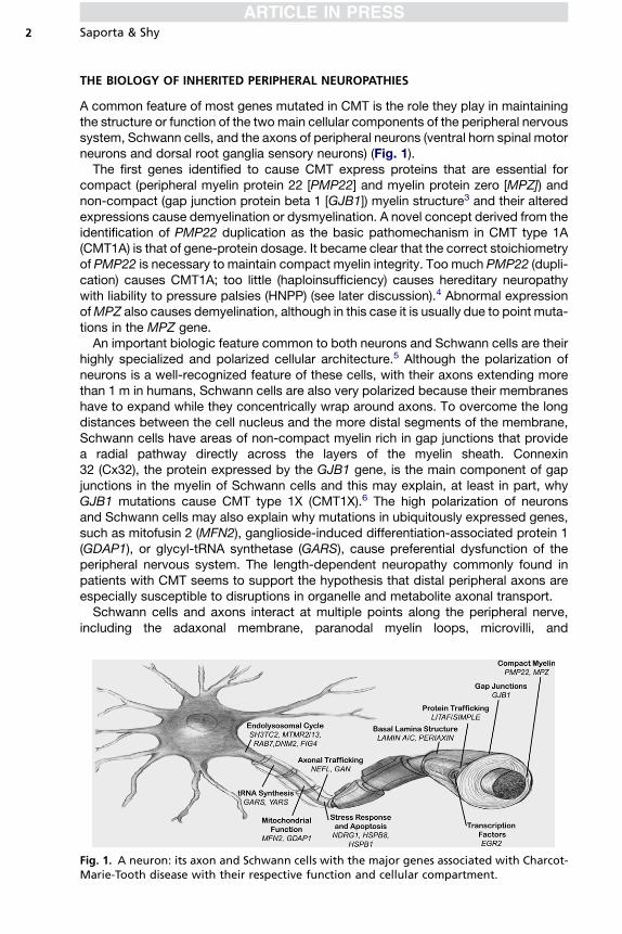
A common feature of most genes mutated in CMT is the role they play in maintainingthe structure or function of the two main cellular components of the peripheral nervoussystem, Schwann cells, and the axons of peripheral neurons (ventral horn spinal motorneurons and dorsal root ganglia sensory neurons) (Fig. 1).The first genes identified to cause CMT express proteins that are essential for
compact (peripheral myelin protein 22 [PMP22] and myelin protein zero [MPZ]) andnon-compact (gap junction protein beta 1 [GJB1]) myelin structure3 and their alteredexpressions cause demyelination or dysmyelination. A novel concept derived from theidentification of PMP22 duplication as the basic pathomechanism in CMT type 1A(CMT1A) is that of gene-protein dosage. It became clear that the correct stoichiometryof PMP22 is necessary to maintain compact myelin integrity. Too much PMP22 (dupli-cation) causes CMT1A; too little (haploinsufficiency) causes hereditary neuropathywith liability to pressure palsies (HNPP) (see later discussion).4 Abnormal expressionofMPZ also causes demyelination, although in this case it is usually due to point muta-tions in the MPZ gene.An important biologic feature common to both neurons and Schwann cells are their
highly specialized and polarized cellular architecture.5 Although the polarization ofneurons is a well-recognized feature of these cells, with their axons extending morethan 1 m in humans, Schwann cells are also very polarized because their membraneshave to expand while they concentrically wrap around axons. To overcome the longdistances between the cell nucleus and the more distal segments of the membrane,Schwann cells have areas of non-compact myelin rich in gap junctions that providea radial pathway directly across the layers of the myelin sheath. Connexin32 (Cx32), the protein expressed by the GJB1 gene, is the main component of gapjunctions in the myelin of Schwann cells and this may explain, at least in part, whyGJB1 mutations cause CMT type 1X (CMT1X).6 The high polarization of neuronsand Schwann cells may also explain why mutations in ubiquitously expressed genes,such as mitofusin 2 (MFN2), ganglioside-induced differentiation-associated protein 1(GDAP1), or glycyl-tRNA synthetase (GARS), cause preferential dysfunction of theperipheral nervous system. The length-dependent neuropathy commonly found inpatients with CMT seems to support the hypothesis that distal peripheral axons areespecially susceptible to disruptions in organelle and metabolite axonal transport.Schwann cells and axons interact at multiple points along the peripheral nerve,
including the adaxonal membrane, paranodal myelin loops, microvilli, and
Fig. 1. A neuron: its axon and Schwann cells with the major genes associated with Charcot-Marie-Tooth disease with their respective function and cellular compartment.
Inherited Peripheral Neuropathies 3
juxtaparanodal basal lamina. These interactions are mutually beneficial, providingtrophic support to the axon and myelinating cues to the Schwann cell. An exampleof this important interaction is the occurrence of secondary axonal degeneration inall forms of demyelinating CMT. This axonal degeneration is deemed to occur asa consequence of ineffective Schwann cell support to the axon and is actually moredirectly related to clinical functional impairment than the demyelination itself.7
Several recent studies have demonstrated a specific susceptibility of Schwanncells to mutations yielding misfolded proteins, as seen in certain PMP228 andMPZ9,10 point mutations. Misfolded proteins accumulate in the endoplasmic reticulum(ER) of Schwann cells inducing a transitory unfolded protein response (UPR), a seriesof cellular events that help the ER to cope with the increased metabolic demandcaused by misfolded protein retention. This, in turn, causes down-regulation of themyelination program genes and dedifferentiation of Schwann cells, a toxic gain offunction that worsens the demyelination and is potentially amenable to therapeuticintervention.11,12
CLINICAL AND NEUROPHYSIOLOGICAL FEATURES
CMT is clinically, as well as genetically, heterogeneous, with variability in the age ofonset, speed of progression, and electrodiagnostic findings. Though both motorand sensory nerves are usually affected, the more prominent phenotypic character-istic is related to motor difficulty in most cases. The classic phenotype includes step-page gait, pes cavus, sensory loss in a stocking or glove distribution, invertedchampagne bottle legs, and atrophy in the hands.13–15 Physical examination alsoshows decreased or absent deep tendon reflexes, often diffusely but virtually alwaysinvolving the Achilles tendon. Findings are usually symmetric.16 Onset is typically inthe first to second decade in classic cases, though this may differ depending on thegenetic subtype, including early-onset, infantile forms (historically designatedDejerine-Sottas syndrome) and late-onset, adult forms. Symptoms are usually slowlyprogressive, especially for the classic and late-onset phenotypes, but can be rathersevere in early-onset forms. Patients usually have impaired proprioception withbalance difficulty. Neuropathic pain affects around 20% of CMT patients.Nerve conductions allow for classification into demyelinating, axonal, or interme-
diate groups, based on the motor nerve conduction velocities (MNCV) and compoundmuscle action potential amplitudes (CMAP). The standard cut off for demyelinatingMNCV in the upper extremities is 38 m/s. Velocities between 35 and 45 m/s may beconsidered intermediately slowed, and greater than 45 m/s are considered axonal ifthere is a decrease in CMAP. Conduction velocities are performed in the armsbecause CMAP amplitudes are often unobtainable in the legs, even for demyelinatingforms of CMT, due to impaired interactions between abnormal myelin and the under-lying axon. CMT can be divided into subtypes based on electrodiagnostic features andinheritance pattern. Patients with autosomal dominant inheritance and a demyelinatingphenotype are said to have CMT1. Patients with autosomal dominant inheritance andan axonal phenotype have CMT type 2 (CMT2) and patients with AR inheritance,regardless of the electrodiagnostic features, have CMT type 4 (CMT4). Patients withCMT inherited in an X-linked fashion have CMT type X (CMTX). The subtypes arefurther divided genetically based on the gene mutated. The gene, or the type of muta-tion in the gene that causes the condition, defines each genetic subtype, as shown onTable 1. The usual electrodiagnostic finding in demyelinating inherited neuropathies iswidespread uniform slowing of conduction velocities, as opposed to the multifocalsegmental slowing found in demyelinating acquired neuropathies in which temporal
Table 1Classification of Charcot-Marie-Tooth disease
Type Gene or Locus Specific Phenotype
AD CMT1
CMT1A Dup 17p (PMP22) Classic CMT1PMP22 (point
mutation)Classic CMT1, DSS, CHN, HNPP
CMT1B MPZ CMT1, DSS, CHN, intermediate, CMT2
CMT1C LITAF Classic CMT1
CMT1D EGR2 Classic CMT1, DSS, CHN
CMT1E NEFL CMT2 but can have slowMNCVs in CMT1 range � early-onset severe disease
HNPP
HNPP Del 17p (PMP22) Typical HNPPPMP22 (point
mutation)Typical HNPP
X-linkedCMT1 (CMT1X)
CMT1X GJB1 Intermediate � patchy MNCVs; male MNCVs less thanfemale MNCVs
AR demyelinating CMT (CMT4)
CMT4A GDAP1 Demyelinating or axonal, usually early onset and severevocal cord and diaphragm paralysis described
Rare AD CMT2 families described
CMT4B1 MTMR2 Severe CMT1, facial, bulbar, focally folded myelin
CMT4B2 SBF2 Severe CMT1, glaucoma, focally folded myelin
CMT4C SH3TC2 Severe CMT1, scoliosis, cytoplasmic expansions
CMT4D (HMSNL) NDRG1 Severe CMT1, gypsy, deafness, tongue atrophy
CMT4E EGR2 Classic CMT1, DSS, CHN
CMT4F PRX CMT1, more sensory, focally folded myelin
CMT4H FGD4 CMT1
CMT4J FIG4 CMT1
CCFDN CTDP1 CMT1, gypsy, cataracts, dysmorphic features
HMSN-Russe 10q22–q23 CMT1
CMT1 PMP22 (pointmutation)
Classic CMT1, DSS, CHN, HNPPs
CMT1 MPZ CMT1, DSS, CHN, intermediate, CMT2
AD CMT 2
CMT2A MFN2 CMT2Usually severeOptic atrophy
CMT2B RAB7A CMT2 with predominant sensory involvement andsensory complications
CMT2C 12q23–q24 CMT2 with vocal cord and respiratory involvement
CMT2D GARS CMT2 with predominant hand wasting, weakness, ordHMN V
CMT2E NEFL CMT2 but can have slowMNCVs in CMT1 range � early-onset severe disease
(continued on next page)
Saporta & Shy4
Table 1(continued)
Type Gene or Locus Specific Phenotype
CMT2F HSPB1 (HSP27) Classic CMT2 or dHMN II
CMT2G 12q12–q13.3 Classic CMT2
CMT2L HSPB8 (HSP22) Classic CMT2 or dHMN II
CMT2 MPZ CMT1, DSS, CHN, intermediate, CMT2
CMT2 (HMSNP) 3q13.1 CMT2 with proximal involvement
AR CMT2 (also called CMT4)
AR CMT2A LMNA CMT2 proximal involvement and rapid progressiondescribed
Also causes muscular dystrophy, cardiomyopathy, orlipodystrophy
AR CMT2B 19q13.1–13.3 Typical CMT2
AR CMT2 GDAP1 CMT1 or CMT2 usually early onset and severeVocal cord and diaphragm paralysis describedRare AD CMT2 families described
DI CMT (DI-CMT)
DI-CMTA 10q24.1–25.1 Typical CMT
DI-CMTB DNM2 Typical CMT
DI-CMTC YARS Typical CMT
Hereditary neuralgic amyotrophy (HNA)
HNA SEPT9 Recurrent neuralgic amyotrophy
Abbreviations: AD, autosomal dominant; CHN, congenital hypomyelinating neuropathy; CTDP1,CTD phosphatase subunit 1; Del, deletion; DMN2, dynamin 2; DSS, Dejerine-Sottas syndrome;Dup, duplication; EGR2, early growth response 2; FGD4, FYVE, RhoGEF, and PH domain containing4; FIG4, FIG 4 homolog; HSP22, heat shock 22 kDa protein 8; HSP27, heat shock 27 kDa protein 1;KIF1Bb, kinesin family member 1B-b; LITAF, lipopolysaccharide-induced tumor necrosis factor;LMNA, lamin A and C; MCV, motor conduction velocity; MTMR2, myotubularin-related protein 2;MTMR13, myotubularin related protein 13; NDRG1, N-myc downstream regulated gene 1; NEFL,neurofilament, light polypeptide 68 kDa; PRX, periaxin; RAB7, RAB7,member RASoncogene family;SEPT9, septin 9; SH3TC2, SH3domain and tetratricopeptide repeats 2; YARS, tyrosyl tRNA synthetase.
From Reilly MM, ShyME. Diagnosis and new treatments in genetic neuropathies. J Neurol Neuro-surg Psychiatry 2009;80(12):1304–14. Copyright 2009, from BMJ Publishing Group Ltd; withpermission.
Inherited Peripheral Neuropathies 5
dispersion and conduction block is frequently seen.17,18 Two exceptions to this ruleare men with CMT1X and patients with HNPP. In these cases, focal demyelinationwith temporal dispersion or conduction block can be seen. In all other cases of demy-elinating CMT the finding of focal slowing should raise the possibility of a superim-posed inflammatory neuropathy, which can benefit from immunosuppressivetherapy.19
GENETIC TESTING STRATEGIES
Strategies for focusing genetic testing have been in place since at least 2001, with flowcharts to help guide testing.20 The distribution of causal genes depends, at least inpart, on the population tested. For European and North American populations, ARCMT comprises less than 10% of all cases and most patients have dominantlyinherited CMT even if their cases are sporadic. Alternatively, populations in whichconsanguinity is high, such as in North Africa, may have up to 40% of their cases being
Saporta & Shy6
AR. Using MNCV and inheritance patterns, several strategies have been publishedsince the 2001 study, mostly based on North American or European populations.21–23
The authors have recently published testing guidelines which included age of onset ofsymptoms to help guide testing.24 Age of onset classifications were infantile (delayedwalking), childhood, or adult. These guidelines dividedMNCV info four categories: lessthan or equal to15 m/s (very slow), between 15 and 35 m/s (slow), between 35 and45 m/s (intermediate), and greater than 45 m/s (axonal). Flow charts were providedusing MNCV as the first level of evidence, with age of onset and inheritance patternsguiding the testing strategy within each category (Figs. 2 and 3). Of patients who hada genetic diagnosis, 92% were found to have changes in one of four genes: PMP22,GJB1,MPZ, andMFN2. Thus, the flow diagrams emphasize testing for these types ofCMT, excepting HNPP, which has a distinctive nerve conduction study (NCS) patternthat differs from those of other forms of CMT and should be recognizable.
MNCV Less than or Equal to 15 m/s
All people with very slow MNCV who walked by 15 months of age had CMT1A; thus,genetic testing for the PMP22 duplication is warranted for these individuals (seeFig. 2A). Of those patients who had delayed walking, most had CMT1A but 32%had CMT1B. Genetic testing for CMT1A and CMT1B is appropriate for people inthis category. If these tests are negative, genetic testing for more rare forms of CMTmay be reasonable.
MNCV Greater than 15 and Less than or Equal to 35 m/s
Approximately 89% of patients with slow MNCV who began walking by 15 months ofage had CMT1A; thus, genetic testing should begin with PMP22 duplication analysis(see Fig. 2B). CMT1X was the next most common type of CMT but should only be per-formed for people who do not have evidence of male-to-male transmission in theirpedigree. CMT1B testing is much less likely to be the cause of the CMT for peoplein this category, but testing may be reasonable if testing if CMT1A and CMT1X arenegative or if there is evidence of male-to-male transmission.
MNCV Greater than 35 and Less than or Equal to 45 m/s
Most people who had intermediate conductions had either CMT1X or CMT1B (seeFig. 3A). If symptoms began in childhood, and no male-to-male transmission ispresent in the pedigree, it is most likely for the person to have CMT1X. If this testingis negative, CMT1B testing may be pursued. However, if the symptom onset was inadulthood, testing for CMT1B is more likely to elicit a positive genetic testing result,with CMT1X being a reasonable follow-up test.
MNCV Greater than 45 m/s or Unobtainable CMAP
People with normal velocities or unobtainable CMAP usually present with CMT1X(usually women), CMT1B, or CMT2A (see Fig. 3B). People with unobtainable CMAPwere usually those with CMT2A, who are often severely affected in infancy and child-hood.25 Thus, for children with early onset or severe CMT, it is proposed to begingenetic testing for CMT2A. For people with axonal CMT who have a classic or adultonset of symptoms, testing should begin with CMT1X in the absence of male-to-male transmission in the pedigree. Testing should begin with CMT1B if male-to-male transmission is present or if CMT1X testing is negative. The authors proposeusing other clinical findings, such as the upper limbs being more severely affectedthan the lower limbs, to help guide additional genetic testing if necessary. For thesepatients, mutations in the GARS gene, causing CMT2D may be appropriate.
Fig. 2. Algorithm for the genetic diagnosis of patients with Charcot-Marie-Tooth disease and very slow (A) or slow (B) upper extremity motor nerveconduction velocities. dup, duplication; EGR2, early growth response 2; LITAF, lipopolysaccharide-induced TNF factor; seq, sequencing. (From SaportaAS, Sottile SL, Miller LJ, et al. Charcot-Marie-Tooth disease subtypes and genetic testing strategies. Ann Neurol 2011;69(1):22–33; with permission.)
Inherite
dPerip
heralNeuropathies
7
Fig. 3. Algorithm for the genetic diagnosis of patients with Charcot-Marie-Tooth disease and intermediate (A) or normal (B) upper extremity MNCVs.NEFL, neurofilament light polypeptide. (From Saporta AS, Sottile SL, Miller LJ, et al. Charcot-Marie-Tooth disease subtypes and genetic testing strate-gies. Ann Neurol 2011;69(1):22–33; with permission.)
Saporta
&Sh
y8
Inherited Peripheral Neuropathies 9
Although a detailed review of the pros and cons for testing is beyond the scope ofthis article, the authors think it reasonable to provide some information about how wepursue genetic testing.26 Clearly, not every patient with a genetic neuropathy wants orneeds testing to identify the genetic cause of their disease. We believe that the ulti-mate decision to undergo genetic testing rests with the patient or the patient’s parentsif a symptomatic child is younger than 18 years of age. Reasons that patients give forobtaining testing include identifying the inheritance pattern of their CMT, makingfamily planning decisions, and obtaining knowledge about the cause and naturalhistory of their form of CMT. Natural history data is available for some forms of CMTsuch as CMT1A27 and CMT1X,28 which can provide guidance for prognosis, recog-nizing that there can be phenotypic variability in these subtypes. Patients with otherforms of CMT frequently choose to undergo genetic testing to contribute to the naturalhistory data collection for other patients with the same subtype. There are alsoreasons why patients do not want genetic testing. These include the high cost ofcommercial testing and fears of discrimination in the workplace or in obtaining healthinsurance. Because there are currently no medications to reverse any form of CMT,many patients decide against testing because their therapies will not depend on theresults. We maintain that is always the patient’s decision whether or not to pursuegenetic testing.Once a genetic diagnosis has been made in a patient, other family members usually
do not need genetic testing but can be identified by clinical evaluation with neurophys-iology. We do not typically test patients for multiple genetic causes of CMT simulta-neously, although we did identify 11 patients with multiple genetic causes of CMT.It is our current policy to only consider performing genetic testing in clinically affectedfamily members of a proband if their phenotype is atypical for the type of CMT in thefamily. In addition, we do not test asymptomatic minors with a family history of CMT,either by electrophysiology or genetic testing owing to the chance for increasedpsychological harm to the child.29 We do routinely perform limited NCSs, thoughnot needle electromyogram (EMG), on symptomatic children with CMT. Becausenerve conduction changes, including slowing, are often uniform and detectable inearly childhood in CMT,17,18 testing of a single nerve is often adequate to guidegenetic testing or determine whether a symptomatic child is affected in a familywith CMT.
SPECIFIC FORMS OF CMTCMT1
CMT1 includes five types of CMT that are caused by four genes when mutated. Thisgroup includes most people with CMT (over 70%). These genes are essential toSchwann cell function and the formation of myelin sheaths surrounding the axon,though they interact in different ways and thus are phenotypically heterogeneous.30
CMT1ACMT1A is the most common type of CMT, affecting 55% of genetically definedpatients.24 It is caused by a 1.4 megabase (Mb) duplication at 17p11.2 including thePMP22 gene, created by unequal crossing over of homologous chromosomes.31,32
People with CMT1A typically have the classic CMT phenotype, with normal age ofonset for walking, development of symptoms in the first two decades, pes cavus,and slowly progressive motor and sensory neuropathy, which rarely progresses towheelchair use later in life.24,33 People with CMT1A have distinctive length dependentsensorimotor demyelinating neuropathies. One study found that more than 90% ofpatients with CMT1A had MNCV in the ulnar nerve between 16 and 35 m/s or less.24
Saporta & Shy10
CMT1A is an autosomal dominant condition, and most patients will have a familyhistory. However, there is a de novo rate of about 10%.34 Therefore, people withoutfamily history with ulnar MNCV under 35 m/s should first be screened for thePMP22 duplication before proceeding with other genetic testing.24 Once one personin a family has been genetically shown to have CMT1A, first and second-degree familymembers can be screened by MNCV. If other family members are shown to have thecharacteristically slowed conductions, it can be assumed that that person also hasCMT1A without needing genetic testing.
HNPPHNPP is caused by the reciprocal deletion of the 1.4 Mb stretch of chromosome17p11.2 containing the PMP22 gene.35 A small percentage of people with HNPPhave this phenotype due to a frameshift, splice site, or point mutation of the PMP22gene (www.molgen.ua.ac.be/cmtmutations). HNPP is the third most common typeof CMT, affecting about 9.1% of genetically diagnosed patients,24 with a de novorate of about 20%.36 The hallmark feature of HNPP is transient and recurrent motorand sensory mononeuropathies. These typically occur at entrapment sites, such asthe carpal tunnel, ulnar groove, and fibular head.37 These palsies may last hours,days, or weeks, or occasionally longer. For some people, HNPP can progress tolong-term peripheral neuropathy phenotypically indistinguishable from CMT1, inwhich patients may require ankle-foot orthoses or wrist splints.37 HNPP can be distin-guished electrodiagnostically by marked slowing of the ulnar and sural sensory nerveconduction velocities, with or without reduced SNAP, and relatively preservedMNCV.38 Distal motor latencies, particularly in the median and peroneal nerves, aretypically prolonged, often out of proportion with the reduction of velocity.39 Conduc-tion block and focal slowing often occur at entrapment sites, particularly during a palsyepisode.37
CMT1BCMT1B is caused by mutations in the MPZ gene40 located at chromosome 1q22,which encodes for MPZ, a major component of the myelin sheath. It affects about8.5% of people with genetically defined CMT.24 People with CMT1B usually presentin a bimodal distribution. One group develops a severe, early onset, demyelinatingneuropathy and the other group develops a late onset, milder, axonal neuropathy.Age of onset of symptoms is useful in determining the subtype of CMT. Most peoplewith early onset CMT1B will have delayed walking and MNCV less than 15 m/s.24
Those with late onset CMT1Bwill walk at a normal age and usually have MNCV greaterthan 35 m/s.24
CMT1CCMT1C is caused by mutations in the SIMPLE gene at chromosome 16p13.3-p12.41
The phenotype of CMT1C seems similar to that of CMT1A, with onset between the firstand third decades and MNCV between 16 and 25 m/s,42,43 as well as progressivesensorimotor nerve involvement. SIMPLEmutations are a rare cause for CMT, makingup 0.6% to 1.2% of demyelinating CMT cohorts.24,42
CMT1DCMT1D is caused by mutations in the EGR2 gene at chromosome 10q21.1-q22.1.44
Patients typically present in infancy with severe symptoms and may have congenitalhypomyelination (hypotonia, delayed motor milestones, MNCV<10 m/s).45 Cranialnerve involvement may also be present.45,46 Recessive inheritance has also beendescribed with this gene causing CMT4E, which seems to have a similar phenotype.
CMT1EPoint mutations in the PMP22 gene cause CMT1E or HNPP, depending on the func-tion of the mutation. Those with CMT1E tend to have earlier onset and more severesymptoms than those with CMT1A, but this is not invariable.47,48 MNCV in severelyaffected patients are markedly reduced, usually less than 10 m/s.47 Onset within thefirst 2 years of life presenting with delayed walking is not uncommon. CMT1E isa rare form of CMT, accounting for about 1% of people with genetically confirmedCMT.24
CMT2
CMT2ACMT2A is caused by mutations in the MFN2 gene.49 This is the most common type ofCMT2, accounting for approximately 21% of axonal CMT.25 People with CMT2Ausually, though not always, have a severe phenotype, with onset in infancy or earlychildhood and usually needing a wheelchair for ambulation by 20 years of age.25,49
It may be difficult to perform NCSs and obtain responses for those with severe muscleatrophy and thus people who have severe symptoms without recordable potentialsshould be screened for CMT2A. The minority of patients may present with a mild ormoderate axonal phenotype.25 There are many polymorphisms in MFN2 so thatcare must be taken to ensure that mutations are disease-causing. Most disease-causing mutations are in the GTPase domain, coiled-coil domains, or in other evolu-tionary conserved regions of the protein.25
CMT2BCMT2B is caused by mutations in the RAB7 gene.50 This type of CMT is distinguishedby distal sensory loss that often leads to foot ulcerations and subsequently infectionsand amputations50–53 in addition to typical motor signs. Nerve conductions often havereduced amplitude with normal or near-normal velocities.50–53 Sensory loss is oftensevere such that patients may be clinically indistinguishable from those with hereditarysensory and autonomic neuropathy (HSAN) type 1.
CMT2CCMT2C is caused by mutations in the TRPV4 gene.54–56 CMT2C is characterized bya predominantly motor axonal neuropathy and vocal cord and diaphragmatic paresis,often presenting with hoarseness or stridor.54–57 Sensorineural hearing loss andbladder urgency and incontinence have been reported.54 CMT2C is allelic with spon-dylometaphyseal dysplasia, metatropic dysplasia, and brachyolmia, and thus mayhave some overlapping characteristics such as short statures and scoliosis.55,58
CMT2DCMT2D is an axonal neuropathy caused by mutations in the GARS gene.59 Peoplewith CMT2D typically have upper extremity weakness greater than and/or beforelower extremity weakness and wasting, with a split-hand appearance of more atrophyin the FDI and thenar eminences and less so in the hypothenar eminence.59,60 CMT2Dis allelic with distal spinal muscular atrophy type V (dSMA-V), with the distinguishingfeature being lack of sensory involvement in dSMA-V.60
CMT2ECMT2E is caused by mutations in the neurofilaments light polypeptide (NEFL) gene.61
NCSs may be axonal or in the demyelinating range.62–64 Those with demyelinatingconductions may have a severe early onset or a childhood presentation.64 This is
Saporta & Shy12
considered an axonal form of CMT because neurofilaments are components of theaxon, not myelin.65
CMT2FCMT2F is caused by mutations in the HSPB1 gene, a member of the heat shockprotein (HSP)-27 superfamily.66 Most people with mutations in this gene have distalhereditary motor neuropathy (dHMN), a pure motor phenotype,67,68 though somewill have sensory findings on examination and electrophysiology.69 Impairment typi-cally begins in the distal legs and progresses slowly to the distal arms and then prox-imal legs.68 There has been one report of presumed AR inheritance with mutations inthis gene.68
CMT2LCMT2L is caused by mutations in the HSPB8 gene, also a member of the HSP super-family, and is also known as HSP22.70 Mutations in this gene have also been found tocause dHMN type II.71 Scoliosis and proximal weakness have been reported.72 Muta-tions in this gene are a rare cause of CMT.
CMT2KCMT2K is caused by heterozygous mutations in the GDAP1 gene, though recessiveforms of CMT with mutations in this gene are called CMT4A and are likely morecommon.73 Thus far, five mutations have been found to cause CMT2K: 358 C>T(p. R120W),74,75 469A>C (p.T157P) 75,66 678A>T (p.R226S),76 101C>G (p.S34C),76
and 23delAG (p.G10fs).76 Phenotypes range frommild adult onset and slowly progres-sive to severe childhood onset.73–76 One study found that 3 out of 11 families withCMT2 had a dominantly inherited mutation in GDAP1,76 indicating that this may bea significant cause of axonal CMT.
CMT4
CMT4ACMT4A is caused by two recessive mutations in the GDAP1 gene.73 People withCMT4A typically have an early onset and severe sensorimotor neuropathy73,77 thatmay be demyelinating or axonal in presentation.78,79 Phenotype is typically severe,with first symptoms being noted in childhood and eventual progression to wheelchairnot uncommon.79,80 Vocal cord paralysis or hoarseness has also been reported.79,80
Nerve conductions have been described as axonal or demyelinating, which has led tosome confusion about the cell type of origin for the disease. GDAP1 is a nuclearencoded gene that plays a role in mitochondrial fission or fragmentation, as opposedto MFN2, the causal gene for CMT2A, which plays an important role in mitochondrialfusion.
CMT4B1CMT4B1 is caused by mutations in the myotubularin-related protein (MTMR)-2gene.81 Patients typically have demyelinating MNCV.82 Onset is usually in childhoodand causes distal weakness that progress proximally, often leading to wheelchairuse by adulthood.83 Diaphragmatic and facial weakness may occur.84,85
CMT4B2CMT4B2 is caused by mutations in SBF2, also known as MTMR13.86,87 Nerveconductions are usually demyelinating.86,87 Onset is typically in childhood, thoughlater than in CMT4B1.87 Nerve biopsies showing focally folded myelin are character-istic of CMT4B1 (MTMR2 mutations) and CMT4B2 (MTMR13 mutations).
Inherited Peripheral Neuropathies 13
CMT4CCMT4C is caused by mutations in the SH3TC2 gene.88 In addition to demyelinatingsensorimotor neuropathy, scoliosis or kyphoscoliosis are hallmark features of thiscondition,88–92 though not universally present. Patients often present in childhoodwith delayed walking, distal weakness, foot deformities, or scoliosis.88–91,93 Cranialnerve involvements may also be present.90,91,93 Although prevalence numbers arenot known in all populations, there is evidence that CMT4C may be the most commonof the AR-inherited neuropathies.92
CMT4FCMT4F is caused by mutations in the PERIAXIN gene (PRX).94,95 Patients have demy-elinating conductions and severe early onset sensorimotor neuropathy.94,96 Sensoryataxia may be present,94,96,97 as might scoliosis.96 Many sequence changes in thePRX gene have been found to be benign variants (www.molgen.ua.ac.be/cmtmutations), and variants of uncertain significance within the gene should be furtherinvestigated before determining if they are disease-causing mutations.
CMT4JCMT4J is caused by mutations in the FIG4 gene.98 Patients may have demyelinatingconductions and a severe motor phenotype, possibly asymmetric, with onset in earlychildhood. Rapid progression to a wheelchair in adulthood has been described forpatients who were only mildly affected in their first two decades of life.98,99 Early deathhas been reported (47 years of age).99 Abnormalities on EMG may be similar to thoseseen in motor neuron disease, including fibrillations, positive waves, and reducedmotor unit action potentials of long durations. However (see previous discussion),NCS may be in the demyelinating range despite these EMG changes.99
CMTX
CMT1XCMT1X is caused by mutations in theGJB1 gene, encoding the protein Cx32.6 CMT1Xis the second most common form of CMT, found in at least 10% of all patients.24 Mentypically have more severe symptoms than women with the condition,100 and tend tohave marked atrophy of the intrinsic hand muscles and all compartments of the calfmuscles. Most men will have symptoms in childhood, though about 20% have a laterage of onset.24 Men with CMT1X have been reported to have transient stroke-likeepisodes with MRI changes following a stressor.101 Whereas two out of three womenwith CMT1X will have slowly progressive mild symptoms, one out of three do havemoderate neuropathy more similar to men with the condition.100 Men with MNCV oftenpresent between 25 and 45 m/s, whereas women usually have MNCV greater than35 m/s.24
HSAN
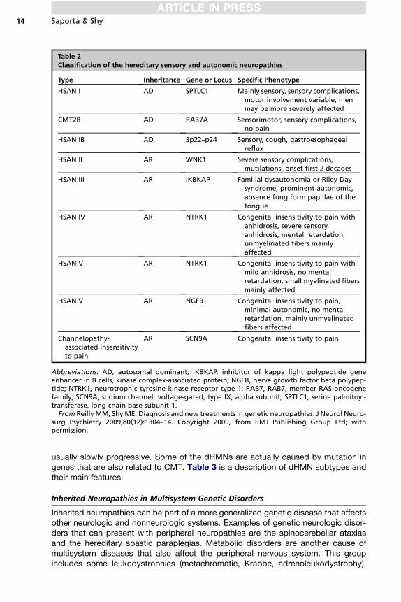
The HSANs are characterized by a predominant (although not always exclusive)sensory presentation. Patients may develop distinct clinical phenotypes accordingto the genetic abnormality, including distal lower limb sensory loss and neuropathicpain, congenital insensitivity to pain, or pure autonomic dysfunction. Most HSANsyndromes are AR and early-onset, although some can be autosomal dominant.HSAN subtypes are described in Table 2.
dHMNs
The dHMNs are inherited neuropathies that are exclusively motor in nature but aresimilar to CMT in any other way. Specifically, they are also length-dependent and
Table 2Classification of the hereditary sensory and autonomic neuropathies
Type Inheritance Gene or Locus Specific Phenotype
HSAN I AD SPTLC1 Mainly sensory, sensory complications,motor involvement variable, menmay be more severely affected
CMT2B AD RAB7A Sensorimotor, sensory complications,no pain
HSAN IB AD 3p22–p24 Sensory, cough, gastroesophagealreflux
HSAN II AR WNK1 Severe sensory complications,mutilations, onset first 2 decades
HSAN III AR IKBKAP Familial dysautonomia or Riley-Daysyndrome, prominent autonomic,absence fungiform papillae of thetongue
HSAN IV AR NTRK1 Congenital insensitivity to pain withanhidrosis, severe sensory,anhidrosis, mental retardation,unmyelinated fibers mainlyaffected
HSAN V AR NTRK1 Congenital insensitivity to pain withmild anhidrosis, no mentalretardation, small myelinated fibersmainly affected
HSAN V AR NGFB Congenital insensitivity to pain,minimal autonomic, no mentalretardation, mainly unmyelinatedfibers affected
Channelopathy-associated insensitivityto pain
AR SCN9A Congenital insensitivity to pain
Abbreviations: AD, autosomal dominant; IKBKAP, inhibitor of kappa light polypeptide geneenhancer in B cells, kinase complex-associated protein; NGFB, nerve growth factor beta polypep-tide; NTRK1, neurotrophic tyrosine kinase receptor type 1; RAB7, RAB7, member RAS oncogenefamily; SCN9A, sodium channel, voltage-gated, type IX, alpha subunit; SPTLC1, serine palmitoyl-transferase, long-chain base subunit-1.
From Reilly MM, ShyME. Diagnosis and new treatments in genetic neuropathies. J Neurol Neuro-surg Psychiatry 2009;80(12):1304–14. Copyright 2009, from BMJ Publishing Group Ltd; withpermission.
Saporta & Shy14
usually slowly progressive. Some of the dHMNs are actually caused by mutation ingenes that are also related to CMT. Table 3 is a description of dHMN subtypes andtheir main features.
Inherited Neuropathies in Multisystem Genetic Disorders
Inherited neuropathies can be part of a more generalized genetic disease that affectsother neurologic and nonneurologic systems. Examples of genetic neurologic disor-ders that can present with peripheral neuropathies are the spinocerebellar ataxiasand the hereditary spastic paraplegias. Metabolic disorders are another cause ofmultisystem diseases that also affect the peripheral nervous system. This groupincludes some leukodystrophies (metachromatic, Krabbe, adrenoleukodystrophy),
Table 3Classification of the dHMNs
Type Inheritance Gene or Locus Specific Phenotype
dHMN I AD Unknown Juvenile-onset dHMN
dHMN II AD HSPB1 (HSP27) Adult-onset typical dHMN, CMT2F
dHMN II AD HSPB8 (HSP22) Adult-onset typical dHMN, CMT2L
dHMN III AR 11q13 Early onset, slowly progressive
dHMN IV AR 11q13 Juvenile onset, diaphragmatic involvement
dHMN V AD GARS Upper limb onset, slowly progressive, CMT2D
dHMN V AD BSCL2 Upper limb onset, � spasticity lower limbs,Silver-Russell syndrome
dHMN VI AR IGHMBP2 Spinal muscle atrophy with respiratory distress,infantile-onset respiratory distress
dHMN VIIA AD 2q14 Adult onset, vocal cord paralysis
dHMN VIIB AD DCTN1 Adult onset, vocal cord paralysis, facial weakness
dHMN, ALS4 AD SETX Early onset, pyramidal signs
dHMN J AR 9p21.1–p12 Juvenile onset, pyramidal features, Jerash
FromReillyMM,ShyME.Diagnosisandnewtreatments ingeneticneuropathies. JNeurolNeurosurgPsychiatry 2009;80(12):1304–14. Copyright 2009, from BMJ Publishing Group Ltd; with permission.
Inherited Peripheral Neuropathies 15
peroxisomal diseases (Fabry, Refsum), lipoprotein deficiencies (Tangier, Cerebroten-dinous xanthomatosis), porphyrias, mitochondrial diseases, and the familial amyloidneuropathies. A comprehensive review of these conditions is beyond the scope ofthis article; however, it is important to include this group of diseases in the differentialdiagnosis of patients with inherited neuropathies and signs of dysfunction beyond theperipheral nervous system.
THERAPEUTIC STRATEGIES AND FUTURE DIRECTIONS
Despite the great improvement in our biologic understanding of inherited neuropa-thies, derived mostly from developments in molecular biology and transgenic animalmodels in the last 25 years, there is still no treatment available for any type of CMT.Physical therapy, occupational therapy, and a few orthopedic procedures are stillthe cornerstone of CMT treatment.A dedicated, multidisciplinary rehabilitation team can significantly contribute to the
management of patients with CMT and improve functionality and quality of life. Phys-ical therapy strategies to maintain muscle strength and tone, prevent muscle contrac-tures, and improve balance are a common need for most patients with CMT. Orthoticsare also an important component of treating these patients, providing support andimproving balance for ambulation. Occupational therapy focused on developing toolsand strategies to help patients with activities of daily living will benefit patients withCMT, especially those with hand weakness. Tendon lengthening and tendon transferscan benefit a subset of CMT patients with muscle contractures and tendon shorteningand patients with significant weakness in functionally relevant muscles, respectively;however, the optimal timing of such procedures is still controversial.
Saporta & Shy16
Reducing the expression of pmp22 in Schwann cells (hence treating the overex-pression of pmp22) is a biologic strategy being tested to treat CMT1A. High-doseascorbic acid (vitamin C) was shown to decrease pmp22 levels and symptoms inmice with CMT1A, so that they were able to stay on a rotating rod longer, crossa beam more rapidly, and grip for longer than untreated mice.102 Several studieshave been performed in humans with CMT1A, testing different doses of vitamin C(1–4 g/d) for up to 2 years. Unfortunately, all studies failed to meet their primaryoutcome measures and did not show a significant effect on phenotype.103–106 Proges-terone antagonists have also been shown to decrease pmp22 expression in a ratmodel of CMT1A, improving their phenotype (specifically, the axonal loss seen duringdisease progression).107,108 Unfortunately, onapristone, the compound shown to havetherapeutic effects in this study, is toxic to humans. Efforts to develop bioequivalentcompounds with a better safety profile are ongoing.Recent studies have demonstrated the role of ER accumulation of misfolded
proteins and UPR activation in the pathogenesis of several animal models of CMTassociated with point mutations in myelin-related genes, including pmp228 andMPZ.9,10 Furthermore, treatment with an agent that relieves ER stress (curcumin)improved the phenotype of both models.11,12 Therefore, compounds that either relieveER stress or reduced UPR activation are promising therapeutic strategies to treatpatients with mutations that cause misfolded proteins to accumulate in the ER ofSchwann cells.Treatment strategies for axonal forms of CMT have not been as easily identified as
for demyelinating forms. Recently, histone deacetylase-6 inhibitors have been shownto correct axonal transport defects in a mouse model of CMT2F associated with pointmutations in theHSPB1 gene, rescuing the axonal loss and clinical phenotype of thesemice.109 It remains to be shown whether this same strategy could be useful in otherforms of axonal CMT, but correcting axonal transport defects may be a common treat-ment option for most of these CMT types.Two new technologies recently developed hold enormous potential in the search for
compounds to treat CMT: cellular reprogramming and high-throughput drugscreening. Cellular reprogramming is a technique that allows the generation of specificcell types (including stem cell–like cells, neurons, and glia) by genetically modifyingreadily available somatic cells such as fibroblasts or lymphocytes.110,111 Using thistechnology, researchers are able to generate unlimited supplies of patient-specificcell lines for use in mechanistic studies and drug development.112 These patient-specific cells lines will be particularly useful when combined with high-throughputscreening of drug libraries containing thousands of compounds. In these highly auto-mated platforms, the process of identifying compounds capable of correcting certaindisease-related cell phenotypes is streamlined, allowing for a faster target selection ofcompounds to be tested in phase 1 animal studies. The use of patient-derived humancells offer the theoretical advantage of a more translational platform, which could facil-itate the process ofmoving fromphase 1 studies to human clinical trials.Whether this isactually true, remains to be proven. A recent study using cellular reprogrammingsuccessfully generated human neural crest progenitors derived from a patient withHSAN type III.113 These cells are the precursors of sensory and autonomic neurons,the cell typesmost affected by this condition. Interestingly, patient-derived neural crestprecursors expressed very low levels of normal inhibitor of kappa light polypeptidegene enhancer in B cells, kinase complex-associated protein (IKBKAP) transcript,while also displaying marked defects in neuronal differentiation and migration. Theinvestigators were also able to find compounds that, at least partially, rescued thisphenotype, validating this platform for drug discovery in inherited neuropathies.
Inherited Peripheral Neuropathies 17
SUMMARY
Although CMT is a genetically heterogeneous condition, it is often possible to deter-mine the type of CMT a person has by distinguishing characteristics. The prevalenceof the various mutations, inheritance pattern, nerve conductions, and age of onsetshould be taken into account when deciding what genetic testing should be ordered.New genes causing CMT continue to be found, prevalence continues to be studied,and recommendations for testing will continue to evolve over time. Our increasingunderstanding of biologic processes involved in CMT has offered new therapeutictargets for drug development and new tools recently developed hold the promise ofeven faster drug discovery in CMT.
ACKNOWLEDGMENTS
MES is supported in part by research grants from the NINDS/ORD, MuscularDystrophy Association, and the Charcot-Marie-Tooth Association. The authors wouldlike to thank Luis Saporta for artwork prepared for this article.
Case study
A 25-year-old man with no family history of neuropathy had been weak since infancy. He wasable to stand independently by 3 years of age but was never able to run normally and alwayshad an abnormal gait. He is currently only able to walk if wearing ankle-foot orthoses. He alsohas pronounced weakness with fine movements of his fingers and is unable to button hisclothes, cut his own food or perform activities such as turning a key in his front door. His neuro-logical function has been relatively stable since his teenage years. Nerve conduction studiesshowed markedly slowed NCVs (<10 m/s) in his upper extremities; NCV in his legs were unob-tainable at routine recording sites. Compound muscle amplitude potentials were significantlyreduced in the arms and absent in the legs. Sensory nerve action potentials were absent in thearms and legs. Genetic testing revealed an Arg98Cys mutation in MPZ (myelin protein zero)leading to a diagnosis of severe CMT1B.
Comment: In North America, if one has a genetically diagnosable form of CMT it is likely thatthe causal mutation is in one of four genes (PMP22, MPZ, GJB1 or MFN2) unless the familyhistory strongly suggests an autosomal recessive inheritance pattern (multiple affectedsiblings with no parents affected). CMT1A, the most common form of CMT typically hasNCV around 20 m/sec in the arms and a classic CMT phenotype with normal early milestonesand gradual weakness developing in the first two decades of life. Delayed early milestonesand NCV<10 m/s are suggestive of an early onset form of CMT1B. GJB1 mutations causingCMT1X typically have intermediately slowed NCV (35-45 m/s) with an x-linked inheritance.MFN2 mutations cause the most frequent form of CMT2. Another group of patients withCMT1B often present symptoms in adulthood, with intermediate to normal NCV.
REFERENCES
1. Skre H. Genetic and clinical aspects of Charcot-Marie-Tooth’s disease. ClinGenet 1974;6(2):98–118.
2. Dyck PJ, Oviatt KF, Lambert EH. Intensive evaluation of referred unclassifiedneuropathies yields improved diagnosis. Ann Neurol 1981;10:222–6.
3. Trapp BD, Pfeiffer SE, Anitei A, et al. Cell biology and myelin assembly. In:Lazzarini RA, editor. Myelin biology and disorders. San Diego (CA), London:Elsevier Academic Press; 2003. p. 29–56.
4. Niemann A, Berger P, Suter U. Pathomechanisms of mutant proteins in Charcot-Marie-Tooth disease. Neuromolecular Med 2006;8(1–2):217–42.
Saporta & Shy18
5. Li J. Hypothesis of double polarization. J Neurol Sci 2008;275(1–2):33–6.6. Bergoffen J, Scherer SS, Wang S, et al. Connexin mutations in X-linked Charcot-
Marie-Tooth disease. Science 1993;262(5142):2039–42.7. Krajewski KM, Lewis RA, Fuerst DR, et al. Neurological dysfunction and axonal
degeneration in Charcot-Marie-Tooth disease type 1A. Brain 2000;123(7):1516–27.
8. Colby J, Nicholson R, Dickson KM, et al. PMP22 carrying the trembler ortrembler-J mutation is intracellularly retained in myelinating Schwann cells. Neu-robiol Dis 2000;7:561–73.
9. Pennuto M, Tinelli E, Malaguti M, et al. Ablation of the UPR-mediator CHOPrestores motor function and reduces demyelination in Charcot-Marie-Tooth1BMice. Neuron 2008;57:393–405.
10. Saporta MA, Shy BR, Patzko A, et al. MpzR98C arrests Schwann cell develop-ment in a mouse model of early-onset Charcot-Marie-Tooth disease type 1B.Brain 2012;135(7):2032–47.
11. Khajavi M, Shiga K, Wiszniewski W, et al. Oral curcumin mitigates the clinicaland neuropathologic phenotype of the Trembler-J mouse: a potential therapyfor inherited neuropathy. Am J Hum Genet 2007;81:438–53.
12. Patzko A, Bai Y, Saporta M, et al. Curcumin derivatives promote Schwann celldifferentiation and improve neuropathy in R98C CMT1B mice. Brain 2012;135(12):3551–66.
13. Harding AE, Thomas PK. Genetic aspects of hereditary motor and sensoryneuropathy (types I and II). J Med Genet 1980;17(5):329–36.
14. Harding AE, Thomas PK. The clinical features of hereditary motor and sensoryneuropathy types I and II. Brain 1980;103(2):259–80.
15. Thomas PK, Harding AE. Inherited neuropathies: the interface between molec-ular genetics and pathology. Brain Pathol 1993;3(2):129–33.
16. Michels VV, Dyck PJ. Mendelian inheritance and basis of classification of hered-itary neuropathy with neuronal atrophy and degeneration. In: Dyck P,Thomas PK, Lambert EH, et al, editors. Peripheral neuropathy. 2nd edition. Phil-adelphia: W.B. Saunders Company; 1984. p. 1512–24.
17. Lewis RA, Sumner AJ. The electrodiagnostic distinctions between chronicfamilial and acquired demyelinative neuropathies. Neurology 1982;32(6):592–6.
18. Lewis RA, Sumner AJ, Shy ME. Electrophysiological features of inherited demy-elinating neuropathies: a reappraisal in the era of molecular diagnosis. MuscleNerve 2000;23(10):1472–87.
19. Ginsberg L, Malik O, Kenton AR, et al. Coexistent hereditary and inflammatoryneuropathy. Brain 2004;127(1):193–202.
20. Dubourg O, Tardieu S, Birouk N, et al. The frequency of 17p11.2 duplication andConnexin 32 mutations in 282 Charcot-Marie-Tooth families in relation to themode of inheritance and motor nerve conduction velocity. Neuromuscul Disord2001;11(5):458–63.
21. England JD, Gronseth GS, Franklin G, et al. Practice parameter: the evaluationof distal symmetric polyneuropathy: the role of laboratory and genetic testing(an evidence-based review). Report of the American Academy of Neurology,the American Association of Neuromuscular and Electrodiagnostic Medicine,and the American Academy of Physical Medicine and Rehabilitation. PM R2009;1(1):5–13.
22. EnglandJD,GronsethGS,FranklinG,et al. Practiceparameter: evaluationofdistalsymmetric polyneuropathy: role of laboratory and genetic testing (an evidence-based review). Report of the American Academy of Neurology, American
Inherited Peripheral Neuropathies 19
Association of Neuromuscular and Electrodiagnostic Medicine, and AmericanAcademy of Physical Medicine and Rehabilitation. Neurology 2009;72(2):185–92.
23. Burgunder JM, Schols L, Baets J, et al. EFNS guidelines for the molecular diag-nosis of neurogenetic disorders: motoneuron, peripheral nerve and muscledisorders. Eur J Neurol 2011;18(2):207–17.
24. Saporta AS, Sottile SL, Miller LJ, et al. Charcot-Marie-Tooth disease subtypesand genetic testing strategies. Ann Neurol 2011;69(1):22–33.
25. Feely SM, Laura M, Siskind CE, et al. MFN2 mutations cause severe phenotypesin most patients with CMT2A. Neurology 2011;76(20):1690–6.
26. Krajewski KM, Shy ME. Genetic testing in neuromuscular disease. Neurol Clin2004;22(3):481–508, v.
27. Shy ME, Chen L, Swan ER, et al. Neuropathy progression in Charcot-Marie-Tooth disease type 1A. Neurology 2008;70(5):378–83.
28. Shy ME, Siskind C, Swan ER, et al. CMT1X phenotypes represent loss of GJB1gene function. Neurology 2007;68(11):849–55.
29. Points to consider: ethical, legal, and psychosocial implications of genetictesting in children and adolescents. American Society of Human GeneticsBoard of Directors, American College of Medical Genetics Board of Directors.Am J Hum Genet 1995;57(5):1233–41.
30. Kamholz J, Menichella D, Jani A, et al. Charcot-Marie-Tooth disease type 1:molecular pathogenesis to gene therapy. Brain 2000;123(Pt 2):222–33.
31. Lupski JR, de Oca-Luna RM, Slaugenhaupt S, et al. DNA duplication associatedwith Charcot-Marie-Tooth disease type 1A. Cell 1991;66(2):219–32.
32. Raeymaekers P, Timmerman V, Nelis E, et al. Duplication in chromosome17p11.2 in Charcot-Marie-Tooth neuropathy type 1a (CMT 1a). The HMSNCollaborative Research Group. Neuromuscul Disord 1991;1(2):93–7.
33. Sheth S, Francies K, Siskind CE, et al. Diabetes mellitus exacerbates motor andsensory impairment in CMT1A. J Peripher Nerv Syst 2008;13(4):299–304.
34. Blair IP, Nash J, Gordon MJ, et al. Prevalence and origin of de novo duplicationsin Charcot-Marie-Tooth disease type 1A: first report of a de novo duplication witha maternal origin. Am J Hum Genet 1996;58(3):472–6.
35. Chance PF, Alderson MK, Leppig KA, et al. DNA deletion associated with hered-itary neuropathy with liability to pressure palsies. Cell 1993;72(1):143–51.
36. Infante J, Garcia A, Combarros O, et al. Diagnostic strategy for familial andsporadic cases of neuropathy associated with 17p11.2 deletion. Muscle Nerve2001;24(9):1149–55.
37. Stogbauer F, Young P, Kuhlenbaumer G, et al. Hereditary recurrent focal neurop-athies: clinical and molecular features. Neurology 2000;54(3):546–51.
38. Andersson PB, Yuen E, Parko K, et al. Electrodiagnostic features of hereditaryneuropathy with liability to pressure palsies. Neurology 2000;54(1):40–4.
39. Li J, Krajewski K, Shy ME, et al. Hereditary neuropathy with liability to pressurepalsy: the electrophysiology fits the name. Neurology 2002;58(12):1769–73.
40. Hayasaka K, Himoro M, Sato W, et al. Charcot-Marie-Tooth neuropathytype 1B is associated with mutations of the myelin P0 gene. Nat Genet 1993;5(1):31–4.
41. Street VA, Bennett CL, Goldy JD, et al. Mutation of a putative protein degrada-tion gene LITAF/SIMPLE in Charcot-Marie-Tooth disease 1C. Neurology 2003;60(1):22–6.
42. Latour P, Gonnaud PM, Ollagnon E, et al. SIMPLE mutation analysis in dominantdemyelinating Charcot-Marie-Tooth disease: three novel mutations. J PeripherNerv Syst 2006;11(2):148–55.
Saporta & Shy20
43. Saifi GM, Szigeti K, Wiszniewski W, et al. SIMPLE mutations in Charcot-Marie-Tooth disease and the potential role of its protein product in protein degradation.Hum Mutat 2005;25(4):372–83.
44. Warner LE, Mancias P, Butler IJ, et al. Mutations in the early growth response2 (EGR2) gene are associated with hereditary myelinopathies. Nat Genet1998;18(4):382–4.
45. Vandenberghe N, Upadhyaya M, Gatignol A, et al. Frequency of mutations in theearly growth response 2 gene associated with peripheral demyelinating neurop-athies. J Med Genet 2002;39(12):e81.
46. Pareyson D, Taroni F, Botti S, et al. Cranial nerve involvement in CMT diseasetype 1 due to early growth response 2 gene mutation. Neurology 2000;54(8):1696–8.
47. Boerkoel CF, Takashima H, Garcia CA, et al. Charcot-Marie-Tooth disease andrelated neuropathies: mutation distribution and genotype-phenotype correla-tion. Ann Neurol 2002;51(2):190–201.
48. Russo M, Laura M, Polke JM, et al. Variable phenotypes are associated withPMP22 missense mutations. Neuromuscul Disord 2011;21(2):106–14.
49. Zuchner S, Mersiyanova IV, Muglia M, et al. Mutations in the mitochondrialGTPase mitofusin 2 cause Charcot-Marie-Tooth neuropathy type 2A. Nat Genet2004;36(5):449–51.
50. Verhoeven K, De Jonghe P, Coen K, et al. Mutations in the small GTP-ase lateendosomal protein RAB7 cause Charcot-Marie-Tooth type 2B neuropathy. AmJ Hum Genet 2003;72(3):722–7.
51. Auer-Grumbach M, De Jonghe P, Wagner K, et al. Phenotype-genotype correla-tions in a CMT2B family with refined 3q13-q22 locus. Neurology 2000;55(10):1552–7.
52. De Jonghe P, Timmerman V, FitzPatrick D, et al. Mutilating neuropathic ulcera-tions in a chromosome 3q13-q22 linked Charcot-Marie-Tooth disease type 2Bfamily. J Neurol Neurosurg Psychiatry 1997;62(6):570–3.
53. Kwon JM, Elliott JL, Yee WC, et al. Assignment of a second Charcot-Marie-Toothtype II locus to chromosome 3q. Am J Hum Genet 1995;57(4):853–8.
54. Landoure G, Zdebik AA, Martinez TL, et al. Mutations in TRPV4 cause Charcot-Marie-Tooth disease type 2C. Nat Genet 2010;42(2):170–4.
55. Chen DH, Sul Y, Weiss M, et al. CMT2C with vocal cord paresis associated withshort stature and mutations in the TRPV4 gene. Neurology 2010;75(22):1968–75.
56. Deng HX, Klein CJ, Yan J, et al. Scapuloperoneal spinal muscular atrophy andCMT2C are allelic disorders caused by alterations in TRPV4. Nat Genet 2010;42(2):165–9.
57. Dyck PJ, Litchy WJ, Minnerath S, et al. Hereditary motor and sensory neurop-athy with diaphragm and vocal cord paresis. Ann Neurol 1994;35(5):608–15.
58. Klein CJ, Cunningham JM, Atkinson EJ, et al. The gene for HMSN2C maps to12q23-24: a region of neuromuscular disorders. Neurology 2003;60(7):1151–6.
59. Antonellis A, Ellsworth RE, Sambuughin N, et al. Glycyl tRNA synthetase muta-tions in Charcot-Marie-Tooth disease type 2D and distal spinal muscular atrophytype V. Am J Hum Genet 2003;72(5):1293–9.
60. Sivakumar K, Kyriakides T, Puls I, et al. Phenotypic spectrum of disorders asso-ciated with glycyl-tRNA synthetase mutations. Brain 2005;128(Pt 10):2304–14.
61. Mersiyanova IV, Perepelov AV, Polyakov AV, et al. A new variant of Charcot-Marie-Tooth disease type 2 is probably the result of a mutation in theneurofilament-light gene. Am J Hum Genet 2000;67(1):37–46.
Inherited Peripheral Neuropathies 21
62. Georgiou DM, Zidar J, Korosec M, et al. A novel NF-L mutation Pro22Ser is asso-ciated with CMT2 in a large Slovenian family. Neurogenetics 2002;4(2):93–6.
63. Yoshihara T, Yamamoto M, Hattori N, et al. Identification of novel sequence vari-ants in the neurofilament-light gene in a Japanese population: analysis ofCharcot-Marie-Tooth disease patients and normal individuals. J Peripher NervSyst 2002;7(4):221–4.
64. Jordanova A, De Jonghe P, Boerkoel CF, et al. Mutations in the neurofilamentlight chain gene (NEFL) cause early onset severe Charcot-Marie-Tooth disease.Brain 2003;126(Pt 3):590–7.
65. Perez-Olle R, Leung CL, Liem RK. Effects of Charcot-Marie-Tooth-linked muta-tions of the neurofilament light subunit on intermediate filament formation.J Cell Sci 2002;115(Pt 24):4937–46.
66. Evgrafov OV, Mersiyanova I, Irobi J, et al. Mutant small heat-shock protein 27causes axonal Charcot-Marie-Tooth disease and distal hereditary motor neurop-athy. Nat Genet 2004;36(6):602–6.
67. Chung KW, Kim SB, Cho SY, et al. Distal hereditary motor neuropathy in Koreanpatients with a small heat shock protein 27 mutation. Exp Mol Med 2008;40(3):304–12.
68. Houlden H, Laura M, Wavrant-De Vrieze F, et al. Mutations in the HSP27(HSPB1) gene cause dominant, recessive, and sporadic distal HMN/CMTtype 2. Neurology 2008;71(21):1660–8.
69. Solla P, Vannelli A, Bolino A, et al. Heat shock protein 27 R127W mutation:evidence of a continuum between axonal Charcot-Marie-Tooth and distalhereditary motor neuropathy. J Neurol Neurosurg Psychiatry 2010;81(9):958–62.
70. Tang BS, Zhao GH, Luo W, et al. Small heat-shock protein 22 mutated in auto-somal dominant Charcot-Marie-Tooth disease type 2L. Hum Genet 2005;116(3):222–4.
71. Irobi J, Van Impe K, Seeman P, et al. Hot-spot residue in small heat-shockprotein 22 causes distal motor neuropathy. Nat Genet 2004;36(6):597–601.
72. Tang BS, Luo W, Xia K, et al. A new locus for autosomal dominant Charcot-Marie-Tooth disease type 2 (CMT2L) maps to chromosome 12q24. Hum Genet2004;114(6):527–33.
73. Baxter RV, Ben Othmane K, Rochelle JM, et al. Ganglioside-induceddifferentiation-associated protein-1 is mutant in Charcot-Marie-Tooth diseasetype 4A/8q21. Nat Genet 2002;30(1):21–2.
74. Sivera R, Espinos C, Vilchez JJ, et al. Phenotypical features of the p.R120Wmutation in the GDAP1 gene causing autosomal dominant Charcot-Marie-Tooth disease. J Peripher Nerv Syst 2010;15(4):334–44.
75. Claramunt R, Pedrola L, Sevilla T, et al. Genetics of Charcot-Marie-Tooth diseasetype 4A: mutations, inheritance, phenotypic variability, and founder effect.J Med Genet 2005;42(4):358–65.
76. Crimella C, Tonelli A, Airoldi G, et al. The GST domain of GDAP1 is a frequenttarget of mutations in the dominant form of axonal Charcot Marie Tooth type2K. J Med Genet 2010;47(10):712–6.
77. Senderek J, Bergmann C, Ramaekers VT, et al. Mutations in the ganglioside-induced differentiation-associated protein-1 (GDAP1) gene in intermediate typeautosomal recessive Charcot-Marie-Tooth neuropathy. Brain 2003;126(Pt 3):642–9.
78. Nelis E, Erdem S, Van Den Bergh PY, et al. Mutations in GDAP1: autosomalrecessive CMT with demyelination and axonopathy. Neurology 2002;59(12):1865–72.
Saporta & Shy22
79. Sevilla T, Cuesta A, Chumillas MJ, et al. Clinical, electrophysiological andmorphological findings of Charcot-Marie-Tooth neuropathy with vocal cord palsyand mutations in the GDAP1 gene. Brain 2003;126(Pt 9):2023–33.
80. Boerkoel CF, Takashima H, Nakagawa M, et al. CMT4A: identification ofa Hispanic GDAP1 founder mutation. Ann Neurol 2003;53(3):400–5.
81. Bolino A, Muglia M, Conforti FL, et al. Charcot-Marie-Tooth type 4B is caused bymutations in the gene encoding myotubularin-related protein-2. Nat Genet 2000;25(1):17–9.
82. Houlden H, King RH, Wood NW, et al. Mutations in the 5’ region of themyotubularin-related protein 2 (MTMR2) gene in autosomal recessive hereditaryneuropathy with focally folded myelin. Brain 2001;124(Pt 5):907–15.
83. Parman Y, Battaloglu E, Baris I, et al. Clinicopathological and genetic study ofearly-onset demyelinating neuropathy. Brain 2004;127(Pt 11):2540–50.
84. Tyson J, Ellis D, Fairbrother U, et al. Hereditary demyelinating neuropathy ofinfancy. A genetically complex syndrome. Brain 1997;120(Pt 1):47–63.
85. Verny C, Ravise N, Leutenegger AL, et al. Coincidence of two genetic formsof Charcot-Marie-Tooth disease in a single family. Neurology 2004;63(8):1527–9.
86. Senderek J, Bergmann C, Weber S, et al. Mutation of the SBF2 gene, encodinga novel member of the myotubularin family, in Charcot-Marie-Tooth neuropathytype 4B2/11p15. Hum Mol Genet 2003;12(3):349–56.
87. Azzedine H, Bolino A, Taieb T, et al. Mutations in MTMR13, a new pseudophos-phatase homologue of MTMR2 and Sbf1, in two families with an autosomalrecessive demyelinating form of Charcot-Marie-Tooth disease associated withearly-onset glaucoma. Am J Hum Genet 2003;72(5):1141–53.
88. Senderek J, Bergmann C, Stendel C, et al. Mutations in a gene encoding a novelSH3/TPR domain protein cause autosomal recessive Charcot-Marie-Tooth type4C neuropathy. Am J Hum Genet 2003;73(5):1106–19.
89. Gooding R, Colomer J, King R, et al. A novel Gypsy founder mutation,p.Arg1109X in the CMT4C gene, causes variable peripheral neuropathy pheno-types. J Med Genet 2005;42(12):e69.
90. Colomer J, Gooding R, Angelicheva D, et al. Clinical spectrum of CMT4Cdisease in patients homozygous for the p.Arg1109X mutation in SH3TC2. Neu-romuscul Disord 2006;16(7):449–53.
91. Azzedine H, Ravise N, Verny C, et al. Spine deformities in Charcot-Marie-Tooth4C caused by SH3TC2 gene mutations. Neurology 2006;67(4):602–6.
92. Lassuthova P, Mazanec R, Vondracek P, et al. High frequency of SH3TC2 muta-tions in Czech HMSN I patients. Clin Genet 2011;80(4):334–45.
93. Houlden H, Laura M, Ginsberg L, et al. The phenotype of Charcot-Marie-Toothdisease type 4C due to SH3TC2 mutations and possible predisposition to aninflammatory neuropathy. Neuromuscul Disord 2009;19(4):264–9.
94. Boerkoel CF, Takashima H, Stankiewicz P, et al. Periaxin mutations cause reces-sive Dejerine-Sottas neuropathy. Am J Hum Genet 2001;68(2):325–33.
95. Guilbot A, Williams A, Ravise N, et al. A mutation in periaxin is responsible forCMT4F, an autosomal recessive form of Charcot-Marie-Tooth disease. HumMol Genet 2001;10(4):415–21.
96. Marchesi C, Milani M, Morbin M, et al. Four novel cases of periaxin-relatedneuropathy and review of the literature. Neurology 2010;75(20):1830–8.
97. Kabzinska D, Drac H, Sherman DL, et al. Charcot-Marie-Tooth type 4F diseasecaused by S399fsx410 mutation in the PRX gene. Neurology 2006;66(5):745–7.
Inherited Peripheral Neuropathies 23
98. Chow CY, Zhang Y, Dowling JJ, et al. Mutation of FIG4 causes neurodegenera-tion in the pale tremor mouse and patients with CMT4J. Nature 2007;448(7149):68–72.
99. Zhang X, Chow CY, Sahenk Z, et al. Mutation of FIG4 causes a rapidly progres-sive, asymmetric neuronal degeneration. Brain 2008;131(Pt 8):1990–2001.
100. Siskind C, Murphy SM, Ovens R, et al. Phenotype expression in women withCMT1X. J Peripher Nerv Syst 2011;16:102–7.
101. Siskind C, Feely SM, Bernes S, et al. Persistent CNS dysfunction in a boy withCMT1X. J Neurol Sci 2009;279(1–2):109–13.
102. Passage E, Norreel JC, Noack-Fraissignes P, et al. Ascorbic acid treatmentcorrects the phenotype of a mouse model of Charcot-Marie-Tooth disease.Nat Med 2004;10(4):396–401.
103. Verhamme C, de Haan RJ, Vermeulen M, et al. Oral high dose ascorbic acidtreatment for one year in young CMT1A patients: a randomised, double-blind,placebo-controlled phase II trial. BMC Med 2009;7:70.
104. Burns J, Ouvrier RA, Yiu EM, et al. Ascorbic acid for Charcot-Marie-Toothdisease type 1A in children: a randomised, double-blind, placebo-controlled,safety and efficacy trial. Lancet Neurol 2009;8(6):537–44.
105. Micallef J, Attarian S, Dubourg O, et al. Effect of ascorbic acid in patients withCharcot-Marie-Tooth disease type 1A: a multicentre, randomised, double-blind,placebo-controlled trial. Lancet Neurol 2009;8(12):1103–10.
106. Pareyson D, Reilly MM, Schenone A, et al. Ascorbic acid in Charcot-Marie-Toothdisease type 1A (CMT-TRIAAL and CMT-TRAUK): a double-blind randomisedtrial. Lancet Neurol 2011;10(4):320–8.
107. Sereda MW, Meyer zu Horste G, Suter U, et al. Therapeutic administration ofprogesterone antagonist in a model of Charcot-Marie-Tooth disease (CMT-1A).Nat Med 2003;9(12):1533–7.
108. Meyer zu Horste G, Prukop T, Liebetanz D, et al. Antiprogesterone therapyuncouples axonal loss from demyelination in a transgenic rat model of CMT1Aneuropathy. Ann Neurol 2007;61(1):61–72.
109. d’Ydewalle C, Krishnan J, Chiheb DM, et al. HDAC6 inhibitors reverse axonalloss in a mouse model of mutant HSPB1-induced Charcot-Marie-Tooth disease.Nat Med 2011;17(8):968–74.
110. Takahashi K, Yamanaka S. Induction of pluripotent stem cells from mouseembryonic and adult fibroblast cultures by defined factors. Cell 2006;126(4):663–76.
111. Dimos JT, Rodolfa KT, Niakan KK, et al. Induced pluripotent stem cells gener-ated from patients with ALS can be differentiated into motor neurons. Science2008;321(5893):1218–21.
112. Saporta MA, Grskovic M, Dimos JT. Induced pluripotent stem cells in the studyof neurological diseases. Stem Cell Res Ther 2011;2(5):37.
113. Lee G, Papapetrou EP, Kim H, et al. Modelling pathogenesis and treatment offamilial dysautonomia using patient-specific iPSCs. Nature 2009;461(7262):402–6.