Page 1
ORIGINAL PAPER
Inhibition and dispersal of Agrobacterium tumefaciens biofilmsby a small diffusible Pseudomonas aeruginosa exoproduct(s)
Michael E. Hibbing • Clay Fuqua
Received: 30 July 2011 / Revised: 12 October 2011 / Accepted: 14 October 2011 / Published online: 22 November 2011
� Springer-Verlag 2011
Abstract Environmental biofilms often contain mixed
populations of different species. In these dense communities,
competition between biofilm residents for limited nutrients
such as iron can be fierce, leading to the evolution of com-
petitive factors that affect the ability of competitors to grow
or form biofilms. We have discovered a compound(s) present
in the conditioned culture fluids of Pseudomonas aeruginosa
that disperses and inhibits the formation of biofilms pro-
duced by the facultative plant pathogen Agrobacterium
tumefaciens. The inhibitory activity is strongly induced
when P. aeruginosa is cultivated in iron-limited conditions,
but it does not function through iron sequestration. In addi-
tion, the production of the biofilm inhibitory activity is not
regulated by the global iron regulatory protein Fur, the iron-
responsive extracytoplasmic function r factor PvdS, or three
of the recognized P. aeruginosa quorum-sensing systems. In
addition, the compound(s) responsible for the inhibition and
dispersal of A. tumefaciens biofilm formation is likely dis-
tinct from the recently identified P. aeruginosa dispersal
factor, cis-2-decenoic acid (CDA), as dialysis of the culture
fluids showed that the inhibitory compound was larger than
CDA and culture fluids that dispersed and inhibited biofilm
formation by A. tumefaciens had no effect on biofilm for-
mation by P. aeruginosa.
Keywords Biofilms � Inhibition � Dispersal � Iron
Introduction
There is a growing appreciation that bacteria in many
environments exist primarily as multicellular communities
known as biofilms. These bacterial aggregates assemble at
interfaces and generally are encased in an extracellular
polymeric matrix (Sutherland 2001; Whitchurch et al.
2002). In contrast to planktonically growing bacteria, the
cells in a biofilm are typically more recalcitrant to anti-
microbial treatment and predatory grazing, capable of
maintaining extremely high local cell densities, and thus
able to potentially facilitate the expression of quorum-
sensing controlled functions and horizontal gene transfer
(Maeda et al. 2006; Horswill et al. 2007). These charac-
teristics are of particular concern in many medically
relevant contexts where biofilms often play a role in
implant-associated infections, waterborne diseases, and the
establishment of chronic infections (Costerton et al. 1999;
Rather 2005; Lim et al. 2006). Biofilms are also prob-
lematic in a wide range of industrial and agricultural
contexts where they are responsible for biofouling and
spoilage (Danhorn et al. 2004; Coetser and Cloete 2005;
Verran et al. 2008). Compounds that are capable of dis-
persing bacterial biofilms are therefore of clear practical
application. One potential approach for the discovery and
mechanistic elucidation of biofilm inhibitory compounds is
to examine the competitive interactions that occur in single
and multispecies biofilms. The high population density
present in most biofilm communities represents a double-
edged sword as bacteria in close proximity may benefit by
cooperating but also compete for the limited resources
available (Platt and Bever 2009; Hibbing et al. 2010).
Communicated by John Helmann.
Electronic supplementary material The online version of thisarticle (doi:10.1007/s00203-011-0767-9) contains supplementarymaterial, which is available to authorized users.
M. E. Hibbing � C. Fuqua (&)
Department of Biology, Indiana University, 1001 E. 3rd St.,
Jordan Hall 142, Bloomington, IN 47405-1847, USA
e-mail: [email protected]
123
Arch Microbiol (2012) 194:391–403
DOI 10.1007/s00203-011-0767-9
Page 2
Because of this, biofilms are likely to foster intense intra-
and interspecies competition among the diverse strains and
species present.
Iron is a necessary nutrient for most bacteria and is one
of the major limiting resources for which microorganisms
compete. This element is extremely abundant but biologi-
cally unavailable in oxygen-rich conditions at neutral pH
and is even more limited in the context of pathogenesis,
where host organisms often sequester iron to limit the
growth of pathogens (Chipperfield and Ratledge 2000;
Weinberg 2009). Bacteria have evolved a range of com-
petitive behaviors that facilitate the acquisition of iron from
the environment and competing organisms. Many bacteria
produce small molecules called siderophores to scavenge
iron from the environment (Krewulak and Vogel 2008). In
addition to their own siderophores, bacteria are often able
to use those produced by other organisms as iron sources
(Andrews et al. 2003; Rodionov et al. 2006). These mol-
ecules can mediate intra- and interspecies competition
depending on differing iron affinities and the ability of
competing organisms to use the siderophores produced by
one another (Carson et al. 2000; Joshi et al. 2006; Buckling
et al. 2007). In addition to the indirect competition for iron
involving siderophores, Pseudomonas aeruginosa has been
shown to kill Staphylococcus aureus and use the released
iron (Mashburn et al. 2005). Bacteria also compete with
one another for favorable locations in the environment.
There are several facets to this form of competition, with
bacteria producing adhesins to increase their chances of
attachment, surface components to block invading bacteria,
and secreted compounds to kill or disperse the previous
biofilm inhabitants (Horie et al. 2002; Rao et al. 2006;
Kolodkin-Gal et al. 2010; Martı́nez-Gil et al. 2010).
The opportunistic pathogen P. aeruginosa is a ubiqui-
tous soil- and water-dwelling organism that has been
intensively studied in the context of its virulence and serves
as a model organism for quorum sensing and biofilm for-
mation. P. aeruginosa has also been used to study multi-
species competitive behaviors and is capable of producing
a wide range of secreted compounds that can potentially
serve to mediate competitive interactions (D. An and
M. Parsek unpublished data). These compounds can func-
tion in a variety of ways to kill or inhibit the growth of
competing organisms (Schuster et al. 2003; D. An and M.
Parsek unpublished data). At least two different com-
pounds secreted by P. aeruginosa have been shown to
influence biofilm formation in a competitive context. The
amphipathic rhamnolipids are responsible for modulating
the structure and dispersal of P. aeruginosa biofilm as well
as for dispersing biofilms formed by Bordetella bronchi-
septica (Boles et al. 2005; Irie et al. 2005; Glick et al.
2010). The short-chain fatty acid cis-2-decenoic acid
(CDA), also produced by P. aeruginosa, has been shown to
disperse biofilms formed by a variety of prokaryotes and
the fungus Candida albicans (Davies and Marques 2009).
The a-proteobacterium Agrobacterium tumefaciens is a
ubiquitous soil microorganism that is best known as the
causative agent of crown-gall neoplasia on dicotyledonous
plants via cross-kingdom horizontal gene transfer (Escobar
and Dandekar 2003). A. tumefaciens can be isolated from
many of the same environments from which P. aeruginosa
can be isolated, and these bacteria have been used previously
as a model for the examination of dual-species interactions in
biofilms (An et al. 2006). In these assays, P. aeruginosa was
shown to dominate the biofilms formed in both static and
flowing conditions via a higher growth rate and to further
outcompete A. tumefaciens during stationary phase in a
quorum-sensing-dependent fashion (An et al. 2006).
In this study, we examined the effects of compounds
present in the conditioned culture fluids of P. aeruginosa
on the biofilm formation of A. tumefaciens. In the work by
An et al., it was shown that a non-motile, aflagellate mutant
of A. tumefaciens was able to produce slightly more
adherent biomass in a continuous flow biofilm co-culture
context (An et al. 2006). This observation led us to
hypothesize that P. aeruginosa was producing a compound
that stimulated A. tumefaciens to disperse from biofilms,
and that the aflagellate mutant was unable to emigrate as
efficiently, resulting in the increased biomass. We have
found that P. aeruginosa produces a compound(s) that is
capable of dispersing and inhibiting the formation of
A. tumefaciens biofilms and that this activity is dramatically
increased when P. aeruginosa is grown in iron-limited
conditions. We also show that the inhibition of A. tum-
efaciens biofilm formation is not due to iron sequestration,
nor is it regulated by the P. aeruginosa quorum-sensing
systems or the result of any of the other recognized
mechanisms by which P. aeruginosa directly competes
with other microbes.
Materials and methods
Bacterial strains, culture conditions, and reagents
The strains used in this study are listed in Table 1. The
P. aeruginosa strains were acquired from the strain collec-
tions of Matthew R. Parsek and E. Peter Greenberg. All
media components and general reagents were purchased
from Fisher Scientific (Pittsburgh, PA) and Sigma-Aldrich
(St. Louis, MO). Both A. tumefaciens and P. aeruginosa
strains were grown in Agrobacterium tumefaciens minimal
medium with 0.5% (wt/vol) glucose and 15 mM ammo-
nium sulfate as a nitrogen source (ATGN) (Tempe et al.
1977). The FeSO4 called for in the original recipe was
omitted for routine cultivation of bacteria with no effects
392 Arch Microbiol (2012) 194:391–403
123
Page 3
on A. tumefaciens growth (Merritt et al. 2007). Synthetic
cis-2-decenoic acid (CDA) was purchased from Carbo-
synth Limited, Berkshire, United Kingdom and suspended
in 10% ethanol to a stock concentration of 50 mM.
Preparation of A. tumefaciens and P. aeruginosa cell-
free culture fluids
Cultures of P. aeruginosa wild-type and mutant strains and
wild-type A. tumefaciens were grown in ATGN with or
without 22 lM FeSO4 at 28�C with shaking to late sta-
tionary phase (for 72–96 h). Bacterial cells were removed
from culture volumes ranging from 5 ml to 500 ml by two
centrifugations at 12,0009g for 10 min followed by fil-
tration through a 0.22-lm filter (Pall Life Sciences, Port
Washington, NY). Filtered culture fluid was stored at 4�C
until ready for use.
Growth and analysis of static biofilm formation assays
P. aeruginosa static culture biofilm and pellicle assays were
performed as described previously (O’Toole and Kolter
1998; Friedman and Kolter 2004). Overnight cultures of
P. aeruginosa were grown in ATGN with no added FeSO4.
These cultures were subcultured, to a final OD600 of 0.05,
into ATGN with or without 22 lM FeSO4 and with or
without 50% (vol/vol) P. aeruginosa culture fluids resulting
in four different static biofilm assay inocula. These inocula
were added to 12-well polystyrene tissue culture plate wells
with polyvinyl chloride (PVC) coverslips placed upright in
the wells for simultaneous examination of surface biofilm
and pellicle formation. These cultures were incubated for
24–72 h at room temperature. The surface-adhered biofilm
was visualized by crystal violet (CV) staining, and the pel-
licle biofilms were photographed.
A. tumefaciens static culture microtiter plate biofilm
assays were performed as previously described (Ramey
et al. 2004). For the biofilm assays to which culture fluids
were added, overnight cultures of A. tumefaciens were
grown in ATGN without FeSO4. Cultures were diluted to
an OD600 of 0.05 in ATGN containing 22 lM FeSO4 and
varying concentrations of culture fluids. Concentrated
ATGN was diluted with the appropriate amounts of culture
fluids, synthetic CDA and/or water to ensure that there was
always at least a 19 concentration of nutrients in the initial
biofilm inoculum. For the high iron biofilms, the final
Table 1 Bacterial strains
Strain name Genotype Reference
A. tumefaciens C58 Wild type (Watson et al. 1975)
P. aeruginosa PAO1 Wild type (Pesci et al. 1997)
P. aeruginosa PTL5032 ? pMRP9-1 pchA- ? gfp (Carb) U. of Washington (Jacobs
et al. 2003) and (Kaneko
et al. 2007)
P. aeruginosa DpvdA ? pMRP9-1 DpvdA ? gfp (Carb) (Banin et al. 2005; Kaneko
et al. 2007)
P. aeruginosa DpchEF DpvdD ? pMRO9-1 DpchEF DpvdD ? gfp (Carb) Urs Ochsner, Replidyne
Inc., Louisville, CO and
(Kaneko et al. 2007)
P. aeruginosa furC6Tc furC6Tc (Barton et al. 1996;
Ochsner et al. 1999)
P. aeruginosa DpvdS DpvdS (Ochsner and Vasil 1996)
P. aeruginosa DhcnB hcnB::ISlacZ/hah Tet Dingding An, Unpublished
P. aeruginosa PTL47305 phzA1::ISphoA/hah Tet U. of Washington (Jacobs
et al. 2003)
P. aeruginosa DrhlAB DrhlAB Gm (Shrout et al. 2006)
P. aeruginosa AHP4C-GFP hcnC::ISlacZ/hahphzA1::ISphoA/hahrhlB::hah miniTn7-gfp2-XGm
Colin Manoil (Jacobs et al.
2003)
P. aeruginosa PAO-JP1 DlasI (Pesci et al. 1997)
P. aeruginosa PDO100 DrhlI (Pesci et al. 1997)
P. aeruginosa PAO-JP2 DlasI DrhlI (Pesci et al. 1997)
P. aeruginosa DlasR DrhlR-GFP DlasR DrhlR mini
Tn7-gfp3-SmXKm
Michael Givskov
P. aeruginosa PTL17628 pqsA::Tet U. of Washington (Jacobs
et al. 2003)
Arch Microbiol (2012) 194:391–403 393
123
Page 4
concentration of FeSO4 varied from 0–1,100 lM in AGTN
amended with 20% (vol/vol) culture fluids. The inoculated
cultures were incubated in PVC 96-well microtiter plates at
room temperature for 48 h. The amount of planktonic
growth in the wells was estimated by OD600. The adherent
biomass was stained for 10 min by the addition of 0.1%
(wt/vol) CV for a final CV concentration of 0.014% (wt/
vol). The stained biofilms were rinsed with distilled water,
and the stain was solubilized by the addition of 33% (vol/
vol) acetic acid and quantified by reading the A600. Spec-
trophotometric measurements of the biofilms were taken
using a Bio-Tek Synergy HT microplate reader (Winooski,
VT). Data are reported as the means and standard devia-
tions of at least three technical replicates.
Physical and enzymatic treatment of the P. aeruginosa
culture fluids
Aliquots of culture fluids were boiled or exposed to ultra-
violet radiation at 254 nm in a Germfree laminar airflow
workstation (Ormond Beach, FL) for 60 min. Enzymatic
treatment of culture fluids was performed as described
previously (Berne et al. 2010). Aliquots of culture fluids
were incubated with 5 lg/ml of DNase, RNase, or Pronase
beads overnight at room temperature. The RNase and
DNase were inactivated by heating to 75�C for 20 min.
Pronase beads were removed by centrifugation at
18,0009g for 1 min. The treated culture fluids were used in
A. tumefaciens static culture biofilm assays as previously
described to determine the impact of each treatment.
Dialysis of culture fluids and synthetic CDA
Aliquots of culture fluids (3 ml) and a suspension of syn-
thetic CDA (52 mM) were placed in dialysis membrane
cassettes (2,000- and 7,000-Da molecular weight cutoff,
MWCO; Slide-a-Lyzer, Thermo Fisher Scientific, Rock-
ford, IL) according to the manufacturer’s instructions. The
cassettes were then suspended in four liters of AT-N
dialysis buffer (ATGN with the glucose omitted) overnight
at room temperature with gentle agitation resulting in a
1,333-fold dilution of the dialyzable molecules present in
the culture fluids. The dialyzed suspensions were removed
from dialysis cassettes and used in A. tumefaciens static
culture biofilm assays to determine the remaining activity,
compared with the same dilutions of undialyzed material
incubated in parallel during dialysis.
Static biofilm dispersal assay
Static culture biofilms of A. tumefaciens were prepared as
described above but incubated for only 24 h at room
temperature. At 24 h postinoculation, culture fluids were
added to a final concentration of 50% (vol/vol). An equal
volume of ATGN was added to control wells. The OD600 of
the bacteria in the planktonic phase was measured, and
adherent biomass was stained by the addition of CV at 0, 5,
10, 15, and 60 min postaddition. The CV was removed and
the stained wells were rinsed two times with distilled water
10 min after the addition of CV. Immediately after rinsing,
33% (vol/vol) acetic acid was added and the wells were
incubated for 10 min to solubilize the CV-stained adherent
biomass. The amount of solubilized CV was quantified by
reading the A600 as described above.
Results
Pseudomonas aeruginosa produces a secreted inhibitor
of Agrobacterium tumefaciens biofilm formation
A study from An et al. suggested that P. aeruginosa might
produce a soluble inhibitor of A. tumefaciens biofilm for-
mation (An et al. 2006). To test this, filtered cell-free
culture fluids were prepared from stationary-phase
P. aeruginosa cultures (growth yields of OD600 between 1.0
and 2.0). This culture fluid was then used as a supplement
in A. tumefaciens biofilm formation assays. The presence
of P. aeruginosa culture fluids resulted in a dramatic
decrease in the crystal violet–stained adherent biomass
observed in these biofilm assays (Fig. 1a). This decrease
was observable with the addition of 1% (vol/vol) culture
fluids and reached a maximum between 10 and 30% (vol/
vol). At higher concentrations, the addition of these culture
fluids also inhibited the planktonic growth of A. tumefac-
iens (Fig. 1b). The addition of culture fluids prepared
identically from A. tumefaciens cultures had no effect on
biofilm formation (Fig. 1a, b).
The biofilm effect of the P. aeruginosa culture fluids
was not due to nutrient depletion as the amendments were
prepared in concentrated media and diluted with the
addition of either culture fluids or water to provide a 19
concentration of ATGN in addition to the nutrients
remaining in the P. aeruginosa conditioned cell-free cul-
ture fluids. The inhibition was also not the result of pH
changes as the non-inhibitory A. tumefaciens fluids and the
inhibitory P. aeruginosa culture fluids were both pH 6.5
compared with 6.7 for un-inoculated ATGN.
Inhibitor production is regulated by iron availability,
but does not act through iron sequestration
P. aeruginosa is an aggressive competitor for iron, and iron
levels have a significant impact on its multicellular
behaviors (Banin et al. 2005; Mashburn et al. 2005). Iron
levels also have a profound effect on A. tumefaciens
394 Arch Microbiol (2012) 194:391–403
123
Page 5
biofilm formation (Hibbing et al., manuscript in prepara-
tion). We therefore utilized a range of iron concentrations
during the growth of P. aeruginosa and tested the effect of
this variable on the production of the A. tumefaciens bio-
film inhibitory activity. We observed that supplementing
ATGN medium with FeSO4 dramatically decreased the
production of the biofilm inhibitory activity (Fig. 1a). The
color of the culture fluids also changed from yellow in the
unsupplemented culture to clear in the iron-replete culture,
suggesting decreased production of the siderophore pyov-
erdine. In addition, after 72–96 h, the growth yield of the
P. aeruginosa culture with iron-supplemented media
increased to an OD600 of 2.0–5.0 (data not shown),
approximately double that observed from the cultures
grown in unsupplemented media.
The dramatic effect of iron levels on the production of
the biofilm inhibitory activity from P. aeruginosa cultures
suggested that it might be due to one or more of the sid-
erophores produced by P. aeruginosa, effectively limiting
iron in the A. tumefaciens cultures and thereby inhibiting
biofilm formation (Hibbing et al., manuscript in prepara-
tion). We tested the first of these hypotheses using culture
fluids prepared from the cultures of P. aeruginosa mutants
that were unable to produce pyoverdine, pyochelin (the
other major siderophore produced by strain PAO1), or both
of the siderophores. None of these mutants were signifi-
cantly abrogated for inhibitor production (Supplemental
Table 1). To test whether iron sequestration by an alternate
mechanism was causing the biofilm inhibition, we added
additional FeSO4 to A. tumefaciens biofilm assays with or
without supplemented culture fluids prepared from iron-
limited, wild-type P. aeruginosa cultures. Iron concentra-
tions from 22 to 220 lM FeSO4 did not diminish the effect
on the biofilm inhibitory activity of the culture fluid. Iron
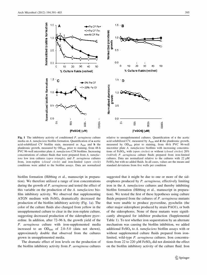
Fig. 1 The inhibitory activity of conditioned P. aeruginosa culture
media on A. tumefaciens biofilm formation. Quantification of a acetic
acid-solubilized CV biofilm stain, measured as A600 and b the
planktonic growth, measured by OD600, prior to staining, from 48-h
PVC 96-well microtiter plate A. tumefaciens C58 biofilms. Increasing
concentrations of culture fluids that were prepared from A. tumefac-iens low iron cultures (open triangle), and P. aeruginosa cultures
from, iron-replete (closed circle) and iron-limited (open circle)
conditions were added to the biofilm assays. Data are normalized
relative to unsupplemented cultures. Quantification of c the acetic
acid-solubilized CV, measured by A600 and d the planktonic growth,
measured by OD600 prior to staining, from 48-h PVC 96-well
microtiter plate A. tumefaciens biofilms with increasing concentra-
tions of FeSO4, with (open circles) or without (closed circles) 20%
(vol/vol) P. aeruginosa culture fluids prepared from iron-limited
cultures. Data are normalized relative to the cultures with 22 lM
FeSO4 but with no added fluids. In all cases, values are the means and
standard deviations from five wells per condition
Arch Microbiol (2012) 194:391–403 395
123
Page 6
concentrations greater than 220 lM inhibited the biofilm
formation and planktonic growth regardless of the presence
of the P. aeruginosa culture fluids (Fig. 1c, d).
Mutations in the global P. aeruginosa iron regulators
fur and pvdS do not abolish iron-responsive control
of its biofilm inhibitory activity
A common global iron-responsive regulator for many
bacteria including P. aeruginosa is Fur, the ferric uptake
repressor (Vasil and Ochsner 1999). The P. aeruginosa fur
homolog is believed to be essential but missense mutations
in this gene have been isolated that exhibit constitutive
pyoverdine production and partially desensitize P. aeru-
ginosa biofilm formation to iron limitation (Barton et al.
1996; Banin et al. 2005). In contrast, the deletion of the
extracytoplasmic function (ECF) r-factor pvdS, involved in
the response to iron limitation, resulted in the loss of
pyoverdine production and formation of structurally aber-
rant biofilms in flow cells under iron-replete conditions
(Banin et al. 2005).
We hypothesized that one or both of these mutations
would disrupt iron-responsive control of biofilm inhibitor
production. Culture fluids were prepared from P. aeru-
ginosa furC6Tc (a missense mutation that decreases fur
activity) and DpvdS strains grown in minimal medium with
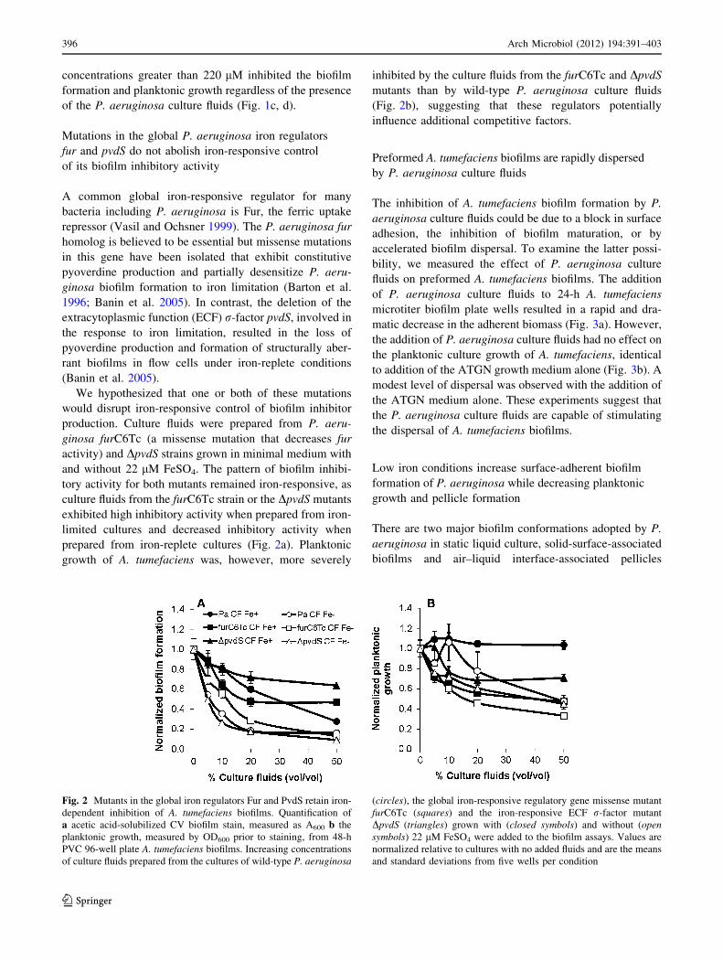
and without 22 lM FeSO4. The pattern of biofilm inhibi-
tory activity for both mutants remained iron-responsive, as
culture fluids from the furC6Tc strain or the DpvdS mutants
exhibited high inhibitory activity when prepared from iron-
limited cultures and decreased inhibitory activity when
prepared from iron-replete cultures (Fig. 2a). Planktonic
growth of A. tumefaciens was, however, more severely
inhibited by the culture fluids from the furC6Tc and DpvdS
mutants than by wild-type P. aeruginosa culture fluids
(Fig. 2b), suggesting that these regulators potentially
influence additional competitive factors.
Preformed A. tumefaciens biofilms are rapidly dispersed
by P. aeruginosa culture fluids
The inhibition of A. tumefaciens biofilm formation by P.
aeruginosa culture fluids could be due to a block in surface
adhesion, the inhibition of biofilm maturation, or by
accelerated biofilm dispersal. To examine the latter possi-
bility, we measured the effect of P. aeruginosa culture
fluids on preformed A. tumefaciens biofilms. The addition
of P. aeruginosa culture fluids to 24-h A. tumefaciens
microtiter biofilm plate wells resulted in a rapid and dra-
matic decrease in the adherent biomass (Fig. 3a). However,
the addition of P. aeruginosa culture fluids had no effect on
the planktonic culture growth of A. tumefaciens, identical
to addition of the ATGN growth medium alone (Fig. 3b). A
modest level of dispersal was observed with the addition of
the ATGN medium alone. These experiments suggest that
the P. aeruginosa culture fluids are capable of stimulating
the dispersal of A. tumefaciens biofilms.
Low iron conditions increase surface-adherent biofilm
formation of P. aeruginosa while decreasing planktonic
growth and pellicle formation
There are two major biofilm conformations adopted by P.
aeruginosa in static liquid culture, solid-surface-associated
biofilms and air–liquid interface-associated pellicles
Fig. 2 Mutants in the global iron regulators Fur and PvdS retain iron-
dependent inhibition of A. tumefaciens biofilms. Quantification of
a acetic acid-solubilized CV biofilm stain, measured as A600 b the
planktonic growth, measured by OD600 prior to staining, from 48-h
PVC 96-well plate A. tumefaciens biofilms. Increasing concentrations
of culture fluids prepared from the cultures of wild-type P. aeruginosa
(circles), the global iron-responsive regulatory gene missense mutant
furC6Tc (squares) and the iron-responsive ECF r-factor mutant
DpvdS (triangles) grown with (closed symbols) and without (opensymbols) 22 lM FeSO4 were added to the biofilm assays. Values are
normalized relative to cultures with no added fluids and are the means
and standard deviations from five wells per condition
396 Arch Microbiol (2012) 194:391–403
123
Page 7
(Friedman and Kolter 2004). In the conditions used in this
study, P. aeruginosa grown in static cultures for 24 h with
22 lM FeSO4 produced thick pellicles with limited sur-
face-attached biofilm and robust planktonic growth. In
contrast, those grown in static culture with no exogenous
iron demonstrated less pellicle formation and planktonic
growth while forming dense surface-associated biofilms
(Fig. 4). After 48 h, pellicle formation in the 22-lM FeSO4
conditions had dissipated and the small amount of surface-
adherent biomass had further decreased. The elevated
amount of surface-adhered biomass in the iron-limited
conditions was still present at 72 h but the pellicle was
completely absent (Fig. 4).
P. aeruginosa static biofilm formation does not respond
to the culture fluids that inhibit A. tumefaciens
In order to test the effect of the inhibitory culture fluids on
P. aeruginosa biofilm formation, static culture coverslip
biofilms were cultivated in the presence or absence of 50%
(vol/vol) culture fluids with or without added 22 lM
FeSO4. P. aeruginosa exhibited increased biofilm forma-
tion and decreased planktonic growth under iron limitation,
but the culture fluids had no apparent impact (Fig. 4). The
addition of 50% (vol/vol) of these same culture fluids
completely abolishes A. tumefaciens biofilm formation
with or without additional FeSO4 (Fig. 1a and data not
shown).
Inhibitor production is not regulated by the
P. aeruginosa quorum-sensing systems
The P. aeruginosa quorum-sensing systems typically play
a critical role in the regulation of its secreted virulence
factors, antimicrobial agents, and antibiofilm compounds.
We therefore evaluated the possibility that the inhibitor
might be regulated by quorum sensing as well. To test
this hypothesis, culture fluids were prepared from five
P. aeruginosa quorum-sensing mutant strains, DlasI, DrhlI,
DlasI DrhlI, DlasR DrhlR, and pqsA::tet. None of these
mutants were found to be deficient for the inhibition of
A. tumefaciens biofilm formation or growth (Fig. 5a, b).
Several recognized bioactive secreted compounds
from P. aeruginosa are not responsible for biofilm
inhibition
P. aeruginosa produces an array of secreted compounds
with antimicrobial or antibiofilm activity, including
hydrogen cyanide, pyocyanin, and rhamnolipids. It seemed
plausible that these compounds might be acting alone or in
combination to inhibit the biofilm formation by A. tum-
efaciens. To test this hypothesis, we prepared culture fluids
from P. aeruginosa individual mutants that were unable to
produce each of these compounds alone, as well as a triple
mutant that could not produce any of these compounds. We
found that inhibitor production was identical to wild type in
all of the mutants tested (Supplemental Table 1).
The P. aeruginosa inhibitory activity is physically
and enzymatically stable
The biofilm inhibitory activity was found to be fully
resistant to boiling and exposure to UV radiation for 1 h.
The inhibitory activity was also completely stable over
nearly a year stored at 4�C (Supplemental Table 2, data not
shown). Exposure of the P. aeruginosa culture fluids to
DNase, RNase, and Pronase enzymes for over 15 h at room
Fig. 3 The addition of P. aeruginosa culture fluids causes the
dispersal of preformed A. tumefaciens biofilms. Static culture 24-h
biofilms of A. tumefaciens. At time 0, ATGN (closed circles) or
P. aeruginosa culture fluids (open circles) were added to the cultures
resulting in the treatment of the preformed biofilms with 50% (vol/
vol) culture fluids or ATGN. At each time point a the wells were
stained with CV, incubated 10 min, the stain was solubilized with
acetic acid, and measured at A600 and b the planktonic growth of the
culture, OD600 prior to staining. Values are normalized relative to the
value at time 0 and are the means and standard deviations from four
wells per time point
Arch Microbiol (2012) 194:391–403 397
123
Page 8
Fig. 4 P. aeruginosa biofilm
formation is not influenced by
its own culture fluids but is
impacted by iron levels. P.aeruginosa static culture
biofilms were grown in 12-well
microtiter dishes with PCV
coverslips sitting vertically in
the wells. After incubating for
24 h, 48 h, and 72 h, the
coverslips were removed and
the adherent biomass was
stained with CV, and the OD600
of the remaining planktonic
culture was measured. The
bacteria at the air–liquid
interface were disrupted and
photographed. The border
between the pellicle suspended
at the air–liquid interface (thelighter regions) and the
planktonic bacteria (the darkerregions) is marked with an
arrow. The images shown are
representative of three
coverslips. The OD600 values
presented are the mean and
(standard deviation) from three
wells
Fig. 5 Quorum-sensing-deficient mutants direct wild-type levels of
biofilm inhibition. a acetic acid-solubilized CV biofilm stain,
measured as A600 and b the planktonic growth, measured by OD600
prior to staining, from 48-h PVC 96-well plate A. tumefaciensbiofilms. Increasing concentrations of culture fluids prepared from
wild-type (closed circles) and the five quorum-sensing-deficient
mutants used in this study, DlasI (closed squares), DrhlI (closedtriangles), pqsA::tet (open circles), DlasI DrhlI (open squares), and
DlasR DrhlR (open triangles), prepared from iron-limited cultures
were added to the biofilm assays. Values are normalized relative to
cultures with no added fluids and are the means and standard
deviations of five wells per condition
398 Arch Microbiol (2012) 194:391–403
123
Page 9
temperature did not significantly diminish the biofilm
inhibitory activity, although these enzymes maintained their
activity over this incubation period (Supplemental Table 2).
The inhibitory activity is predominantly due to a low
molecular weight compound(s) that is distinct
from CDA
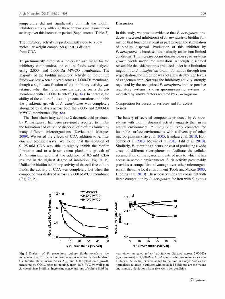
To preliminarily establish a molecular size range for the
inhibitory compound(s), the culture fluids were dialyzed
using 2,000- and 7,000-Da MWCO membranes. The
majority of the biofilm inhibitory activity of the culture
fluids was lost when dialyzed across a 7,000-Da membrane,
though a significant fraction of the inhibitory activity was
retained when the fluids were dialyzed across a dialysis
membrane with a 2,000-Da cutoff (Fig. 6a). In contrast, the
ability of the culture fluids at high concentrations to inhibit
the planktonic growth of A. tumefaciens was completely
abrogated by dialysis across both the 7,000- and 2,000-Da
MWCO membranes (Fig. 6b).
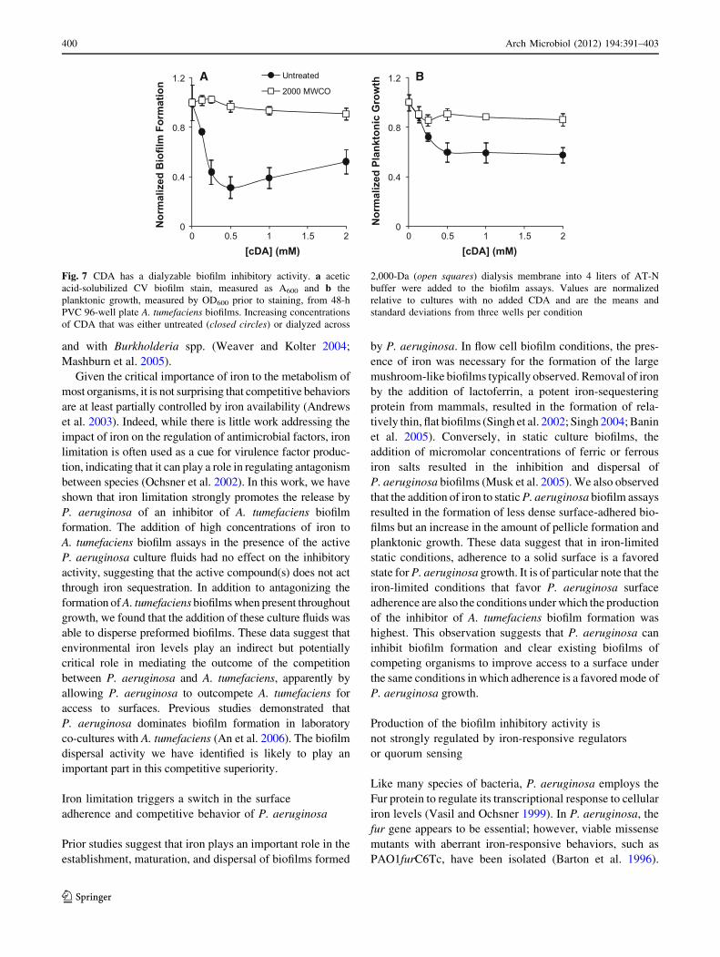
The short-chain fatty acid cis-2-decenoic acid produced
by P. aeruginosa has been previously reported to inhibit
the formation and cause the dispersal of biofilms formed by
many different microorganisms (Davies and Marques
2009). We tested the effects of CDA addition to A. tum-
efaciens biofilm assays. We found that the addition of
0.125 mM CDA was able to slightly inhibit the biofilm
formation and to a lesser extent planktonic growth of
A. tumefaciens and that the addition of 0.5 mM CDA
resulted in the highest degree of inhibition (Fig. 7a, b).
Unlike the biofilm inhibitory activity of the cell-free culture
fluids, the activity of CDA was completely lost when this
compound was dialyzed across a 2,000 MWCO membrane
(Fig. 7a, b).
Discussion
In this study, we provide evidence that P. aeruginosa pro-
duces a secreted inhibitor(s) of A. tumefaciens biofilm for-
mation that functions at least in part through the stimulation
of biofilm dispersal. Production of this inhibitor by
P. aeruginosa is increased dramatically under iron-limited
conditions. This increase occurs despite lower P. aeruginosa
growth yields under iron limitation. Although it seemed
reasonable that siderophores produced under iron limitation
might inhibit A. tumefaciens biofilm formation through iron
sequestration, the inhibition was not alleviated by high levels
of exogenous iron. Nor was the inhibitory activity strongly
regulated by the recognized P. aeruginosa iron-responsive
regulatory systems, known quorum-sensing systems, or
mediated by known factors secreted by P. aeruginosa.
Competition for access to surfaces and for access
to iron
The battery of secreted compounds produced by P. aeru-
ginosa with biofilm dispersal activity suggests that, in its
natural environment, P. aeruginosa likely competes for
favorable surface environments with a diversity of other
microorganisms (Irie et al. 2005; Bandara et al. 2010; Hol-
combe et al. 2010; Mowat et al. 2010; Pihl et al. 2010).
Similarly, P. aeruginosa incurs the cost of producing a wide
array of different siderophores to facilitate the cellular
accumulation of the scarce amounts of iron to which it has
access in aerobic environments. Such activity presumably
provides a competitive advantage over other microorgan-
isms in the same local environment (Poole and McKay 2003;
Hibbing et al. 2010). These observations are consistent with
fierce competition by P. aeruginosa for iron with S. aureus
Fig. 6 Dialysis of P. aeruginosa culture fluids reveals a low
molecular size for the active compound(s) a acetic acid-solubilized
CV biofilm stain, measured as A600 and b the planktonic growth,
measured by OD600 prior to staining, from 48-h PVC 96-well plate
A. tumefaciens biofilms. Increasing concentrations of culture fluid that
was either untreated (closed circles) or dialyzed across 2,000-Da
(open squares) or 7,000-Da (closed squares) dialysis membranes into
4 liters of AT-N buffer were added to the biofilm assays. Values are
normalized relative to cultures with no added fluids and are the means
and standard deviations from five wells per condition
Arch Microbiol (2012) 194:391–403 399
123
Page 10
and with Burkholderia spp. (Weaver and Kolter 2004;
Mashburn et al. 2005).
Given the critical importance of iron to the metabolism of
most organisms, it is not surprising that competitive behaviors
are at least partially controlled by iron availability (Andrews
et al. 2003). Indeed, while there is little work addressing the
impact of iron on the regulation of antimicrobial factors, iron
limitation is often used as a cue for virulence factor produc-
tion, indicating that it can play a role in regulating antagonism
between species (Ochsner et al. 2002). In this work, we have
shown that iron limitation strongly promotes the release by
P. aeruginosa of an inhibitor of A. tumefaciens biofilm
formation. The addition of high concentrations of iron to
A. tumefaciens biofilm assays in the presence of the active
P. aeruginosa culture fluids had no effect on the inhibitory
activity, suggesting that the active compound(s) does not act
through iron sequestration. In addition to antagonizing the
formation of A. tumefaciens biofilms when present throughout
growth, we found that the addition of these culture fluids was
able to disperse preformed biofilms. These data suggest that
environmental iron levels play an indirect but potentially
critical role in mediating the outcome of the competition
between P. aeruginosa and A. tumefaciens, apparently by
allowing P. aeruginosa to outcompete A. tumefaciens for
access to surfaces. Previous studies demonstrated that
P. aeruginosa dominates biofilm formation in laboratory
co-cultures with A. tumefaciens (An et al. 2006). The biofilm
dispersal activity we have identified is likely to play an
important part in this competitive superiority.
Iron limitation triggers a switch in the surface
adherence and competitive behavior of P. aeruginosa
Prior studies suggest that iron plays an important role in the
establishment, maturation, and dispersal of biofilms formed
by P. aeruginosa. In flow cell biofilm conditions, the pres-
ence of iron was necessary for the formation of the large
mushroom-like biofilms typically observed. Removal of iron
by the addition of lactoferrin, a potent iron-sequestering
protein from mammals, resulted in the formation of rela-
tively thin, flat biofilms (Singh et al. 2002; Singh 2004; Banin
et al. 2005). Conversely, in static culture biofilms, the
addition of micromolar concentrations of ferric or ferrous
iron salts resulted in the inhibition and dispersal of
P. aeruginosa biofilms (Musk et al. 2005). We also observed
that the addition of iron to static P. aeruginosa biofilm assays
resulted in the formation of less dense surface-adhered bio-
films but an increase in the amount of pellicle formation and
planktonic growth. These data suggest that in iron-limited
static conditions, adherence to a solid surface is a favored
state for P. aeruginosa growth. It is of particular note that the
iron-limited conditions that favor P. aeruginosa surface
adherence are also the conditions under which the production
of the inhibitor of A. tumefaciens biofilm formation was
highest. This observation suggests that P. aeruginosa can
inhibit biofilm formation and clear existing biofilms of
competing organisms to improve access to a surface under
the same conditions in which adherence is a favored mode of
P. aeruginosa growth.
Production of the biofilm inhibitory activity is
not strongly regulated by iron-responsive regulators
or quorum sensing
Like many species of bacteria, P. aeruginosa employs the
Fur protein to regulate its transcriptional response to cellular
iron levels (Vasil and Ochsner 1999). In P. aeruginosa, the
fur gene appears to be essential; however, viable missense
mutants with aberrant iron-responsive behaviors, such as
PAO1furC6Tc, have been isolated (Barton et al. 1996).
0
0.4
0.8
1.2
0 0.5 1 1.5 2
Nor
mal
ized
Bio
film
For
mat
ion
[cDA] (mM)
Untreated
2000 MWCO
0
0.4
0.8
1.2
0 0.5 1 1.5 2
Nor
mal
ized
Pla
nkto
nic
Gro
wth
[cDA] (mM)
A B
Fig. 7 CDA has a dialyzable biofilm inhibitory activity. a acetic
acid-solubilized CV biofilm stain, measured as A600 and b the
planktonic growth, measured by OD600 prior to staining, from 48-h
PVC 96-well plate A. tumefaciens biofilms. Increasing concentrations
of CDA that was either untreated (closed circles) or dialyzed across
2,000-Da (open squares) dialysis membrane into 4 liters of AT-N
buffer were added to the biofilm assays. Values are normalized
relative to cultures with no added CDA and are the means and
standard deviations from three wells per condition
400 Arch Microbiol (2012) 194:391–403
123
Page 11
P. aeruginosa also uses specific iron-responsive ECF r-factors
to control iron uptake, including pvdS that is responsible for
controlling the production of the siderophore pyoverdine
(Leoni et al. 2000). Surprisingly, deletion of pvdS and the
furC6Tc missense mutation, both of which result in iron-
insensitive flowcell biofilm phenotypes, had no appreciable
effects on iron-stimulated production of the A. tumefaciens
biofilm inhibitor(s). These data show that the iron-dependent
regulation of inhibitor production is not controlled by the
major iron-responsive regulators of P. aeruginosa.
P. aeruginosa uses a complex quorum-sensing-dependent
regulatory system to mediate a stationary-phase dominance
phenotype over A. tumefaciens in co-culture and to control
the expression of diverse biologically active compounds (An
et al. 2006; D. An and M. Parsek unpublished data). These
compounds can function alone or in combination to affect the
physiology, behavior, and viability of competing organisms
(D. An and M. Parsek unpublished data). We found that
neither the quorum-sensing-controlled effectors nor the
quorum-sensing systems themselves had any effect on the
production of the inhibitory compound. This indicates that
the previously observed ability of P. aeruginosa to out
compete A. tumefaciens in laboratory co-cultures depends
on a variety of diversely regulated P. aeruginosa factors
including the dispersal promoting and biofilm inhibiting
factor we report here.
The biofilm inhibitory factor is small, stable,
and unlikely to be conferred by extracellular DNA,
RNA, or proteins
To better understand the mechanism of biofilm inhibition
and dispersal, and because of the potential importance and
wide applicability of biofilm inhibitors, we attempted to
examine the physical characteristics of the inhibitor. We
showed that the inhibitory activity was stable over the
course of extended storage, resistant to heat and UV irra-
diation, and not degraded by nucleic acid- or protein-
degrading enzymes, suggesting that the inhibitory activity
was the result of a bacterially produced small molecule. In
addition, the complete loss of activity when the culture
fluids were dialyzed across a 7,000 MWCO membrane and
partial loss of activity across a 2,000 MWCO membrane
suggest that the biofilm inhibitory activity may be the result
of multiple small molecules, with molecular masses
both greater and less than 2,000 Da. However, none of the
small molecules that have been shown to be produced by
P. aeruginosa and to influence iron acquisition, biofilm
formation or multispecies interactions appear to play a role
in the observed inhibitory activity, with the exception of
CDA (Boles et al. 2005; Irie et al. 2005; Glick et al. 2010;
Davies and Marques 2009; D. An and M. Parsek unpub-
lished data).
A novel agent capable of inhibiting and dispersing
A. tumefaciens biofilms
A biofilm-dispersing activity from P. aeruginosa, now
identified to be CDA, was initially observed to trigger the
auto-dispersal of biofilms formed by P. aeruginosa as well
as a wide range of microorganisms including the yeast
Candida albicans (Davies and Marques 2009). Thus, we
were interested to determine whether the A. tumefaciens
biofilm-dispersing activity in iron-limited P. aeruginosa
culture fluids would be able to disperse P. aeruginosa
biofilms. We observed that culturing static P. aeruginosa
biofilms in the presence or absence of 50% (vol/vol) cul-
ture fluids from iron-limited cultures had no observable
effect on the accumulation of surface-adherent biomass.
The only condition we examined that altered P. aeruginosa
biofilm formation was the presence of added iron, which
greatly reduced the accumulation of surface-adherent bio-
mass. In contrast, the biofilm formation of A. tumefaciens
was essentially eliminated in the presence of 50% (vol/vol)
culture fluids and severely compromised at much lower
amounts. A concentration of as little as 1 nM synthetic
CDA was shown to be sufficient to induce the dispersal of
P. aeruginosa biofilms, and the concentration of CDA in
spent medium was measured at 2.5 nM (Davies and Mar-
ques 2009). Our results, showing that over 100 lM CDA
is required for the inhibition of biofilm formation by
A. tumefaciens and that the inhibitory activity of CDA was
completely eliminated by dialysis, suggest that CDA is not
the primary inhibitory compound in the cell-free culture
fluids. In addition, there is no indication that CDA syn-
thesis is stimulated under iron-limited conditions (D.
Davies, personal communication). In fact, CDA is typically
prepared from P. aeruginosa cultures grown in iron-replete
media, whereas the primary activity that inhibits and dis-
perses A. tumefaciens biofilms is strikingly elevated in
iron-limited P. aeruginosa cultures (Davies and Marques
2009). Collectively, the lack of activity of the culture fluids
against P. aeruginosa biofilms and the dialysis profile of
the inhibitory activity suggest that we have identified a
novel compound(s) produced by P. aeruginosa. The pro-
duction of this compound is induced under iron-limiting
conditions, but the compound is distinct from siderophores
and is capable of efficiently inhibiting and disrupting
A. tumefaciens biofilms.
Conclusions
Bacteria in the environment must compete with one
another for scarce resources such as nutrients and access to
surface environments. We have found that batch cultures of
P. aeruginosa grown in a defined medium produce a
Arch Microbiol (2012) 194:391–403 401
123
Page 12
secreted factor(s) that inhibits A. tumefaciens biofilms,
production of which is stimulated by iron limitation. These
same conditions also foster the formation of surface-
adhered biofilms of P. aeruginosa. The biofilm inhibitory
activity is clearly not the result of rhamnolipid production
and appears to be distinct from CDA, thus likely repre-
senting a novel biofilm inhibitory activity. Future work will
be directed toward identifying the chemical nature of the
agent(s), its mode of action, and the regulatory basis for the
iron-responsive production of the inhibitor.
Acknowledgments We wish to acknowledge Andrew Philips and
Ying Cao for valuable input on this project. Matthew Parsek was
particularly helpful in providing strains of P. aeruginosa. Thomas
Platt provided useful suggestions on the manuscript. M.E.H. was
funded on the Indiana University Genetics, Molecular and Cellular
Sciences Training Grant T32-GM007757. This study was supported
by National Institutes of Health grant RO1-GM080546 (C.F.) and
through a grant from the Indiana University META-Cyt program
funded in part by a major endowment from the Lilly Foundation
(C.F.).
References
An DD, Danhorn T, Fuqua C, Parsek MR (2006) Quorum sensing and
motility mediate interactions between Pseudomonas aeruginosaand Agrobacterium tumefaciens in biofilm cocultures. Proc Natl
Acad Sci USA 103:3828–3833
Andrews SC, Robinson AK, Rodriguez-Quinones F (2003) Bacterial
iron homeostasis. FEMS Microbiol Rev 27:215–237
Bandara H, Yau JYY, Watt RM, Jin LJ, Samaranayake LP (2010)
Pseudomonas aeruginosa inhibits in vitro Candida biofilm
development. BMC Microbiol 10:125–133
Banin E, Vasil ML, Greenberg EP (2005) Iron and Pseudomonasaeruginosa biofilm formation. Proc Natl Acad Sci USA
102:11076–11081
Barton HA, Johnson Z, Cox CD, Vasil AI, Vasil ML (1996) Ferric
uptake regulator mutants of Pseudomonas aeruginosa with
distinct alterations in the iron-dependent repression of exotoxin
A and siderophores in aerobic and microaerobic environments.
Mol Microbiol 21:1001–1017
Berne C, Kysela DT, Brun YV (2010) A bacterial extracellular DNA
inhibits settling of motile progeny cells within a biofilm. Mol
Microbiol 77:815–829
Boles BR, Thoendel M, Singh PK (2005) Rhamnolipids mediate
detachment of Pseudomonas aeruginosa from biofilms. Mol
Microbiol 57:1210–1223
Buckling A et al (2007) Siderophore-mediated cooperation and
virulence in Pseudomonas aeruginosa. FEMS Microbiol Ecol
62:135–141
Carson KC, Meyer JM, Dilworth MJ (2000) Hydroxamate sidero-
phores of root nodule bacteria. Soil Biol Biochem 32:11–21
Chipperfield JR, Ratledge C (2000) Salicylic acid is not a bacterial
siderophore: a theoretical study. Biometals 13:165–168
Coetser SE, Cloete TE (2005) Biofouling and biocorrosion in
industrial water systems. Crit Rev Microbiol 31:213–232
Costerton JW, Stewart PS, Greenberg EP (1999) Bacterial biofilms: a
common cause of persistent infections. Science 284:1318–1322
Danhorn T, Hentzer M, Givskov M, Parsek MR, Fuqua C (2004)
Phosphorus limitation enhances biofilm formation of the plant
pathogen Agrobacterium tumefaciens through the PhoR-PhoB
regulatory system. J Bacteriol 186:4492–4501
Davies DG, Marques CNH (2009) A fatty acid messenger is
responsible for inducing dispersion in microbial biofilms.
J Bacteriol 191:1393–1403
Escobar MA, Dandekar AM (2003) Agrobacterium tumefaciens as an
agent of disease. Trends Plant Sci 8:380–386
Friedman L, Kolter R (2004) Genes involved in matrix formation in
Pseudomonas aeruginosa PA14 biofilms. Mol Microbiol
51:675–690
Glick R et al (2010) Increase in rhamnolipid synthesis under iron-
limiting conditions influences surface motility and biofilm
formation in Pseudomonas aeruginosa. J Bacteriol
192:2973–2980
Hibbing ME, Fuqua C, Parsek MR, Peterson SB (2010) Bacterial
competition: surviving and thriving in the microbial jungle. Nat
Rev Microbiol 8:15–25
Holcombe LJ et al (2010) Pseudomonas aeruginosa secreted factors
impair biofilm development in Candida albicans. Soc Gen
Microbiol 156:1476–1486
Horie M, Ishiyama A, Fujihira-Ueki Y, Sillanpaa J, Korhonen TK,
Toba T (2002) Inhibition of the adherence of Escherichia colistrains to basement membrane by Lactobacillus crispatus
expressing an S-layer. J Appl Microbiol 92:396–403
Horswill AR, Stoodley P, Stewart PS, Parsek MR (2007) The effect of
the chemical, biological, and physical environment on quorum
sensing in structured microbial communities. Anal Bioanal
Chem 387:371–380
Irie Y, O’Toole GA, Yuk MH (2005) Pseudomonas aeruginosarhamnolipids disperse Bordetella bronchiseptica biofilms. FEMS
Microbiol Lett 250:237–243
Jacobs MA et al (2003) Comprehensive transposon mutant library of
Pseudomonas aeruginosa. Proc Natl Acad Sci 100:14339–14344
Joshi F, Archana G, Desai A (2006) Siderophore cross-utilization
amongst rhizospheric bacteria and the role of their differential
affinities for Fe3? on growth stimulation under iron-limited
conditions. Curr Microbiol 53:141–147
Kaneko Y, Thoendel M, Olakanmi O, Britigan BE, Singh PK (2007)
The transition metal gallium disrupts Pseudomonas aeruginosairon metabolism and has antimicrobial and antibiofilm activity.
J Clin Invest 117:877–888
Kolodkin-Gal I, Romero D, Cao S, Clardy J, Kolter R, Losick R
(2010) d-Amino acids trigger biofilm disassembly. Science
328:627–629
Krewulak KD, Vogel HJ (2008) Structural biology of bacterial iron
uptake. Biochimica Et Biophysica Acta-Biomembranes
1778:1781–1804
Leoni L, Orsi N, de Lorenzo V, Visca P (2000) Functional analysis of
PvdS, an iron starvation sigma factor of Pseudomonas aerugin-osa. J Bacteriol 182:1481–1491
Lim B, Beyhan S, Meir J, Yildiz FH (2006) Cyclic-diGMP signal
transduction systems in Vibrio cholerae: modulation of rugosity
and biofilm formation. Mol Microbiol 60:331–348
Maeda S et al (2006) Horizontal transfer of nonconjugative plasmids
in a colony biofilm of Escherichia coli. FEMS Microbiol Lett
255:115–120
Martı́nez-Gil M, Yousef-Coronado F, Espinosa-Urgel M (2010)
LapF, the second largest Pseudomonas putida protein, contrib-
utes to plant root colonization and determines biofilm architec-
ture. Mol Microbiol 77:549–561
Mashburn LM, Jett AM, Akins DR, Whiteley M (2005) Staphylo-coccus aureus serves as an iron source for Pseudomonasaeruginosa during in vivo coculture. J Bacteriol 187:554–566
Merritt PA, Danhorn T, Fuqua C (2007) Motility and chemotaxis in
Agrobacterium tumefaciens surface attachment and Biofilm
formation. J Bacteriol 189:8005–8014
402 Arch Microbiol (2012) 194:391–403
123
Page 13
Mowat E et al (2010) Pseudomonas aeruginosa and their small
diffusible extracellular molecules inhibit Aspergillus fumigatusbiofilm formation. FEMS Microbiol Lett 313:96–102
Musk DJ, Banko DA, Hergenrother PJ (2005) Iron salts perturb
biofilm formation and disrupt existing biofilms of Pseudomonasaeruginosa. Chem Biol 12:789–796
Ochsner UA, Vasil ML (1996) Gene repression by the ferric uptake
regulator in Pseudomonas aeruginosa: Cycle selection of iron-
regulated genes. Proc Natl Acad Sci USA 93:4409–4414
Ochsner UA, Vasil AI, Johnson Z, Vasil ML (1999) Pseudomonasaeruginosa fur overlaps with a gene encoding a novel outer
membrane lipoprotein. OmlA J Bacteriol 181:1099–1109
Ochsner UA, Wilderman PJ, Vasil AI, Vasil ML (2002) Gene-
Chip((R)) expression analysis of the iron starvation response in
Pseudomonas aeruginosa: identification of novel pyoverdine
biosynthesis genes. Mol Microbiol 45:1277–1287
O’Toole GA, Kolter R (1998) Initiation of biofilm formation in
Pseudomonas fluorescens WCS365 proceeds via multiple,
convergent signalling pathways: a genetic analysis. Mol Micro-
biol 28:449–461
Pesci E, Pearson J, Seed P, Iglewski B (1997) Regulation of las and
rhl quorum sensing in Pseudomonas aeruginosa. J Bacteriol
179:3127–3132
Pihl M, Davies JR, de Paz LEC, Svensater G (2010) Differential
effects of Pseudomonas aeruginosa on biofilm formation by
different strains of Staphylococcus epidermidis. FEMS Immunol
Med Microbiol 59:439–446
Platt TG, Bever JD (2009) Kin competition and the evolution of
cooperation. Trends Ecol Evol 24:370–377
Poole K, McKay GA (2003) Iron acquisition and its control in
Pseudomonas aeruginosa: many roads lead to Rome. Frontiers
Biosci 8:D661–D686
Ramey BE, Matthysse AG, Fuqua C (2004) The FNR-type transcrip-
tional regulator SinR controls maturation of Agrobacteriumtumefaciens biofilms. Mol Microbiol 52:1495–1511
Rao D, Webb JS, Kjelleberg S (2006) Microbial colonization and
competition on the marine alga Ulva australis. Appl Environ
Microbiol 72:5547–5555
Rather PN (2005) Swarmer cell differentiation in Proteus mirabilis.
Environ Microbiol 7:1065–1073
Rodionov DA, Gelfand MS, Todd JD, Curson ARJ, Johnston AWB
(2006) Computational reconstruction of iron- and manganese-
responsive transcriptional networks in a-proteobacteria. PLoS
Comp Biol 2:1568–1585
Schuster M, Lostroh CP, Ogi T, Greenberg EP (2003) Identification,
timing, and signal specificity of Pseudomonas aeruginosaquorum-controlled genes: a transcriptome analysis. J Bacteriol
185:2066–2079
Shrout JD, Chopp DL, Just CL, Hentzer M, Givskov M, Parsek MR
(2006) The impact of quorum sensing and swarming motility on
Pseudomonas aeruginosa biofilm formation is nutritionally
conditional. Mol Microbiol 62:1264–1277
Singh PK (2004) Iron sequestration by human lactoferrin stimulates
P. aeruginosa surface motility and blocks biofilm formation.
Biometals 17:267–270
Singh PK, Parsek MR, Greenberg EP, Welsh MJ (2002) A component
of innate immunity prevents bacterial biofilm development.
Nature 417:552–555
Sutherland IW (2001) Biofilm exopolysaccharides: a strong and
sticky framework. Microbiology 147:3–9
Tempe J, Petit A, Holsters M, Montagu MV, Schell J (1977)
Thermosensitive step associated with transfer of Ti plasmid
during conjugation—possible relation to transformation in crown
gall. Proc Natl Acad Sci USA 74:2848–2849
Vasil ML, Ochsner UA (1999) The response of Pseudomonasaeruginosa to iron: genetics, biochemistry and virulence. Mol
Microbiol 34:399–413
Verran J, Airey P, Packer A, Whitehead KA (2008) Chapter 8
microbial retention on open food contact surfaces and implica-
tions for food contamination. In: Allen I, Laskin SS, Geoffrey
MG (eds) Advances in applied microbiology. Academic Press,
Waltham, pp 223–246
Watson B, Currier TC, Gordon MP, Chilton MD, Nester EW (1975)
Plasmid required for virulence of Agrobacterium tumefaciens.
J Bacteriol 123:255–264
Weaver VB, Kolter R (2004) Burkholderia spp. alter Pseudomonasaeruginosa physiology through iron sequestration. J Bacteriol
186:2376–2384
Weinberg ED (2009) Iron availability and infection. Biochimica Et
Biophysica Acta-General Subjects 1790:600–605
Whitchurch CB, Tolker-Nielsen T, Ragas PC, Mattick JS (2002)
Extracellular DNA required for bacterial biofilm formation.
Science 295:1487–1487
Arch Microbiol (2012) 194:391–403 403
123