The Journal of Clinical Investigation | November 2003 | Volume 112 | Number 9 1395 Introduction Accumulating evidence suggests that oxidative stress plays an important role in mediating pathologic responses in cardiac myocytes and various diseases in the heart (reviewed in refs. 1, 2). Production of reactive oxy- gen species by mitochondrial sources as well as non- phagocytic oxidases is enhanced in various pathologic stimuli in the heart (1, 2). To counteract increased levels of oxidative stress, cells have antioxidant mechanisms that include superoxide dismutases (SODs) — which are present in mitochondria, such as manganese SOD (MnSOD), cytosol, such as copper zinc SOD (CuZn- SOD), and plasma membrane and extracellular spaces (such as extracellular SOD) — catalase, glutathione per- oxidases, peroxiredoxins, and thioredoxin (Trx) (1, 2). Trx1 is a 12-kDa multifunctional protein with a redox- active disulfide/dithiol within the conserved active site 32 Cys-Gly-Pro- 35 Cys (reviewed in refs. 3, 4). Trx1, Trx reductase, and NADPH, collectively called the Trx system, operate as a powerful protein–disulfide oxidoreductase system (3, 4). Trx1 seems unique among antioxidants in that it interacts directly with various intracellular-signal- ing molecules as well as transcription factors, thereby affecting cell growth and cell survival in some cell types (3, 4). Recent evidence suggests that either protein expres- sion or the activity of Trx1 is modified in some cardio- vascular diseases (5–8). Surprisingly, however, the impor- tance of endogenous Trx1 as an antioxidant and the in vivo role of Trx1 in cell growth responses are not clearly under- stood in any organs, including the heart. We hypothesized that endogenous Trx1 plays an important role in regulating the tissue level of oxida- tive stress, thereby controlling cardiac myocyte growth responses. Although mice systemically deficient in Trx1 have been generated by homologous recombination, Inhibition of endogenous thioredoxin in the heart increases oxidative stress and cardiac hypertrophy Mitsutaka Yamamoto, 1 Guiping Yang, 1 Chull Hong, 1 Jing Liu, 1 Eric Holle, 2 Xianzhong Yu, 2 Thomas Wagner, 2 Stephen F. Vatner, 1 and Junichi Sadoshima 1 1 Cardiovascular Research Institute, Department of Cell Biology and Molecular Medicine and Department of Medicine, University of Medicine and Dentistry of New Jersey, New Jersey Medical School, Newark, New Jersey, USA 2 Oncology Research Institute, Greenville, South Carolina, USA Thioredoxin 1 (Trx1) has redox-sensitive cysteine residues and acts as an antioxidant in cells. However, the extent of Trx1 contribution to overall antioxidant mechanisms is unknown in any organs. We gen- erated transgenic mice with cardiac-specific overexpression of a dominant negative (DN) mutant (C32S/C35S) of Trx1 (Tg-DN-Trx1 mice), in which the activity of endogenous Trx was diminished. Mark- ers of oxidative stress were significantly increased in hearts from Tg-DN-Trx1 mice compared with those from nontransgenic (NTg) mice. Tg-DN-Trx1 mice exhibited cardiac hypertrophy with maintained car- diac function at baseline. Intraperitoneal injection of N-2-mercaptopropionyl glycine, an antioxidant, normalized cardiac hypertrophy in Tg-DN-Trx1 mice. Thoracic aortic banding caused greater increases in myocardial oxidative stress and enhanced hypertrophy in Tg-DN-Trx1 compared with NTg mice. In contrast, transgenic mice with cardiac-specific overexpression of wild-type Trx1 did not show cardiac hypertrophy at baseline but exhibited reduced levels of hypertrophy and oxidative stress in response to pressure overload. These results demonstrate that endogenous Trx1 is an essential component of the cel- lular antioxidant mechanisms and plays a critical role in regulating oxidative stress in the heart in vivo. Furthermore, inhibition of endogenous Trx1 in the heart primarily stimulates hypertrophy, both under basal conditions and in response to pressure overload through redox-sensitive mechanisms. J. Clin. Invest. 112:1395–1406 (2003). doi:10.1172/JCI200317700. Received for publication December 30, 2002, and accepted in revised form September 9, 2003. Address correspondence to: Junichi Sadoshima, Cardiovascular Research Institute, University of Medicine and Dentistry of New Jersey, New Jersey Medical School, 185 South Orange Avenue, Medical Science Building G-609, Newark, New Jersey 07103, USA. Phone: (973) 972-8619; Fax: (973) 972-8919; E-mail: [email protected]. A preliminary form of this work was presented at the Late- Breaking Science Session of the American Heart Association Scientific Sessions 2002 in Chicago, Illinois, USA, on November 17, 2002. Conflict of interest: The authors have declared that no conflict of interest exists. Nonstandard abbreviations used: superoxide dismutase (SOD); manganese SOD (MnSOD); copper zinc SOD (CuZnSOD); thioredoxin (Trx); human Trx1 (hTrx1); dominant negative hTrx1 (DN-hTrx1); DN-hTrx1 transgenic (Tg-DN-Trx1); wild- type hTrx1 transgenic (Tg-Trx1); malondialdehyde (MDA); 4-hydroxyalkenals (4-HAE); reduced glutathione (GSH); oxidized glutathione (GSSG); atrial natriuretic factor (ANF); left ventricle/left ventricular (LV); LV end-diastolic diameter (LVEDD); LV end-systolic diameter (LVESD); LV ejection fraction (LVEF); percent fractional shortening (%FS); 8-hydroxy-2′- deoxyguanosine (8-OHdG); nontransgenic (NTg); N-2-mercaptopropionyl glycine (MPG); LV weight/body weight ratio (LVW/BW); LV weight/tibial length ratio (LVW/TL); extracellular signal–regulated kinase (ERK); vitamin D3–upregulated protein-1 (VDUP1).
Transcript
The Journal of Clinical Investigation | November 2003 | Volume 112 | Number 9 1395
IntroductionAccumulating evidence suggests that oxidative stressplays an important role in mediating pathologicresponses in cardiac myocytes and various diseases in theheart (reviewed in refs. 1, 2). Production of reactive oxy-
gen species by mitochondrial sources as well as non-phagocytic oxidases is enhanced in various pathologicstimuli in the heart (1, 2). To counteract increased levelsof oxidative stress, cells have antioxidant mechanismsthat include superoxide dismutases (SODs) — which arepresent in mitochondria, such as manganese SOD(MnSOD), cytosol, such as copper zinc SOD (CuZn-SOD), and plasma membrane and extracellular spaces(such as extracellular SOD) — catalase, glutathione per-oxidases, peroxiredoxins, and thioredoxin (Trx) (1, 2).
Trx1 is a 12-kDa multifunctional protein with a redox-active disulfide/dithiol within the conserved active site32Cys-Gly-Pro-35Cys (reviewed in refs. 3, 4). Trx1, Trxreductase, and NADPH, collectively called the Trx system,operate as a powerful protein–disulfide oxidoreductasesystem (3, 4). Trx1 seems unique among antioxidants inthat it interacts directly with various intracellular-signal-ing molecules as well as transcription factors, therebyaffecting cell growth and cell survival in some cell types(3, 4). Recent evidence suggests that either protein expres-sion or the activity of Trx1 is modified in some cardio-vascular diseases (5–8). Surprisingly, however, the impor-tance of endogenous Trx1 as an antioxidant and the in vivorole of Trx1 in cell growth responses are not clearly under-stood in any organs, including the heart.
We hypothesized that endogenous Trx1 plays animportant role in regulating the tissue level of oxida-tive stress, thereby controlling cardiac myocyte growthresponses. Although mice systemically deficient in Trx1have been generated by homologous recombination,
Inhibition of endogenous thioredoxin in the heart increases oxidative stress and cardiac hypertrophy
Mitsutaka Yamamoto,1 Guiping Yang,1 Chull Hong,1 Jing Liu,1 Eric Holle,2
Xianzhong Yu,2 Thomas Wagner,2 Stephen F. Vatner,1 and Junichi Sadoshima1
1Cardiovascular Research Institute, Department of Cell Biology and Molecular Medicine and Department of Medicine,University of Medicine and Dentistry of New Jersey, New Jersey Medical School, Newark, New Jersey, USA
2Oncology Research Institute, Greenville, South Carolina, USA
Thioredoxin 1 (Trx1) has redox-sensitive cysteine residues and acts as an antioxidant in cells. However,the extent of Trx1 contribution to overall antioxidant mechanisms is unknown in any organs. We gen-erated transgenic mice with cardiac-specific overexpression of a dominant negative (DN) mutant(C32S/C35S) of Trx1 (Tg-DN-Trx1 mice), in which the activity of endogenous Trx was diminished. Mark-ers of oxidative stress were significantly increased in hearts from Tg-DN-Trx1 mice compared with thosefrom nontransgenic (NTg) mice. Tg-DN-Trx1 mice exhibited cardiac hypertrophy with maintained car-diac function at baseline. Intraperitoneal injection of N-2-mercaptopropionyl glycine, an antioxidant,normalized cardiac hypertrophy in Tg-DN-Trx1 mice. Thoracic aortic banding caused greater increasesin myocardial oxidative stress and enhanced hypertrophy in Tg-DN-Trx1 compared with NTg mice. Incontrast, transgenic mice with cardiac-specific overexpression of wild-type Trx1 did not show cardiachypertrophy at baseline but exhibited reduced levels of hypertrophy and oxidative stress in response topressure overload. These results demonstrate that endogenous Trx1 is an essential component of the cel-lular antioxidant mechanisms and plays a critical role in regulating oxidative stress in the heart in vivo.Furthermore, inhibition of endogenous Trx1 in the heart primarily stimulates hypertrophy, both underbasal conditions and in response to pressure overload through redox-sensitive mechanisms.
J. Clin. Invest. 112:1395–1406 (2003). doi:10.1172/JCI200317700.
Received for publication December 30, 2002, and accepted in revised formSeptember 9, 2003.
Address correspondence to: Junichi Sadoshima, CardiovascularResearch Institute, University of Medicine and Dentistry of NewJersey, New Jersey Medical School, 185 South Orange Avenue,Medical Science Building G-609, Newark, New Jersey 07103, USA.Phone: (973) 972-8619; Fax: (973) 972-8919; E-mail: [email protected] preliminary form of this work was presented at the Late-Breaking Science Session of the American Heart AssociationScientific Sessions 2002 in Chicago, Illinois, USA, on November17, 2002.Conflict of interest: The authors have declared that no conflict ofinterest exists.Nonstandard abbreviations used: superoxide dismutase (SOD);manganese SOD (MnSOD); copper zinc SOD (CuZnSOD);thioredoxin (Trx); human Trx1 (hTrx1); dominant negativehTrx1 (DN-hTrx1); DN-hTrx1 transgenic (Tg-DN-Trx1); wild-type hTrx1 transgenic (Tg-Trx1); malondialdehyde (MDA); 4-hydroxyalkenals (4-HAE); reduced glutathione (GSH); oxidizedglutathione (GSSG); atrial natriuretic factor (ANF); leftventricle/left ventricular (LV); LV end-diastolic diameter(LVEDD); LV end-systolic diameter (LVESD); LV ejection fraction(LVEF); percent fractional shortening (%FS); 8-hydroxy-2′-deoxyguanosine (8-OHdG); nontransgenic (NTg); N-2-mercaptopropionyl glycine (MPG); LV weight/body weightratio (LVW/BW); LV weight/tibial length ratio (LVW/TL);extracellular signal–regulated kinase (ERK); vitaminD3–upregulated protein-1 (VDUP1).
1396 The Journal of Clinical Investigation | November 2003 | Volume 112 | Number 9
they are embryonic lethal (9). In order to examine therole of endogenous Trx1 in the heart in vivo, we gener-ated transgenic mice with cardiac-specific overexpres-sion of dominant negative human Trx1 (DN-hTrx1), inwhich the disulfide oxidoreductase activity of Trx isselectively diminished among cellular antioxidantmechanisms. We examined whether inhibition ofendogenous Trx1 increases tissue levels of oxidativestress and whether it affects any cardiac phenotype,including cardiac hypertrophy, under basal conditionsas well as in response to pressure overload.
MethodsTransgenic mice. DN-hTrx1 was generated by mutationof 32Cys and 35Cys of hTrx1 to Ser using QuikChange(Stratagene, La Jolla, California, USA). This redox-inactive mutant of Trx1 has been shown to work as adominant negative for endogenous Trx1 in a breastcancer cell line (10). DN-hTrx1 transgenic mice (here-after designated as Tg-DN-Trx1) as well as wild-typehTrx1 mice (hereafter designated as Tg-Trx1) were gen-erated on an FVB background using the α-myosinheavy chain promoter (courtesy of J. Robbins, Univer-sity of Cincinnati, Cincinnati, Ohio, USA) to achievecardiac-specific expression.
Immunoblot analyses. Tissue homogenates were preparedin buffer A, containing 150 mM NaCl, 50 mM Tris (pH7.5), 1% Triton X-100, 10% glycerol, 5 mM EDTA, 1 mMNa3VO4, 10 mM NaF, 0.5 mM 4-(2-aminoethyl)benzene-sulfonyl fluoride hydrochloride, 0.5 µg/ml aprotinin, and0.5 µg/ml leupeptin. We used anti-hTrx1 mAb (clones2G11 and 4H9; BD Pharmingen, San Diego, California,USA), anti-CuZnSOD Ab (BD Pharmingen), anti-MnSOD Ab (Upstate Biotechnology Inc., Lake Placid,New York, USA), and anti-catalase Ab (Abcam Ltd., Cam-bridge, United Kingdom) as primary Ab’s. All anti-phos-phospecific and corresponding non-phosphospecificAb’s against protein kinases were obtained from Cell Sig-naling Technology Inc. (Beverly, Massachusetts, USA).
Detection of oxidative stress and antioxidant mechanisms.Tissue homogenates were prepared using 20 mMphosphate buffer (pH 7.4) with 5 mM butylatedhydroxytoluene. Tissue levels of malondialdehyde(MDA) and 4-hydroxyalkenals (4-HAE) were deter-mined using a Bioxytech LPO-586 kit (Oxis Interna-tional Inc., Portland, Oregon, USA) (11). The tissuelevel of reduced glutathione/oxidized glutathione(GSH/GSSG) was determined using a BioxytechGSH/GSSG-412 kit (Oxis International Inc.). Formeasurement of GSSG, the thiol-scavenging reagent1-methyl-2-vinylpyridium trifluoromethanesulfonatewas included in the homogenization buffer to mini-mize oxidation of GSH to GSSG during sample prepa-ration, and only fresh samples were used (12).
RT-PCR. Total RNA was prepared using TRIzol (Invit-rogen Corp., Carlsbad, California, USA) and then sub-jected to RT-PCR using the First-Strand cDNA Synthesiskit (Invitrogen Corp.) as previously described (13). Thefollowing oligonucleotide primers specific for mouse car-
diac genes were used in this study: atrial natriuretic fac-tor (ANF), sense 5′-ATGGGCTCCTTCTCCATCAC-3′ andantisense 5′-TCTTCGGTACCGGAAGCT-3′; α-skeletal actin,sense 5′-TATTCCTTCGTGACCACAGCTGAACGT-3′ andantisense 5′-CGCGAACGCAGACGCGAGTGCGC-3′; andGAPDH, 5′-TTCTTGTGCAGTGCCAGCCTCGTC-3′ andantisense 5′-TAGGAACAGGGAAGG-CCATGCCAG-3′. Wealso used oligonucleotide primers common to mouseand human Trx1, sense 5′-GGTGTGGACCTTGCAAAAT-GATC-3′ and antisense 5′-GGCTTCAAGCTTTTCCTT-3′.
Insulin reduction assay for Trx. The activity of Trx in theheart was determined by the insulin reduction assay,according to the method described by Holmgren andBjornstedt with a slight modification (14). Hearts werehomogenated with ice-cold PBS containing 0.5 mM 4-(2-aminoethyl)benzenesulfonyl fluoride hydrochloride,0.5 µg/ml aprotinin, and 0.5 µg/ml leupeptin. An equalamount of protein (50 µg) in a volume of 8 µl was prein-cubated with 2 µl of the DTT activation buffer (100 mMTris-Cl [pH 7.5], 2 mM EDTA, 1 mg/ml BSA, and 2 mMDTT) at 37°C for 15 minutes. The samples were thenmixed with 110 µl of reaction mixture (100 mM Tris-Cl[pH 7.5], 2.0 mM EDTA, 0.2 mM NADPH, 1.0 µg humanTrx reductase [American Diagnostica Inc., Greenwich,Connecticut, USA], and 140 µM insulin) and incubatedat 25°C. Reduction in absorbance at 340 nm, indicatingoxidation of NADPH, was measured using a spec-trophotometer at 30-second intervals. As a control, thesamples were mixed with the reaction mixture withoutinsulin. Changes in absorbance in the absence of insulinwere subtracted from those in the presence of insulin.
Echocardiography. Mice were anesthetized andechocardiography was performed using ultrasonog-raphy (Acuson Sequoia C256, Siemens Medical Solu-tions USA Inc., Malvern, Pennsylvania, USA) as previ-ously described (15). A 13-MHz linear ultrasoundtransducer was used. M-mode measurements of leftventricular (LV) internal diameter were taken frommore than three beats and averaged. LV end-diastolicdiameter (LVEDD) was measured at the time of theapparent maximal LV diastolic dimension, while LVend-systolic diameter (LVESD) was measured at thetime of the most anterior systolic excursion of theposterior wall. LV ejection fraction (LVEF) and per-cent fractional shortening (%FS) were calculated asfollows: LVEF = [(LVEDD)3 – (LVESD)3]/(LVEDD)3;%FS = (LVEDD – LVESD)/LVEDD × 100.
Histological analyses. The LV accompanied by the sep-tum was cut into base, midportion, and apex, fixedwith 10% formalin, embedded in paraffin, and sec-tioned at 6 µm thickness. The sections were incubatedin 3% H2O2 in PBS to prevent endogenous peroxidationand blocked with 5% BSA in PBS. Anti–8-hydroxy-2′-deoxyguanosine (anti–8-OHdG) Ab (Oxis Internation-al Inc.) was diluted to 7.5 µg/ml in PBS and applied tothe sections for 1 hour at 37°C. After washing, biotiny-lated secondary Ab (anti-mouse IgG; BD Pharmingen)was applied for 1 hour, followed by streptavidin-HRP(BD Pharmingen) for 30 minutes at room temperature.
The Journal of Clinical Investigation | November 2003 | Volume 112 | Number 9 1397
Myocyte cross-sectional area was measured fromimages captured from silver-stained 1-µm-thickmethacrylate sections as previously described (13). Theoutline of 100–200 myocytes was traced in each section.
Antioxidant treatment. Age-matched Tg-DN-Trx1 andnontransgenic (NTg) mice were randomized to 4 weeksof treatment with either N-2-mercaptopropionylglycine (MPG; 100 mg/kg/d by intraperitoneal injec-tion) (16) or vehicle (PBS).
Aortic banding. The method to impose pressure over-load in mice has been described (13). Mice were anes-thetized with a mixture of ketamine (0.065 mg/g),xylazine (0.013 mg/g), and acepromazine (0.002 mg/g)and mechanically ventilated. The left chest was openedat the second intercostal space. Aortic constriction wasperformed by ligation of the transverse thoracic aortabetween the innominate artery and left common carotidartery with a 28-gauge needle using a 7-0 braided poly-ester suture. Sham operation was performed withoutconstricting the aorta. To measure arterial pressure gra-dients, high-fidelity micromanometer catheters (1.4French; Millar Instruments Inc., Houston, Texas, USA)were used as previously described (13).
Measurement of Ras activation. The heart homogenateswere prepared in lysis buffer containing 125 mM HEPES(pH 7.5), 750 mM NaCl, 5% NP-40, 50 mM MgCl2, 5 mMEDTA, 25 mM NaF, 1 mM Na3VO4, 10% glycerol, 10µg/ml aprotinin, and 10 µg/ml leupeptin. The GTP-bound form of Ras was determined using the Ras Acti-vation Assay Kit (Upstate Biotechnology Inc.), whichuses binding of the GTP-bound form of Ras to the Ras-binding domain in Raf-1 conjugated with agarose, andsubsequent immunoblotting with anti-Ras Ab.
S-thiolation assays. COS-7 cells were transfected witheither empty vector (pcDNA3.1) or mammalian expres-sion vectors containing DN-hTrx1, antisense rat Trx1,or wild-type hTrx1 (2 µg in 60-mm dishes). Rat Trx1was cloned by RT-PCR using total RNA prepared fromneonatal rat hearts. The antisense construct was madeby subcloning rat Trx1 into pcDNA3.1 in reverse ori-entation. Forty-eight hours after transfection, cellswere incubated with culture medium containing 0.5mM biotinylated cysteine (SynPep Corp., Dublin, Cal-ifornia, USA) for 10 minutes. Cells were lysed with 1 mllysis buffer containing 25 mM HEPES (pH 7.5), 150mM NaCl, 1% NP-40, 10 mM MgCl2, 1 mM EDTA, 10%
Figure 1(a) Heart homogenates were pre-pared from Tg-DN-Trx1 and NTgmice. Immunoblot analyses wereconducted using anti-hTrx1 Ab.Short (15-second) and long (5-minute) exposures of the immuno-blot are shown. After long expo-sure, endogenous mouse Trx1 wasdetected. Note that the anti-hTrx1Ab (clone 2G11) does not detectmouse Trx1 as efficiently as itdetects hTrx1. (b) Tissue homo-genates were prepared from vari-ous organs. Immunoblot analyseswere conducted using anti-hTrx1Ab. (c) RT-PCR analyses of Trx1and GAPDH. Total RNA wasextracted from Tg-DN-Trx1 (lineno. 13) and NTg mice. The lowerleft panel indicates protein expres-sion of total Trx1, determined usinganti-hTrx1 Ab (clone 4H9), whichdetects both mouse Trx1 andhTrx1. (d) The disulfide oxidore-ductase activity of Trx was deter-mined by the insulin reductionassay. Time-dependent reductionof NADPH, determined by spec-trophotometry, is shown. *P < 0.01compared with NTg. #P < 0.05, ##P < 0.01 compared with 0 min.
1398 The Journal of Clinical Investigation | November 2003 | Volume 112 | Number 9
glycerol, 25 mM NaF, 1 mM Na3VO4, 10 µg/ml apro-tinin, and 10 µg/ml leupeptin, and the samples wereincubated with 40 µl slurry of streptavidin-agarose at4°C for 1 hour. After three washes with 1 ml of the lysisbuffer, the samples were eluted with Laemmli buffer at95°C for 5 minutes and then subjected to SDS-PAGEand immunoblot analyses with anti-Ras Ab.
Evaluation of cardiac myocyte hypertrophy in vitro. Prima-ry cultures of neonatal rat cardiac myocytes from 1-day-old Crl:(WI)BR-Wistar rats (Charles River Laboratories,Wilmington, Massachusetts, USA) were prepared as pre-viously described (17). Total myocyte protein contentwas determined as previously described (18). A plasmidcontaining a 638-bp fragment of the rat ANF promoterlinked to firefly luciferase was used to determine thetranscriptional activity of the ANF gene as previouslydescribed (17). An SV40-β-galactosidase construct wascotransfected to determine the transfection efficiency.
Morpholino oligo. The sequence of Morpholino antisenseoligo designed for rat Trx1 was 5′-GGCAGAACCCGATG-GAAATGGAT-3′, while that of control Morpholino anti-sense oligo was 5′-CCTCTTACCTCAGTTACAATTTATA-3′.Morpholino antisense oligo (2 µM) was applied to car-diac myocytes grown in either 12- or 24-well plates usingthe osmotic-loading method according to the manufac-turer’s instructions (GeneTools LLC, Philomath, Oregon,USA). Twelve hours after initial application, the culturemedium was replaced with serum-free cardiac myocyteculture medium without Morpholino antisense oligo,and myocytes were cultured for an additional 36 hours.
Statistics. All values are expressed as mean ± SEM. Statis-tical analyses were performed using ANOVA (StatView;SAS Institute Inc., Cary, North Carolina, USA), and, whenF values were significant at a 95% confidence limit, differ-ences among group means were evaluated using the Fish-er projected least significant difference post-test proce-dure for group data, with P < 0.05 considered significant.
ResultsGeneration of Tg-DN-Trx1 mice. In order to examine thefunction of endogenous Trx1 in the mouse heart in vivo,transgenic mice with cardiac-specific overexpression ofdominant negative hTrx1 (Tg-DN-Trx1 mice) were gen-erated using the α-myosin heavy chain promoter. Weidentified five transgenic founders by Southern blotanalysis and established four independent lines.Although anti-hTrx1 Ab (2G11) cross-reacts less effi-ciently with mouse Trx1, endogenous mouse Trx1 wasdetected by anti-hTrx1 Ab after a long exposure of theblot in NTg mice. DN-hTrx1 is overexpressed in theheart in all lines of Tg-DN-Trx1 mice (Figure 1a and datanot shown). The relative levels of DN-hTrx1 expressionin the different lines were line no. 13 > 8 > 25 > 24. In thisstudy, we predominantly characterized line no. 13,unless otherwise stated. We confirmed that DN-hTrx1 isselectively overexpressed in the heart (Figure 1b). Theresult of RT-PCR indicated that the level of DN-hTrx1expression was 4.1-fold higher than that of endogenousmouse Trx1 in line no. 13 (Figure 1c). Protein expression
of DN-hTrx1 in line no. 13 was 3.5-fold higher than thatof endogenous Trx1, as determined using another anti-Trx1 Ab (4H9) that cross-reacts with both human andrat Trx1. DN-hTrx1 sequestrates NADPH and Trx reduc-tase, essential components of the Trx system, therebyinhibiting disulfide oxidoreductase activity of endoge-nous Trx1. To test this notion, insulin reduction assayswere performed. Although the Trx activity determinedby consumption of NADPH was readily detected in NTgmice, it was significantly reduced in Tg-DN-Trx1 mice(Figure 1d), suggesting that the activity of endogenousTrx1 is reduced in Tg-DN-Trx1 mice.
Myocardial tissue levels of oxidative stress are elevated inTg-DN-Trx1 mice. Myocardial tissue levels of MDA andMDA plus 4-HAE, sensitive indicators of lipid perox-
Figure 2(a) Heart homogenates were prepared from Tg-DN-Trx1 mice and NTglittermates. Tissue levels of MDA alone and MDA plus 4-HAE, markersof lipid peroxidation, were found to be increased in Tg-DN-Trx1 mice.(b) LV myocardial sections were subjected to immunostaining with 8-OHdG, a marker of oxidative DNA damage, which was increased inTg-DN-Trx1 mice. The result is representative of three experiments. (c)Heart homogenates were prepared from Tg-DN-Trx1 mice and NTg lit-termates. Immunoblot analyses of MnSOD, CuZnSOD, and catalaseare shown. Note that neither the level of MnSOD nor the level of cata-lase differed between Tg-DN-Trx1 and NTg mice, while that of CuZn-SOD was higher (about 2.5-fold) in Tg-DN-Trx1 mice. n = 3. (d) Tissuelevels of GSSG and GSH were determined using fresh samples.
The Journal of Clinical Investigation | November 2003 | Volume 112 | Number 9 1399
idation, were significantly elevated in Tg-DN-Trx1mice compared with those in NTg littermates (Figure2a). Myocardial levels of 8-OHdG, a sensitive indica-tor of oxidative DNA damage, were also elevated inTg-DN-Trx1 mice (Figure 2b). These results suggestthat the tissue levels of oxidative stress are signifi-cantly elevated in Tg-DN-Trx1 mice.
Other antioxidant mechanisms are not downregulated in Tg-DN-Trx1 mice. We examined whether overexpressionof DN-hTrx1 affects other antioxidant mechanisms inthe heart. Immunoblot analyses indicated that expres-sion of MnSOD and catalase was not significantly dif-ferent between Tg-DN-Trx1 mice and NTg littermates(Figure 2c). By contrast, expression of CuZnSOD wasslightly higher (twofold) in Tg-DN-Trx1 mice (Figure2c). Although the myocardial level of GSSG, an oxi-dized form, was significantly elevated in Tg-DN-Trx1mice, the amount of GSH was not significantly differ-ent between Tg-DN-Trx1 and NTg mice (Figure 2d).
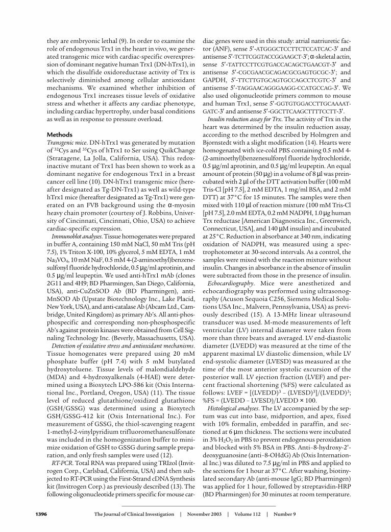
Tg-DN-Trx1 mice show base-line cardiac hypertrophy. At 3months, both the LV weight/body weight ratio
(LVW/BW) and the LV weight/tibial length ratio(LVW/TL) were significantly greater in Tg-DN-Trx1mice (line no. 13) than in NTg littermates (Figure 3a).Smaller but significant hypertrophy was also observedin the other three lines (nos. 8, 24, and 25) (Figure 3aand data not shown). LV cardiac myocyte cross-section-al area was significantly greater in Tg-DN-Trx1 micethan in NTg littermates (Figure 3, b and c). The resultsof RT-PCR analyses indicated that mRNA expression ofANF and α-skeletal actin, the fetal-type genes, was sig-nificantly elevated in Tg-DN-Trx1 mice (Figure 3, d ande). H&E staining of the LV showed that there was no sig-nificant infiltration of inflammatory cells in Tg-DN-Trx1 mice. Picric acid Sirius red staining indicated thatthere was no significant interstitial fibrosis in Tg-DN-Trx1 mice (data not shown).
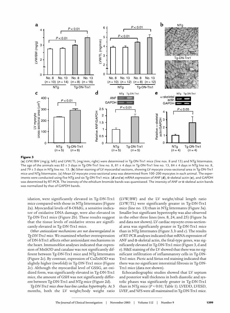
Echocardiographic studies showed that LV septumand posterior wall thickness in both diastolic and sys-tolic phases was significantly greater in Tg-DN-Trx1than in NTg mice (P < 0.01; Table 1). LVEDD, LVESD,LVEF, and %FS were all maintained in Tg-DN-Trx1 mice.
Figure 3(a) LVW/BW (mg/g; left) and LVW/TL (mg/mm; right) were determined in Tg-DN-Trx1 mice (line nos. 8 and 13) and NTg littermates.The age of the animals was 85 ± 3 days in Tg-DN-Trx1 line no. 8, 81 ± 4 days in Tg-DN-Trx1 line no. 13, 84 ± 4 days in NTg line no. 8,and 79 ± 5 days in NTg line no. 13. (b) Silver staining of LV myocardial sections, showing LV myocyte cross-sectional area in Tg-DN-Trx1mice and NTg littermates. (c) Mean LV myocyte cross-sectional area was determined from 100–200 myocytes in each animal. The exper-iments were conducted using five NTg and six Tg-DN-Trx1 mice. (d and e) mRNA expression of ANF (d), α-skeletal actin (e), and GAPDHwas determined by RT-PCR. The intensity of the ethidium bromide bands was quantitated. The intensity of ANF or α-skeletal actin bandswas normalized by that of GAPDH bands.
1400 The Journal of Clinical Investigation | November 2003 | Volume 112 | Number 9
These results suggest that Tg-DN-Trx1 mice exhibit car-diac hypertrophy with well-maintained LV function.
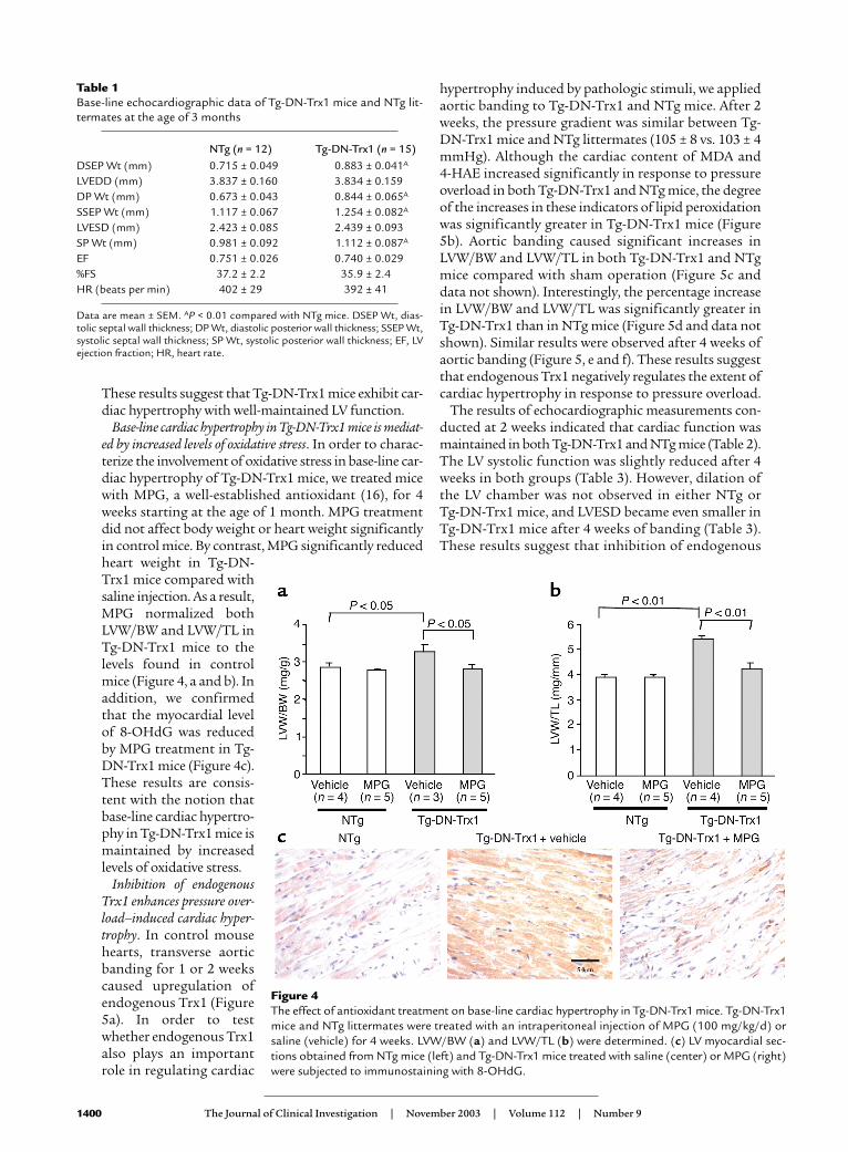
Base-line cardiac hypertrophy in Tg-DN-Trx1 mice is mediat-ed by increased levels of oxidative stress. In order to charac-terize the involvement of oxidative stress in base-line car-diac hypertrophy of Tg-DN-Trx1 mice, we treated micewith MPG, a well-established antioxidant (16), for 4weeks starting at the age of 1 month. MPG treatmentdid not affect body weight or heart weight significantlyin control mice. By contrast, MPG significantly reducedheart weight in Tg-DN-Trx1 mice compared withsaline injection. As a result,MPG normalized bothLVW/BW and LVW/TL inTg-DN-Trx1 mice to thelevels found in controlmice (Figure 4, a and b). Inaddition, we confirmedthat the myocardial levelof 8-OHdG was reducedby MPG treatment in Tg-DN-Trx1 mice (Figure 4c).These results are consis-tent with the notion thatbase-line cardiac hypertro-phy in Tg-DN-Trx1 mice ismaintained by increasedlevels of oxidative stress.
Inhibition of endogenousTrx1 enhances pressure over-load–induced cardiac hyper-trophy. In control mousehearts, transverse aorticbanding for 1 or 2 weekscaused upregulation ofendogenous Trx1 (Figure5a). In order to testwhether endogenous Trx1also plays an importantrole in regulating cardiac
hypertrophy induced by pathologic stimuli, we appliedaortic banding to Tg-DN-Trx1 and NTg mice. After 2weeks, the pressure gradient was similar between Tg-DN-Trx1 mice and NTg littermates (105 ± 8 vs. 103 ± 4mmHg). Although the cardiac content of MDA and 4-HAE increased significantly in response to pressureoverload in both Tg-DN-Trx1 and NTg mice, the degreeof the increases in these indicators of lipid peroxidationwas significantly greater in Tg-DN-Trx1 mice (Figure5b). Aortic banding caused significant increases inLVW/BW and LVW/TL in both Tg-DN-Trx1 and NTgmice compared with sham operation (Figure 5c anddata not shown). Interestingly, the percentage increasein LVW/BW and LVW/TL was significantly greater inTg-DN-Trx1 than in NTg mice (Figure 5d and data notshown). Similar results were observed after 4 weeks ofaortic banding (Figure 5, e and f). These results suggestthat endogenous Trx1 negatively regulates the extent ofcardiac hypertrophy in response to pressure overload.
The results of echocardiographic measurements con-ducted at 2 weeks indicated that cardiac function wasmaintained in both Tg-DN-Trx1 and NTg mice (Table 2).The LV systolic function was slightly reduced after 4weeks in both groups (Table 3). However, dilation ofthe LV chamber was not observed in either NTg or Tg-DN-Trx1 mice, and LVESD became even smaller inTg-DN-Trx1 mice after 4 weeks of banding (Table 3).These results suggest that inhibition of endogenous
Table 1Base-line echocardiographic data of Tg-DN-Trx1 mice and NTg lit-termates at the age of 3 months
Data are mean ± SEM. AP < 0.01 compared with NTg mice. DSEP Wt, dias-tolic septal wall thickness; DP Wt, diastolic posterior wall thickness; SSEP Wt,systolic septal wall thickness; SP Wt, systolic posterior wall thickness; EF, LVejection fraction; HR, heart rate.
Figure 4The effect of antioxidant treatment on base-line cardiac hypertrophy in Tg-DN-Trx1 mice. Tg-DN-Trx1mice and NTg littermates were treated with an intraperitoneal injection of MPG (100 mg/kg/d) orsaline (vehicle) for 4 weeks. LVW/BW (a) and LVW/TL (b) were determined. (c) LV myocardial sec-tions obtained from NTg mice (left) and Tg-DN-Trx1 mice treated with saline (center) or MPG (right)were subjected to immunostaining with 8-OHdG.
The Journal of Clinical Investigation | November 2003 | Volume 112 | Number 9 1401
Trx stimulates a concentric form of cardiac hypertro-phy in response to pressure overload.
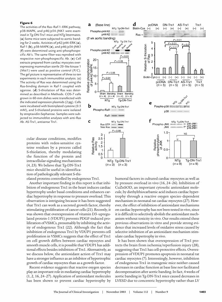
Activity of the Ras–Raf-1–ERK pathway, rather than the JNKor p38-MAPK pathway, is enhanced in the hearts of Tg-DN-Trx1 mice. In order to examine the mechanism of cardiachypertrophy in Tg-DN-Trx1 mice, we examined whethermembers of the MAPK family are activated in Tg-DN-Trx1 hearts. Interestingly, activity of extracellular sig-nal–regulated kinase (ERK) and that of its upstreamregulators, Raf-1 and Ras, were significantly elevated inTg-DN-Trx1 compared with NTg hearts (Figure 6, a–c).ERK activation in Tg-DN-Trx1 mice was furtherenhanced by aortic banding (Figure 6a, lower panel). Ithas been shown that oxidation of the cysteine residues(S-thiolation) activates Ras (19). To examine whetherRas undergoes S-thiolation in the presence of DN-hTrx1, S-thiolation assays were conducted using COS-7cells. S-thiolation of Ras was observed in the presence ofDN-hTrx1, but not either empty vector or wild-typeTrx1. S-thiolation of Ras was also observed in the pres-ence of antisense Trx1, suggesting that decreases in theactivity of Trx1 cause S-thiolation of Ras (Figure 6d).Although it has been shown that DN-hTrx1 activatesASK1 (20), an upstream kinase of p38-MAPK and JNKs,activities of JNKs and p38-MAPK were not significant-ly elevated in Tg-DN-Trx1 mice (Figure 6, e and f).
Transgenic mice with cardiac-specific overexpression of Trx1do not develop cardiac hypertrophy. Cardiac-specific over-expression of DN-hTrx1 may stimulate hypertrophy
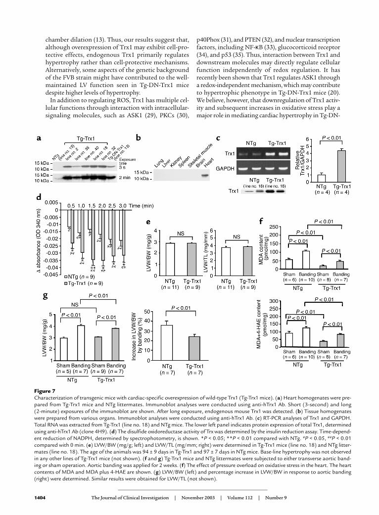
through redox-independent protein-protein interac-tions (4). In addition, wild-type Trx1 works as a growthfactor in some cell types (21, 22). In order to testwhether cardiac hypertrophy seen in Tg-DN-Trx1 miceis mediated by redox-independent effects of Trx1, wegenerated transgenic mice with cardiac-specific overex-pression of wild-type Trx1 (Tg-Trx1 mice). We generat-ed five transgenic lines and confirmed that wild-typeTrx1 is overexpressed in a heart-specific manner (Fig-ures 7, a and b). We predominantly characterized lineno. 18, where expression of wild-type Trx1 in the heart,determined by RT-PCR, was 4.5-fold higher than in NTgmice (Figure 7c). Protein expression of wild-type Trx1 inline no. 18, determined using anti-Trx1 Ab (4H9), was3.4-fold higher than that of endogenous Trx1. We con-firmed that the Trx activity is significantly elevated inTg-Trx1 hearts, which contrasted markedly with the Trxactivity in Tg-DN-Trx1 hearts (Figure 7d). At 3 months,neither LVW/BW nor LVW/TL in Tg-Trx1 mice was dif-ferent from that in NTg mice (Figure 7e). Similar resultswere confirmed in all other Tg-Trx1 lines generated(data not shown). Echocardiographic measurementsindicated that chamber sizes and cardiac function in Tg-Trx1 mice were normal, and wall thickness in Tg-Trx1 did not differ from that in NTg mice (Table 4).If the prohypertrophic effect of DN-hTrx1 is mediatedby decreases in endogenous Trx1 activity, overexpres-sion of Trx1 should exhibit antihypertrophic effects. Totest this hypothesis, aortic banding was conducted for
Figure 5Tg-DN-Trx1 mice and NTg littermates were subjected to either transverse aortic banding or sham operation. (a) The effect of pressure over-load on expression of endogenous mouse Trx1 in NTg mice. The result is representative of four experiments. (b–d) Aortic banding was appliedfor 2 weeks. (b) The effect of pressure overload on oxidative stress in the heart. The heart content of MDA and of MDA plus 4-HAE, the per-centage increase in MDA, and the percentage increase in MDA plus 4-HAE are shown. (c and d) LVW/BW (c) and percentage increase inLVW/BW in response to aortic banding (d) were determined. Similar results were obtained for LVW/TL. The percentage increase in LVW/BWand LVW/TL (not shown) was significantly greater in Tg-DN-Trx1 than in NTg mice in response to pressure overload. (e and f) Aortic band-ing was applied for 4 weeks. LVW/BW (e) and percentage increase in LVW/BW in response to aortic banding (f) were determined. The per-centage increase in LVW/BW was significantly greater in Tg-DN-Trx1 than in NTg mice.
1402 The Journal of Clinical Investigation | November 2003 | Volume 112 | Number 9
2 weeks using Tg-Trx1 and NTg mice, and the pressuregradient was similar between the two groups. As expect-ed, increases in oxidative stress (MDA alone and MDAplus 4-HAE) and in LV hypertrophy (LVW/BW),induced by pressure overload, were smaller in Tg-Trx1than in NTg mice (Figure 7, f and g).
Antisense inhibition of Trx1 causes hypertrophy in culturedcardiac myocytes. In order to confirm that the hyper-trophic effect of DN-hTrx1 is mediated by decreases inthe endogenous Trx1 activity, we examined the effectof antisense inhibition of Trx1. We confirmed thatantisense inhibition by either Morpholino oligo orplasmid significantly decreased expression of endoge-nous Trx1 in cardiac myocytes and COS-7 cells (Figure8, a and d). Morpholino antisense Trx1 oligo treatmentsignificantly increased the total protein content (Fig-ure 8b) as well as MDA and MDA plus 4-HAE content(Figure 8c) in cardiac myocytes compared with controlMorpholino oligo treatment. Cotransfection assaysindicated that both DN-hTrx1 and antisense Trx1 sig-nificantly increased the transcriptional activity of ANF,a cardiac-hypertrophy marker gene, while empty vectorand wild-type Trx1 did not (Figure 8e). These results
suggest that antisense inhibition of Trx1 exhibitshypertrophic effects similar to those of DN-hTrx1 incultured cardiac myocytes.
DiscussionAlthough it has been suggested that one of the mostimportant functions of Trx1 is as an antioxidant (4),the importance of Trx1 among cellular antioxidantmechanisms has not been demonstrated clearly in anyorgans in vivo. Our results suggest that cardiac-specif-ic overexpression of redox-inactive Trx1 in transgenicmice reduced disulfide oxidoreductase activity ofendogenous Trx in the heart. In this animal model,oxidative stress in the heart was significantly increasedunder both basal and pathologic conditions. This clear-ly indicates that Trx1 plays an important role as a reg-ulator of oxidative stress in the heart in vivo.
The cellular environment is maintained in reducingconditions, and therefore intracellular proteins containfree thiol, rather than disulfides, under physiologicalconditions (2). Trx1 reduces intracellular proteins withdisulfides through its disulfide oxidoreductase activity(4). Oxidative stress in the heart, in various cardiovas-
Table 2Echocardiographic data of Tg-DN-Trx1 mice and NTg littermates before and after aortic banding for 2 weeks
NTg (n = 4) Tg-DN-Trx1 (n = 7)
Before banding After banding Before banding After bandingDSEP Wt (mm) 0.613 ± 0.037 0.829 ± 0.035A 0.733 ± 0.019B 1.137 ± 0.021A,C
Data are mean ± SEM. AP < 0.05 compared with base line. BP < 0.05, CP < 0.01 compared with NTg mice.
The Journal of Clinical Investigation | November 2003 | Volume 112 | Number 9 1403
cular disease conditions, modifiesproteins with redox-sensitive cys-teine residues by a process called S-thiolation, thereby modulatingthe function of the protein andintracellular-signaling mechanisms(4, 23). We believe that Tg-DN-Trx1mice should be useful in identifica-tion of pathologically relevant S-thi-olated proteins controlled by endogenous Trx1.
Another important finding in this report is that inhi-bition of endogenous Trx1 in the heart induces cardiachypertrophy under basal conditions and enhances car-diac hypertrophy in response to pressure overload. Thisobservation is intriguing because it has been suggestedthat Trx1 can work as a secreted growth factor, therebystimulating proliferation of cancer cells (21). Recently, itwas shown that overexpression of vitamin D3–upregu-lated protein-1 (VDUP1) prevents PDGF-induced pro-liferation of VSMCs, presumably by inhibiting the activ-ity of endogenous Trx1 (22). Although the fact thatinhibition of endogenous Trx1 by VDUP1 prevents cellproliferation in VSMCs suggests that the effect of Trx1on cell growth differs between cardiac myocytes andsmooth muscle cells, it is possible that VDUP1 has addi-tional effects besides inhibition of Trx1. Alternatively, aswe discuss below, the antioxidant action of Trx1 mayhave a stronger influence as an inhibitor of hypertrophicgrowth of cardiac myocytes than as a growth factor.
Recent evidence suggests that reactive oxygen speciesplay an important role in mediating cardiac hypertrophy(1, 2, 16, 24–27). Application of antioxidant moleculeshas been shown to prevent cardiac hypertrophy by
humoral factors in cultured cardiac myocytes as well asby pressure overload in vivo (16, 24–26). Inhibition ofCuZnSOD, an important cytosolic antioxidant mole-cule, by diethylthiocarbamic acid induces cardiac hyper-trophy through a reactive oxygen species–dependentmechanism in neonatal rat cardiac myocytes (27). How-ever, the effect of inhibition of antioxidant mechanismson cardiac hypertrophy has not been tested in vivo, sinceit is difficult to selectively abolish the antioxidant mech-anism without toxicity in vivo. Our results extend theseprevious observations in vitro and provide strong evi-dence that increased levels of oxidative stress caused byselective inhibition of an antioxidant mechanism stim-ulate cardiac hypertrophy in vivo.
It has been shown that overexpression of Trx1 pro-tects the brain from ischemia/reperfusion injury (28),suggesting that Trx1 has cell-protective effects. Overex-pression of VDUP1 promotes apoptosis in neonatal ratcardiac myocytes (7). Interestingly, however, inhibitionof endogenous Trx1 in transgenic mice neither causeddecreases in cardiac function at base line nor facilitateddecompensation after aortic banding. In fact, 4 weeks ofaortic banding in Tg-DN-Trx1 mice caused decreases inLVESD due to concentric hypertrophy rather than LV
Figure 6The activities of the Ras–Raf-1–ERK pathway,p38-MAPK, and p46/p54–JNK1 were exam-ined in Tg-DN-Trx1 mice and NTg littermates.(a) Some mice were subjected to aortic band-ing for 2 weeks. Activities of p42/p44–ERK (a),Raf-1 (b), p38-MAPK (e), and p46/p54–JNK1(f) were determined using anti-phosphospe-cific Ab’s. The same filter was reprobed withrespective non-phosphospecific Ab. (e) Cellextracts prepared from cardiac myocytes over-expressing mammalian sterile 20–like kinase 1(Mst1) were used as positive control (P/C).The gel picture is representative of three to tenexperiments in each immunoblot analysis. (c)The activity of Ras was determined using theRas-binding domain in Raf-1 coupled withagarose. (d) S-thiolation of Ras was deter-mined as described in Methods. COS-7 cellsgrown in 60-mm dishes were transfected withthe indicated expression plasmids (2 µg). Cellswere incubated with biotinylated cysteine (0.5mM), and S-thiolated proteins were isolatedby streptavidin-Sepharose. Samples were sub-jected to immunoblot analyses with anti-RasAb. AS-Trx1, antisense Trx1.
1404 The Journal of Clinical Investigation | November 2003 | Volume 112 | Number 9
chamber dilation (13). Thus, our results suggest that,although overexpression of Trx1 may exhibit cell-pro-tective effects, endogenous Trx1 primarily regulateshypertrophy rather than cell-protective mechanisms.Alternatively, some aspects of the genetic backgroundof the FVB strain might have contributed to the well-maintained LV function seen in Tg-DN-Trx1 micedespite higher levels of hypertrophy.
In addition to regulating ROS, Trx1 has multiple cel-lular functions through interaction with intracellular-signaling molecules, such as ASK1 (29), PKCs (30),
p40Phox (31), and PTEN (32), and nuclear transcriptionfactors, including NF-κB (33), glucocorticoid receptor(34), and p53 (35). Thus, interaction between Trx1 anddownstream molecules may directly regulate cellularfunction independently of redox regulation. It hasrecently been shown that Trx1 regulates ASK1 througha redox-independent mechanism, which may contributeto hypertrophic phenotype in Tg-DN-Trx1 mice (20).We believe, however, that downregulation of Trx1 activ-ity and subsequent increases in oxidative stress play amajor role in mediating cardiac hypertrophy in Tg-DN-
Figure 7Characterization of transgenic mice with cardiac-specific overexpression of wild-type Trx1 (Tg-Trx1 mice). (a) Heart homogenates were pre-pared from Tg-Trx1 mice and NTg littermates. Immunoblot analyses were conducted using anti-hTrx1 Ab. Short (3-second) and long (2-minute) exposures of the immunoblot are shown. After long exposure, endogenous mouse Trx1 was detected. (b) Tissue homogenateswere prepared from various organs. Immunoblot analyses were conducted using anti-hTrx1 Ab. (c) RT-PCR analyses of Trx1 and GAPDH.Total RNA was extracted from Tg-Trx1 (line no. 18) and NTg mice. The lower left panel indicates protein expression of total Trx1, determinedusing anti-hTrx1 Ab (clone 4H9). (d) The disulfide oxidoreductase activity of Trx was determined by the insulin reduction assay. Time-depend-ent reduction of NADPH, determined by spectrophotometry, is shown. *P < 0.05; **P < 0.01 compared with NTg. #P < 0.05, ##P < 0.01compared with 0 min. (e) LVW/BW (mg/g; left) and LVW/TL (mg/mm; right) were determined in Tg-Trx1 mice (line no. 18) and NTg litter-mates (line no. 18). The age of the animals was 94 ± 9 days in Tg-Trx1 and 97 ± 7 days in NTg mice. Base-line hypertrophy was not observedin any other lines of Tg-Trx1 mice (not shown). (f and g) Tg-Trx1 mice and NTg littermates were subjected to either transverse aortic band-ing or sham operation. Aortic banding was applied for 2 weeks. (f) The effect of pressure overload on oxidative stress in the heart. The heartcontents of MDA and MDA plus 4-HAE are shown. (g) LVW/BW (left) and percentage increase in LVW/BW in response to aortic banding(right) were determined. Similar results were obtained for LVW/TL (not shown).
The Journal of Clinical Investigation | November 2003 | Volume 112 | Number 9 1405
Trx1 mice, because Tg-Trx1 mice exhibited oppositeresponses to pressure overload, namely reduced levels ofcardiac hypertrophy and oxidative stress. Furthermore,downregulation of Trx1 by antisense plasmid and Mor-pholino oligo also caused cardiac hypertrophy in cul-tured cardiac myocytes, consistent with the notion thatthe Trx1 activity is a critical regulator of cardiac hyper-
trophy. It should be noted, however, that the contribu-tion of the signaling mechanisms mediated by directinteraction with Trx1 to cardiac phenotype of Tg-DN-Trx1 mice remains to be elucidated.
A search for downstream signaling mechanisms caus-ing cardiac hypertrophy in Tg-DN-Trx1 mice indicatedthat the Ras–Raf-1–ERK pathway, rather than stress-responsive protein kinases, is activated in Tg-DN-Trx1mice. A well-compensated concentric form of cardiachypertrophy in Tg-DN-Trx1 mice is similar to that intransgenic mice with cardiac-specific overexpression ofMAP kinase kinase 1 (36). It has been shown that oxi-dation of Cys118 in Ras increases GTPase activity ofRas (19). In fact, our results indicated that Ras is S-thi-olated in the presence of DN-hTrx1 or antisense Trx1.
It has been recently shown that mice deficient in Trx2,a Trx1 homolog localized in mitochondria, are embry-onic lethal because of massive apoptosis (37). AlthoughTrx2 is expressed in the heart, we do not know whetherthe activity of Trx2 in mitochondria is affected in Tg-DN-Trx1 mice, because of technical difficulties inisolating Trx2 from the heart samples. However, sincethe cellular effect of Trx2 is predominantly cell protec-
Table 4Base-line echocardiographic data of Tg-Trx1 mice and NTg litter-mates at the age of 3 months
Figure 8(a–c) Neonatal rat cardiac myocytes were treated with either control Morpholino oligo or Morpholino antisense oligo for rat Trx1 (2 µM)using osmotic delivery. Myocytes were harvested 48 hours after application of the oligo. (a) Expression of Trx1 was determined by immunoblot-ting with anti-Trx1 Ab (4H9). (b) Total cardiac myocyte protein content was determined. The mean value in control Morpholino oligo–treat-ed myocytes was designated as 1. (c) Cellular content of MDA and MDA plus 4-HAE was determined as described in Methods. (d) COS-7 cellsgrown in 60-mm dishes were transfected with either empty vector plasmid or pcDNA3.1 harboring DN-hTrx1, antisense rat Trx1 (AS-Trx1), orTrx1. Forty-eight hours after transfection, cells were harvested, and expression of Trx1 was determined by immunoblot analysis. The level ofTrx1 expression quantitated by densitometric analyses is shown. (e) Cardiac myocytes were transfected with ANF-luciferase (-638) (ANF-luciferase reporter gene containing 638 base pairs upstream of the rat ANF gene transcription start site) and SV40-β-galactosidase, togetherwith pcDNA3.1 alone (empty vector) or with pcDNA3.1 harboring DN-hTrx1, antisense rat Trx1, or Trx1. Forty-eight hours after transfection,activities of luciferase and β-galactosidase were determined. The activity of luciferase was normalized by that of β-galactosidase.
1406 The Journal of Clinical Investigation | November 2003 | Volume 112 | Number 9
tive (37), if the activity of Trx2 is significantly inhibited,a well-compensated cardiac hypertrophy in Tg-DN-Trx1mice would be unexpected. It would be interesting togenerate mice with cardiac-specific deletion of Trx2 orexpression of dominant negative Trx2 and comparetheir phenotype with that of Tg-DN-Trx1 mice.
Recent evidence suggests that the activity of endoge-nous Trx1 can be posttranslationally modified throughinteraction with other antioxidant mechanisms and oxi-dant species. For example, Cys72 of Trx1 undergoes glu-tathiolation in response to oxidative stress, which abol-ishes the enzymatic activity of Trx1 (38). By contrast,nitric oxide S-nitrosylates Cys69 of Trx1, thereby stim-ulating the activity of Trx1 (39). The activity of Trx1 isalso subjected to regulation by the Trx1-interacting pro-teins, such as VDUP1 (22). Thus, Tg-DN-Trx1 and Tg-Trx1 mice are useful to elucidate functional conse-quences of Trx1 modulation in the heart in vivo.
In summary, our results suggest that endogenousTrx1 is an important antioxidant in the mouse heart,and that increases in oxidative stress caused by decreas-es in the Trx1 activity stimulate a concentric form ofcardiac hypertrophy in vivo. Thus, both Trx1 and itsdownstream target proteins may be an important ther-apeutic focus for treatment of cardiac hypertrophy.
AcknowledgmentsWe thank Jeffrey Robbins for providing us with the α-myosin heavy chain promoter construct. We thankDaniela Zablocki and Dennis Quinio for critical readingof the manuscript. This work was supported in part bygrants from the NIH (HL-59139, HL-33107, HL-33065,HL-65182, HL-65183, AG-14121, HL-69020, HL-67724,and HL-67727) and an Established Investigator Awardfrom the American Heart Association (0340123N).
1. Sawyer, D.B., et al. 2002. Role of oxidative stress in myocardial hypertro-phy and failure. J. Mol. Cell. Cardiol. 34:379–388.
2. Sorescu, D., and Griendling, K.K. 2002. Reactive oxygen species, mito-chondria, and NAD(P)H oxidases in the development and progression ofheart failure. Congest. Heart Fail. 8:132–140.
3. Nakamura, H., Nakamura, K., and Yodoi, J. 1997. Redox regulation of cel-lular activation. Annu. Rev. Immunol. 15:351–369.
4. Masutani, H., and Yodoi, J. 2002. Thioredoxin. Overview. Methods Enzymol.347:279–286.
5. Kishimoto, C., et al. 2001. Serum thioredoxin (TRX) levels in patients withheart failure. Jpn. Circ. J. 65:491–494.
6. Shioji, K., et al. 2000. Upregulation of thioredoxin (TRX) expression ingiant cell myocarditis in rats. FEBS Lett. 472:109–113.
7. Wang, Y., De Keulenaer, G.W., and Lee, R.T. 2002. Vitamin D(3)-up-regu-lated protein-1 is a stress-responsive gene that regulates cardiomyocyte via-bility through interaction with thioredoxin. J. Biol. Chem. 277:26496–26500.
8. Shioji, K., et al. 2002. Overexpression of thioredoxin-1 in transgenic miceattenuates adriamycin-induced cardiotoxicity. Circulation. 106:1403–1409.
9. Nakamura, H., Mitsui, A., and Yodoi, J. 2002. Thioredoxin overexpressionin transgenic mice. Methods Enzymol. 347:436–440.
10. Gallegos, A., et al. 1996. Transfection with human thioredoxin increasescell proliferation and a dominant-negative mutant thioredoxin reversesthe transformed phenotype of human breast cancer cells. Cancer Res.56:5765–5770.
11. Esterbauer, H., Schaur, R.J., and Zollner, H. 1991. Chemistry and bio-chemistry of 4-hydroxynonenal: malonaldehyde and related aldehydes.Free Radic. Biol. Med. 11:81–128.
12. Liang, Q., et al. 2002. Overexpression of metallothionein reduces diabeticcardiomyopathy. Diabetes. 51:174–181.
13. Sadoshima, J., et al. 2002. The MEKK1-JNK pathway plays a protectiverole in pressure overload but does not mediate cardiac hypertrophy.
J. Clin. Invest. 110:271–279. doi:10.1172/JCI200214938.14. Holmgren, A., and Bjornstedt, M. 1995. Thioredoxin and thioredoxin
reductase. Methods Enzymol. 252:199–208.15. Asai, K., et al. 1999. β-Adrenergic receptor blockade arrests myocyte dam-
age and preserves cardiac function in the transgenic Gsα mouse. J. Clin.Invest. 104:551–558.
16. Date, M.O., et al. 2002. The antioxidant N-2-mercaptopropionyl glycineattenuates left ventricular hypertrophy in in vivo murine pressure-over-load model. J. Am. Coll. Cardiol. 39:907–912.
17. Tomita, H., et al. 2003. Inducible cAMP early repressor (ICER) is a nega-tive-feedback regulator of cardiac hypertrophy and an important media-tor of cardiac myocyte apoptosis in response to beta-adrenergic receptorstimulation. Circ. Res. 93:12–22.
18. Sadoshima, J., and Izumo, S. 1995. Rapamycin selectively inhibitsangiotensin II-induced increase in protein synthesis in cardiac myocytesin vitro. Potential role of 70-kD S6 kinase in angiotensin II-induced car-diac hypertrophy. Circ. Res. 77:1040–1052.
19. Lander, H.M., et al. 1996. Redox regulation of cell signalling. Nature.381:380–381.
20. Liu, Y., and Min, W. 2002. Thioredoxin promotes ASK1 ubiquitinationand degradation to inhibit ASK1-mediated apoptosis in a redox activity-independent manner. Circ. Res. 90:1259–1266.
21. Wakasugi, N., et al. 1990. Adult T-cell leukemia-derived factor/thioredox-in, produced by both human T-lymphotropic virus type I- and Epstein-Barrvirus-transformed lymphocytes, acts as an autocrine growth factor and syn-ergizes with interleukin 1 and interleukin 2. Proc. Natl. Acad. Sci. U. S. A.87:8282–8286.
22. Schulze, P.C., De Keulenaer, G.W., Yoshioka, J., Kassik, K.A., and Lee, R.T.2002. Vitamin D3-upregulated protein-1 (VDUP-1) regulates redox-dependent vascular smooth muscle cell proliferation through interactionwith thioredoxin. Circ. Res. 91:689–695.
23. Eaton, P., Byers, H.L., Leeds, N., Ward, M.A., and Shattock, M.J. 2002. Detec-tion, quantitation, purification, and identification of cardiac proteins S-thiolated during ischemia and reperfusion. J. Biol. Chem. 277:9806–9811.
24. Xie, Z., et al. 1999. Intracellular reactive oxygen species mediate the link-age of Na+/K+-ATPase to hypertrophy and its marker genes in cardiacmyocytes. J. Biol. Chem. 274:19323–19328.
25. Nakamura, K., et al. 1998. Inhibitory effects of antioxidants on neonatalrat cardiac myocyte hypertrophy induced by tumor necrosis factor-alphaand angiotensin II. Circulation. 98:794–799.
26. Amin, J.K., et al. 2001. Reactive oxygen species mediate alpha-adrenergicreceptor-stimulated hypertrophy in adult rat ventricular myocytes. J. Mol.Cell. Cardiol. 33:131–139.
27. Siwik, D.A., et al. 1999. Inhibition of copper-zinc superoxide dismutaseinduces cell growth, hypertrophic phenotype, and apoptosis in neonatalrat cardiac myocytes in vitro. Circ. Res. 85:147–153.
28. Takagi, Y., et al. 1999. Overexpression of thioredoxin in transgenic miceattenuates focal ischemic brain damage. Proc. Natl. Acad. Sci. U. S. A.96:4131–4136.
29. Saitoh, M., et al. 1998. Mammalian thioredoxin is a direct inhibitor ofapoptosis signal-regulating kinase (ASK) 1. EMBO J. 17:2596–2606.
30. Watson, J.A., Rumsby, M.G., and Wolowacz, R.G. 1999. Phage display iden-tifies thioredoxin and superoxide dismutase as novel protein kinase C-interacting proteins: thioredoxin inhibits protein kinase C-mediated phos-phorylation of histone. Biochem. J. 343:301–305.
31. Nishiyama, A., et al. 1999. Demonstration of the interaction of thiore-doxin with p40phox, a phagocyte oxidase component, using a yeast two-hybrid system. Immunol. Lett. 68:155–159.
32. Lee, S.R., et al. 2002. Reversible inactivation of the tumor suppressor PTENby H2O2. J. Biol. Chem. 277:20336–20342.
33. Hirota, K., et al. 1999. Distinct roles of thioredoxin in the cytoplasm andin the nucleus. A two-step mechanism of redox regulation of transcriptionfactor NF-kappaB. J. Biol. Chem. 274:27891–27897.
34. Makino, Y., et al. 1999. Direct association with thioredoxin allowsredox regulation of glucocorticoid receptor function. J. Biol. Chem.274:3182–3188.
35. Ueno, M., et al. 1999. Thioredoxin-dependent redox regulation of p53-mediated p21 activation. J. Biol. Chem. 274:35809–35815.
36. Bueno, O.F., et al. 2000. The MEK1-ERK1/2 signaling pathway pro-motes compensated cardiac hypertrophy in transgenic mice. EMBO J.19:6341–6350.
37. Nonn, L., Williams, R.R., Erickson, R.P., and Powis, G. 2003. The absenceof mitochondrial thioredoxin 2 causes massive apoptosis, exencephaly,and early embryonic lethality in homozygous mice. Mol. Cell. Biol.23:916–922.
38. Casagrande, S., et al. 2002. Glutathionylation of human thioredoxin: apossible crosstalk between the glutathione and thioredoxin systems. Proc.Natl. Acad. Sci. U. S. A. 99:9745–9749.
39. Haendeler, J., et al. 2002. Redox regulatory and anti-apoptotic functionsof thioredoxin depend on S-nitrosylation at cysteine 69. Nat. Cell Biol.4:743–749.