Inhibition of IL-10 Production by Maternal Antibodies against Group B Streptococcus GAPDH Confers Immunity to Offspring by Favoring Neutrophil Recruitment Pedro Madureira 1,2 , Elva Bonifa ´ cio Andrade 1,2 , Bernardo Gama 1 , Liliana Oliveira 1,2 , Susana Moreira 3 , Adı´liaRibeiro 1,2 , Margarida Correia-Neves 4,5 , Patrick Trieu-Cuot 6 , Manuel Vilanova 1,2 , Paula Ferreira 1,2 * 1 ICBAS – Instituto de Cie ˆncias Biome ´dicas de Abel Salazar, Universidade do Porto, Porto, Portugal, 2 IBMC – Instituto de Biologia Molecular e Celular, Porto, Portugal, 3 IBB, Institute for Biotechnology and Bioengineering, Centre of Biological Engineering, University of Minho, Braga, Portugal, 4 Life and Health Sciences Research Institute (ICVS), School of Health Sciences, University of Minho, Braga, Portugal, 5 ICVS/3B’s, PT Government Associate Laboratory, Braga/Guimara ˜es, Portugal, 6 Institut Pasteur, Unite ´ de Biologie des Bacte ´ries Pathoge `nes a ` Gram-Positif, CNRS URA 2172, Paris, France Abstract Group B Streptococcus (GBS) is the leading cause of neonatal pneumonia, septicemia, and meningitis. We have previously shown that in adult mice GBS glycolytic enzyme glyceraldehyde-3-phosphate dehydrogenase (GAPDH) is an extracellular virulence factor that induces production of the immunosuppressive cytokine interleukin-10 (IL-10) by the host early upon bacterial infection. Here, we investigate whether immunity to neonatal GBS infection could be achieved through maternal vaccination against bacterial GAPDH. Female BALB/c mice were immunized with rGAPDH and the progeny was infected with a lethal inoculum of GBS strains. Neonatal mice born from mothers immunized with rGAPDH were protected against infection with GBS strains, including the ST-17 highly virulent clone. A similar protective effect was observed in newborns passively immunized with anti-rGAPDH IgG antibodies, or F(ab’) 2 fragments, indicating that protection achieved with rGAPDH vaccination is independent of opsonophagocytic killing of bacteria. Protection against lethal GBS infection through rGAPDH maternal vaccination was due to neutralization of IL-10 production soon after infection. Consequently, IL-10 deficient (IL-10 2/2 ) mice pups were as resistant to GBS infection as pups born from vaccinated mothers. We observed that protection was correlated with increased neutrophil trafficking to infected organs. Thus, anti-rGAPDH or anti-IL-10R treatment of mice pups before GBS infection resulted in increased neutrophil numbers and lower bacterial load in infected organs, as compared to newborn mice treated with the respective control antibodies. We showed that mothers immunized with rGAPDH produce neutralizing antibodies that are sufficient to decrease IL-10 production and induce neutrophil recruitment into infected tissues in newborn mice. These results uncover a novel mechanism for GBS virulence in a neonatal host that could be neutralized by vaccination or immunotherapy. As GBS GAPDH is a structurally conserved enzyme that is metabolically essential for bacterial growth in media containing glucose as the sole carbon source (i.e., the blood), this protein constitutes a powerful candidate for the development of a human vaccine against this pathogen. Citation: Madureira P, Andrade EB, Gama B, Oliveira L, Moreira S, et al. (2011) Inhibition of IL-10 Production by Maternal Antibodies against Group B Streptococcus GAPDH Confers Immunity to Offspring by Favoring Neutrophil Recruitment. PLoS Pathog 7(11): e1002363. doi:10.1371/journal.ppat.1002363 Editor: Michael R. Wessels, Children’s Hospital Boston, United States of America Received February 24, 2011; Accepted September 22, 2011; Published November 17, 2011 Copyright: ß 2011 Madureira et al. This is an open-access article distributed under the terms of the Creative Commons Attribution License, which permits unrestricted use, distribution, and reproduction in any medium, provided the original author and source are credited. Funding: This work was supported by SUDOE-FEDER IMMUNONET SOE1/P1/E014 and COMPETE-FEDER FCT Grant PTDC/SAU-MIC/111387/2009. Pedro Madureira was supported by a Post-Doctoral fellowship under the project PTDC/SAU-MIC/111387/2009. Elva BonifA ˜ ¡cio Andrade and Liliana Oliveira were supported by the FCT fellowships SFRH/BD/38380/2007 and SFRH/BD/38121/2007, respectively. The funders had no role in study design, data collection and analysis, decision to publish, or preparation of the manuscript. Competing Interests: The authors have declared that no competing interests exist. * E-mail: [email protected]Introduction Streptococcus agalactiae, also named Group B Streptococcus (GBS), is a Gram-positive encapsulated commensal bacterium of the human intestine that colonizes the vagina of up to 30% of healthy women. This bacterium is the leading cause of neonatal pneumonia, septicemia, and meningitis [1,2,3,4]. Neonatal GBS infections are acquired through maternal transmission and may result in early-onset disease (EOD), which occurs within the first week of life, or in late-onset disease (LOD), that occurs after the first week and accounts for most meningitis cases and deaths [3,5,6]. Despite early antimicrobial treatment and improvement in neonatal intensive care, up to 10% of neonatal invasive GBS infections are lethal and 25 to 35% of surviving infants with meningitis experience permanent neurological sequelae [3]. Because recommendations for intrapartum antibiotic prophylaxis (IAP) for mothers in labor at risk for GBS infection have been widely implemented in many countries, the incidence of EOD has declined to ,1/1,000 births, but the incidence of LOD has slowly increased in the last decade [7]. An unexpected burden of case fatalities among children aged less than 90 days caused by GBS infection was recently reported in different European countries [8,9,10]. Moreover, recent reports described the emergence of antibiotic-resistant GBS strains likely caused by the widespread use of IAP [11,12]. Maternal vaccination is the best alternative to IAP to deal with GBS neonatal infections. Vaccines to prevent GBS disease have been initially developed by coupling capsular polysaccharide (CPS) PLoS Pathogens | www.plospathogens.org 1 November 2011 | Volume 7 | Issue 11 | e1002363
Transcript
Inhibition of IL-10 Production by Maternal Antibodiesagainst Group B Streptococcus GAPDH Confers Immunityto Offspring by Favoring Neutrophil RecruitmentPedro Madureira1,2, Elva Bonifacio Andrade1,2, Bernardo Gama1, Liliana Oliveira1,2, Susana Moreira3,
Adılia Ribeiro1,2, Margarida Correia-Neves4,5, Patrick Trieu-Cuot6, Manuel Vilanova1,2, Paula Ferreira1,2*
1 ICBAS – Instituto de Ciencias Biomedicas de Abel Salazar, Universidade do Porto, Porto, Portugal, 2 IBMC – Instituto de Biologia Molecular e Celular, Porto, Portugal,
3 IBB, Institute for Biotechnology and Bioengineering, Centre of Biological Engineering, University of Minho, Braga, Portugal, 4 Life and Health Sciences Research Institute
(ICVS), School of Health Sciences, University of Minho, Braga, Portugal, 5 ICVS/3B’s, PT Government Associate Laboratory, Braga/Guimaraes, Portugal, 6 Institut Pasteur,
Unite de Biologie des Bacteries Pathogenes a Gram-Positif, CNRS URA 2172, Paris, France
Abstract
Group B Streptococcus (GBS) is the leading cause of neonatal pneumonia, septicemia, and meningitis. We have previouslyshown that in adult mice GBS glycolytic enzyme glyceraldehyde-3-phosphate dehydrogenase (GAPDH) is an extracellularvirulence factor that induces production of the immunosuppressive cytokine interleukin-10 (IL-10) by the host early uponbacterial infection. Here, we investigate whether immunity to neonatal GBS infection could be achieved through maternalvaccination against bacterial GAPDH. Female BALB/c mice were immunized with rGAPDH and the progeny was infectedwith a lethal inoculum of GBS strains. Neonatal mice born from mothers immunized with rGAPDH were protected againstinfection with GBS strains, including the ST-17 highly virulent clone. A similar protective effect was observed in newbornspassively immunized with anti-rGAPDH IgG antibodies, or F(ab’)2 fragments, indicating that protection achieved withrGAPDH vaccination is independent of opsonophagocytic killing of bacteria. Protection against lethal GBS infection throughrGAPDH maternal vaccination was due to neutralization of IL-10 production soon after infection. Consequently, IL-10deficient (IL-102/2) mice pups were as resistant to GBS infection as pups born from vaccinated mothers. We observed thatprotection was correlated with increased neutrophil trafficking to infected organs. Thus, anti-rGAPDH or anti-IL-10Rtreatment of mice pups before GBS infection resulted in increased neutrophil numbers and lower bacterial load in infectedorgans, as compared to newborn mice treated with the respective control antibodies. We showed that mothers immunizedwith rGAPDH produce neutralizing antibodies that are sufficient to decrease IL-10 production and induce neutrophilrecruitment into infected tissues in newborn mice. These results uncover a novel mechanism for GBS virulence in a neonatalhost that could be neutralized by vaccination or immunotherapy. As GBS GAPDH is a structurally conserved enzyme that ismetabolically essential for bacterial growth in media containing glucose as the sole carbon source (i.e., the blood), thisprotein constitutes a powerful candidate for the development of a human vaccine against this pathogen.
Citation: Madureira P, Andrade EB, Gama B, Oliveira L, Moreira S, et al. (2011) Inhibition of IL-10 Production by Maternal Antibodies against Group BStreptococcus GAPDH Confers Immunity to Offspring by Favoring Neutrophil Recruitment. PLoS Pathog 7(11): e1002363. doi:10.1371/journal.ppat.1002363
Editor: Michael R. Wessels, Children’s Hospital Boston, United States of America
Received February 24, 2011; Accepted September 22, 2011; Published November 17, 2011
Copyright: � 2011 Madureira et al. This is an open-access article distributed under the terms of the Creative Commons Attribution License, which permitsunrestricted use, distribution, and reproduction in any medium, provided the original author and source are credited.
Funding: This work was supported by SUDOE-FEDER IMMUNONET SOE1/P1/E014 and COMPETE-FEDER FCT Grant PTDC/SAU-MIC/111387/2009. Pedro Madureirawas supported by a Post-Doctoral fellowship under the project PTDC/SAU-MIC/111387/2009. Elva BonifA¡cio Andrade and Liliana Oliveira were supported by theFCT fellowships SFRH/BD/38380/2007 and SFRH/BD/38121/2007, respectively. The funders had no role in study design, data collection and analysis, decision topublish, or preparation of the manuscript.
Competing Interests: The authors have declared that no competing interests exist.
also acts as a GBS extracellular virulence factor that induces
rapid production of interleukin-10 (IL-10) by the host [29]. Adult
C57BL/6 mice (resistant to GBS infection) infected with a GBS
mutant strain that over-express GAPDH (oeGAPDH) had
increased bacterial colonization compared to mice infected with
wild-type (WT) GBS. Increased bacterial burden in oeGAPDH
infected C57BL/6 mice was accompanied by elevated serum
levels of IL-10. Consequently, acquired susceptibility of C57BL/6
mice to oeGAPDH infection was completely reverted in IL-10-
deficient animals [29]. This suggested that an exacerbated
production of IL-10 during GBS infection might facilitate
pathogen immune evasion. We demonstrate here that maternal
immunization with rGAPDH confers protection against GBS
infection in neonatal mice by abrogating the early IL-10
production detected upon the bacterial challenge. We also
demonstrate that blocking GAPDH-induced early IL-10 produc-
tion restores the recruitment of neutrophils in infected organs,
which is essential for pathogen elimination and host protection
against GBS infection.
Since GBS GAPDH is a structurally conserved enzyme that is
metabolically essential for bacterial growth in blood, it constitutes
an attractive target for the development of a human vaccine.
Results
GAPDH, a structurally conserved GBS proteinGAPDH, a key enzyme of the glycolytic pathway, is structurally
conserved in all 8 published GBS genomes (identity.99.8%).
Anti-rGAPDH IgG antibodies purified from sera of rGAPDH
immunized mice or rabbits were thus used to demonstrate the
presence of GAPDH in culture supernatants of ten unrelated GBS
clinical isolates (Figure 1A) belonging to different serotypes and/or
MLSTypes (Table S1). GBS GAPDH displays 44.7, 45.8 and
44.0% amino acid identity with rabbit, mice, and human
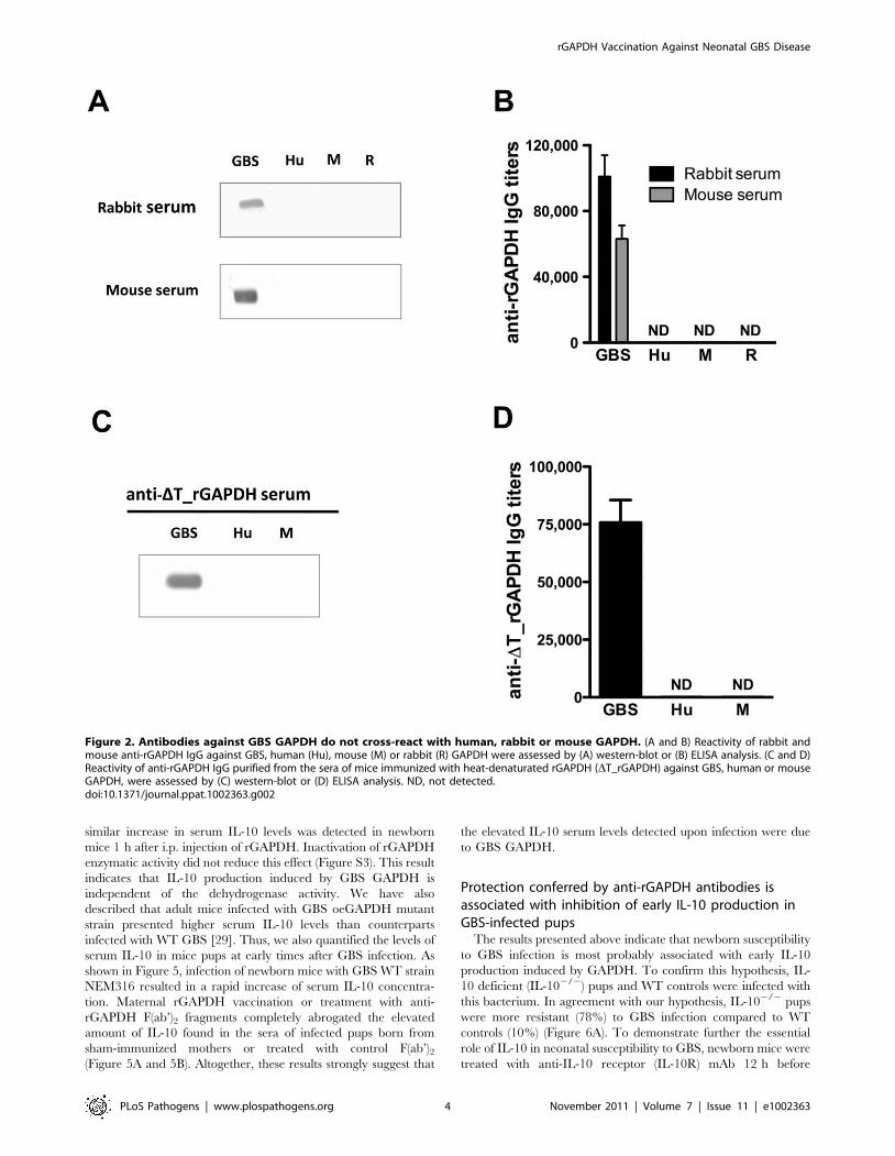
GAPDH, respectively (Figure 1B). However, western blot and
ELISA analysis revealed that rabbits and mice antibodies directed
against GBS rGAPDH do not react with human, mouse, or rabbit
GAPDH (Figure 2A and 2B). To favor the production of
antibodies recognizing linear buried epitopes, mice were immu-
nized with heat-denaturated rGAPDH (DT_rGAPDH). Anti-
DT_rGAPDH antibodies purified from the sera of these animals
did not show any cross-reactivity against mouse (self cross-
reactivity) or human GAPDH when analyzed by western blot
and ELISA (Figure 2C and 2E). These results are consistent with
the fact that the longest identical stretches observed between
eukaryotic and prokaryotic GADPH sequences are only 10-
aminoacid long (Figure 1B).
Maternal vaccination with rGAPDH protects neonatesfrom GBS infection
To test whether maternal immunization with rGAPDH
conferred protection to the offspring against GBS infection,
female BALB/c mice were immunized with rGAPDH in alum
adjuvant. Control mice were sham-immunized with the adjuvant
alone.
Pups born from sham-immunized or rGAPDH-immunized
females were infected intraperitoneally (i.p.) 48 h after birth with
56106 colony-forming units (CFU) of serotype III virulent GBS
strain NEM316. All but one mouse born from rGAPDH-
immunized mothers survived the infection (95% survival) whereas
22 out of 27 infected pups succumbed to GBS challenge in the
control group (18.5% survival) (Figure 3A). Most of the cases of
GBS meningitis and LOD are caused by a serotype III hyper
virulent clone, defined by multilocus sequence typing as ST-17
[30,31,32]. To better assess the effectiveness of maternal
vaccination with rGAPDH, pups born from sham- or rGAPDH-
immunized progenitors were i.p. infected 48 h after birth with
106 CFU of BM110, a serotype III GBS hyper virulent strain ST-
17 (Table S1). All ST-17 GBS challenged neonates born from
Author Summary
Streptococcus agalactiae (Group B streptococcus, GBS) isthe leading infectious cause of morbidity and mortalityamong neonates. However, there is still no satisfactoryexplanation of why neonates are so susceptible to GBSinfections. Intrapartum antibiotic prophylaxis (IAP) wasimplemented in many countries but led to the emergenceof antibiotic-resistant GBS strains. Therefore, maternalvaccination represents an attractive alternative to IAP.Here, we show that the high susceptibility of newbornmice to GBS infections is associated with their propensityto produce elevated amounts of immunosuppressivecytokine IL-10. We also demonstrate that IL-10 impairsneutrophil recruitment into infected organs thus prevent-ing bacterial clearance. We identified extracellular GAPDHas the GBS factor that induces the high IL-10 productiondetected early upon neonatal infection. We show thatmaternal vaccination with recombinant GAPDH confersrobust protective immunity against lethal infection with aGBS hyper-virulent strain in mice offspring. This protectioncan also be obtained either by antibody neutralization ofGBS GAPDH or by blocking IL-10 binding to its receptor. AsGBS GAPDH is an essential protein for bacterial growth, itis present in all GBS strains and thus constitutes anappropriate target antigen for a global effective vaccineagainst this pathogen.
sham-immunized mothers died whereas mortality rate dropped to
21.4% in neonates born from rGAPDH-immunized mothers
(Figure 3B). The protective effect conferred by rGAPDH maternal
immunization was also observed in neonate mice infected by the
subcutaneous (s.c.) route with 2.56104 BM110 CFU. As shown in
Figure 3C, none of the mice born from sham-immunized mothers
survived this infectious challenge whereas only 23% of mice born
from rGAPDH-immunized mothers died. Altogether, these results
show that maternal vaccination with rGAPDH protected the
offspring against GBS infections, including those caused by the
hyper virulent strain BM110.
Passive immunization with anti-rGAPDH F(ab’)2-fragments protects newborns against GBS ST-17challenge
Pups born from rGAPDH-immunized mothers presented
increased serum titers of anti-rGAPDH IgG antibodies when
compared with those born from sham-immunized mothers (Figure
S1). To evaluate the importance of these maternal antibodies in
the newborn protection against GBS infection, neonatal mice were
passively immunized with purified anti-rGAPDH IgG antibodies
12 h prior to GBS challenge. The passive antibody transfer
conferred protection against infection caused by the virulent
NEM316 or hyper virulent BM110 strains (Figure 4A and 4B).
Anti-rGAPDH IgG antibodies conferred a similar protection to
neonate mice infected by the s.c. route (data not shown). As
described by others [33], we observed that GAPDH is present at
the cell surface of GBS strains (Figure S2) and, it was therefore
conceivable that protection conferred by anti-rGAPDH antibodies
could be due to an enhanced opsonophagocytosis-mediated killing
of GBS. However, anti-rGAPDH IgG antibodies did not
enhanced in vitro phagocytosis or complement-mediated killing of
GBS BM110 cells (Figure 4C). This indicated that protection
conferred by anti-rGAPDH antibodies was not mediated by these
mechanisms. Furthermore, complete protection against GBS
infection was observed in neonate mice treated with purified
anti-rGAPDH F(ab’)2 fragments 12 h before i.p. infection with
BM110 strain. In contrast, all pups that received the same amount
of control F(ab’)2 fragments died within the first 3 days upon the
infectious challenge (Figure 4D). Altogether, these results demon-
strate that enhanced opsonophagocytic killing or complement
activation did not mediate the observed protective effect of anti-
rGAPDH antibodies.
GBS GAPDH induces early IL-10 production in newbornmice
We have previously described a rise in IL-10 serum levels in
adult mice treated with rGAPDH [29]. As shown in Figure S3, a
Figure 1. GAPDH is a conserved GBS protein. (A) Extracellular proteins from culture supernatants of different GBS clinical isolates wereseparated by SDS-page and analyzed by western-blot using anti-rGAPDH IgG obtained from rGAPDH-immunized rabbits. rGAPDH was used as apositive control. The data are representative of four independent experiments. (B) Multiple alignment of the aminoacid sequences of the human,rabbit, mice, and GBS GAPDH. The green vertical bars below the consensus sequences indicate identical aminoacid at the same position in all foursequences. The two heavy black lines delineate the two conserved 10-aminoacid stretches of identity.doi:10.1371/journal.ppat.1002363.g001
similar increase in serum IL-10 levels was detected in newborn
mice 1 h after i.p. injection of rGAPDH. Inactivation of rGAPDH
enzymatic activity did not reduce this effect (Figure S3). This result
indicates that IL-10 production induced by GBS GAPDH is
independent of the dehydrogenase activity. We have also
described that adult mice infected with GBS oeGAPDH mutant
strain presented higher serum IL-10 levels than counterparts
infected with WT GBS [29]. Thus, we also quantified the levels of
serum IL-10 in mice pups at early times after GBS infection. As
shown in Figure 5, infection of newborn mice with GBS WT strain
NEM316 resulted in a rapid increase of serum IL-10 concentra-
tion. Maternal rGAPDH vaccination or treatment with anti-
rGAPDH F(ab’)2 fragments completely abrogated the elevated
amount of IL-10 found in the sera of infected pups born from
sham-immunized mothers or treated with control F(ab’)2(Figure 5A and 5B). Altogether, these results strongly suggest that
the elevated IL-10 serum levels detected upon infection were due
to GBS GAPDH.
Protection conferred by anti-rGAPDH antibodies isassociated with inhibition of early IL-10 production inGBS-infected pups
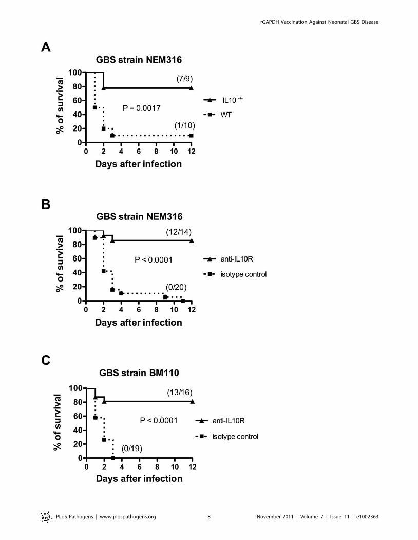
The results presented above indicate that newborn susceptibility
to GBS infection is most probably associated with early IL-10
production induced by GAPDH. To confirm this hypothesis, IL-
10 deficient (IL-102/2) pups and WT controls were infected with
this bacterium. In agreement with our hypothesis, IL-102/2 pups
were more resistant (78%) to GBS infection compared to WT
controls (10%) (Figure 6A). To demonstrate further the essential
role of IL-10 in neonatal susceptibility to GBS, newborn mice were
treated with anti-IL-10 receptor (IL-10R) mAb 12 h before
Figure 2. Antibodies against GBS GAPDH do not cross-react with human, rabbit or mouse GAPDH. (A and B) Reactivity of rabbit andmouse anti-rGAPDH IgG against GBS, human (Hu), mouse (M) or rabbit (R) GAPDH were assessed by (A) western-blot or (B) ELISA analysis. (C and D)Reactivity of anti-rGAPDH IgG purified from the sera of mice immunized with heat-denaturated rGAPDH (DT_rGAPDH) against GBS, human or mouseGAPDH, were assessed by (C) western-blot or (D) ELISA analysis. ND, not detected.doi:10.1371/journal.ppat.1002363.g002
NEM316 or BM110 GBS challenge. As expected, most pups
treated with anti-IL-10R mAb survived (86% or 82%, respectively)
while all control pups died (Figure 6B and 6C). No additional
protection was observed when newborn mice were treated
simultaneously with anti-IL10R mAb and anti-rGAPDH IgG
(Figure 7). Altogether, these results indicate that protection
achieved using anti-GAPDH antibodies is due to inhibition of
host IL-10 production. Several studies have shown that GBS can
survive for prolonged periods within the phagolysosome of
macrophages [34,35,36,37]. Interestingly, we observed that the
simultaneous addition of anti-rGAPDH IgG’s, or anti-IL10R
mAb, and GBS cells to bone marrow-derived macrophages
(BMMw) cultures inhibits the bacterial survival (Table 1). This
result, combined with those shown in Figure S3, indicates that the
Figure 3. Maternal immunization with rGAPDH protects newborn mice from GBS-induced death. Pups born from sham- or rGAPDH-immunized mothers were infected i.p. 48 h after birth with (A) 56106 NEM316 CFU or with (B) 106 CFU of the ST-17 hyper virulent strain BM110. (C)Mice pups were infected s.c. 48 h after birth with 2.56104 CFU of BM110. The results represent data pooled from three independent experiments. Inall figures depicting survival experiments, the numbers between parentheses represent the number of animals that survived the different infectiouschallenges versus the total number of infected animals. Statistical differences (P values) between immunized versus sham-immunized groups areindicated.doi:10.1371/journal.ppat.1002363.g003
inability of the macrophages to kill the intracellular GBS is due to
IL-10 production induced by GBS GAPDH.
GAPDH blocks neutrophil recruitment in injured organsupon GBS infection
A plausible explanation for the observed protection induced by
maternal vaccination with rGAPDH could be that GAPDH-
mediated early IL-10 production, elicited upon the GBS challenge
in newborns, inhibits the initiation of a host-protective inflamma-
tory response. Neutrophil recruitment is an early event associated
with protection against bacterial infection in neonates. Moreover,
lack of neutrophil recruitment into infected organs has already
been associated with neonatal susceptibility against GBS infections
[21,22,23,38]. To confirm that neutrophil recruitment into
infected organs is an essential event for newborn protection
against GBS infection, neutrophils of newborn mice were depleted
by treatment with anti-Ly6G (1A8 clone) monoclonal antibodies
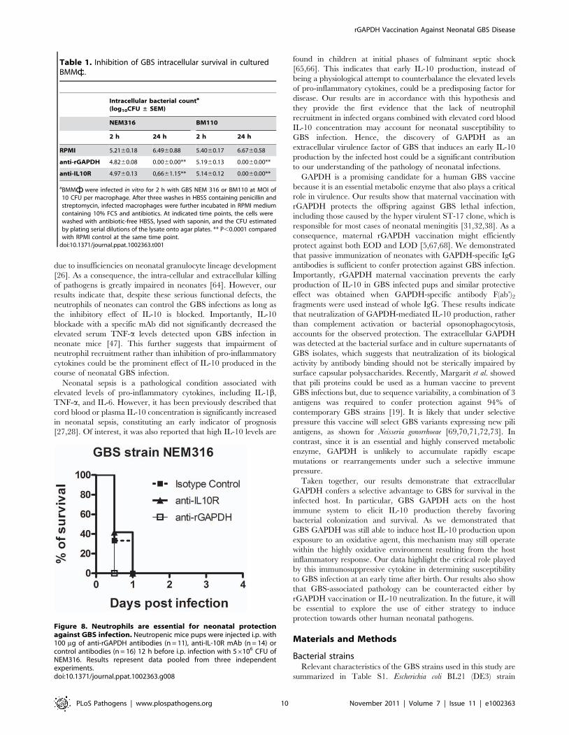
(Figure S4). We observed that either blocking IL-10 signaling with
neutralizing antibodies or anti-rGAPDH antibody treatment was
not sufficient to protect neutropenic pups infected with a lethal
inoculum of GBS (Figure 8).
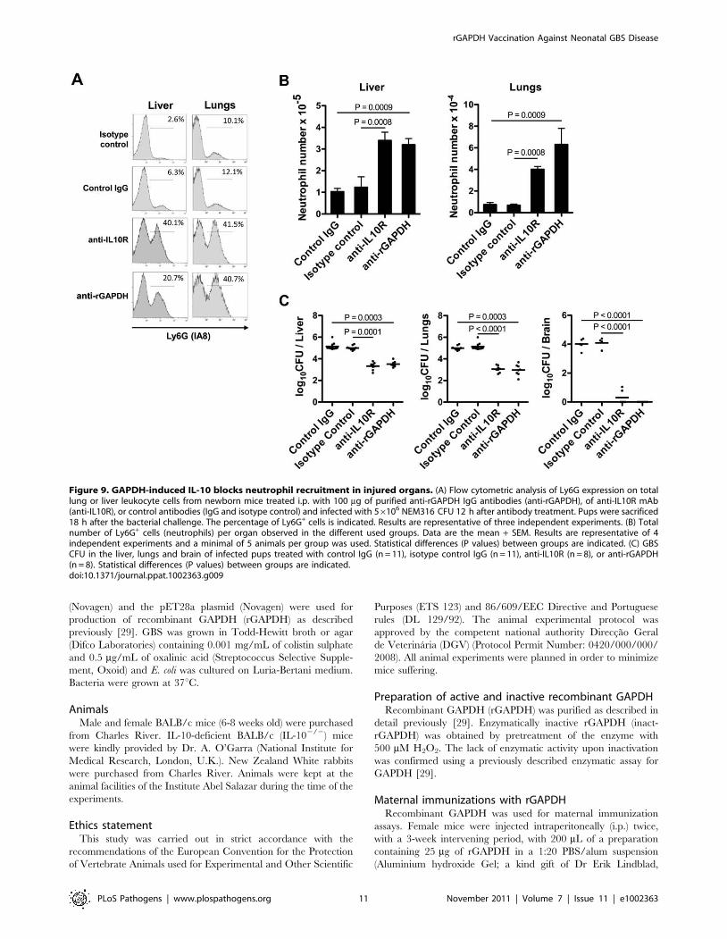
In addition, we assessed the numbers and frequency of
neutrophils in the liver and lungs of GBS NEM316-infected pups
previously treated with anti-rGAPDH IgG or anti-IL10R mAb.
The frequency and the total number of neutrophils quantified
18 h after GBS challenge in the analyzed organs of infected pups
Figure 4. Passive immunization with purified anti-rGAPDH antibodies protects newborn mice from GBS-induced death. anti-rGAPDHIgG or control IgG (80 mg) were injected i.p. into mice pups and 12 h after the immunization they were infected i.p. with (A) 56106 NEM316 CFU orwith (B) 106 CFU of the ST-17 hyper virulent strain BM110. The results represent data pooled from two independent experiments. (C) Upper panel:BMM were stimulated in vitro with 106 CFU of GBS NEM316 (or BM110) plus 25 mg/mL of anti-rGAPDH IgG’s, or 10% of serum containing anti-GBSantibodies, and incubated for 30 min at 37uC in 5% CO2. Data represent the mean + SEM. Results are representative of 3 independent experiments.Statistical differences (P values) are indicated. Lower panel: complement-mediated killing of GBS. Mice peripheral blood leukocytes were stimulatedwith 106 CFU of GBS NEM316 (or BM110) plus 25 mg/mL of anti-rGAPDH IgG’s, or 10% of anti-GBS serum, and 5% of rabbit serum was added to themixture as a source of complement. After 2 h of incubation period at 37uC, GBS CFU were evaluated on agar plates. Data represent the mean + SEM.Results are representative of 2 independent experiments. Statistical differences (P values) are indicated. Similar results were obtained when a higherconcentration (100 mg/mL) of anti-rGAPDH IgG’s was used. (D) Pups were passively immunized with 80 mg of anti-rGAPDH F(ab’)2 fragments, orcontrol (Fab’)2 and infected 12 h later with 106 CFU of the ST-17 hyper virulent strain BM110. The results represent data pooled from twoindependent experiments.doi:10.1371/journal.ppat.1002363.g004
zation. Moreover, no bacterial colonization was detected three
weeks after GBS infection in the brain or in any other organ of
pups born from rGAPDH-immunized mothers and infected with
NEM316 or with the ST-17 hyper-virulent strain BM110 (data not
shown). These results indicate that rGAPDH-maternal vaccination
is also effective in preventing LOD.
Figure 5. GAPDH neutralization abolishes IL-10 production observed in newborn mice early upon GBS infection. Serum IL-10concentration in mice pups was measured 1 h and 4 h after i.p. infection with 56106 NEM316 CFU (A) born from sham- or rGAPDH-immunized damsor (B) passively immunized with anti-rGAPDH F(ab’)2 fragments or control F(ab’)2. Data represent the mean + SEM. Results are a representativeexample out of 3 independent experiments. A minimum of 8 animals per group was used in each experiment. Statistical differences (P values)between immunized versus sham-immunized groups (A) or between pups treated with anti-rGAPDH F(ab’)2 fragments versus controls (B) areindicated.doi:10.1371/journal.ppat.1002363.g005
leukocytes are highly committed to produce increased amounts
of IL-10 [44,45], as also shown in human neonates [41,43,46]. As
report in this work, high levels of serum IL-10 could be detected in
the sera of GBS infected newborn mice early (1 h and 4 h) upon
infection. This result is in agreement with a previous report by
Cusumano et al. [47], showing that elevated levels of plasma IL-10
were detected in newborn mice 24 h and 48 h upon GBS
challenge. These authors suggested a host protective role for IL-10
in the outcome of neonatal GBS sepsis as pre-treatment of
newborn mice with recombinant IL-10 improved their survival
upon a lethal s.c. GBS challenge [47]. Nevertheless, they also
showed that a therapeutic administration of this cytokine (24 h
after the bacterial challenge) did not improve survival. This would
limit its use in human therapies because neonatal GBS infection is
usually acquired before or during labor [3,5,6].
Our results revealed that blocking IL-10 signaling through anti-
IL-10R mAb administration was sufficient to confer protection
against a bacterial challenge using either the s.c. or i.p. routes.
Thus, they contrastingly indicate that IL-10 has a deleterious effect
in the newborn host resistance to GBS infection. The increased
resistance of IL-10-deficient neonates to GBS infection reported
here constitutes further support for a deleterious effect of IL-10 in
host resistance to GBS. As shown here, the GBS GAPDH induced
host IL-10 production detected early after bacterial infection.
IL-10 is produced by multiple cell types and inhibits leukocyte
activation, pro-inflammatory cytokine production and down-
regulates the expression of anti-microbial molecules on activated
phagocytes [48,49,50,51]. IL-10 also inhibits production of CC
and CXC chemokines by activated monocytes [52,53,54]. Since
these chemokines are implicated in the recruitment of leukocytes
during inflammation, IL-10 production indirectly inhibits leuko-
cyte trafficking to inflamed tissues [50,55,56].
IL-10 production was already associated with host susceptibility
against different pathogens [24,45,57,58,59,60,61]. We show here
that treatment of pups with either anti-IL10R mAb or anti-
rGAPDH IgG prior to the GBS challenge increased the neutrophil
recruitment in liver and lungs that is triggered upon infection.
Neutrophil recruitment is a crucial event in the host effector
immune response to GBS [21,62,63] and, consequently, lack of
neutrophil infiltration in infected sites has been reported in cases of
severe early-onset GBS sepsis [21,22,23,38]. Thus, neutralization
of GAPDH, and hence blockade of the induced IL-10 production,
allowed an effective immune response at an early stage of infection
that prevented death of pups. Moreover, pups protected by
maternal immunization with rGAPDH presented no GBS CFU in
the brain, lungs, and liver 3 weeks upon the infectious challenge.
This indicates that protection achieved by this vaccination strategy
might prevent LOD.
The recruitment of neutrophils into infected tissues is very
important to restrain bacterial replication. Thus, qualitative and
quantitative deficiencies in the neutrophils of newborns may
explain the observed susceptibility to GBS infections. Indeed,
newborn neutrophils have reduced adhesion capabilities due to
reduced expression of adhesion molecules [24,25] and they
produced a limited number of microbicidal molecules. Moreover,
the number of these cells is also reduced when compared to adults
Figure 7. Simultaneous injection of anti-rGAPDH IgG’s and anti-IL10R mAb does not increase survival of newborn mice infectedwith GBS BM110. Newborn mice were treated i.p. with 100 mg of anti-IL10R mAb, anti-rGAPDH IgG’s, or simultaneous with anti-IL10R mAb andanti-rGAPDH IgG’s (anti-rGAPDH + anti-IL10R) 12 h before s.c. injection of 2.56104 CFU of GBS BM110. Control pups received 100 mg of control IgG’s.P,0.0001 between anti-IL10R-, anti-rGAPDH- and anti-rGAPDH+antiIL-10R-treated pups and controls.doi:10.1371/journal.ppat.1002363.g007
Figure 6. Impairment of IL-10 signaling confers protection to newborn mice against GBS infection. (A) IL102/2 and WT pups wereinfected i.p. with 56106 NEM316 CFU (results represent data pooled from two independent experiments). (B) Newborn mice were injected i.p. withanti-IL10R mAb (anti-IL10R) or isotype control IgG (100 mg) and 12 h later were challenged i.p. with 56106 NEM316 CFU (results represent datapooled from three independent experiments). (C) Newborn mice were injected i.p. with anti-IL10R mAb (anti-IL10R) or isotype control IgG (100 mg)and 12 h later were challenged s.c. with 2.56104 BM110 CFU (results represent data pooled from four independent experiments). Statisticaldifferences (P values) between IL-102/2 and WT pups (A) or between immunized versus sham-immunized groups (B and C) are indicated.doi:10.1371/journal.ppat.1002363.g006
aBMM were infected in vitro for 2 h with GBS NEM 316 or BM110 at MOI of10 CFU per macrophage. After three washes in HBSS containing penicillin andstreptomycin, infected macrophages were further incubated in RPMI mediumcontaining 10% FCS and antibiotics. At indicated time points, the cells werewashed with antibiotic-free HBSS, lysed with saponin, and the CFU estimatedby plating serial dilutions of the lysate onto agar plates. ** P,0.0001 comparedwith RPMI control at the same time point.
doi:10.1371/journal.ppat.1002363.t001
Figure 8. Neutrophils are essential for neonatal protectionagainst GBS infection. Neutropenic mice pups were injected i.p. with100 mg of anti-rGAPDH antibodies (n = 11), anti-IL-10R mAb (n = 14) orcontrol antibodies (n = 16) 12 h before i.p. infection with 56106 CFU ofNEM316. Results represent data pooled from three independentexperiments.doi:10.1371/journal.ppat.1002363.g008
rGAPDH) was obtained by pretreatment of the enzyme with
500 mM H2O2. The lack of enzymatic activity upon inactivation
was confirmed using a previously described enzymatic assay for
GAPDH [29].
Maternal immunizations with rGAPDHRecombinant GAPDH was used for maternal immunization
assays. Female mice were injected intraperitoneally (i.p.) twice,
with a 3-week intervening period, with 200 mL of a preparation
containing 25 mg of rGAPDH in a 1:20 PBS/alum suspension
(Aluminium hydroxide Gel; a kind gift of Dr Erik Lindblad,
Figure 9. GAPDH-induced IL-10 blocks neutrophil recruitment in injured organs. (A) Flow cytometric analysis of Ly6G expression on totallung or liver leukocyte cells from newborn mice treated i.p. with 100 mg of purified anti-rGAPDH IgG antibodies (anti-rGAPDH), of anti-IL10R mAb(anti-IL10R), or control antibodies (IgG and isotype control) and infected with 56106 NEM316 CFU 12 h after antibody treatment. Pups were sacrificed18 h after the bacterial challenge. The percentage of Ly6G+ cells is indicated. Results are representative of three independent experiments. (B) Totalnumber of Ly6G+ cells (neutrophils) per organ observed in the different used groups. Data are the mean + SEM. Results are representative of 4independent experiments and a minimal of 5 animals per group was used. Statistical differences (P values) between groups are indicated. (C) GBSCFU in the liver, lungs and brain of infected pups treated with control IgG (n = 11), isotype control IgG (n = 11), anti-IL10R (n = 8), or anti-rGAPDH(n = 8). Statistical differences (P values) between groups are indicated.doi:10.1371/journal.ppat.1002363.g009
erythrocytes, rabbit erythrocytes or mouse muscle as previously
described [75,76].
Statistical analysisStudent’s T test was used to analyze the differences between
groups. Survival studies were analyzed with the log-rank test. A P
value,0.05 was considered statistically significant.
Supporting Information
Figure S1 Increased anti-rGAPDH IgG serum titers inmice pups born from rGAPDH-vaccinated mothers.Newborn mice from rGAPDH-immunized mothers present higher
serum anti-rGAPDH IgG antibody titers than controls born from
sham-immunized mothers. Anti-rGAPDH antibody titers were
determined by ELISA. Results are pooled data from three
independent experiments (n = 13 or 17 for pups born from
sham-immunized or rGAPDH-immunized mothers, respectively).
(TIF)
Figure S2 GAPDH is present at GBS cell surface.Fluorescence microscopy analysis of GBS cells using anti-
rGAPDH polyclonal antibodies purified from rGAPDH-immu-
nized rabbits and revealed with FITC-conjugated anti-rabbit IgG
(Green). Bacterial DNA was stained with DAPI (blue). GBS cells
incubated with (A) secondary antibody only, (B) with anti-
rGAPDH plus secondary antibody, or (C) with anti-rGAPDH
plus rGAPDH to inhibit antibody binding to surface-localized
antigen.
(TIF)
Figure S3 Active or enzymatically inactive rGAPDHinduces IL-10 production. IL-10 concentration in the sera of
newborn mice 1 h after i.p. injection with 50 mg of rGAPDH or
rGAPDH pre-treated with 500 mM H2O2 (inact_rGAPDH).
Control mice were injected with PBS. Results are pooled data
from two independent experiments (n = 9 for controls, 8 for
rGAPDH and 7 for pups treated with inact_rGAPDH). Statistical
differences (P values) between groups are indicated.
(TIF)
Figure S4 Treatment of newborn mice with anti-Ly6Gantibodies induces neutropenia. Pups were treated i.p. 36 h
and 48 h after birth with 40 mg of anti-Ly6G (clone 1A8) mAb or
with the same amount of an isotype matched control antibody.
The frequency of blood neutrophils was determined 4 h after the
last injection by FACS analysis using anti-Gr-1 mAb (clone RB6-
8C5).
(TIF)
Table S1 Phenotypic and genotypic characteristics ofthe GBS human isolates used in this study.(DOC)
Acknowledgments
We thank Anne O’Garra and Rui Appelberg for scientific discussion.
Author Contributions
Conceived and designed the experiments: PF PTC MCN. Performed the
experiments: PM EBA BG LO SM AR. Analyzed the data: PM PF.
Contributed reagents/materials/analysis tools: PF PTC MCN. Wrote the
paper: PF PM MV PTC.
References
1. Baker CJ, Barrett FF, Gordon RC, Yow MD (1973) Suppurative meningitis dueto streptococci of Lancefield group B: a study of 33 infants. J Pediatr 82:
724–729.
2. Barton LL, Feigin RD, Lins R (1973) Group B beta hemolytic streptococcalmeningitis in infants. J Pediatr 82: 719–723.
3. Edwards MS (2006) Issues of antimicrobial resistance in group B streptococcus
in the era of intrapartum antibiotic prophylaxis. Semin Pediatr Infect Dis 17:149–152.
4. Heath PT, Schuchat A (2007) Perinatal group B streptococcal disease. Best PractRes Clin Obstet Gynaecol 21: 411–424.
5. Johri AK, Paoletti LC, Glaser P, Dua M, Sharma PK, et al. (2006) Group B
Streptococcus: global incidence and vaccine development. Nat Rev Microbiol 4:932–942.
6. Zangwill KM, Schuchat A, Wenger JD (1992) Group B streptococcal disease in
the United States, 1990: report from a multistate active surveillance system.MMWR CDC Surveill Summ 41: 25–32.
7. Jordan HT, Farley MM, Craig A, Mohle-Boetani J, Harrison LH, et al. (2008)
Revisiting the need for vaccine prevention of late-onset neonatal group Bstreptococcal disease: a multistate, population-based analysis. Pediatr Infect Dis J
27: 1057–1064.
8. Fluegge K, Siedler A, Heinrich B, Schulte-Moenting J, Moennig MJ, et al.(2006) Incidence and clinical presentation of invasive neonatal group B
streptococcal infections in Germany. Pediatrics 117: e1139–1145.
9. Hajdu A, Blystad H, Hoiby EA, Klouman E, Schimmer B, et al. (2006)Unexpected increase in case fatality of invasive group B streptococcal infections
in infants in Norway, January-July 2006. Euro Surveill 11: E060727 060722.10. Heath PT, Balfour G, Weisner AM, Efstratiou A, Lamagni TL, et al. (2004)
Group B streptococcal disease in UK and Irish infants younger than 90 days.
Lancet 363: 292–294.11. Baltimore RS (2007) Consequences of prophylaxis for group B streptococcal
infections of the neonate. Semin Perinatol 31: 33–38.
12. Castor ML, Whitney CG, Como-Sabetti K, Facklam RR, Ferrieri P, et al. (2008)Antibiotic resistance patterns in invasive group B streptococcal isolates. Infect
Dis Obstet Gynecol 727505 2008: 727505.
13. Doran KS, Nizet V (2004) Molecular pathogenesis of neonatal group Bstreptococcal infection: no longer in its infancy. Mol Microbiol 54: 23–31.
14. Amundson NR, Flores AE, Hillier SL, Baker CJ, Ferrieri P (2005) DNA
macrorestriction analysis of nontypeable group B streptococcal isolates: clonalevolution of nontypeable and type V isolates. J Clin Microbiol 43: 572–576.
15. Kong F, Lambertsen LM, Slotved HC, Ko D, Wang H, et al. (2008) Use of
phenotypic and molecular serotype identification methods to characterize
previously nonserotypeable group B streptococci. J Clin Microbiol 46:
29. Madureira P, Baptista M, Vieira M, Magalhaes V, Camelo A, et al. (2007)
Streptococcus agalactiae GAPDH is a virulence-associated immunomodulatoryprotein. J Immunol 178: 1379–1387.
30. Jones N, Bohnsack JF, Takahashi S, Oliver KA, Chan MS, et al. (2003)
Multilocus sequence typing system for group B streptococcus. J Clin Microbiol41: 2530–2536.
31. Lamy MC, Dramsi S, Billoet A, Reglier-Poupet H, Tazi A, et al. (2006) Rapid
detection of the ‘‘highly virulent’’ group B Streptococcus ST-17 clone. MicrobesInfect 8: 1714–1722.
32. Poyart C, Reglier-Poupet H, Tazi A, Billoet A, Dmytruk N, et al. (2008) Invasivegroup B streptococcal infections in infants, France. Emerg Infect Dis 14:
41. Belderbos ME, van Bleek GM, Levy O, Blanken MO, Houben ML, et al. (2009)
Skewed pattern of Toll-like receptor 4-mediated cytokine production in human
neonatal blood: low LPS-induced IL-12p70 and high IL-10 persist throughoutthe first month of life. Clin Immunol 133: 228–237.
42. Chelvarajan L, Popa D, Liu Y, Getchell TV, Stromberg AJ, et al. (2007)Molecular mechanisms underlying anti-inflammatory phenotype of neonatal
splenic macrophages. J Leukoc Biol 82: 403–416.
43. Kollmann TR, Crabtree J, Rein-Weston A, Blimkie D, Thommai F, et al. (2009)Neonatal innate TLR-mediated responses are distinct from those of adults.
J Immunol 183: 7150–7160.
44. Sun CM, Deriaud E, Leclerc C, Lo-Man R (2005) Upon TLR9 signaling, CD5+B cells control the IL-12-dependent Th1-priming capacity of neonatal DCs.
Immunity 22: 467–477.
45. Genovese F, Mancuso G, Cuzzola M, Biondo C, Beninati C, et al. (1999) Role ofIL-10 in a neonatal mouse listeriosis model. J Immunol 163: 2777–2782.
46. Rainsford E, Reen DJ (2002) Interleukin 10, produced in abundance by humannewborn T cells, may be the regulator of increased tolerance associated with
47. Cusumano V, Genovese F, Mancuso G, Carbone M, Fera MT, et al. (1996)Interleukin-10 protects neonatal mice from lethal group B streptococcal
infection. Infect Immun 64: 2850–2852.
48. Fiorentino DF, Zlotnik A, Mosmann TR, Howard M, O’Garra A (1991) IL-10inhibits cytokine production by activated macrophages. J Immunol 147:
3815–3822.
49. Fiorentino DF, Zlotnik A, Vieira P, Mosmann TR, Howard M, et al. (1991) IL-
10 acts on the antigen-presenting cell to inhibit cytokine production by Th1 cells.
J Immunol 146: 3444–3451.
50. Moore KW, de Waal Malefyt R, Coffman RL, O’Garra A (2001) Interleukin-10
and the interleukin-10 receptor. Annu Rev Immunol 19: 683–765.
51. Saraiva M, O’Garra A (2010) The regulation of IL-10 production by immunecells. Nat Rev Immunol 10: 170–181.
52. Berkman N, John M, Roesems G, Jose PJ, Barnes PJ, et al. (1995) Inhibition ofmacrophage inflammatory protein-1 alpha expression by IL-10. Differential
sensitivities in human blood monocytes and alveolar macrophages. J Immunol
155: 4412–4418.
53. Kopydlowski KM, Salkowski CA, Cody MJ, van Rooijen N, Major J, et al.
(1999) Regulation of macrophage chemokine expression by lipopolysaccharidein vitro and in vivo. J Immunol 163: 1537–1544.
54. Marfaing-Koka A, Maravic M, Humbert M, Galanaud P, Emilie D (1996)
Contrasting effects of IL-4, IL-10 and corticosteroids on RANTES productionby human monocytes. Int Immunol 8: 1587–1594.
55. Zuany-Amorim C, Creminon C, Nevers MC, Nahori MA, Vargaftig BB, et al.(1996) Modulation by IL-10 of antigen-induced IL-5 generation, and CD4+ T
lymphocyte and eosinophil infiltration into the mouse peritoneal cavity.
J Immunol 157: 377–384.56. Zuany-Amorim C, Haile S, Leduc D, Dumarey C, Huerre M, et al. (1995)
Interleukin-10 inhibits antigen-induced cellular recruitment into the airways ofsensitized mice. J Clin Invest 95: 2644–2651.
57. Belkaid Y, Hoffmann KF, Mendez S, Kamhawi S, Udey MC, et al. (2001) Therole of interleukin (IL)-10 in the persistence of Leishmania major in the skin after
healing and the therapeutic potential of anti-IL-10 receptor antibody for sterile
cure. J Exp Med 194: 1497–1506.58. Murphy ML, Wille U, Villegas EN, Hunter CA, Farrell JP (2001) IL-10
mediates susceptibility to Leishmania donovani infection. Eur J Immunol 31:2848–2856.
mediates susceptibility to Trypanosoma cruzi infection. J Immunol 153:3135–3140.
60. Roque S, Nobrega C, Appelberg R, Correia-Neves M (2007) IL-10 underliesdistinct susceptibility of BALB/c and C57BL/6 mice to Mycobacterium avium
infection and influences efficacy of antibiotic therapy. J Immunol 178:8028–8035.
61. Silva RA, Appelberg R (2001) Blocking the receptor for interleukin 10 protects
mice from lethal listeriosis. Antimicrob Agents Chemother 45: 1312–1314.62. Cleat PH, Wells C, Coid CR (1984) Electron microscopic evidence of antibody
entry into neutrophils after phagocytosis of highly virulent group B streptococci.J Gen Microbiol 130: 3059–3061.
63. Schuit KE, DeBiasio R (1980) Kinetics of phagocyte response to group B
streptococcal infections in newborn rats. Infect Immun 28: 319–324.64. Levy O, Martin S, Eichenwald E, Ganz T, Valore E, et al. (1999) Impaired
innate immunity in the newborn: newborn neutrophils are deficient inbactericidal/permeability-increasing protein. Pediatrics 104: 1327–1333.
65. Derkx B, Marchant A, Goldman M, Bijlmer R, van Deventer S (1995) Highlevels of interleukin-10 during the initial phase of fulminant meningococcal
septic shock. J Infect Dis 171: 229–232.
66. Lehmann AK, Halstensen A, Sornes S, Rokke O, Waage A (1995) High levels ofinterleukin 10 in serum are associated with fatality in meningococcal disease.
Infect Immun 63: 2109–2112.67. Schrag SJ, Zywicki S, Farley MM, Reingold AL, Harrison LH, et al. (2000)
Group B streptococcal disease in the era of intrapartum antibiotic prophylaxis.
N Engl J Med 342: 15–20.68. Schuchat A (1998) Epidemiology of group B streptococcal disease in the United
States: shifting paradigms. Clin Microbiol Rev 11: 497–513.69. Abu-Asab MS, Laassri M, Amri H (2010) Algorithmic assessment of vaccine-
induced selective pressure and its implications on future vaccine candidates. AdvBioinformatics 178069 2010: 178069.
70. Haas R, Meyer TF (1986) The repertoire of silent pilus genes in Neisseria
gonorrhoeae: evidence for gene conversion. Cell 44: 107–115.71. Helm RA, Seifert HS (2009) Pilin antigenic variation occurs independently of
the RecBCD pathway in Neisseria gonorrhoeae. J Bacteriol 191: 5613–5621.72. Hill SA, Davies JK (2009) Pilin gene variation in Neisseria gonorrhoeae: reassessing
the old paradigms. FEMS Microbiol Rev 33: 521–530.
73. Kline KA, Criss AK, Wallace A, Seifert HS (2007) Transposon mutagenesisidentifies sites upstream of the Neisseria gonorrhoeae pilE gene that modulate pilin
antigenic variation. J Bacteriol 189: 3462–3470.74. Tushinski RJ, Oliver IT, Guilbert LJ, Tynan PW, Warner JR, et al. (1982)
Survival of mononuclear phagocytes depends on a lineage-specific growth factor
that the differentiated cells selectively destroy. Cell 28: 71–81.75. He RQ, Yang MD, Zheng X, Zhou JX (1995) Isolation and some properties of
glycated D-glyceraldehyde-3-phosphate dehydrogenase from rabbit muscle.Biochem J 309(Pt 1): 133–139.
76. Mountassif D, Baibai T, Fourrat L, Moutaouakkil A, Iddar A, et al. (2009)Immunoaffinity purification and characterization of glyceraldehyde-3-phosphate
dehydrogenase from human erythrocytes. Acta Biochim Biophys Sin (Shanghai)