Page 1
i
Inhibition of Polyphenoloxidase and Melanosis in Pacific White Shrimp
(Litopenaeus vannamei) by Phenolic Compounds
Nilesh Prakash Nirmal
A Thesis Submitted in Fulfillment of the Requirements for the Degree of
Doctor of Philosophy in Food Science and Technology
Prince of Songkla University
2011
Copyright of Prince of Songkla University
Page 3
iii
Thesis Title Inhibition of Polyphenoloxidase and Melanosis in Pacific White
Shrimp (Litopenaeus vannamei) by Phenolic Compounds
Author Mr.Nilesh Prakash Nirmal
Major Program Food Science and Technology
Academic Year 2010
ABSTRACT
Effects of ferulic acid and catechin on polyphenoloxidase (PPO) from
Pacific white shrimp (Litopenaeus vannamei) were evaluated. Both compounds
showed PPO inhibitory activity in a dose dependent manner (P < 0.05). When whole
shrimp were treated with ferulic acid solution (1 or 2%) or catechin solution (0.05 or
0.1 %) and stored in ice for 10 days, the increase in psychrotrophic bacteria count
(PBC), pH, total volatile base (TVB) content and thiobarbituric acid reactive
substances (TBARS) were retarded, in comparison with the control and those treated
with 1.25% sodium metabisulfite (SMS) (P < 0.05). After 10 days of storage, shrimp
treated with 2% ferulic acid or 0.1% catechin had the lower melanosis score and
higher likeness, compared with the control and SMS treated shrimp (P < 0.05).
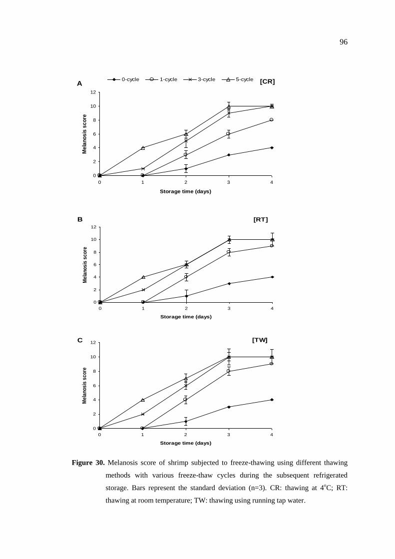
Shrimp subjected to freeze-thawing with different thawing methods
and various cycles showed the increase in melanosis during subsequent refrigerated
storage (4oC) up to 4 days. Melanosis score was lower in shrimp thawed at 4oC,
compared with that found in samples thawed at room temperature or using tap water
(P < 0.05). Shrimp treated with catechin (0.05, 0.1 and 0.2% (w/v)) or ferulic acid (1,
2 and 3% (w/v)) and subjected to freeze-thawing with various cycles had lower
melanosis and quality changes during the subsequent refrigerated storage of 4 days,
compared with the control (P < 0.05). Thus, either catechin or ferulic acid could be
used as the potential additive to lower melanosis of shrimp with prior freeze-thawing.
Green tea extract (GTE) was used as a natural source of catechin with
PPO inhibitory activity. GTE with and without prior chlorophyll removal showed the
higher PPO inhibitory activity, compared with mulberry tea extract. Additionally,
GTE had the higher antioxidant activity, compared to mulberry tea extract (P < 0.05).
Whole shrimp treated with GTE with prior chlorophyll removal at concentrations of
Page 4
iv
0.5 and 1 % (w/v) and stored in ice for 12 days lowered PBC, lipid oxidation, and
melanosis formation, compared with the control and shrimp treated with 1.25 % SMS
(P < 0.05). Furthermore, GTE with prior chlorophyll removal had no adverse impact
on sensory attributes of treated shrimp. When GTE (0.1%) was used in combination
with ascorbic acid (AA; 0.005 or 0.1%), the greater PPO inhibitory activity was
achieved as compared to GTE alone (P < 0.05). Shrimp treated with 0.1 % GTE in
combination with AA (0.005 or 0.01%) (GTE + AA) and stored in iced had the
lowered quality changes, in comparison with the control and those treated with 1.25
% SMS (P < 0.05). Shrimp without treatment stored under modified atmosphere
packaging (MAP) had lowered microbial and chemical changes, in comparison with
shrimp stored in air (control) (P < 0.05). When shrimp were treated with GTE in
combination with AA and stored under MAP, higher inhibition on melanosis
formation and microbial growth was obtained, compared with other treatments (P <
0.05). Therefore, shrimp treated with GTE in combination with AA prior to MAP had
the lowest losses in quality during refrigerated storage.
Lead (Leucaena leucocephala) brown seed extract was studied for
PPO inhibitory activity. Lead seed extract powder (LSEP) was prepared using
distilled water as a medium. LSEP (0.05, 0.1, 0.25, 0.5, and 1 %, w/v) showed PPO
inhibitory activity in a dose dependent manner. When the whole shrimp were treated
with 0.25 and 0.5 % (w/v) LSEP, the shrimp treated with 0.5 % LSEP had the lower
melanosis score and showed a higher likeness, compared with the control and 1.25 %
SMS treated samples at day 12 of iced storage (P < 0.05). Thus, LSEP can be used as
a novel melanosis inhibitor for Pacific white shrimp.
Biochemical properties of PPO from cephalothorax of Pacific white
shrimp were investigated. PPO showed the maximal activity using L- -(3, 4
dihydroxylphenyl) alanine (L-DOPA) as a substrate at pH 6 and 55oC. PPO was stable
over a pH range of 5-10 but was unstable at a temperature greater than 60oC. Based
on the activity staining with L-DOPA, the apparent molecular weight of PPO was 210
kDa. The Michaelis constant (Km) of PPO for the oxidation of L-DOPA was 2.43
mM. Trypsin, copper acetate, and sodium dodecyl sulfate (SDS) were unable to
activate PPO, suggesting that the enzyme was in the active form.
Page 5
v
Inhibition kinetics and mode of catechin, ferulic acid and mimosine
towards PPO from cephalothorax of Pacific white shrimp were investigated. Catechin
and mimosine showed mixed type reversible inhibition with Ki value of 1.4 and 3.7
mM, respectively. Inhibition kinetic study of ferulic acid exhibited non-competitive
reversible inhibition on PPO with Ki value of 37 mM. With increasing concentrations,
catechin or ferulic acid or mimosine had higher copper (Cu2+) reduction and copper
chelating capacity (P < 0.05). Addition of catechin or ferulic acid or mimosine, into
browning reaction could prevent dopachrome formation by inactivation of PPO or by
binding with browning product.
Therefore, phenolic compounds including catechin, ferulic acid and
mimosine could be a safe natural substitute for the synthetic chemical used in shrimp
processing industry to lower melanosis and quality losses of shrimp during extended
iced or refrigerated storage.
Page 6
viii
CONTENTS
Page
Contents................................................................................................................. viii
List of Tables......................................................................................................... xviii
List of Figures........................................................................................................ xx
Chapter
1. Introduction and Review of Literature
1.1 Introduction………….……...…………………………………...……… 1
1.2 Review of literature…………………………………………………...… 3
1.2.1 Polyphenoloxidase (PPO) ………………………………………... 3
1.2.1.1 Distribution (localization) of PPO in crustaceans…………. 3
1.2.1.2 Molecular structure………………………………………… 5
1.2.1.3 Enzyme mechanisms ………………………………………. 7
1.2.1.3.1 Monophenol oxidase…………………………………. 7
1.2.1.3.2 Diphenol oxidase …………………………………….. 9
1.2.1.4 Characteristics of PPO from crustaceans………………… 10
1.2.1.4.1 Molecular weight ………………………………… 10
1.2.1.4.2 pH optima and stability …………………………… 10
1.2.1.4.3 Temperature optima and stability …………………. 10
1.2.2 Melanosis and factors influencing melanosis in crustaceans……. 12
1.2.2.1 Species ………………………………………………… .... 13
1.2.2.2 Method of capture and season ……………………...…….. 13
1.2.2.3 Metal ion ………………………………………………….. 13
1.2.2.4 Protease and some chemicals ……………………………… 14
1.2.3 Melanosis/ PPO inhibitors………………………………………… 15
1.2.3.1 Acidulants………………………………………………….. 15
1.2.3.2 Chelating agents ………………………………………… 16
1.2.3.3 Reducing agents …………………………………………… 16
1.2.3.4 PPO inhibitor (4-Hexylresorcinol) ………………………… 18
Page 7
ix
CONTENTS (Continued)
Chapter Page
1.2.3.5 Miscellaneous …………………………………………… 20
1.2.4 Changes in quality of shrimp during post mortem storage………. 22
1.2.4.1 Microbiological changes during storage ………………….. 22
1.2.4.2 Chemical changes during storage ………………………… 23
1.2.4.3 Physical and Sensorial changes during storage…………… 24
1.2.5 Modified atmosphere packaging (MAP) ………………………… 25
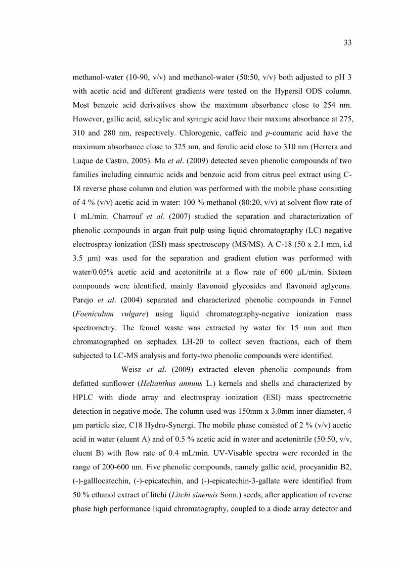
1.2.6 Phenolic compounds …………………………………………….. 27
1.2.6.1 Extraction of phenolic compounds ………………………… 31
1.2.6.2 Identification and characterization of phenolic compounds .. 32
1.2.6.3 Antioxidant activity of phenolic compounds……………….. 34
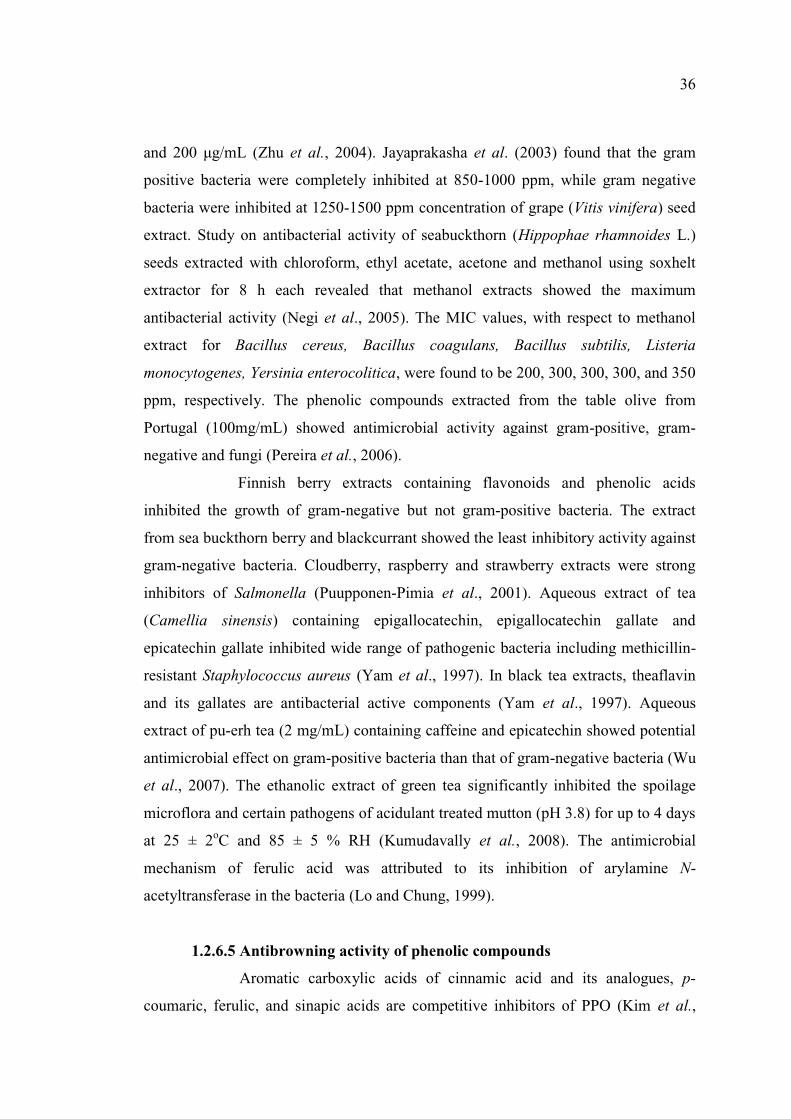
1.2.6.4 Antimicrobial activity of phenolic compounds …………….. 35
1.2.6.5 Antibrowning activity of phenolic compounds …………….. 36
1.2.7 Mimosine…………………………………………………………... 38
1.3 Objectives ……………………………………………………………… 40
2. Effects of ferulic acid on inhibition of polyphenoloxidase and quality
changes of Pacific white shrimp (Litopenaeus vannamei) during iced
storage
2.1 Abstract………………………………………………..……………… 41
2.2 Introduction……………………………………………………………… 41
2.3 Material and Methods……………………………………………………. 43
2.4 Results and Discussion…………………………………………………. 50
2.4.1 Effect of ferulic acid on the inhibition of PPO…………………… 50
2.4.2 Effect of FA on the microbiological changes of Pacific white
shrimp during iced storage………………………………………… 53
Page 8
x
CONTENTS (Continued)
Chapter Page
2.4.3. Effect of FA on the chemical changes of Pacific white shrimp
during iced storage ……………………………………………… 55
2.4.3.1 pH ………………………………………………………… 55
2.4.3.2 TVB and TMA contents…………………………………… 56
2.4.3.3 Peroxide value and thiobarbituric acid reactive substances
(TBARS) value ……………………………………………… 58
2.4.4 Effect of FA on melanosis of Pacific white shrimp during
iced storage ………………………………………………………… 60
2.4.5 Effect of FA on sensory properties of Pacific white shrimp
during iced storage ………………………………………………….. 61
2.5 Conclusions……………………………………………………………… 62
3. Melanosis and quality changes of Pacific white shrimp (Litopenaeus
vannamei) treated with catechin during iced storage
3.1 Abstract………………………………………………..………………… 63
3.2 Introduction……………………………………………………………… 63
3.3 Material and Methods……………………………………………………. 65
3.4 Results and Discussion…………………………………………………… 70
3.4.1 Effect of catechin treatment on microbiological changes of Pacific
white shrimp during iced storage ……………………………………. 70
3.4.2 Effect of catechin treatment on chemical changes of Pacific white
shrimp during iced storage ……………………………………………. 74
3.4.2.1 pH ……………………………………………………………. 74
3.4.2.2 TVB contents ……………………………………………….. 74
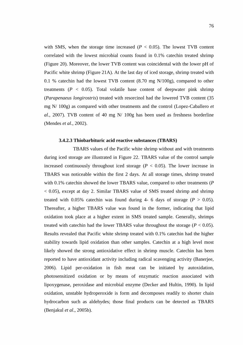
3.4.2.3 Thiobarbituric acid reactive substances (TBARS) ………… 76
3.4.2.4 K-value ………………………………………………………. 77
Page 9
xi
CONTENTS (Continued)
Chapter Page
3.4.3 Effect of catechin treatment on physical changes of Pacific
white shrimp during iced storage …………………………………… 79
3.4.3.1 Shear force …………………………………………………… 79
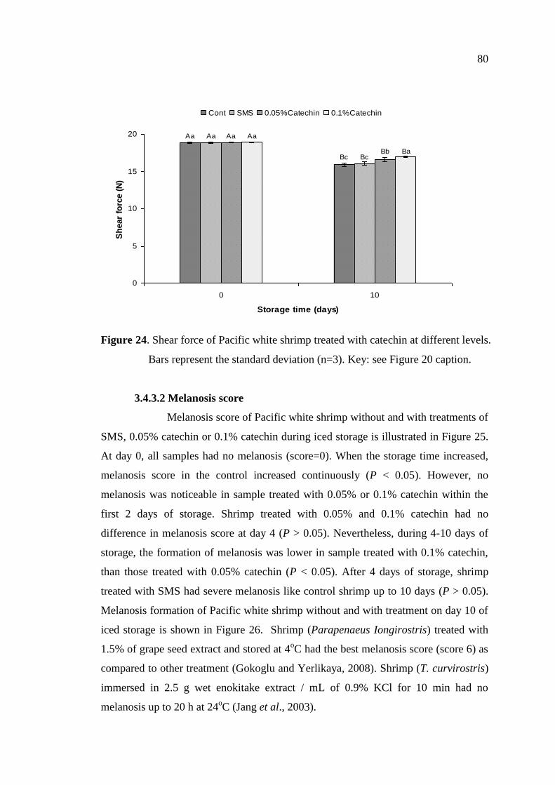
3.4.3.2 Melanosis score ……………………………………………… 80
3.4.4 Effect of catechin on PPO inhibition ………………………………. 82
3.5 Conclusions …………………………………………………………… 83
4. Effect of catechin and ferulic acid on melanosis and quality of Pacific
white shrimp subjected to prior freeze-thawing during refrigerated
storage
4.1 Abstract………………………………………………..………………… 84
4.2 Introduction……………………………………………………………… 84
4.3 Material and Methods…………………………………………………… 86
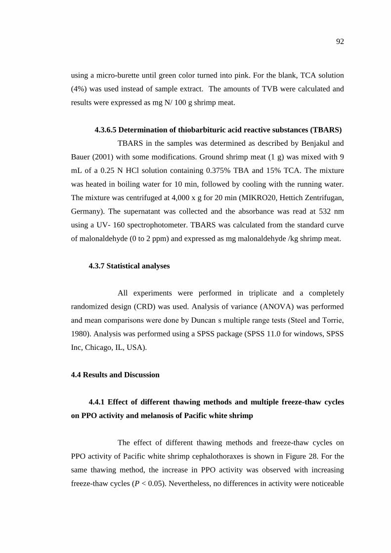
4.4 Results and Discussion…………………………………………………… 92
4.4.1 Effect of different thawing methods and multiple freeze-thaw
cycles on PPO activity and melanosis of Pacific white shrimp ……… 92
4.4.2 Effect of catechin and ferulic acid on melanosis and the quality
changes of Pacific white shrimp with prior freeze-thawing during
the extended refrigerated storage …………………………………….. 97
4.4.2.1 Changes in melanosis ………………………………………… 97
4.4.2.2 Changes in psychrotrophic bacterial count …………………... 101
4.4.2.3 Changes in TVB content …………………………………….. 103
4.4.2.4 Changes in TBARS ………………………………………….. 103
4.5 Conclusions …………………………………………………………….. 104
Page 10
xii
CONTENTS (Continued)
Chapter Page
5. Use of tea extracts for inhibition of polyphenoloxidase and retardation
of quality loss of Pacific white shrimp during iced storage
5.1 Abstract………………………………………………..………………… 105
5.2 Introduction……………………………………………………………… 106
5.3 Material and Methods…………………………………………………… 107
5.4 Results and Discussion…………………………………………………… 114
5.4.1 Characteristics and PPO inhibitory activity of green tea and
mulberry tea extracts prepared under different conditions ………… 114
5.4.1.1 Yield, total phenolic content and total chlorophyll content …. 114
5.4.1.2 PPO inhibitory activity ……………………………………… 116
5.4.2 Antioxidant activities of green tea and mulberry tea extracts
prepared under different conditions ……………………………… 118
5.4.2.1 Reducing power …………………………………………… 118
5.4.2.2 DPPH radical-scavenging activity ………………………… 120
5.4.2.3 Copper chelating activity…………………………………… 121
5.4.3 Identification of phenolic compound in green tea extract ………… 122
5.4.4 Effect of ethanolic green tea extract treatment on melanosis
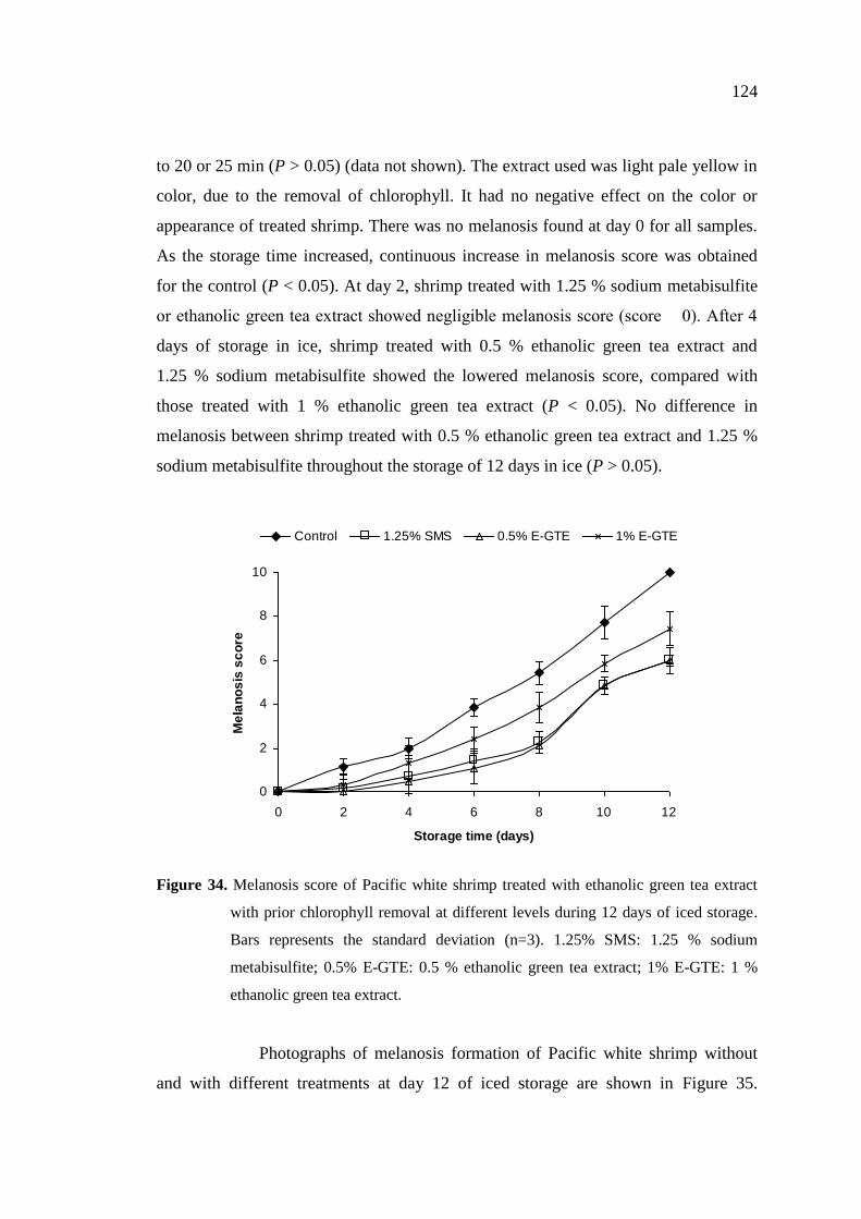
and quality of Pacific white shrimp during iced storage …………. 123
5.4.4.1 Melanosis …………………………………………………… 123
5.4.4.2 Lipid oxidation …………………………………………….. 126
5.4.4.3 Psychrotrophic bacterial count ……………………………… 127
5.4.4.4 Sensory properties…………………………………………… 128
5.5 Conclusions……………………………………………………………... 129
Page 11
xiii
CONTENTS (Continued)
Chapter Page
6. Effect of green tea extract in combination with ascorbic acid on the
retardation of melanosis and quality changes of Pacific white shrimp
during iced storage
6.1 Abstract………………………………………………..………………… 131
6.2 Introduction……………………………………………………………… 131
6.3 Material and Methods…………………………………………………… 133
6.4 Results and Discussion…………………………………………………… 138
6.4.1 Effect of green tea extract without and with different additives
on PPO inhibition……………………………………………………. 138
6.4.2 Effect of green tea extract without and with ascorbic acid on
microbial changes of Pacific white shrimp during iced storage ……... 141
6.4.3 Effect of green tea extract without and with ascorbic acid on
chemical changes of Pacific white shrimp during iced storage………. 144
6.4.4 Effect of green tea extract without and with ascorbic acid on
melanosis of Pacific white shrimp during iced storage ……………… 147
6.4.5 Effect of green tea extract without and with ascorbic acid on
sensory properties of Pacific white shrimp stored in ice …………….. 148
6.5 Conclusions……………………………………………………………… 150
7. Retardation of quality changes of Pacific white shrimp by green
tea extract treatment and modified atmosphere packaging during
refrigerated storage
7.1 Abstract………………………………………………..………………… 151
7.2 Introduction……………………………………………………………… 152
7.3 Material and Methods…………………………………………………… 153
7.4 Results and Discussion…………………………………………………… 157
Page 12
xiv
CONTENTS (Continued)
Chapter Page
7.4.1 Combined effect of GTE with or without AA treatment and MAP
on microbiological changes of Pacific white shrimp during
refrigerated storage …………………………………………………. 157
7.4.2 Combined effect of GTE with or without AA treatment and MAP
on chemical changes of Pacific white shrimp during refrigerated
storage ……………………………………………………………… 162
7.4.2.1 pH …………………………………………………………….. 162
7.4.2.2 Total volatile base (TVB) content …………………………… 163
7.4.2.3 Thiobarbituric acid reactive substances (TBARS) ………….... 164
7.4.3 Combined effect of GTE with or without AA treatment and MAP
on melanosis of Pacific white shrimp during refrigerated storage …. 166
7.4.4 Combined effect of GTE with or without AA treatment and MAP
on sensory properties of Pacific white shrimp during refrigerated
storage ………………………………………………………………. 167
7.5 Conclusions...……………………………………………………………. 169
8. Inhibition of melanosis formation in Pacific white shrimp by the
extract of lead (Leucaena leucocephala) seed
8.1 Abstract………………………………………………..………………… 170
8.2 Introduction……………………………………………………………… 170
8.3 Material and Methods…………………………………………………… 172
8.4 Results and Discussion………………………………………………… 177
8.4.1 Characteristic and PPO inhibitory activity of LSEP………………. 177
8.4.1.1 Extraction yield, total phenolic and mimosine contents ……. 177
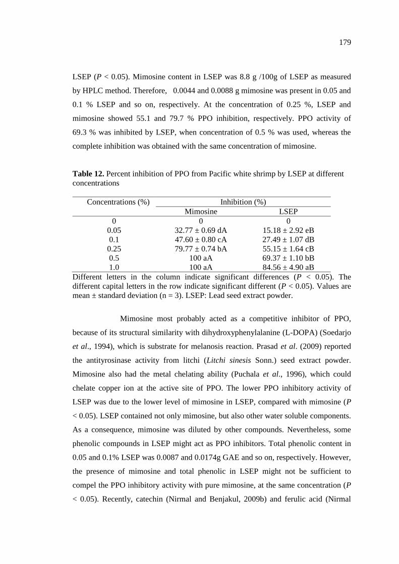
8.4.1.2 PPO inhibitory activity ……………………………………… 178
8.4.2 Effect of LSEP treatment on melanosis of Pacific white shrimp
during iced storage ………………………………………………… 180
Page 13
xv
CONTENTS (Continued)
Chapter Page
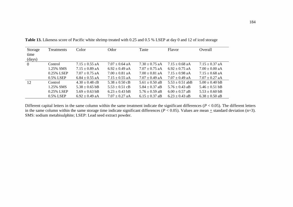
8.4.3 Effect of LSEP treatment on sensory properties of Pacific white
shrimp during iced storage ………………………………………… 183
8.5 Conclusions.……………………………………………………………. 185
9. Biochemical properties of polyphenoloxidase from cephalothorax
of Pacific white shrimp (Litopenaeus vannamei)
9.1 Abstract………………………………………………..………………… 186
9.2 Introduction……………………………………………………………… 186
9.3 Material and Methods…………………………………………………… 187
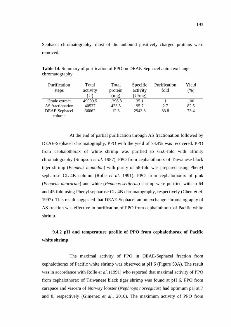
9.4 Results and Discussion………………………………………………… 192
9.4.1 Extraction and partial purification of PPO from cephalothorax of
Pacific white shrimp ………………………………………………… 192
9.4.2 pH and temperature profile of PPO from cephalothorax of Pacific
white shrimp…………………………………………………………. 193
9.4.3 pH and temperature stability of PPO from cephalothorax of Pacific
white shrimp ………………………………………………………… 195
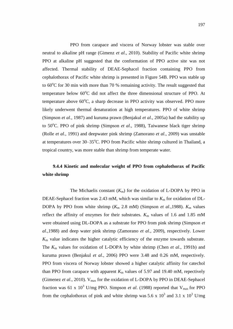
9.4.4 Kinetic and molecular weight of PPO from cephalothorax of Pacific
white shrimp ………………………………………………………… 197
9.4.5 Effect of some chemicals on the activity of PPO from
cephalothorax of Pacific white shrimp …………………………….. 199
9.5 Conclusions……………………………………………………………….. 202
10. Inhibition mode of catechin and ferulic acid on polyphenoloxidase
from cephalothorax of Pacific white shrimp (Litopenaeus vannamei)
10.1 Abstract……………………………………………..………………… 203
10.2 Introduction……………………………………………………………… 203
Page 14
xvi
CONTENTS (Continued)
Chapter Page
10.3 Material and Methods………………………………………………… 204
10.4 Results and Discussion………………………………………………… 209
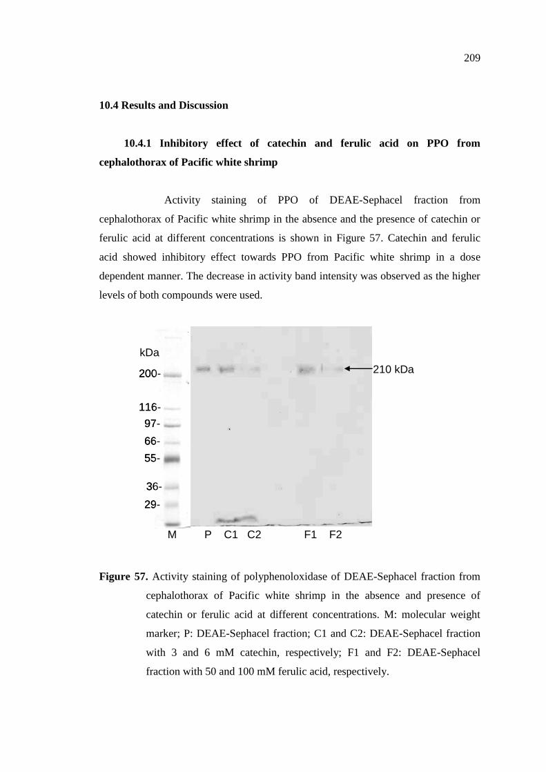
10.4.1 Inhibitory effect of catechin and ferulic acid on PPO from
cephalothorax of Pacific white shrimp………………………….. 209
10.4.2 Inhibition kinetics of catechin and ferulic acid towards PPO ….. 210
10.4.3 Copper reduction capability of catechin and ferulic acid ………. 213
10.4.4 Copper chelating activity of catechin and feurlic acid …………. 214
10.4.5 Effect of catechin and ferulic acid on browning reaction …...….. 215
10.5 Conclusions...……………………………… ………………………….. 216
11. Inhibitory effect of mimosine on polyphenoloxidase from
cephalothoraxes of Pacific white shrimp (Litopenaeus vannamei)
11.1 Abstract……………………………………………..………………… 217
11.2 Introduction……………………………………………………………… 217
11.3 Material and Methods………………………………………………… 219
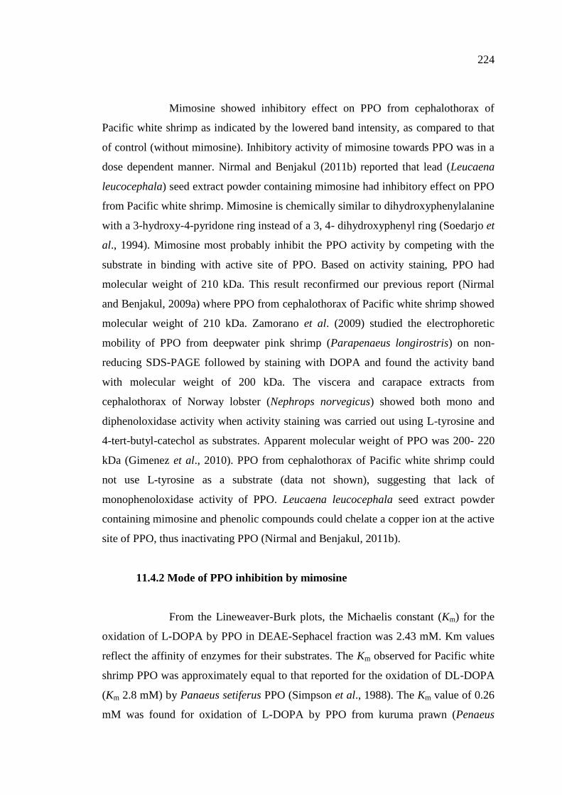
11.4 Results and Discussion………………………………………………… 223
11.4.1 Effect of mimosine on PPO from cephalothorax of Pacific
white shrimp …………………………………………………….. 223
11.4.2 Mode of PPO inhibition by mimosine…………………………… 224
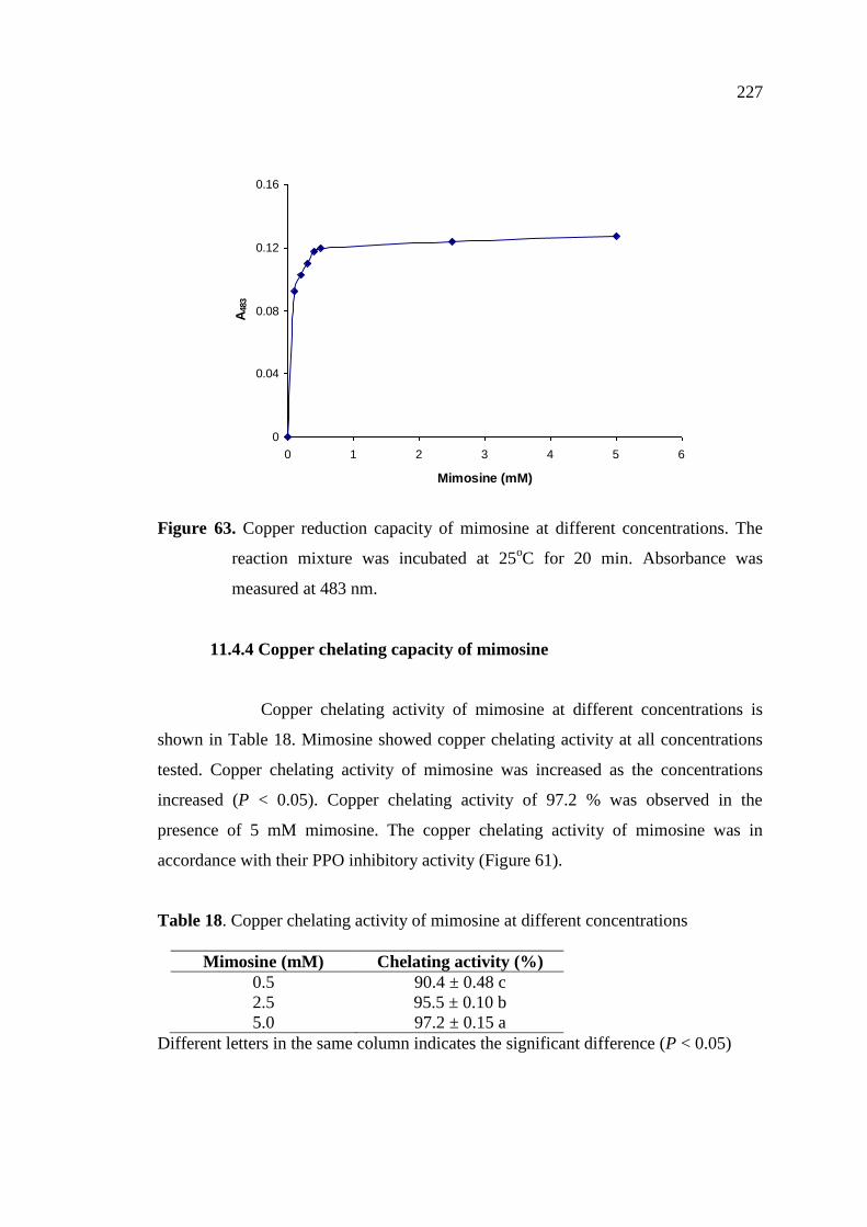
11.4.3 Copper reduction capacity of mimosine ………………………… 226
11.4.4 Copper chelating capacity of mimosine ………………………… 227
11.4.5 Effect of mimosine on browning reaction ……..……..………… 228
11.5 Conclusions..………………………………………………………….. 229
Page 15
xvii
CONTENTS (Continued)
Chapter Page
12. Summary and future works
12.1 Summary….………….……...……………………………………...… 230
12.2 Future works……..…………………………………………………… 231
References……………………….….…………………………………...……... 232
Vitae……….…………………….….……………………………...………...…. 264
Page 16
xviii
LIST OF TABLES
Table Page
1. Classes of phenolic compounds in plants ………………………… 28
2. Effect of FA treatment on likeness score of Pacific white shrimp
before and after 10 days of iced storage………………………….. 62
3. Melanosis score of Pacific white shrimp treated without and with
catechin and ferulic acid at different levels and subjected to freeze-
thawing at various cycles during the subsequent refrigerated storage. 100
4. Psychrotrophic bacterial count (PSC), total volatile base (TVB) and
thiobarbituric acid reactive substances (TBARS) of shrimp treated
without and with 0.2% catechin or 3% ferulic acid and subjected to
freeze-thawing at various cycles during the subsequent refrigerated
storage……………………………………………………………. 102
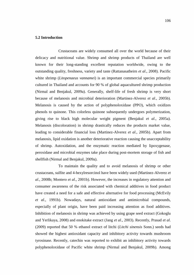
5. Extraction yield, total phenolic content and total chlorophyll content
of water and ethanolic extracts from green tea and mulberry tea with
and without prior chlorophyll removal…………………………… 115
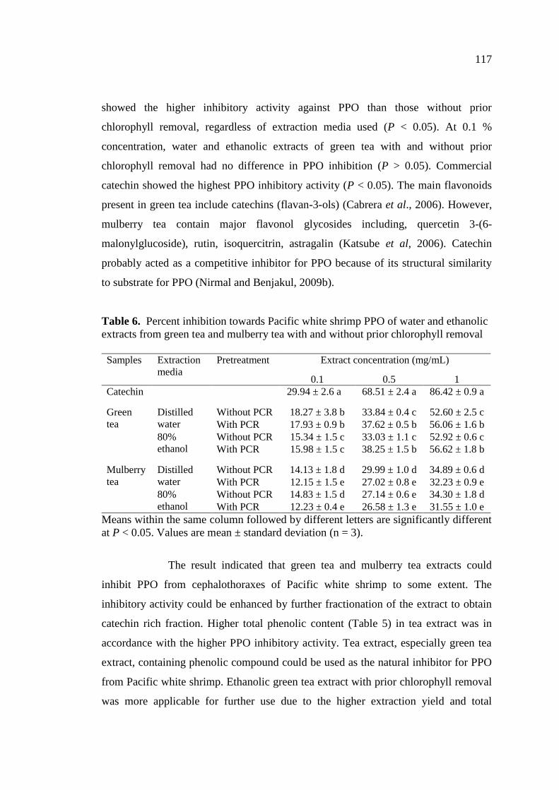
6. Percent inhibition towards Pacific white shrimp PPO of water and
ethanolic extracts from green tea and mulberry tea with and without
prior chlorophyll removal…………………………………………… 117
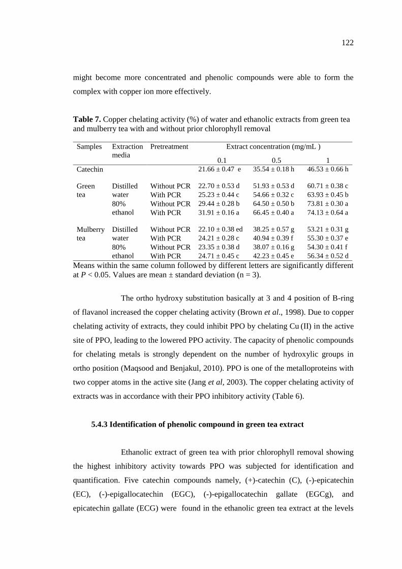
7. Copper chelating activity (%) of water and ethanolic extracts from
green tea and mulberry tea with and without prior chlorophyll
removal……………………………………………………………… 122
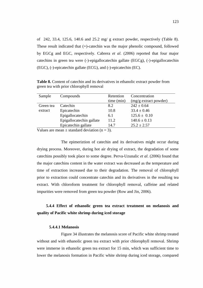
8. Content of catechin and its derivatives in ethanolic extract powder
from green tea with prior chlorophyll removal ……………………. 123
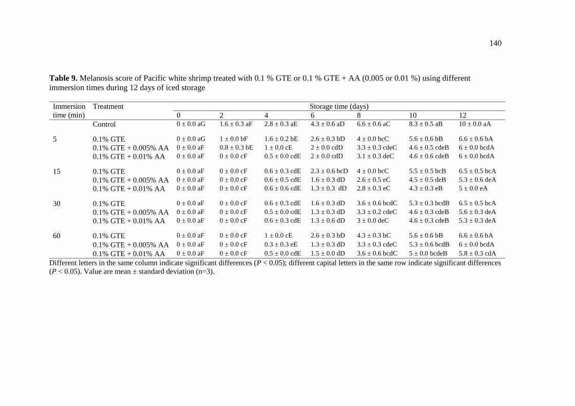
9. Melanosis score of Pacific white shrimp treated with 0.1 % GTE or
0.1 % GTE + AA (0.005 or 0.01 %) using different immersion times
during 12 days of iced storage………………………………………. 140
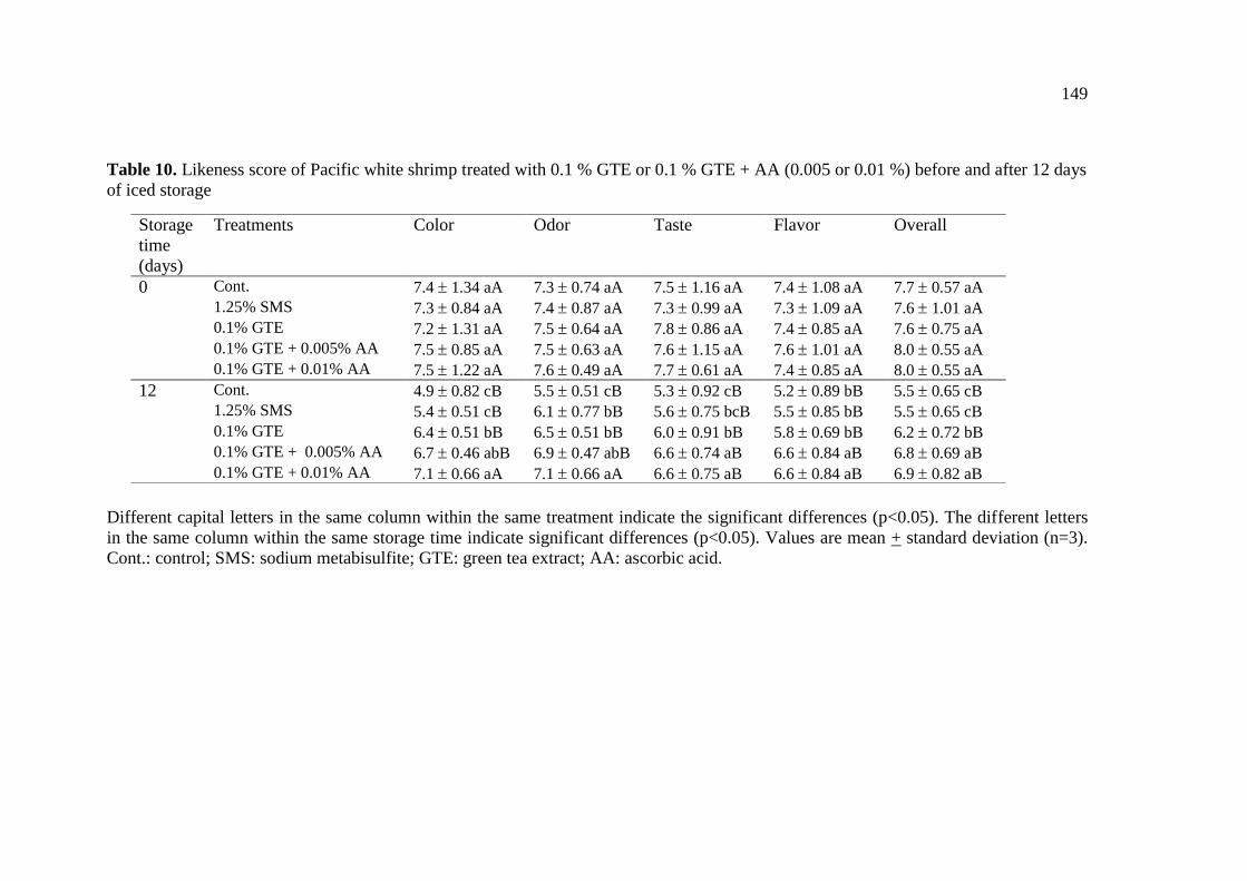
10. Likeness score of Pacific white shrimp treated with 0.1 % GTE or
0.1 % GTE + AA (0.005 or 0.01 %) before and after 12 days of iced
storage………………………………………………………………. 149
Page 17
xix
LIST OF TABLES (Continued)
Table Page
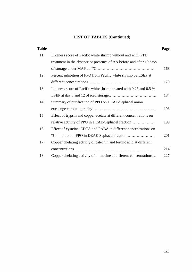
11. Likeness score of Pacific white shrimp without and with GTE
treatment in the absence or presence of AA before and after 10 days
of storage under MAP at 4oC……………………………………….. 168
12. Percent inhibition of PPO from Pacific white shrimp by LSEP at
different concentrations…………………………………………….. 179
13. Likeness score of Pacific white shrimp treated with 0.25 and 0.5 %
LSEP at day 0 and 12 of iced storage………………………………. 184
14. Summary of purification of PPO on DEAE-Sephacel anion
exchange chromatography………………………………………….. 193
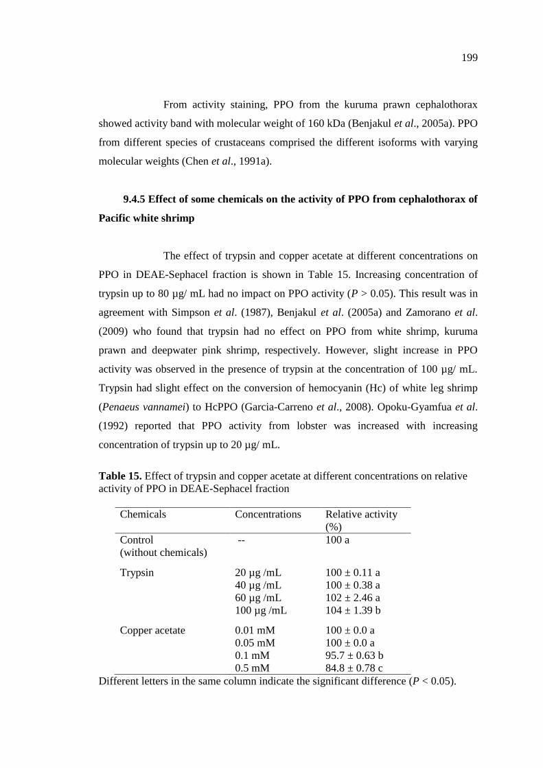
15. Effect of trypsin and copper acetate at different concentrations on
relative activity of PPO in DEAE-Sephacel fraction………………. 199
16. Effect of cysteine, EDTA and PABA at different concentrations on
% inhibition of PPO in DEAE-Sephacel fraction………………….. 201
17. Copper chelating activity of catechin and ferulic acid at different
concentrations……………………………………………………… 214
18. Copper chelating activity of mimosine at different concentrations… 227
Page 18
xx
LIST OF FIGURES
Figure Page
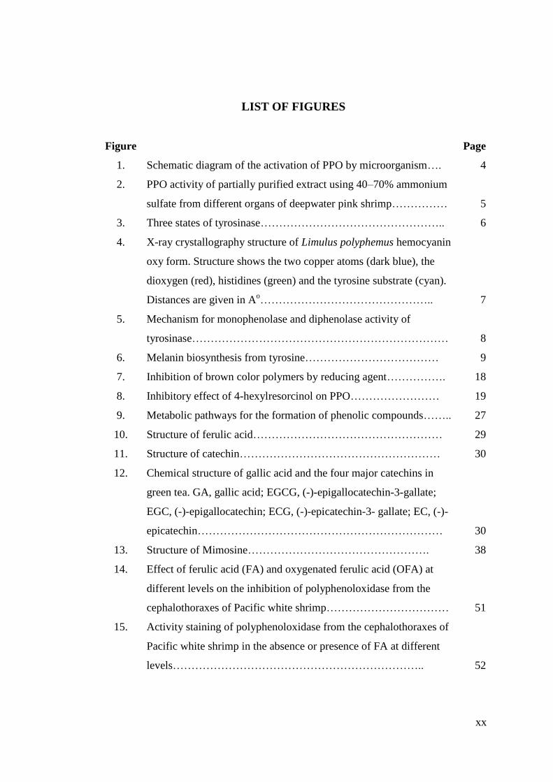
1. Schematic diagram of the activation of PPO by microorganism…. 4
2. PPO activity of partially purified extract using 40–70% ammonium
sulfate from different organs of deepwater pink shrimp…………… 5
3. Three states of tyrosinase………………………………………….. 6
4. X-ray crystallography structure of Limulus polyphemus hemocyanin
oxy form. Structure shows the two copper atoms (dark blue), the
dioxygen (red), histidines (green) and the tyrosine substrate (cyan).
Distances are given in Ao……………………………………….. 7
5. Mechanism for monophenolase and diphenolase activity of
tyrosinase…………………………………………………………… 8
6. Melanin biosynthesis from tyrosine……………………………… 9
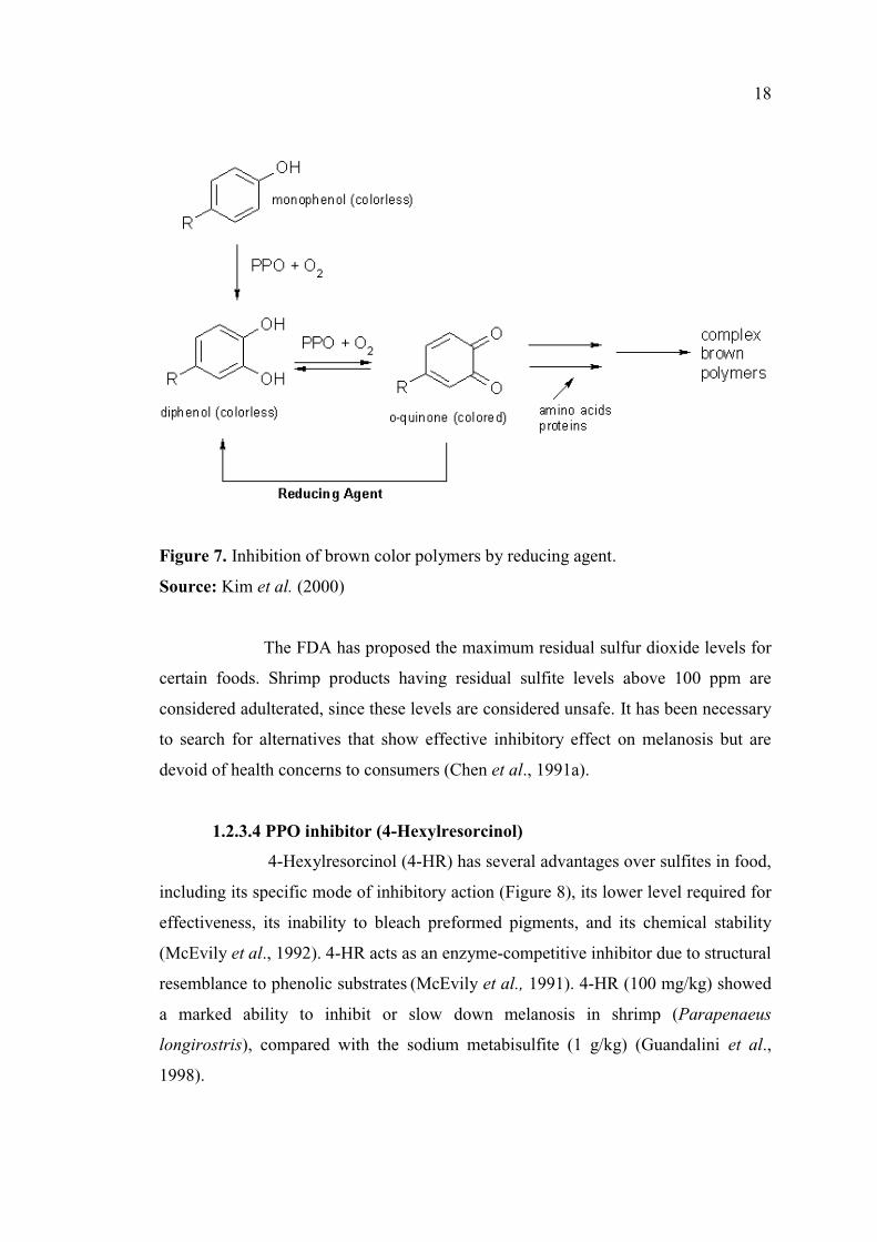
7. Inhibition of brown color polymers by reducing agent……………. 18
8. Inhibitory effect of 4-hexylresorcinol on PPO…………………… 19
9. Metabolic pathways for the formation of phenolic compounds…….. 27
10. Structure of ferulic acid…………………………………………… 29
11. Structure of catechin……………………………………………… 30
12. Chemical structure of gallic acid and the four major catechins in
green tea. GA, gallic acid; EGCG, (-)-epigallocatechin-3-gallate;
EGC, (-)-epigallocatechin; ECG, (-)-epicatechin-3- gallate; EC, (-)-
epicatechin………………………………………………………… 30
13. Structure of Mimosine…………………………………………. 38
14. Effect of ferulic acid (FA) and oxygenated ferulic acid (OFA) at
different levels on the inhibition of polyphenoloxidase from the
cephalothoraxes of Pacific white shrimp…………………………… 51
15. Activity staining of polyphenoloxidase from the cephalothoraxes of
Pacific white shrimp in the absence or presence of FA at different
levels………………………………………………………….. 52
Page 19
xxi
LIST OF FIGURES (Continued)
Figure Page
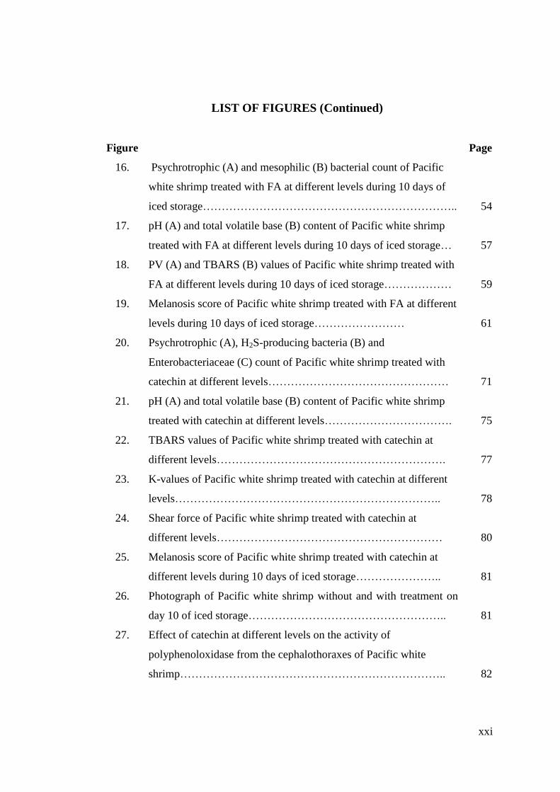
16. Psychrotrophic (A) and mesophilic (B) bacterial count of Pacific
white shrimp treated with FA at different levels during 10 days of
iced storage………………………………………………………….. 54
17. pH (A) and total volatile base (B) content of Pacific white shrimp
treated with FA at different levels during 10 days of iced storage… 57
18. PV (A) and TBARS (B) values of Pacific white shrimp treated with
FA at different levels during 10 days of iced storage……………… 59
19. Melanosis score of Pacific white shrimp treated with FA at different
levels during 10 days of iced storage…………………… 61
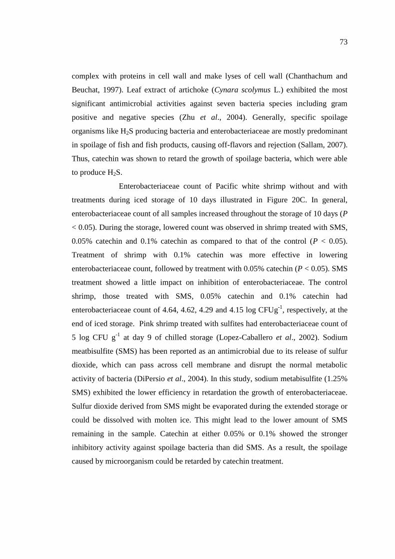
20. Psychrotrophic (A), H2S-producing bacteria (B) and
Enterobacteriaceae (C) count of Pacific white shrimp treated with
catechin at different levels………………………………………… 71
21. pH (A) and total volatile base (B) content of Pacific white shrimp
treated with catechin at different levels……………………………. 75
22. TBARS values of Pacific white shrimp treated with catechin at
different levels……………………………………………………. 77
23. K-values of Pacific white shrimp treated with catechin at different
levels…………………………………………………………….. 78
24. Shear force of Pacific white shrimp treated with catechin at
different levels…………………………………………………… 80
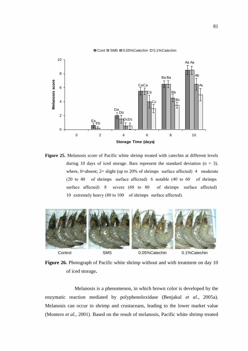
25. Melanosis score of Pacific white shrimp treated with catechin at
different levels during 10 days of iced storage………………….. 81
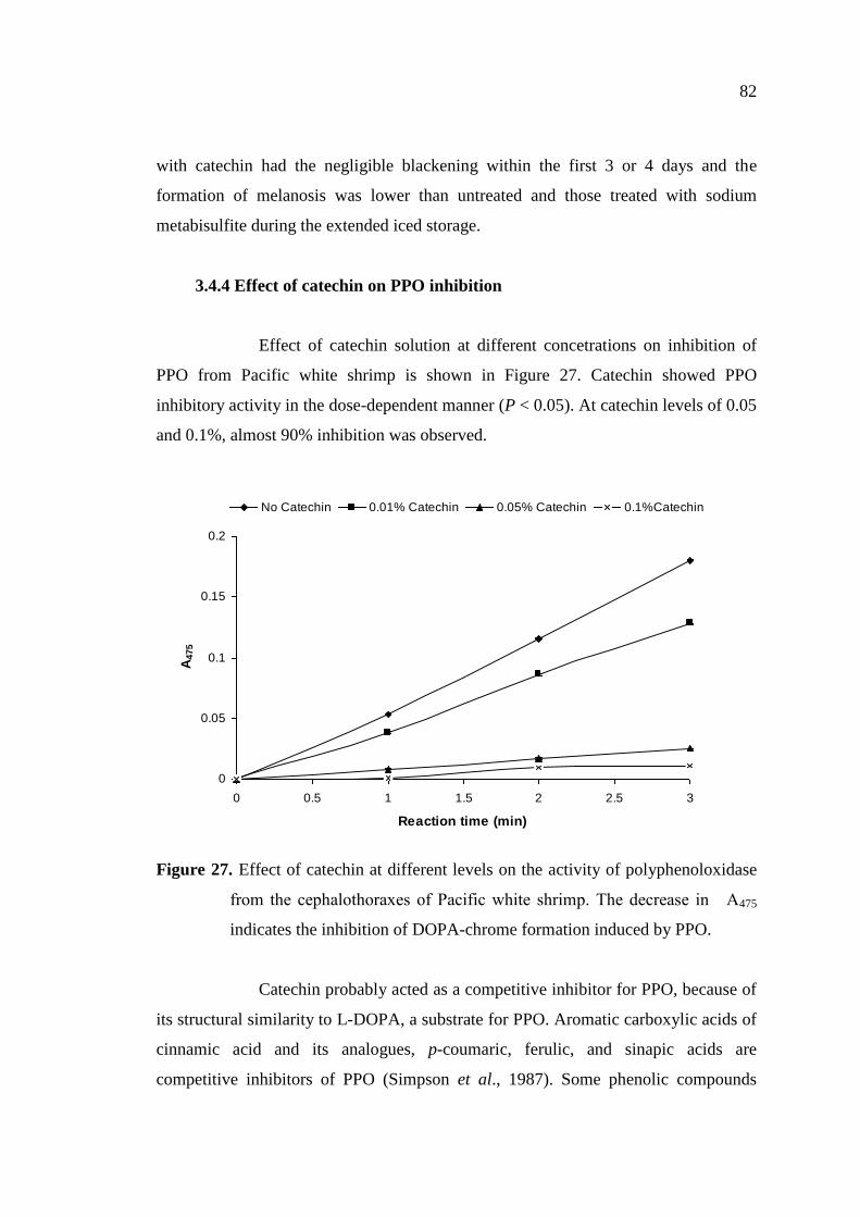
26. Photograph of Pacific white shrimp without and with treatment on
day 10 of iced storage…………………………………………….. 81
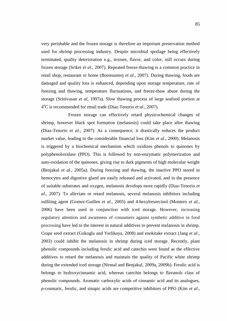
27. Effect of catechin at different levels on the activity of
polyphenoloxidase from the cephalothoraxes of Pacific white
shrimp…………………………………………………………….. 82
Page 20
xxii
LIST OF FIGURES (Continued)
Figure Page
28. Polyphenoloxidase activity of Pacific white shrimp subjected to
freeze-thawing using different thawing methods with various
freeze-thaw cycles………………………………………………. 93
29. Activity staining of polyphenoloxidase of white shrimp subjected to
freeze-thawing with different cycles. Freeze shrimp were thawed by
leaving the sample at 4oC for 6 h………………………………… 94
30. Melanosis score of shrimp subjected to freeze-thawing using
different thawing methods with various freeze-thaw cycles during
the subsequent refrigerated storage………………………………… 96
31. Melanosis score of shrimp treated with catechin (A) and ferulic acid
(B) at different levels during 8 days of refrigerated storage………. 98
32. Reducing power of water and ethanolic extract from green tea (A)
and mulberry tea (B) with and without prior chlorophyll removal… 119
33. DPPH radical scavenging activity of water and ethanolic extract
from green tea (A) and mulberry tea (B) with and without prior
chlorophyll removal……………………………………………….. 120
34 Melanosis score of Pacific white shrimp treated with ethanolic
green tea extract with prior chlorophyll removal at different levels
during 12 days of iced storage. …………………………………… 124
35. Photographs of Pacific white shrimp treated without and with
ethanolic green tea extract with prior chlorophyll removal after 12
days of iced storage………………………………………………… 125
36. Thiobarbituric acid reactive substances of Pacific white shrimp
treated with ethanolic green tea extract with prior chlorophyll
removal at different levels during 12 days of iced storage………… 127
37. Psychrotrophic bacterial count of Pacific white shrimp treated with
ethanolic green tea extract with prior chlorophyll removal at
different levels during 12 days of iced storage…………………….. 128
Page 21
xxiii
LIST OF FIGURES (Continued)
Figure Page
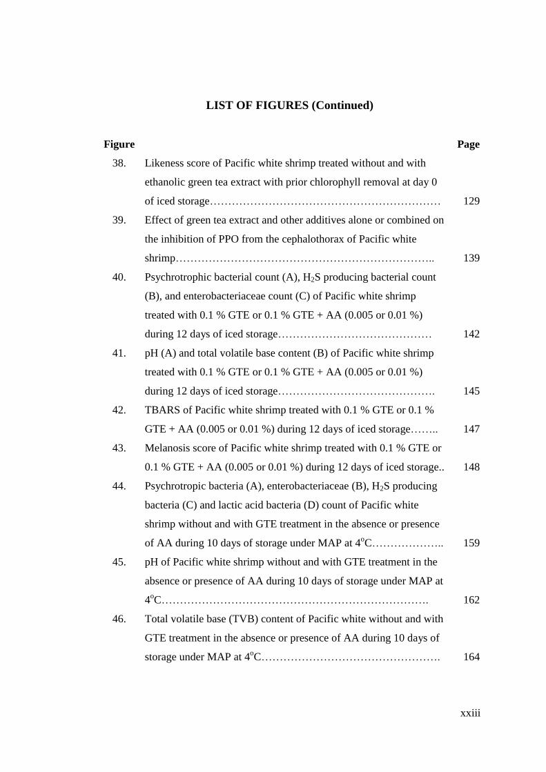
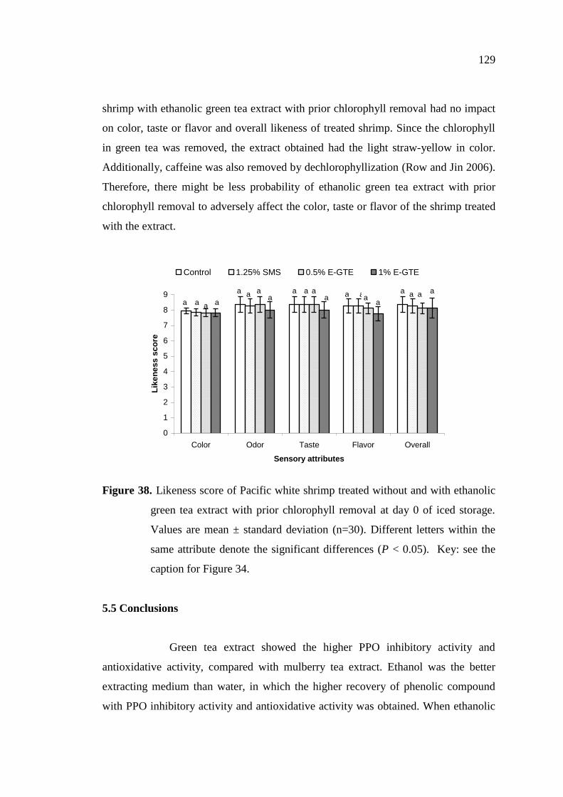
38. Likeness score of Pacific white shrimp treated without and with
ethanolic green tea extract with prior chlorophyll removal at day 0
of iced storage……………………………………………………… 129
39. Effect of green tea extract and other additives alone or combined on
the inhibition of PPO from the cephalothorax of Pacific white
shrimp…………………………………………………………….. 139
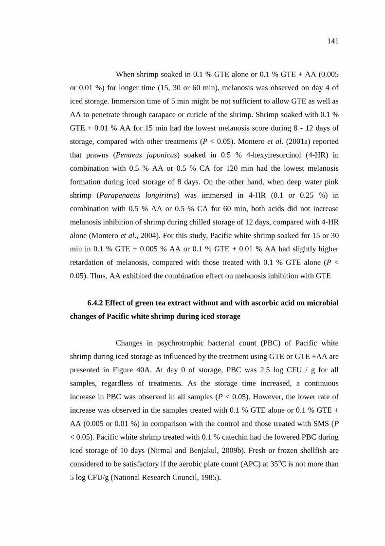
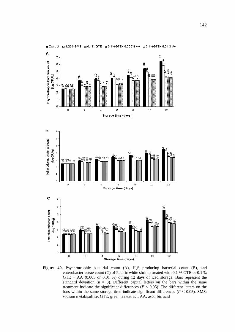
40. Psychrotrophic bacterial count (A), H2S producing bacterial count
(B), and enterobacteriaceae count (C) of Pacific white shrimp
treated with 0.1 % GTE or 0.1 % GTE + AA (0.005 or 0.01 %)
during 12 days of iced storage…………………………………… 142
41. pH (A) and total volatile base content (B) of Pacific white shrimp
treated with 0.1 % GTE or 0.1 % GTE + AA (0.005 or 0.01 %)
during 12 days of iced storage……………………………………. 145
42. TBARS of Pacific white shrimp treated with 0.1 % GTE or 0.1 %
GTE + AA (0.005 or 0.01 %) during 12 days of iced storage…….. 147
43. Melanosis score of Pacific white shrimp treated with 0.1 % GTE or
0.1 % GTE + AA (0.005 or 0.01 %) during 12 days of iced storage.. 148
44. Psychrotropic bacteria (A), enterobacteriaceae (B), H2S producing
bacteria (C) and lactic acid bacteria (D) count of Pacific white
shrimp without and with GTE treatment in the absence or presence
of AA during 10 days of storage under MAP at 4oC……………….. 159
45. pH of Pacific white shrimp without and with GTE treatment in the
absence or presence of AA during 10 days of storage under MAP at
4oC………………………………………………………………. 162
46. Total volatile base (TVB) content of Pacific white without and with
GTE treatment in the absence or presence of AA during 10 days of
storage under MAP at 4oC…………………………………………. 164
Page 22
xxiv
LIST OF FIGURES (Continued)
Figure Page
47. Thiobarbituric acid reactive substances (TBARS) of Pacific white
shrimp without and with GTE treatment in the absence or presence
of AA during 10 days of storage under MAP at 4oC……………… 165
48. Melanosis score of Pacific white shrimp without and with GTE
treatment in the absence or presence of AA during 10 days of
storage under MAP at 4oC……………………………………….. 167
49. HPLC chromatogram of mimosine from LSEP…………………… 178
50. Melanosis score of Pacific white shrimp without and with treatment
of LSEP during 12 days of iced storage…………………………… 180
51. Photographs of Pacific white shrimp without and with treatment of
LSEP at different concentrations after 12 days of iced storage……. 182
52. Mimosine content in the muscle of white shrimp treated with 0.25
and 0.5 % LSEP during 12 days of iced storage………………….. 182
53. pH (A) and temperature (B) profiles of DEAE-Sephacel fraction
containing PPO from cephalothorax of Pacific white shrimp……… 194
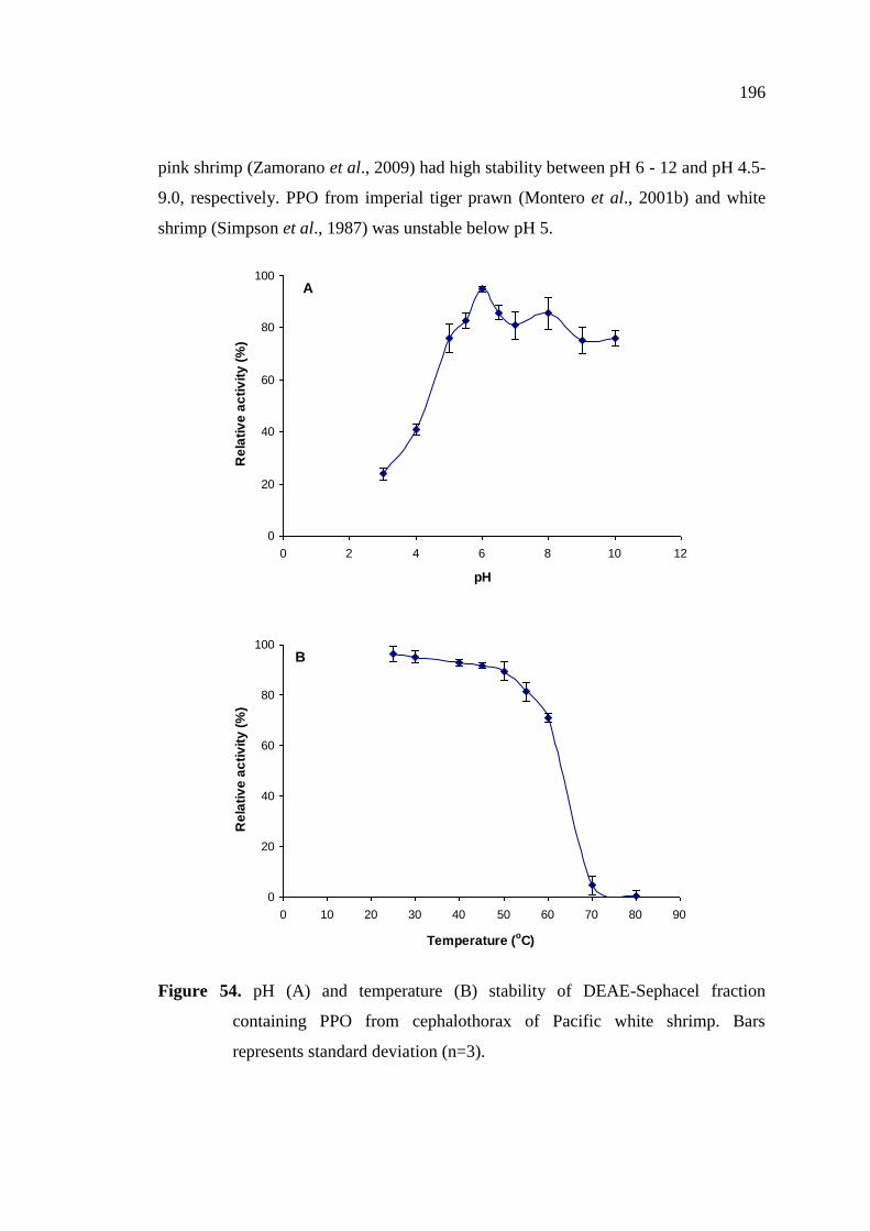
54. pH (A) and temperature (B) stability of DEAE-Sephacel fraction
containing PPO from cephalothorax of Pacific white shrimp…….. 196
55. Activity staining of DEAE-Sephacel fraction containing PPO from
cephalothorax of Pacific white shrimp……………………………. 198
56. Effect of sodium dodecyl sulfate on activity of PPO from DEAE-
Sephacel fraction. The decrease in A475 indicates the inhibition of
DOPA-chrome formation by PPO…………………………………. 201
57. Activity staining of polyphenoloxidase of DEAE-Sephacel fraction
from cephalothorax of Pacific white shrimp in the absence and
presence of catechin or ferulic acid at different concentrations……. 209
Page 23
xxv
LIST OF FIGURES (Continued)
Figure Page
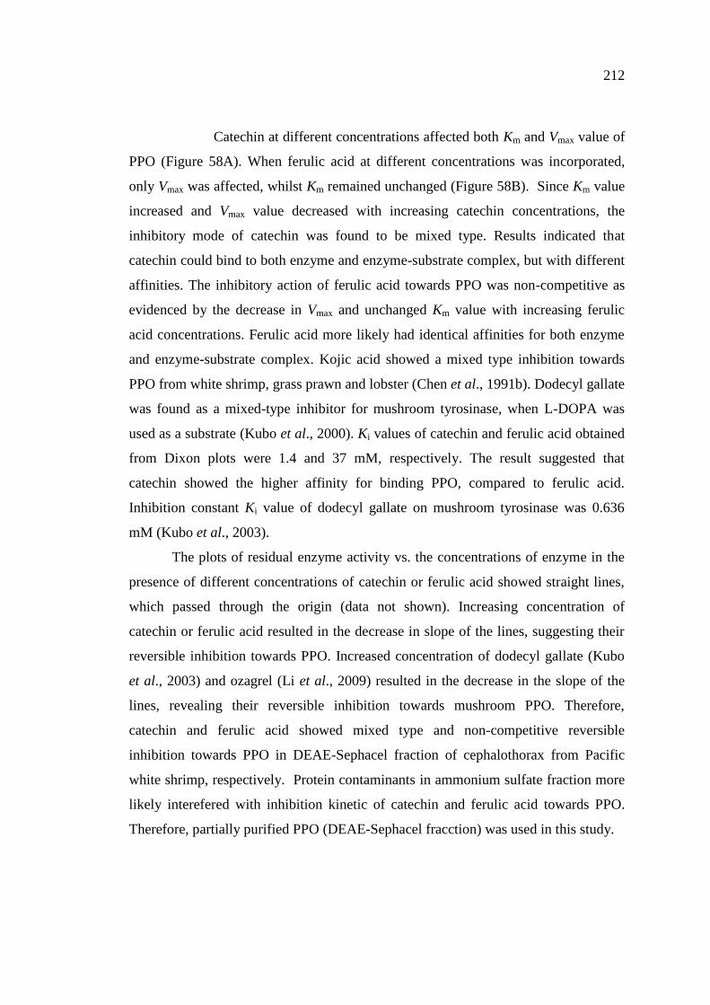
58. Lineaweaver-Burk plots of polyphenoloxidase from cephalothorax
of Pacific white shrimp in the absence and presence of catechin (A)
and ferulic acid (B) at different concentrations. L-DOPA at levels of
0.5- 5 mM were used as substrate………………………………….. 211
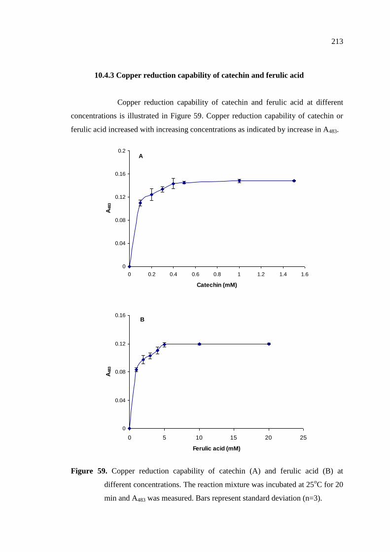
59. Copper reduction capability of catechin (A) and ferulic acid (B) at
different concentrations. The reaction mixture was incubated at
25oC for 20 min and A483 was measured………………………….. 213
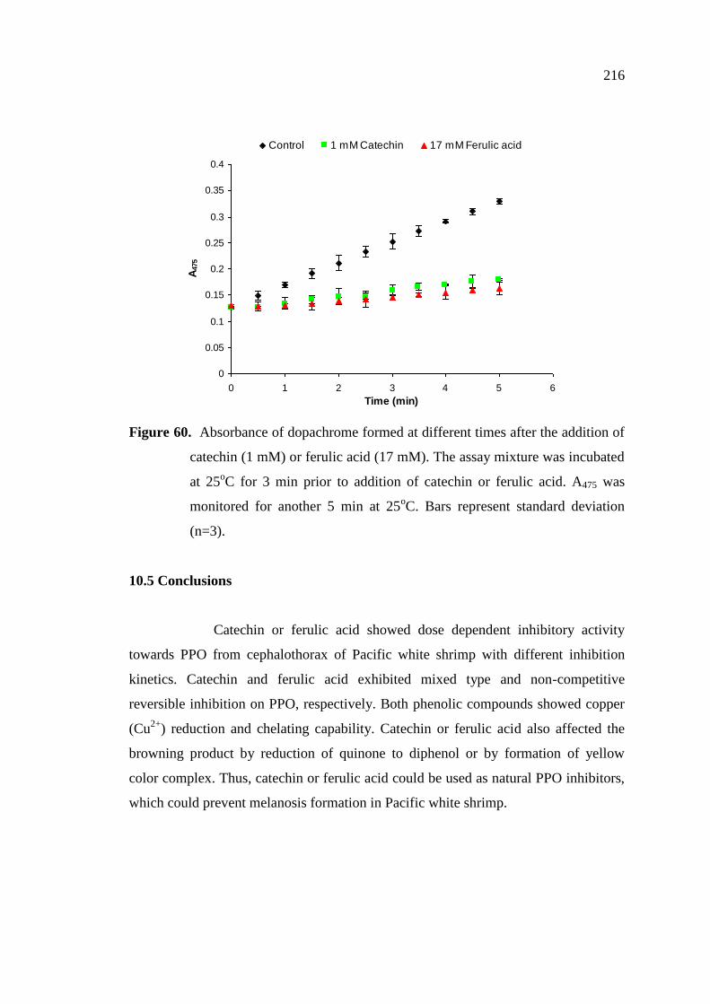
60. Absorbance of dopachrome formed at different times after the
addition of catechin (1 mM) or ferulic acid (17 mM). The assay
mixture was incubated at 25oC for 3 min prior to addition of
catechin or ferulic acid. A475 was monitored for another 5 min at
25oC……………………………………………………………….
216
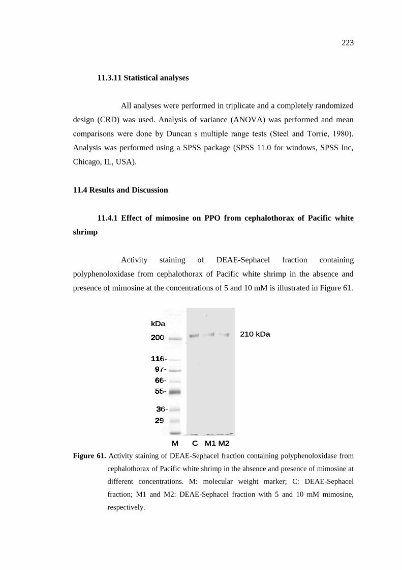
61. Activity staining of DEAE-Sephacel fraction containing
polyphenoloxidase from cephalothorax of Pacific white shrimp in
the absence and presence of mimosine at different concentrations… 223
62 Lineweaver- Burk plot of polyphenoloxidase in DEAE-Sephacel
fraction from cephalothorax of Pacific white shrimp in the absence
and presence of mimosine at different concentrations. L-DOPA at
levels of 0.5- 5 mM were used as substrate………………………… 225
63. Copper reduction capacity of mimosine at different concentrations.
The reaction mixture was incubated at 25oC for 20 min. Absorbance
was measured at 483 nm……………………………………………. 227
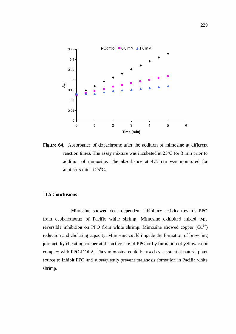
64. Absorbance of dopachrome after the addition of mimosine at
different reaction times. The assay mixture was incubated at 25oC
for 3 min prior to addition of mimosine. The absorbance at 475 nm
was monitored for another 5 min at 25oC………………………….. 229
Page 24
1
CHAPTER 1
INTRODUCTION AND REVIEW OF LITERATURE
1.1 Introduction
Pacific white shrimp (Litopenaeus vannamei) accounts for 90% of the
global aquaculture shrimp production. -farming
country and has become the top supplier of farmed shrimp to the United States and
Japan (Wyban, 2007). Pacific white shrimp (Litopenaeus vannamei) is an important
commercial species primarily cultured in Thailand and have become the essential
income generator of the country. Despite their delicacy, shrimp are highly perishable
with the limited shelf-life, mainly associated with melanosis (discoloration) and
microbial spoilage (Gokoglu and Yerlikaya, 2008). Free amino acids and other
soluble non-nitrogenous substances in shrimp serve as digestible nutrient for
microbial growth (Zeng et al., 2005). Spoilage microorganisms contribute to the loss
of essential fatty acids and proteins, production of biogenic amines and formation of
off-odors (Mastromatteo et al., 2010). Melanosis is triggered by a biochemical
mechanism which oxidizes phenols to quinones by polyphenol oxidase (PPO) (Kim et
al., 2000). This is followed by non-enzymatic polymerization and autooxidation of the
quinones, giving rise to dark pigments of high molecular weight (Benjakul et al.,
2005). Although melanosis (black spots) seems to be harmless to consumers, it
causing the considerable financial loss (Montero et al., 2001a). PPO is synthesized as
a zymogen (proPPO) in crustaceans and can be activated by protease cascade
triggered by bacterial cell wall components including lipopolysaccharides,
peptidoglycans and 1, 3- -glucans (Encarnacion et al., 2010). Melanosis and quality
changes of shrimp were retarded during frozen storage, but continued in defrosted
shrimp (Lopez-Caballero et al., 2007). Apart from melanosis, lipid oxidation is
another deteriorative reaction causing the unacceptability of shrimp and shrimp
products. Fish lipids are susceptible to oxidation owing to the high levels of
polyunsaturated fatty acid; this can be initiated by autoxidation, enzymatic reaction
1
Page 25
2
induced by lipoxygenase, peroxidase and microbial enzymes. Lipid oxidation is
associated with physicochemical changes, rancidity and off-flavor in fish meat (Bak et
al., 1999).
To maintain the quality and to avoid melanosis of shrimp or other
crustaceans, melanosis inhibitiors such as sulfite and 4-hexylresorcinol have been
widely used (Martinez-Alverez et al., 2008a; Montero et al., 2001b). However, the
increases in regulatory attention and consumer awareness of the risk associated with
chemical additives in food product have created a need for a safe and effective
additive (McEvily et al., 1991). Therefore, safe compounds from natural origin such
as ascorbic acid, kojic acid (Chen et al, 1991a); ficin (Taoukis et al., 1990), enokitake
extract (Jang et al., 2003); dodecyl gallate (Kubo et al., 2003) and oxalic acid (Son et
al., 2000) have been used as the substitutes of sulfiting agents. Recently, it has been
reported that the grape seed extract could inhibit the melanosis in shrimp
(Parapenaeus longirostris) (Gokoglu and Yerlikaya, 2008). Encarnacion et al. (2010)
found that the dietary supplement of mushroom extract (Flammulina velutipes) in
kuruma shrimp (Marsupenaeus japonicus) could reduce post mortem development of
melanosis.
In addition to melanosis inhibition, plant phenolic compounds may act
as antimicrobial and antioxidant, which could retard the microbial and chemical
spoilage of shrimp. Nevertheless, a little information regarding the use of single
phenolic compound or plant extracts on melanosis prevention as well as shelf-life
extension of white shrimp during post-mortem storage has been reported. The
information gained can provide the important and potential approach for shrimp
processor to maintain the prime quality of white shrimp during handling and storage,
thereby lowering the loss in market value.
Page 26
3
1.2 Review of Literature
1.2.1 Polyphenoloxidase (PPO)
Polyphenoloxidase (PPO) or tyrosinase (monophenol, L-dopa: oxygen
oxidoreductase, EC 1.14.18.1) is a bi-functional, copper containing enzyme widely
distributed throughout the phylogenetic scale (Garcia-Molina et al., 2007). It
catalyzes the hydroxylation of monophenol to o-diphenols and their subsequent
oxidation to o-quinones by molecular oxygen (Likhitwitayawuid, 2008; Garcia-
Molina et al., 2007; Decker and Tuczek, 2000). In all cases, PPO is associated with
dark pigmentation in the organism and has a protective function (Martinez and
Whitaker, 1995). Different PPO obtained from several biological sources has similar
structural and fuctional characteristics (Garcia-Molina et al., 2005). PPO plays an
essential role in physiological functions, particularly for the sclerotization of the
cuticle of crustaceans such as shrimp and lobster. Sclerotization is the hardening of
the chitin shell after molting, which is part of the growing phase for the organism
(Terwilliger, 1999). The highly reactive o-quinones cross-link with histidyl residues
of cuticular proteins and chitin, resulting in hardening of the exoskeleton (Xu et al.,
1997). A second function of polyphenoloxidase is immunity and self-recognition. The
o-quinones are involved in the melanin synthesis, a compound with antimicrobial,
antiviral properties and thus PPO is important components of the innate immune
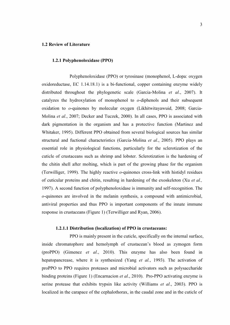
response in crustaceans (Figure 1) (Terwilliger and Ryan, 2006).
1.2.1.1 Distribution (localization) of PPO in crustaceans:
PPO is mainly present in the cuticle, specifically on the internal surface,
inside chromatophore ood as zymogen form
(proPPO) (Gimenez et al., 2010). This enzyme has also been found in
hepatopancrease, where it is synthesized (Yang et al., 1993). The activation of
proPPO to PPO requires proteases and microbial activators such as polysaccharide
binding proteins (Figure 1) (Encarnacion et al., 2010). Pro-PPO activating enzyme is
serine protease that exhibits trypsin like activity (Williams et al., 2003). PPO is
localized in the carapace of the cephalothorax, in the caudal zone and in the cuticle of
Page 27
4
the abdomen, mainly where the cuticle segments are joined and where the cuticle is
connected to the pleopods (Benjakul et al., 2005). PPO from different species, body
location, sex, molting stage and tissue might have different activity levels as well as
varying properties (Ferrer et al., 1989).
Figure 1. Schematic diagram of the activation of PPO by microorganism.
Source: Modified from Kim et al. (2000)
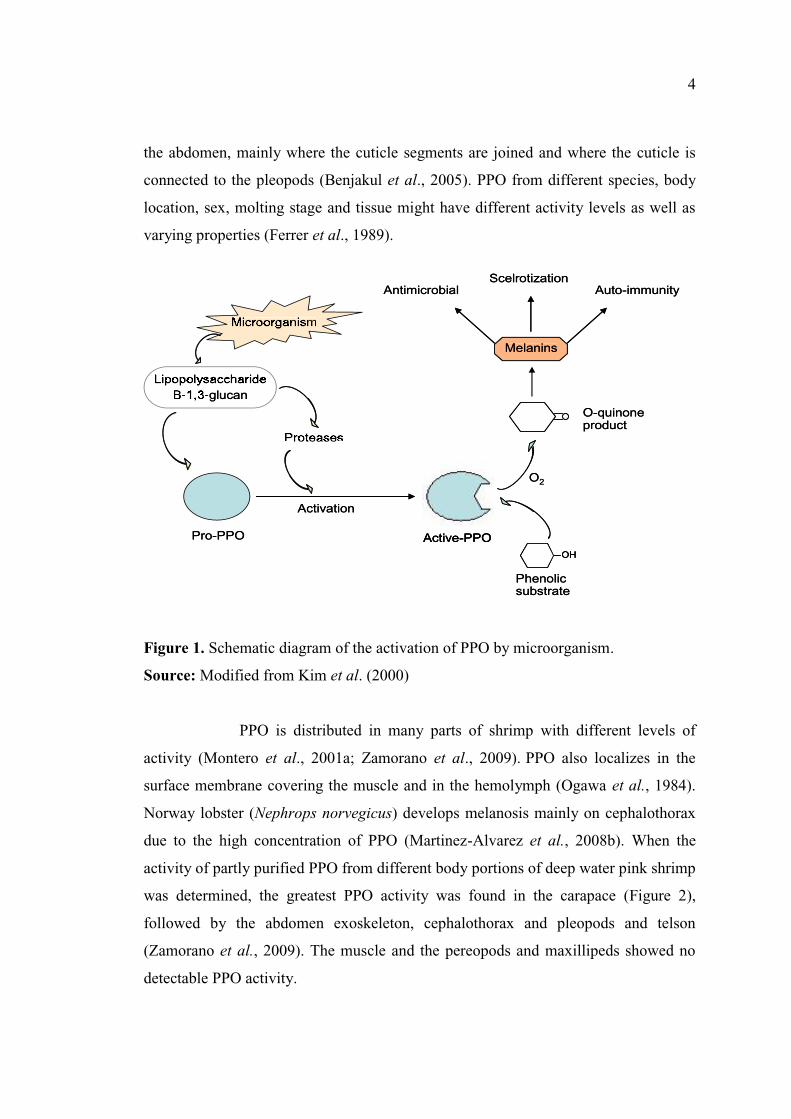
PPO is distributed in many parts of shrimp with different levels of
activity (Montero et al., 2001a; Zamorano et al., 2009). PPO also localizes in the
surface membrane covering the muscle and in the hemolymph (Ogawa et al., 1984).
Norway lobster (Nephrops norvegicus) develops melanosis mainly on cephalothorax
due to the high concentration of PPO (Martinez-Alvarez et al., 2008b). When the
activity of partly purified PPO from different body portions of deep water pink shrimp
was determined, the greatest PPO activity was found in the carapace (Figure 2),
followed by the abdomen exoskeleton, cephalothorax and pleopods and telson
(Zamorano et al., 2009). The muscle and the pereopods and maxillipeds showed no
detectable PPO activity.
Pro-PPO Active-PPO
Phenolicsubstrate
OH
O2
O-quinoneproduct
glucan
AntimicrobialScelrotization
Auto-immunity
Activation
Pro-PPO
ProteasesProteases
Active-PPOActive-PPO
Phenolicsubstrate
OHOH
O2
O-quinoneproduct
MicroorganismMicroorganism
LipopolysaccharideLipopolysaccharide--1,31,3--glucan
MelaninsMelanins
AntimicrobialScelrotization
Auto-immunity
Activation
Page 28
5
Figure 2. PPO activity of partially purified extract using 40 70% ammonium
sulfate from different organs of deepwater pink shrimp.
Source: Modified from Zamorano et al. (2009)
Hemocyanain (Hc) which is closely resemble with the PPO in their
sequence and in their active site can also acquire PPO activity through proteolytic
cleavage in its terminal amino end by serine proteases (Martinez-Alvarez et al.,
2008a). Hc is found in hemolymph as well as in cuticle (Adachi et al., 2005).
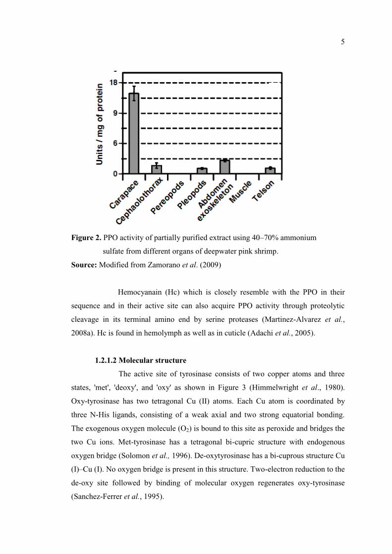
1.2.1.2 Molecular structure
The active site of tyrosinase consists of two copper atoms and three
states, 'met', 'deoxy', and 'oxy' as shown in Figure 3 (Himmelwright et al., 1980).
Oxy-tyrosinase has two tetragonal Cu (II) atoms. Each Cu atom is coordinated by
three N-His ligands, consisting of a weak axial and two strong equatorial bonding.
The exogenous oxygen molecule (O2) is bound to this site as peroxide and bridges the
two Cu ions. Met-tyrosinase has a tetragonal bi-cupric structure with endogenous
oxygen bridge (Solomon et al., 1996). De-oxytyrosinase has a bi-cuprous structure Cu
(I) Cu (I). No oxygen bridge is present in this structure. Two-electron reduction to the
de-oxy site followed by binding of molecular oxygen regenerates oxy-tyrosinase
(Sanchez-Ferrer et al., 1995).
Page 29
6
Figure 3. Three states of tyrosinase.
Source: Modified from Himmelwright et al. (1980)
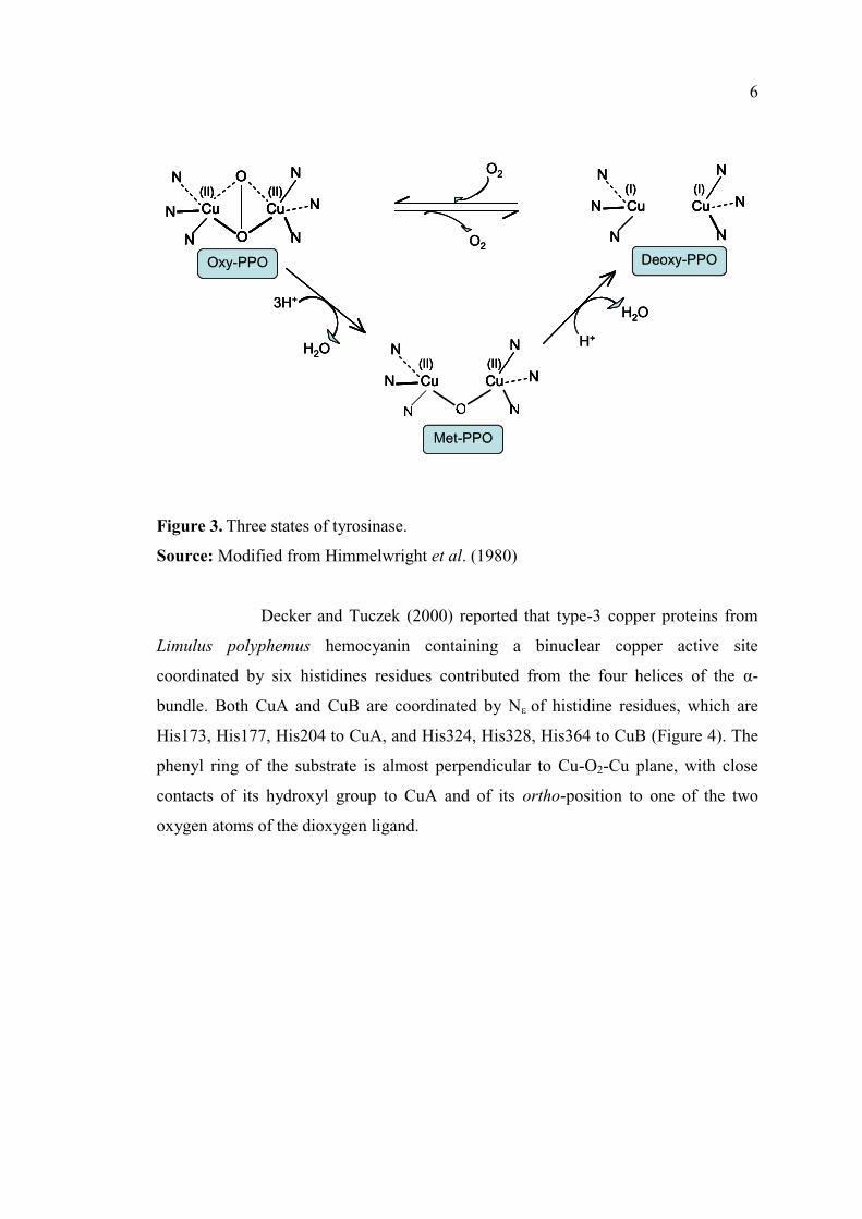
Decker and Tuczek (2000) reported that type-3 copper proteins from
Limulus polyphemus hemocyanin containing a binuclear copper active site
coordinated by six histidines residues contributed from the four helices of the -
bundle. Both CuA and CuB are coordinated by N of histidine residues, which are
His173, His177, His204 to CuA, and His324, His328, His364 to CuB (Figure 4). The
phenyl ring of the substrate is almost perpendicular to Cu-O2-Cu plane, with close
contacts of its hydroxyl group to CuA and of its ortho-position to one of the two
oxygen atoms of the dioxygen ligand.
O2
O2
N Cu Cu(I) (I)
N
N
N
N
N
N Cu Cu(II) (II)
N
N
N
N Cu Cu(II) (II)
O
ON
N
N
N
N
3H+
H2OH+
H2O
O2
O2
O2
O2
N Cu Cu(I) (I)
N
N
N
N
N
N Cu Cu(I) (I)
N
N
N
N
N
N Cu Cu(I) (I)
N
N
N
N
N
DeoxyDeoxy--PPOPPODeoxy-PPO
N Cu Cu(II) (II)
N
N
N
N Cu Cu(II) (II)
N
N
N
N Cu Cu(II) (II)
OOOOOOOONNNNNNN
N
NNNNNNNNNNNNNN
N
N
MetMet--PPOPPOMet-PPO
N Cu Cu(II) (II)
O
ON
N
N
N
N
N Cu Cu(II) (II)
O
ON
N
N
N
N
N Cu Cu(II) (II)
O
ON
N
N
N
N
Cu Cu(II) (II)
O
ON
N
N
N
N
OxyOxy--PPOPPOOxy-PPO
3H+
H2O
3H+
H2OH+
H2O
H+
H2O
Page 30
7
Figure 4. X-ray crystallography structure of Limulus polyphemus hemocyanin oxy
form. Structure shows the two copper atoms (dark blue), the dioxygen (red),
histidines (green) and the tyrosine substrate (cyan). Distances are given in
Ao.
Source: Decker and Tuczek (2000)
1.2.1.3 Enzyme mechanisms
PPO catalyzes two basic reactions, in the presence of molecular
oxygen, including o-hydroxylation of monophenols to give o-diphenols (Monophenol
oxidase, cresolase activity) and the further oxidation of o-diphenols to o-quinones
(Diphenoloxidase, catecholase activity) (Garcia-Molina et al., 2005).
1.2.1.3.1 Monophenol oxidase
It catalyzes the hydroxylation of monophenols to diphenols. As shown
in Figure 5, the monophenolase activity begins with the binding of the substrate
Page 31
8
monophenol to one of the Cu atoms of the oxygenated form (oxy-tyrosinase) to
generate Oxy-tyrosine (Rodriguez-Lopez et al., 1992). Then, o-hydroxylation of the
monophenol by the bound peroxide occurs, and an enzyme-coordinated o-diphenol
structure (Met-D) is formed. It should be noted that monophenol can react with oxy-
tyrosinase, but not with met-tyrosinase, to form the product o-quinone.
Monophenolase activity shows a characteristic lag period. This may be due to the fact
that tyrosinase in the resting form contains 15 % oxy sites, which is the only form that
can react with monophenol substrates (Likhitwitayawuid, 2008).
Figure 5. Mechanism for monophenolase and diphenolase activity of tyrosinase.
Source: Likhitwitayawuid (2008)
Page 32
9
1.2.1.3.2 Diphenol oxidase
It catalyzes the oxidation of the di-phenol to quinones. The reaction of
oxidation of diphenol to quinone is shown in Figure 5. This reaction receives more
attention because of its faster rate than the monophenol oxidase and its association
with the formation of quinones, which polymerize with amino acids / protein or self
polymerize non-enzymetically to form melanin (Figure. 6) (Garcia-Molina et al.,
2005). The o-diphenol can react with both the oxy and the met forms to produce o-
quinone. The reaction of diphenol with met-tyrosinase converts the enzyme to the de-
oxy form, bringing it into the monophenolase cycle (Solomon et al., 1996). When
quinones are formed and undergo polymerization with protein or amino acids or self
polymerized, they form a melanoid compound (dark brown color) (Satoh et al., 1999).
Figure 6. Melanin biosynthesis from tyrosine.
Source: Garcia-Molina et al. (2005)
Page 33
10
1.2.1.4 Characteristics of PPO from crustaceans
1.2.1.4.1 Molecular weight
PPO from different shrimp comprise the different iso-forms with
varying molecular weights (Chen et al., 1991a). Also, the molecular weight of PPO
varied with the molting stage (Ferrer et al., 1989). PPO from the kuruma prawn
cephalothorax had the molecular weight of 160 kDa (Benjakul et al., 2005). Antarctic
krill catechol oxidases had the molecular weights of 75 and 83 kDa (Ohshima and
Nagayama, 1980). For PPO from shrimp (P. setiferus), its molecular weight was 30
kDaA (Simpson et al., 1987). The molecular weights of pink shrimp PPO were 30 and
35 kDa, while those of white shrimp were 20 and 25 kDa (Chen et al., 1997). The
apparent molecular weight of proPPO and PPO from hemolymph of crab Charybdis
japonica was 69.5 and 64.5 kDa, respectively (Liu et al., 2006). Zamorano et al.
(2009) reported that PPO with the molecular weight of 500 kDa and 200 kDa were
found in deep water pink shrimp (Parapenaeus longirostris). PPO from viscera and
carapace extracts of cephalothorax of Norway lobster (Nephrops norvegicus) had
apparent molecular weight about 200- 220 kDa as determined by activity staining
using L-tyrosine and 4-tert-butyl-catechol as substrates (Gimenz et al., 2010).
1.2.1.4.2 pH optima and stability
The pH profile for PPO isolated from different crustaceans varies with
species. In the case of shrimp (Penaeus setiferus), PPO was active in the pH range of
6.5 9 (Simpson et al., 1988). For Penaeus monodon PPO, the maximum activity was
reported at pH 6.0 (Rolle et al., 1991). Lobster (Panulirus argus) PPO had the optimal
pH of 6 6.5 (Ali et al., 1994). The maximal activity of PPO from the kuruma prawn
cephalothorax (Benjakul et al., 2005) and Antractic krill (Ohshima and Nagayama
1980) was observed at pH 6.5. Montero et al. (2001a) reported that PPO from
imperial tiger prawn (Penaeus japonicus) was most active at pH 5 and 8. Zamorano et
al. (2009) found that PPO from deep water pink shrimp (Parapenaeus longirostris)
had the highest activity at pH 4.5. PPO from the carapace of shrimp (P. setiferus)
showed the maximal activity at pH 7.5 (Simpson et al., 1987). PPO from hemolymph
of crab Charybdis japonica showed optimum pH at 6 (Liu et al., 2006). PPO from
Page 34
11
carapace and viscera of Norway lobster (Nephrops norvegicus) had optimum pH at 7
and 8, respectively (Gimenez et al., 2010).
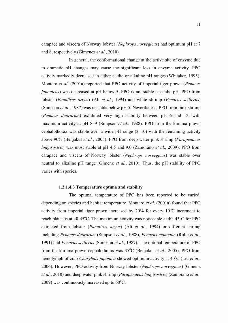
In general, the conformational change at the active site of enzyme due
to dramatic pH changes may cause the significant loss in enzyme activity. PPO
activity markedly decreased in either acidic or alkaline pH ranges (Whitaker, 1995).
Montero et al. (2001a) reported that PPO activity of imperial tiger prawn (Penaeus
japonicus) was decreased at pH below 5. PPO is not stable at acidic pH. PPO from
lobster (Panulirus argus) (Ali et al., 1994) and white shrimp (Penaeus setiferus)
(Simpson et al., 1987) was unstable below pH 5. Nevertheless, PPO from pink shrimp
(Penaeus duorarum) exhibited very high stability between pH 6 and 12, with
maximum activity at pH 8 9 (Simpson et al., 1988). PPO from the kuruma prawn
cephalothorax was stable over a wide pH range (3 10) with the remaining activity
above 90% (Benjakul et al., 2005). PPO from deep water pink shrimp (Parapenaeus
longirostris) was most stable at pH 4.5 and 9.0 (Zamorano et al., 2009). PPO from
carapace and viscera of Norway lobster (Nephrops norvegicus) was stable over
neutral to alkaline pH range (Gimenz et al., 2010). Thus, the pH stability of PPO
varies with species.
1.2.1.4.3 Temperature optima and stability
The optimal temperature of PPO has been reported to be varied,
depending on species and habitat temperature. Montero et al. (2001a) found that PPO
activity from imperial tiger prawn increased by 20% for every 10oC increment to
reach plateaus at 40-45oC. The maximum activity was noticeable at 40 45oC for PPO
extracted from lobster (Panulirus argus) (Ali et al., 1994) or different shrimp
including Penaeus duorarum (Simpson et al., 1988), Penaeus monodon (Rolle et al.,
1991) and Penaeus setiferus (Simpson et al., 1987). The optimal temperature of PPO
from the kuruma prawn cephalothorax was 35oC (Benjakul et al., 2005). PPO from
hemolymph of crab Charybdis japonica showed optimum activity at 40oC (Liu et al.,
2006). However, PPO activity from Norway lobster (Nephrops norvegicus) (Gimenz
et al., 2010) and deep water pink shrimp (Parapenaeus longirostris) (Zamorano et al.,
2009) was continuously increased up to 60oC.
Page 35
12
PPO of shrimp (Penaeus duorarum and Penaeus monodon) was
unstable at temperatures over 30 35oC (Rolle et al., 1991; Simpson et al., 1988). For
PPO of Penaeus setiferus, the upper limit of stability was 50oC (Simpson et al., 1987),
while PPO of lobster (Panulirus argus) had the stability at 40oC (Ali et al., 1994).
Even within the same species, there are differences depending on the state of
activation of enzyme (Ferrer et al., 1989). PPO from Kuruma prawn was stable up to
40oC and slightly lost its stability at 50oC (Benjakul et al., 2005). Montero et al.
(2001a) reported that thermal stability of PPO from imperial tiger prawn was
considerably reduced when the enzyme extract was subjected to heating at
temperatures up to 35oC. PPO from deep water pink shrimp had the stability at 30
35oC (Zamorano et al., 2009).
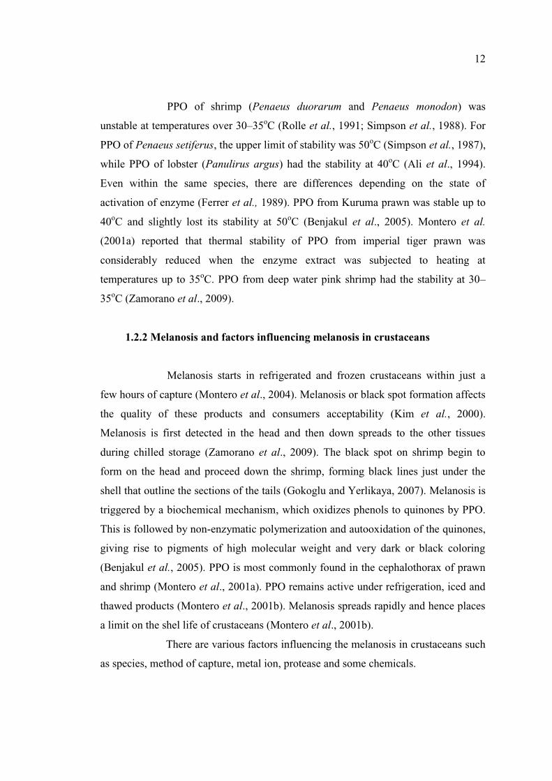
1.2.2 Melanosis and factors influencing melanosis in crustaceans
Melanosis starts in refrigerated and frozen crustaceans within just a
few hours of capture (Montero et al., 2004). Melanosis or black spot formation affects
the quality of these products and consumers acceptability (Kim et al., 2000).
Melanosis is first detected in the head and then down spreads to the other tissues
during chilled storage (Zamorano et al., 2009). The black spot on shrimp begin to
form on the head and proceed down the shrimp, forming black lines just under the
shell that outline the sections of the tails (Gokoglu and Yerlikaya, 2007). Melanosis is
triggered by a biochemical mechanism, which oxidizes phenols to quinones by PPO.
This is followed by non-enzymatic polymerization and autooxidation of the quinones,
giving rise to pigments of high molecular weight and very dark or black coloring
(Benjakul et al., 2005). PPO is most commonly found in the cephalothorax of prawn
and shrimp (Montero et al., 2001a). PPO remains active under refrigeration, iced and
thawed products (Montero et al., 2001b). Melanosis spreads rapidly and hence places
a limit on the shel life of crustaceans (Montero et al., 2001b).
There are various factors influencing the melanosis in crustaceans such
as species, method of capture, metal ion, protease and some chemicals.
Page 36
13
1.2.2.1 Species
Melanosis is associated with PPO activity. PPO activity varies with
species. PPO from pink shrimp was more active than that from white shrimp (Madero
and Finne, 1982; Simpson et al., 1987). In chilled shrimp, the rate of spread of
melanosis differs among various species. This could be related to differences in levels
of substrate or levels of enzyme concentration or enzymatic activity in each species
(Simpson et al., 1987; Montero et al., 2001a). The spread of melanosis in pink shrimp
(Penaeus duorarum) is faster than in white shrimp (Penaeus setiferus) (Simpson et al.,
1987). Melanosis develops very rapidly in deepwater pink shrimp (Martinez-Alvarez
et al., 2008a).
1.2.2.2 Method of capture and season
Capture and
seem to trigger a defense mechanism in shellfish involving the activation of PPO,
resulting ultimately in increased black spot (Kim et al., 2000). Lobster and shrimp can
be induced to form melanin by injuring them while alive (Ogawa, 1987). Danish
Norway lobster processors have reported an annually recurring rise in black spot-
related problems around September each year. Annual drop in catch quality was truly
related to changes in PPO activity (Bartolo and Birk, 1998). Nevertheless,
hemocyanin derived PPO could be responsible for the rapid formation of black spots
in broken clawed legs, parapods or carapace during postmortem handling (Gimenz et
al., 2010).
1.2.2.3 Metal ion
The role of copper in catalysis of oxidation of monophenols and o-
diphenols was elucidated. Some Metal ions such as Cu2+, Zn2+, and Mg2+ have a
significant effect on PPO activity. Liu et al. (2006) reported that PPO activity from
crab (Charybdis japonica) was strongly inhibited by Cu2+, Zn2+, and Mg2+.
Nevertheless, Cu2+ had an obvious recovery effect on the activity of EDTA-pretreated
PPO, but the other metal ions did not have such an effect. Simpson et al. (1987)
reported that PPO activity from shrimp (Penaeus setiferus) increased with the
addition of the copper, whereas Benjakul et al.(2005) found that PPO isolated from
Page 37
14
the kuruma prawn cephalothorax might be in the active form, which did not require
copper ion for PPO activation. The increase in copper ion might cause the
conformational change of enzyme by affecting the ionic interaction stabilizing the
structure of enzyme. Therefore, the copper dependency of PPO depended on species,
tissue, and other intrinsic factors determining the activity. The activity of lobster PPO
was stimulated by the addition of copper but inhibited by EDTA (Opoku-Gyamfua et
al., 1992).
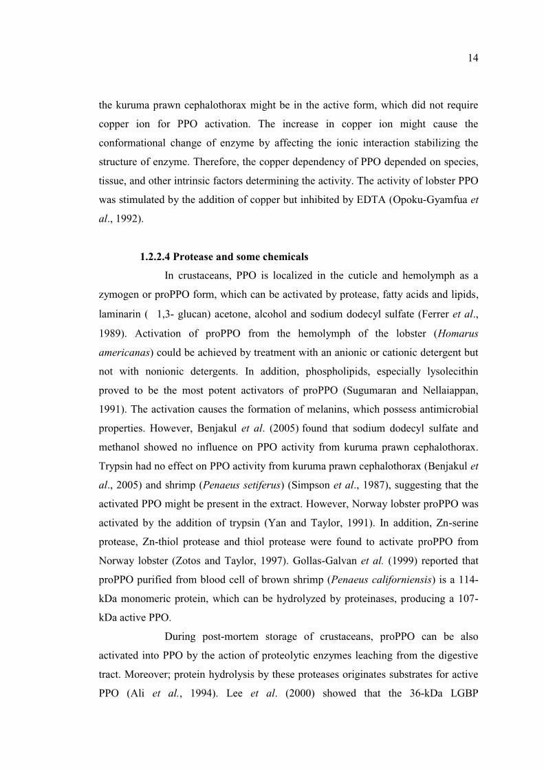
1.2.2.4 Protease and some chemicals
In crustaceans, PPO is localized in the cuticle and hemolymph as a
zymogen or proPPO form, which can be activated by protease, fatty acids and lipids,
laminarin ( 1,3- glucan) acetone, alcohol and sodium dodecyl sulfate (Ferrer et al.,
1989). Activation of proPPO from the hemolymph of the lobster (Homarus
americanas) could be achieved by treatment with an anionic or cationic detergent but
not with nonionic detergents. In addition, phospholipids, especially lysolecithin
proved to be the most potent activators of proPPO (Sugumaran and Nellaiappan,
1991). The activation causes the formation of melanins, which possess antimicrobial
properties. However, Benjakul et al. (2005) found that sodium dodecyl sulfate and
methanol showed no influence on PPO activity from kuruma prawn cephalothorax.
Trypsin had no effect on PPO activity from kuruma prawn cephalothorax (Benjakul et
al., 2005) and shrimp (Penaeus setiferus) (Simpson et al., 1987), suggesting that the
activated PPO might be present in the extract. However, Norway lobster proPPO was
activated by the addition of trypsin (Yan and Taylor, 1991). In addition, Zn-serine
protease, Zn-thiol protease and thiol protease were found to activate proPPO from
Norway lobster (Zotos and Taylor, 1997). Gollas-Galvan et al. (1999) reported that
proPPO purified from blood cell of brown shrimp (Penaeus californiensis) is a 114-
kDa monomeric protein, which can be hydrolyzed by proteinases, producing a 107-
kDa active PPO.
During post-mortem storage of crustaceans, proPPO can be also
activated into PPO by the action of proteolytic enzymes leaching from the digestive
tract. Moreover; protein hydrolysis by these proteases originates substrates for active
PPO (Ali et al., 1994). Lee et al. (2000) showed that the 36-kDa LGBP
Page 38
15
(lipopolysaccharide- -1,3-glucan-binding protein) plays a role in the activation
of the proPO activating system in freshwater crayfish (Pacifastacus leniusculus).
Garcia-Carreno et al. (2008) reported that hemocyanin (Hc) from whiteleg shrimp
(Penaeus vannamei) was converted to HcPPO by SDS treatment.
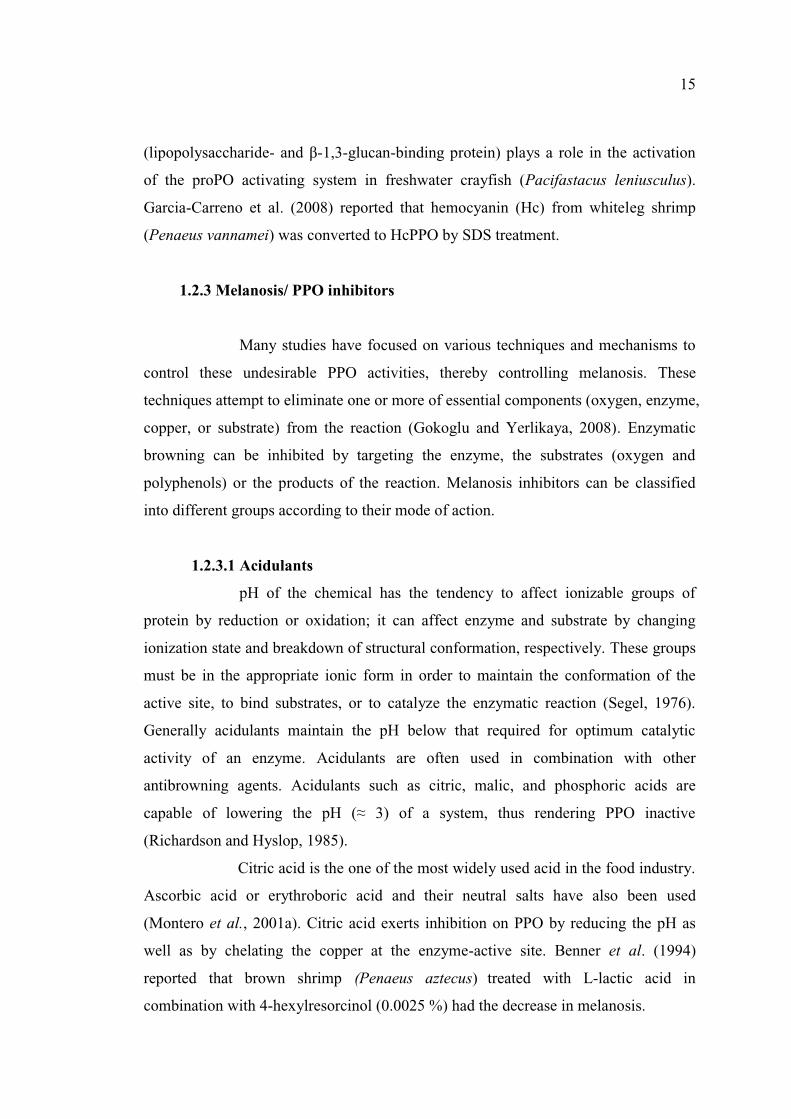
1.2.3 Melanosis/ PPO inhibitors
Many studies have focused on various techniques and mechanisms to
control these undesirable PPO activities, thereby controlling melanosis. These
techniques attempt to eliminate one or more of essential components (oxygen, enzyme,
copper, or substrate) from the reaction (Gokoglu and Yerlikaya, 2008). Enzymatic
browning can be inhibited by targeting the enzyme, the substrates (oxygen and
polyphenols) or the products of the reaction. Melanosis inhibitors can be classified
into different groups according to their mode of action.
1.2.3.1 Acidulants
pH of the chemical has the tendency to affect ionizable groups of
protein by reduction or oxidation; it can affect enzyme and substrate by changing
ionization state and breakdown of structural conformation, respectively. These groups
must be in the appropriate ionic form in order to maintain the conformation of the
active site, to bind substrates, or to catalyze the enzymatic reaction (Segel, 1976).
Generally acidulants maintain the pH below that required for optimum catalytic
activity of an enzyme. Acidulants are often used in combination with other
antibrowning agents. Acidulants such as citric, malic, and phosphoric acids are
(Richardson and Hyslop, 1985).
Citric acid is the one of the most widely used acid in the food industry.
Ascorbic acid or erythroboric acid and their neutral salts have also been used
(Montero et al., 2001a). Citric acid exerts inhibition on PPO by reducing the pH as
well as by chelating the copper at the enzyme-active site. Benner et al. (1994)
reported that brown shrimp (Penaeus aztecus) treated with L-lactic acid in
combination with 4-hexylresorcinol (0.0025 %) had the decrease in melanosis.
Page 39
16
1.2.3.2 Chelating agents
PPO possess metal ion at their active site for the functional activity.
These metal ions are participated in enzyme reaction. Removal of these metal ion by
chelating agents make enzyme inactive. Chelating agents are able to form complex
with PPO activators such as copper and iron ions, through an unshared pair of
electrons in their molecular structures (Kim et al., 2000).
The well known chelating agent is EDTA (ethylenediamine tetra acetic
acid). Chelators used in the food industry include sorbic acid, polycarboxylic acid
(citric, malic, tartaric, oxalic and succinic acids), polyphosphate (triphosphate and
pyrophosphate), macromolecules (porphyrins, proteins) and EDTA. Kojic acid has
potential applicability in the prevention of melanosis in both plant and seafood
products (Chen et al., 1991a). The phenolic derivatives of benzoic acid appear to act
as chelating agents of copper (Montero et al., 2001a). Sodium benzoate was more
effective in lowering the melanosis formation of prawns (Penaeus japonicus) than
ascorbic acid or citric acid during storage at 4oC for 8 days (Montero et al., 2001b).
Opoku-Gyamfua et al. (1992) reported that EDTA inhibited PPO from lobster
(Homarus americanus). PPO from crab Charybdis japonica was totally inhibited by
phenylthiourea and was extremely sensitive to EDTA or diethyldithiocarbamate
(DETC) (Liu et al., 2006)
1.2.3.3 Reducing agents
The most widely used chemicals in preventing enzymatic browning are
reducing agents such as sulfiting agent, ascorbic acid, cysteine and glutathione.
Reducing agents prevent enzymatic browning either by reducing o-quinones to
colorless diphenols, or by reacting irreversibly with o-quinones to form stable
colorless products (Ferrer et al., 1989) (Figure 7). Ascorbic acid is highly water-
soluble, which is acidic and moderately strong reducing compound. Ascorbic acid
also acts as an oxygen scavenger for the removal of molecular oxygen in PPO
reactions. Walker (1977) reported that PPO inhibition by ascorbic acid and cysteine
has been attributed to the reduction of enzymatically formed o-quinones to their
precursor diphenols. The inhibition of melanosis by sulfhydryl compounds, such as
cysteine and glutathione is thought to be due to the formation of colorless thiol-
Page 40
17
conjugated o-quinones (Benjakul et al., 2006). Cysteine-quinone adducts were proved
to be the competitive inhibitors of PPO (Kim et al., 2000). Arias et al. (2007) reported
that ascorbic acid (AA) prevents browning by two different mechanisms. In the
absence of PPO subtstatres AA inactivates PPO irreversibly and in the presence of
PPO substrates, it reduces quinone back to hydroquinone.
Sulfites serve a multifunctional role in foods. They possess
antimicrobial activity and inhibit both enzymatic and non-enzymatic browning
reaction (Januario and Dykes, 2005). Sulfiting agents (sulfur dioxide, SO2; sulfite,
SO3; hydrogen sulfite, HSO3; metabisulfite, S2O5) are the most widely applied
reagents for the control of browning in the food industry (Gokoglu and Yerlikaya,
2008). Bisulfite (HSO3-) is a competitive inhibitor of PPO by binding a sulfhydryl
group on the PPO active site (Madero and Finne, 1982). Inhibition on the PPO
catalyzed melanosis in lobster was accomplished by bisulfite via its reaction with
intermediate quinones forming sulfoquinones, and via its complete inactivation of
PPO (Ferrer et al., 1989). Martinez-Alvarez et al. (2005a) reported that prawns
(Marsupenaeus japonicus) treated with sulfite-based solution had the lowest
melanosis score up to 8 days. Figure 7 depict the inhibition of brown color formation
by reducing agents which convert reactive o-quinone to diphenol. Gomez-Guillen et
al. (2005) used sodium metabisulfite (6.2 to 50 gKg-1) to prevent melanosis in fresh
deep water pink shrimp (Parapenaeus longirostris) by immersion method for 1 h.
Marinez-Alvarez et al. (2008b) reported that Norwegian lobsters (Nephrops
norvegicus) dusted with sulfites had retarded formation of black spot for at least 7
days during chilled storage. Rotllant et al. (2002) reported that the shrimp (Aristeus
anteunatus) treated with increasing concnentration of HQ-Bacterol F containing 40 %
sodium metabisulfite could lower black spot formation up to 27 h but increased the
residual SO2 in th tissue. Prawn (Marsupenaeus japonicus) treated with sulfites
showed initially better protection to lower melanosis and quality losses; however 4-
HR was more effective at the end of storage (Martinez-Alvarez et al., 2005a).
Ascorbic acid and sodium metabisulfite inhibited the activity of both
polyphenoloxidase and hemocyanin from deepwater pink shrimp (Parapenaeus
longirostris) (Martinez-Alvarez et al., 2008a).
Page 41
18
Figure 7. Inhibition of brown color polymers by reducing agent.
Source: Kim et al. (2000)
The FDA has proposed the maximum residual sulfur dioxide levels for
certain foods. Shrimp products having residual sulfite levels above 100 ppm are
considered adulterated, since these levels are considered unsafe. It has been necessary
to search for alternatives that show effective inhibitory effect on melanosis but are
devoid of health concerns to consumers (Chen et al., 1991a).
1.2.3.4 PPO inhibitor (4-Hexylresorcinol)
4-Hexylresorcinol (4-HR) has several advantages over sulfites in food,
including its specific mode of inhibitory action (Figure 8), its lower level required for
effectiveness, its inability to bleach preformed pigments, and its chemical stability
(McEvily et al., 1992). 4-HR acts as an enzyme-competitive inhibitor due to structural
resemblance to phenolic substrates (McEvily et al., 1991). 4-HR (100 mg/kg) showed
a marked ability to inhibit or slow down melanosis in shrimp (Parapenaeus
longirostris), compared with the sodium metabisulfite (1 g/kg) (Guandalini et al.,
1998).
Page 42
19
Figure 8. Inhibitory effect of 4-hexylresorcinol on PPO
Source: Lambrecht (1995)
McEvily et al. (1991) reported that dipping shrimps, brown shrimp
(Penaeus aztecus) and pink shrimp (Penaeus duorarum), into 50 ppm 4-HR in sea
water with subsequent storage on crushed ice inhibited black spot formation up to 14
days. Lambrecht (1995) reported that the headless brown shrimp (Penaeus aztecus)
dipped in 4-HR for 1 min controlled black spot formation for a longer period of time
than the control or those treated with 1.25 % sodium metabisulfite. 4-HR alone or in
combination with ascorbic or citric acid, was effective as inhibitors of melanosis and
microbial spoilage in prawns (Penaeus japonicus) (Montero et al., 2001b). Montero et
al. (2006) studied inhibition of melanosis in pink shrimp (Parapenaeus longirostris)
treated by immersion and dusting method with various concentration (0.0025 to 5 g of
inhibitor per 100 g of shrimp) of 4-HR during chilled storage of 12 days. Melanosis
inhibition increased with inhibitor concentration. PPO activity of Norway lobster
(Nephrops norvegicus) was inhibited with a formulation containing 4-hexylresorcinol
(0.05 and 0.1%) in combination with organic acids and chelating agents (Martinez-
Alvarez et al., 2007). Montero et al. (2001b) reported that 4-HR in combination with
ascorbic or citric acid showed most efffectvie melanosis inhibition in prawns
(Penaeus japonicus) stored at 4oC for 8 days. Shrimp (Penaeus Japonicus) treated
Page 43
20
with 1 % citirc or lactic acid in combination with sodium metabisulfite (0.3%) showed
lowered melanosis formation, compared with the shrimp treated with acids alone
(Gokoglu, 2004). Incubation of mushroom PPO with 4-HR decreased PPO activity
effectively, due to the high affinity of 4-HR for PPO (Arias et al., 2007). 4-HR did not
inhibit the PPO activity of hemocyanin from deepwater pink shrimp (Parapenaeus
longirostris) (Martinez-Alvarez et al., 2008a).
1.2.3.5 Miscellaneous
Chemical and natural compounds have been reported to lower the
enzymatic browning in fruits, vegetables and seafoods. Inorganic halides are known
as the inhibitors of PPO. NaF was the most potent inhibitor, followed by NaCl, NaBr
and NaI (Janovitz-Klapp et al., 1990). Sodium salts of four n-alkyl xanthate
compounds, C2H5OCS2Na (I), C3H7OCS2Na (II), C4H9OCS2Na (III), and
C6H13OCS2Na (IV) exhibited the inhibitory activity towards cresolase and catecholase
of mushroom tyrosinase (Saboury et al., 2007). Amino acids (cysteine, glutathione
and histidine), peptides or proteins can also inhibit browning reaction by reducing
quinone or directly react with PPO (McEvily et al., 1992). Melanosis of pink shrimp
(Penaeus duorarum) was inhibited by treatment with solution of ficin, a sulfhydryl
protease (Taoukis et al., 1990). Richard-Forget et al. (1998) reported that crude
- as cysteine
and a dipeptide cysteine-glutamic acid, which shows the competitive inhibitor
towards PPO.
Honey has been shown to inhibit enzymatic browning. Vela et al.
(2006) studied thirty-six Spanish honeys of different floral origin (nectars and
honeydews) and found that honeydew honeys showed higher antioxidant capacities
and ability to inhibit enzymatic browning in apple homogenate than nectar honeys.
Jeon and Zhao (2005) found that fresh cut apples dip in 10 % honey for 30 min had
antibrowning effect, however vacuum impregnation (75 mmHg for 15 min) with 10 %
honey was more effective in prevention of browning discoloration.
Maillard reaction products (MRP) have also been known to inhibit
PPO from apple and mushroom (Maillard et al., 2007). Cheriot et al. (2007) reported
that MRP from a preheated cysteine-derived compound and a carbonyl component,
Page 44
21
especially hydroxymethylfurfural (HMF), furfural and benzaldehyde, exhibited a
stronger inhibitory potency towards PPO of eggplant, apple, and mushroom.
Matmaroh et al. (2006) had found that the inhibitory activity of MRPs towards
browning in back tiger shrimp, induced by PPO, was most likely due to their reducing
power as well as copper chelating property.
Mimosine inhibited mammalian tyrosinase competitively because of its
structural similarity to the substrate, L-DOPA and its tendency to chelate cupric ion
(Hashiguchi and Takahashi, 1977). Cabanes et al. (1987) reported that L-mimosine
was a slow binding inhibitor of mushroom tyrosinase for oxidation L-DOPA.
Mimosine inhibited polyphenoloxidase from S. rolfsii competitively and decreased the
specific activity by 87 % (Serrano et al., 1983). This indicated that mimosine most
likely inhibit PPO by binding the active site of the PPO. A number of naturally
occurring tyrosinase inhibitors consist of a phenol structure or of metal chelating
agents (Fadimatou et al., 2010). The inhibition of metal-dependent enzymes by L-
mimosine was related to its metal chelating ability (Stunzi et al., 1980). Mimosine and
kojic acid was reported to be a standard inhibitor for mushroom tyrosinase with
competitive type inhibition (Matsumoto-Akanuma et al., 2011; Fadimatou et al.,
2010; Sabudak et al., 2006). Mimosine inhibited both monophenol and
diphenoloxidase activity from European spiny lobster (Palinurus elephas)
competitively (Brack et al., 2008).
A competitive and mixed type inhibition occurred for mushrrom PPO
depending on the phenolic substrates studied (Chen et al., 1991b). Kojic acid showed
a mixed type inhibition towards PPO from white shrimp, grass prawn and lobster
(Chen et al., 1991b). Dodecyl gallate was found as a mixed-type inhibitor for
mushroom tyrosinase, when L-DOPA was used as a substrate (Kubo et al., 2000).
Inhibition constant Ki value of dodecyl gallate on mushroom tyrosinase was 0.636
mM (Kubo et al., 2003). Benjakul et al. (2006) reported that cysteine and glutathione
showed competitive inhibition toward kuruma prawn PPO with Ki values of 0.45-0.46
mM. Ozagrel was a reversible mixed type inhibitor of diphenoloxidase activity of
mushrrom tyrosinase with KS1, KS2, Ki1 and Ki2 was 2.21, 3.89, 0.454 and 0.799
mM, respectively (Li et al., 2009).
Page 45
22
1.2.4 Changes in quality of shrimp during post mortem storage
Storage of fish and shellfish in crushed iced or liquid ice (chilling or
super chilling) has been the routine practice of preserving fish on board and at shore
(Lakshmanan et al., 2002). However, freezing technology has been widely used to
stored crustaceans for export (Lopez-caballero et al., 2007). Although freezing is an
effective method of preserving foods, some deterioration in frozen food quality occur
during storage. The extent of quality loss is dependent upon many factors, including
storage temperature, rate of freezing and thawing, temperature fluctuations, freeze-
thaw abuse during storage (Srinivasan et al., 1997). The impact of iced and frozen
storage on shrimp quality has been dependent on the length of storage (Erickson et al.,
2007).
1.2.4.1 Microbiological changes during storage
Microorganisms are the major cause of spoilage of most seafood
products by formation of amines, sulfides, alcohols, aldehydes, ketones, and organic
acids with unpleasant and unacceptable off-flavors (Gram and Dalgaard, 2002). The
high content of free amino acids and other soluble non-nitrogenous substances, can
serve as easily digestible nutrients for microbial growth (Zeng et al., 2005). However
the specific spoilage organism (SSO) are not the same in every case and the microbial
flora isolated from seafoods differs considerably from one study to another,
depending on the species of fish, their environment, the mode of capture, the type of
fish product as well as the climatic and storage conditions (Gram and Dalgaard, 2002).
Generally, pseudomonas, H2S- producing bacteria and lactic acid bacteria (LAB) are
predominant in spoiled fish flora, while Enterobacteriaceae, a gram negative bacteria
is also frequently present (Sallam, 2007).
On iced storage, the total bacterial load was reduced to one log from
initial load in fresh fish/shrimp due to cold shock (Lakshmanan et al., 2002). Zeng et
al. (2005) had found that shrimp (Pandalus borealis) stored in liquid ice slowed down
microbial growth as compared to shrimp stored in flake ice or brine mixed ice. Black
tiger prawns (Marsupenaeus japonicus) treated with the formulation containing 0.1 %
4-HR in combination with organic acids (citric, ascorbic, and acetic acids), EDTA and
Page 46
23
disodium dihydrogen pyrophosphate (PPi) had the lower microbial growth (total
bacteria count, H2S-producing bacteria, LAB, enterobacteria, and pseudomonas) as
compared to control and sulfite-based treated prawn during chilled storage of 13 days
(Martinez-Alvarez et al., 2005a). Lopez-caballero et al. (2007) reported that thawed
deep water pink shrimp (Parapenaeus longirostris) treated with different formulations
containing 4-HR (0.05 and 0.1 %) in combination with organic acids and chelating
agents, showed lowered total bacterial count in comparison with those treated with a
commercial formula based on sulfites, and a mixture of gluconic acid and commercial
sulfites during 14 days of chilled storage. Mastromatteo et al. (2010) reported that a
slight antimicrobial effect was observed in ready-to-eat peeled shrimp (Palaemon
serratus) when coated with thymol essential oil (500, 1000 and 1500 ppm) and effect
was concentration dependent. Fresh white shrimp (Penaeus vannamei) coated with
antimicrobial film prepared from catfish skin gelatin incorporation with potassium
sorbate and sodium tripolyphosphate could retard microbial growth and prolong shelf
life for up to 10 days of iced storage (Jiang et al., 2011).
1.2.4.2 Chemical changes during storage
Chemical changes in seafoods are mostly related with the microbial
load (Gram and Dalgaard, 2002). Quality of fish and shellfish are generally accessed
by the changes in lipid oxidation (PV, TBARS, etc), total volatile base (TVB),
trimethylamine (TMA), K-value, and pH. Lipid oxidation in fish meat can be
initiated by autoxidation, photosensitized oxidation, or an enzymatic reaction
involving lipoxygenase, peroxidase and microbial enzymes (Bak et al., 1999). The
volatile base found in the shrimp more likely indicated that an autolytic process was
involved during postmortem handling. Adenosine and adenosine monophosphate
(AMP) deaminase might play a major role in this process after capture and
transportation (Lopez-caballero et al., 2007). TMA is produced by some bacteria
capable of using trimethylamine oxide (TMAO) in anaerobic respiration (Gram and
Dalgaard, 2002). K-value has been used as the freshness index in fish and shellfish.
During postmortem handling and storage, endogenous enzymes break down
adenosine triphosphate to different derivatives such as adenosine diphosphate,
adenosine monophosphate (AMP), inosine monophosphate (IMP), inosine (HxR), and
Page 47
24
finally hypoxanthine (Hx) (Yamagata and Low, 1995). The increase in pH was
associated with the accumulation of basic compounds, mainly resulted from microbial
action (Lopez-caballero et al., 2007). In general, a K-value of 60% has been realized
as the rejection limit. TMA acceptability limit for shrimp was reported to be 5 mg
N/100g and TVB content of 40 mg N/100g has been used as the freshness borderline
(Uchiyama and Kakuda, 1984; Mendes et al., 2002).
Thepnuan et al. (2008) found that the shrimp (Litopenaeus vannamei)
pretreated with 2 % pyrophosphate and 0.25 % 4-HR and stored under MAP showed
the lower TBARS value throughout 12 days of storage. Marination of deep water pink
shrimp (Parapenaeus longirostris) with rosemary extract lowered TBA value by 2.7
times than the control at the end of storage day at 1 ± 0.5oC (Cadun et al., 2008).
Lopez-caballero et al. (2007) reported that thawed deep water pink shrimp
(Parapenaeus longirostris) treated with different formulations containing 4-HR in
combination with organic acids and chelating agents, a commercial formula based on
sulfites, and a mixture of gluconic acid and commercial sulfites, did not show any
noticeable differences for TVB and TMA during 14 days of chilled storage. The
control banana shrimp (Penaeus merhuiensis) and those treated with sodium bisulfite
had similar K-values (43%) at the last day (day 6) of iced storage (Yamagata and Low,
1995).
1.2.4.3 Physical and Sensorial changes during storage
Super chilling of muscle food can result in partial freezing, which may
lead to negative changes such as decrease in water holding capacity (WHC) and shear
force (Zeng et al., 2005). Over the extended storage time, the breakdown of structural
protein in shrimp tissue may also cause loss of physical integrity (Rutherford et al.,
2007). Generally, the softening of shrimp or fish muscle is associated with proteolysis
caused by endogenous or microbial proteinases and collagenase (Benjakul et al.,
1997). Lopez-caballero et al. (2007) found that WHC was slightly decreased for
thawed deep water pink shrimp (Parapenaeus longirostris) treated with different
formulations containing 4-HR in combination with organic acids and chelating agents
throughout storage period. Treatment with sulfite based formulations showed the
increase in shear strength at the end of storage (14 day). Thepnuan et al. (2008)
Page 48
25
reported that whole Pacific white shrimp (Litopenaeus vannamei) treated with
pyrophosphate and 4-HR and packaged under MAP-2 (80% CO2, 20% N2) had the
highest shear force within the first 6 days of storage. However, no marked difference
in WHC was observed within different treatments throughout storage.
Erickson et al. (2007) studied sensory attribute for both fresh and
frozen commercially available shrimp and found that only appearance could uniquely
differentiate a specific type of frozen shrimp. Fresh shrimp were characterized as
being sweeter and juicier than frozen shrimp. Zeng et al. (2005) reported that shrimp
stored in liquid ice at -1.5oC had highest sensory scores than shrimp stored at +1.5oC
and in ice at +1.5oC and in brine mixed with ice at -1.5oC. In general, the loss in
sensory properties, especially on appearance, is associated with the melanosis.
Refrigeration alone does not prevent, but slow down black spot formation, because
the PPO remains active during refrigeration, storage on ice, and post freeze-thawing
(Montero et al., 2004). During storage, proPPO can be activated into PPO by the
action of proteolytic enzymes, which may produce substrates for active PPO by
protein hydrolysis (Ali et al., 1994). Marination of deep water pink shrimp
(Parapenaeus longirostris) with rosemary extract had lowered the sensorial quality
loss compared with the control at the end of storage day at 1 ± 0.5oC (Cadun et al.,
2008).
1.2.5 Modified atmosphere packaging (MAP)
Modified atmosphere packaging (MAP) is the removal and/or
replacement of the atmosphere surrounding the product before sealing in vapor-barrier
materials. MAP can be vacuum packaging (VP), which removes most of the air before
the product is enclosed in barrier materials (McMillin, 2008). The combination of
CO2, N2 and O2 is generally used in MAP (Rutherford et al., 2007). CO2 is the most
important gas used in MAP, because of its bacteriostatic properties (Lopez-Caballero
et al., 2002). CO2 is highly soluble in water and fat, and the solubility increases
greatly with decreasing temperature. The solubility of CO2 leads to dissolved CO2 in
the food product and form carbonic acid (Sivertsvik et al., 2002), according to the
following equation:
Page 49
26
CO2 + H2 2CO3 3- + H+
Thus, the resultant decrease in pH can alter bacterial cell membrane,
affecting nutrient uptake and absorption, and can inhibit intracellular enzyme activity
(Rutherford et al., 2007). It has been suggested that atmosphere poor in oxygen and
rich in CO2 could act as a coadjuvent in delaying melanosis and chemical changes
(Goncalves et al., 2003). Nitrogen (N2), an inert and tasteless gas, is mostly used as
filler gas in MAP. The use of low level of oxygen (O2) in MAP is generally
recommended to avoid lipid oxidation and growth of aerobic spoilage bacteria
(Martinez-Alvarez et al., 2005b). The ratio between the volume of gas and volume of
food product (G/P ratio) is usually 2:1 or 3:1. This high G/P ratio is also necessary to
prevent package collapse because of the CO2 solubility in wet foods (Sivertsvik et al.,
2002).
Deepwater pink shrimp (Parapenaeus longirostris) packed under MAP
(40% CO2 / 30% O2 / 30% N2 and 45% CO2 / 5% O2 / 50% N2) and stored at 2oC for 9
days had lowered bacterial count (total bacteria count, H2S-producing microorganism
and enterobacteria) and biochemical indexes (TVB and TMA) as compared to air-
packed or iced stored shrimp (Lopez-Caballero et al., 2002). Deepwater pink shrimp
(Parapenaeus longirostris) treated with sulfite and packed under 45% CO2 / 5% O2 /
50% N2 had lowered black spot formation in comparison with the treated shrimp
packed under 40% CO2 / 30% O2 / 30% N2 on day 7 of iced storage (Goncalves et al.,
2003). Deep water pink shrimp (Parapenaeus longirostris) treated with 0.1 % 4-HR
and kept in MAP (53% CO2 / 7% O2 / 40% N2) showed less melanosis than shrimp
treated with 4-HR and kept in ice (Martinez-Alvarez et al., 2005b). Lu (2009)
reported that whole or decapitated Chinese shrimp (Fenneropenaeus chinensis) stored
under MAP (40% CO2 / 30% O2 / 30% N2) and 100% CO2 after soaking with
bactericide (1 g/L 4-HR, 500 IU/mL nisin and 5 g/L sodium dehydroacetate) had
increased shelf life up to 13 and 17 day as compared to the control (up to 9 days) at
2oC. Thepnuan et al. (2008) found that the whole and decapitated white shrimp
(Litopenaeus vannamei) pretreated with 2% pyrophosphate and 0.25% 4-
hexylresorcinol and stored under MAP (80% CO2 / 10% O2 / 10% N2 or 80% CO2 /
20% N2) showed the lowered microbiological (mesophilic, psychrophilic, lactic acid
Page 50
27
bacteria, coliforms and E.coli); chemical deteriorations (TVB, TMA, TBARS values)
and lower melanosis score throughout the storage of 12 days at 4oC, compared with
those without treatment. Martinez-Alvarez et al. (2005b) reported that shrimp
(Parapenaeus longirostris) treated with 4 % sulfites in combination with MAP (53%
CO2 / 7% O2 / 40% N2) totally inhibited darkening during 20 days of storage at 2oC.
Nevertheless, the application of atmosphere in shrimps without antimelanosis agents
did not inhibit melanosis (Martinez-Alvarez et al., 2005b). Mastromatteo et al. (2010)
reported that thymol coated (1000 ppm) ready-to-eat peeled shrimp (Palaemon
serratus) and kept under MAP (5%O2 : 95%CO2) had lowered microbial growth and
sensorial quality loss, thereby increasing the shelf life up to 14 days, compared to
sample in air (5 days).
1.2.6 Phenolic compounds
Phenolic compounds are naturally occurring secondary metabolites
that are derivatives of the pentose phosphate, shikimate, and phenylpropanoid
pathways in plants (Figure 9) (Ryan and Robards, 1998).
Figure 9. Metabolic pathways for the formation of phenolic compounds.
Source: Ryan and Robards (1998)
Page 51
28
They are present in fruits, vegetables, leaves, nuts, seeds, flowers, and
barks (Sellappan et al., 2002). These compounds are of considerable physiological
and morphological importance in plants (Balasundram et al., 2006).
Chemically, phenolic compounds comprise an aromatic ring, bearing
one or more hydroxyl groups, and range from simple phenolic molecules to highly
polymerized compounds (Dykes and Rooney, 2007). Most naturally occurring
phenolic compounds are present as conjugates with saccharides (mostly mono and
disaccharides), linked to one or more of the phenolic groups by acid-labile hemiacetal
bond (Ryan and Robards, 1998) and may also occur as functional derivatives such as
esters and methyl esters (Balasundram et al., 2006). Despite the structural diversity,
the wide range of phenolic compounds are often referred to as polyphenols and
basically be categorized into several classes as shown in Table 1 (Ryan and Robards,
1998; Naczk and Shahidi, 2004; Balasundram et al., 2006; Dykes and Rooney, 2007).
Table 1. Classes of phenolic compounds in plants
Classes Structure
Simple phenolics, benzoquinones C6
Hydroxybenzoic acids C6-C1
Acethophenones, phenylacetic acids C6-C2
Hydroxycinnamic acids, phenylpropanoids
(coumarins, isocoumarins, chromones, chromenes) C6-C3
Napthoquinones C6-C4
Xanthones C6-C1-C6
Stilbenes, anthraquinones C6-C2-C6
Flavonoids, isoflavonoids C6-C3-C6
Lignans, neolignans (C6-C3)2
Biflavonoids (C6-C3-C6)2
Lignins (C6-C3)n
Condensed tannins (proantocyanidins or flavolans) (C6-C3-C6)n
Source: Balasundram et al. (2006)
Page 52
29
Phenolic acids consist of two subclasses, hydroxybenzoic acids and
hydroxycinnnamic acids. Hydroxybenzoic acids (C6-C1) include gallic, p-
hydroxybenzoic, vanillic, syringic and protocatecguic acids. The hydroxycinnamic
acids have C6-C3 structure and include coumaric, caffeic, ferulic, and sinapic acids
(Dykes and Rooney, 2007). Ferulic acid (4-nydroxy-3-methoxycinnamic acid) is
ubiquitous phenolic acid in plant kingdom (Figure 10) (Ou et al., 2007). Ferulic acid
can be found in the different plant sources at various levels: 0.5 % of wheat bran,
0.9 % of sugar beet pulp, 5 % of corn kernel (Ou and Kwok, 2004; Zhao et al., 2005),
1.5-2.8 % of rice bran oil (Taniguchi et al., 1999) and 1.36-2.58 % of sugarcane
bagasse (Ou et al., 2007).
Figure 10. Structure of ferulic acid.
Source: Balasundram et al. (2006)
The flavonoids are a large class of compounds, ubiquitous in plants,
and usually occurring as glycosides (Rice-Evans et al., 1997). Flavonoids are low
molecular weight compounds, consisting of fifteen carbon atoms, arranged in a C6-C3-
C6 configuration. They contain several phenolic hydroxyl functional groups attached
to ring structures, designated as A, B and C (Balasundram et al., 2006). Variation in
substitution pattern to ring C result in flavanols (e.g. catechin), lacking the 2,3-double
bond and the 4-one structure (Figure11) (Rice-Evans et al., 1997). Green tea is the
most abundant source of polyphenols, mostly in the form of gallic acid and catechin
derivatives. Green tea catechins are structurally flavanols. The four major catechins in
green tea are (-)-epigallocatechin gallate (EGCG), (-)-epigallocatechin (EGC), (-)-
epicatechin gallate (ECG), and (-)-epicatechin (EC) (Figure 12) (Cabrera et al., 2006).
In general, these four major caetchin derivatives together constitute 30 % of the dry
Page 53
30
leaf weight (Gupta et al., 2002). Among these, EGCG represents approximately 59 %
of the total catechins (Cabrera et al., 2006).
Figure 11. Structure of catechin.
Source: Wang and Helliwell (2000).
Figure 12. Chemical structure of gallic acid and the four major catechins in green tea.
GA, gallic acid; EGCG, (-)-epigallocatechin-3-gallate; EGC, (-)-
epigallocatechin; ECG, (-)-epicatechin-3- gallate; EC, (-)-epicatechin.
Source: Cabrera et al. (2006)
Page 54
31
Tannins, the relatively high molecular weight compounds, which
constitute as the third important group of phenolics may be subdivided into
hydrolysable tannin, which is ester of gallic acid and condensed tannins, are polymers
of polyhydroxy flavanol monomers (Naczk and Shahidi, 2004).
The functionality of phenolic compounds is expressed through their
action as an inhibitor or an activator for a large variety of mammalian enzyme system
and as metal chelators as well as scavengers of free radicals (Pringent, 2005; Rice-
Evans et al., 1997; Shahidi et al., 2007). Phenolic compounds have been associated
with the health benefits derived from consuming high levels of fruits and vegetables,
because of their antioxidant activity (Balasundram et al., 2006; Sellappan et al., 2002)
and could therefore be a natural source of antioxidants.
1.2.6.1 Extraction of phenolic compounds
The structural diversity of phenolic compounds varies from simple to
highly polymerized substances that include varying proportions of phenolics,
distributed randomly in plants at the tissue, cellular and subcellular levels. They may
also exist as complexes with carbohydrates, proteins and other plant components
(Luthria et al., 2006). Extraction of phenolic compounds in plant materials is
therefore influenced by their chemical nature, the extraction method employed,
sample particle size, storage time and conditions as well as the presence of interfering
substances (Naczk and Shahidi, 2004). Therefore, phenolic extracts of plant materials
are always mixture of different classes of phenolics that are soluble in the solvent
system used. Solubility of phenolic compounds is governed by the type of solvent
(polarity) used, degree of polymerization of phenolics, as well as interaction of
phenolics with other food constituents and formation of insoluble complexes (Robbins,
2003). Therefore, there is no uniform or completely satisfactory procedure that is
suitable for extraction of all phenolics or a specific class of phenolic substances in
plant materials (Luthria et al., 2006). Methanol, ethanol, acetone, water, ethyl acetate
and, to a lesser extent, propanol, dimethyl formamide, and their combinations are
frequently used for the extraction of phenolics (Naczk and Shahidi, 2004). In some
cases, mechanical means to enhance molecular interaction between the phenolic
compounds and solvent are employed (Robbins, 2003). Shaking, stirring, vortex
Page 55
32
mixing and sonication are four commonly used extraction techniques (Luthria et al.,
2006).
Row and Jin (2006) reported the recovery of catechin compounds from
Korean tea by solvent extraction. The optimum extraction condition was 80oC for 40
min in pure water. The extract was partitioned with water/chloroform (1:1 v/v), which
was suited to remove caffeine impurity from the extract. The resulting extract was
further partitioned with water/ethyl acetate (1:1 v/v) to deeply purify the catechin
compounds. Torre et al. (2008) investigated the release of ferulic acid from corn cobs
by alkaline hydrolysis. Optimum hydrolysis condition for hydroxy-cinnamic acids
content was 0.5 N NaOH and solid/liquid ratio of 0.084 for 6 h. Perva-Uzunalic et al.
(2006) studied the extaction efficiency of caetchins and caffeine from green tea leaves
using different solvents (acetone, ethanol, methanol, acetonitrile, and water) at
different temperature and times. Optimal condition for extraction of caetchin with
water was obtained at 80oC for 20 min. Rusak et al. (2008) extracted phenolics from
bagged and loose leaves of white and green tea by distilled water (80oC), distilled
water (80oC) with 5 ml of lemon juice and aqueous ethanol (10, 40 and 70 %).
Addition of lemon juice could enhance the extraction of phenolic from white tea and
aqueous ethanol (40 %) was most effective in the extraction of catechins. Extracction
of ferulic acid and vanillin from flax shives, wheat bran and corn bran were carried
out by non-pressurised alkaline hydrolysis (0.5 M NaOH) and pressurized solvents
(0.5 M NaOH, water, ethanol and ammonia) (Buranov and Mazza, 2009).
1.2.6.2 Identification and characterization of phenolic compounds
In the last twenty years, the analytical technique that has dominated the
separation and characterization of phenolic compounds is HPLC with reverse phase
(RP) column technology (Robbins, 2003). There is no single wavelength appropriate
to monitor all phenolics since they display the absorbance maxima at different
wavelengths (Herrera and Luque de Castro, 2005). Other less common means of
detection, coupled to LC are through electrochemical detection (EC), fluorescence (F),
and MS (Robbins, 2003).
Phenolic compounds from strawberries were detected by liquid
chromatography with photodiode array UV detection. The mobile phase consisted of
Page 56
33
methanol-water (10-90, v/v) and methanol-water (50:50, v/v) both adjusted to pH 3
with acetic acid and different gradients were tested on the Hypersil ODS column.
Most benzoic acid derivatives show the maximum absorbance close to 254 nm.
However, gallic acid, salicylic and syringic acid have their maxima absorbance at 275,
310 and 280 nm, respectively. Chlorogenic, caffeic and p-coumaric acid have the
maximum absorbance close to 325 nm, and ferulic acid close to 310 nm (Herrera and
Luque de Castro, 2005). Ma et al. (2009) detected seven phenolic compounds of two
families including cinnamic acids and benzoic acid from citrus peel extract using C-
18 reverse phase column and elution was performed with the mobile phase consisting
of 4 % (v/v) acetic acid in water: 100 % methanol (80:20, v/v) at solvent flow rate of
1 mL/min. Charrouf et al. (2007) studied the separation and characterization of
phenolic compounds in argan fruit pulp using liquid chromatography (LC) negative
electrospray ionization (ESI) mass spectroscopy (MS/MS). A C-18 (50 x 2.1 mm, i.d
ed with
water/0.05% acetic acid and acetonitrile
compounds were identified, mainly flavonoid glycosides and flavonoid aglycons.
Parejo et al. (2004) separated and characterized phenolic compounds in Fennel
(Foeniculum vulgare) using liquid chromatography-negative ionization mass
spectrometry. The fennel waste was extracted by water for 15 min and then
chromatographed on sephadex LH-20 to collect seven fractions, each of them
subjected to LC-MS analysis and forty-two phenolic compounds were identified.
Weisz et al. (2009) extracted eleven phenolic compounds from
defatted sunflower (Helianthus annuus L.) kernels and shells and characterized by
HPLC with diode array and electrospray ionization (ESI) mass spectrometric
detection in negative mode. The column used was 150mm x 3.0mm inner diameter, 4
-Synergi. The mobile phase consisted of 2 % (v/v) acetic
acid in water (eluent A) and of 0.5 % acetic acid in water and acetonitrile (50:50, v/v,
eluent B) with flow rate of 0.4 mL/min. UV-Visable spectra were recorded in the
range of 200-600 nm. Five phenolic compounds, namely gallic acid, procyanidin B2,
(-)-galllocatechin, (-)-epicatechin, and (-)-epicatechin-3-gallate were identified from
50 % ethanol extract of litchi (Litchi sinensis Sonn.) seeds, after application of reverse
phase high performance liquid chromatography, coupled to a diode array detector and
Page 57
34
electrospray ionization mass spectra (Prasad et al., 2009). The samples were eluted
with gradient system consisting of solvent A (2 % acetic acid, v/v) and solvent B
(acetonitrile:methanol, 10:15, v/v), with a flow rate of 1 mL/min from C-18 column
rticle size). UV-visible absorption spectra were recorded from
200 to 600 nm during HPLC analysis.
1.2.6.3 Antioxidant activity of phenolic compounds
The chemical activity of phenols in terms of their reducing properties
as hydrogen or electron donating agents predicts their potential for action as
antioxidants (Rice-Evans et al., 1997). Radical-scavenging activity (1,1-diphenyl-2-
picrylhydrazyl method) and total antioxidant activity (phosphomolybdenum method)
of grape (Vitis vinifera) seed extracts of acetone:water:acetic acid (90:9.5:0.5) and
methanol:water:acetic acid (90:9.5:0.5) were determined at 25 and 50 ppm
concentration. It was found that acetone:water:acetic acid (90:9.5:0.5) extract showed
the higher radical scavenger than methanol:water:acetic acid (90:9.5:0.5) extract
(Jayaprakasha et al., 2003). Negi et al. (2005) successively extracted seabuckthorn
(Hippophae rhamnoides L.) seeds with chloroform, ethyl acetate, acetone and
methanol using soxhelt extractor for 8 h each. The reducing power and antioxidant
activities of natural crude methanol extract evaluated using 1,1-diphenyl-2-
picrylhydrazine and liposome model system were the highest.
Shahidi et al. (2007) evaluated the antioxidant activity in hazelnut
kernel (Corylus avellana L.) and hazelnut byproduct ethanol extract. Extracts of
hazelnut byproducts (skin, hard shell, green leafy cover, and tree leaf) exhibited the
stronger antioxidant activities than hazelnut kernel at all concentrations tested.
Hazelnut extracts showed different antioxidative efficacies, related to the presence of
phenolic compounds. Among samples, extracts of hazelnut skin, in general, showed
superior antioxidative efficacy and higher phenolic content as compared to other
extracts. Prasad et al. (2009) reported that 50 % ethanol extract of Litchi sinensis
Sonn. seeds showed total antioxidant capacity, scavenging the 1,1-diphenyl-2-picryl
hydrazyl radical and inhibitory activity against lipid peroxidation, at all
concentrations tested (25-
synthetic antioxidant, butylated hydroxyl toluene. Yerlikaya et al. (2009) reported that
Page 58
35
natural grape seed extract or garlic extract (6 %) in batter coating of shrimp was able
to lower lipid oxidation during frozen storage.
Green tea extract prepared with hot water showed high antioxidant
activity for an oil-in-water emulsion at pH 5.5 during prolonged storage (40 days).
Myricetin showed the higher contribution to the antioxidant activity than EGCG and
ECG (Roedig-Penman and Gordon, 1997). The Tungting oolong tea (Taiwanese tea)
together with Chinese green tea effectively suppressed the prooxidant activities of the
dark meat and skin of blue sprat assessed by peroxide value and carbonyl value during
refrigeration (Seto et al., 2005). The antioxidant activity of Taiwanese tea extract
showed positive correlation with the total catechins, especially with that of EGCG but
a weak correlation with the contents of total polyphenols (Seto et al., 2005). Based on
TEAC, FRAP and hypochlorite scavenging activity, the odrer of phenolics was:
procyanidin dimmer > flavanol > flavonol > hydroxycinnamic acids > simple
phenolic acids (Soobrattee et al., 2005). Gallic acid and rosmarinic acid were the most
potent antioxidants among the simple phenolic and hydroxycinnamic acids
(Soobrattee et al., 2005). Theaflavins in black tea and caetchins in green tea are
equally effective antioxidants, when tested in Cu2+ mediated LDL oxidation (Leuang
et al., 2001).The presence of ferulic acid or similar phenolic compounds reduced the
free radical damage in neural cell (Kanski et al., 2002). Ferulic acid showed high
scavenging activity for hydrogen peroxide, superoxide, hydroxyl radical and nitrogen
dioxide radicals dut to the resonance stabilized phenoxy radical (Ou and Kwok, 2004).
1.2.6.4 Antimicrobial activity of phenolic compounds
Phenolic compounds might disrupt the cell wall of microorganism by
forming complex with proteins in cell wall and make lyses of cell wall (Chanthachum
and Beuchat, 1997). The treatment with 5 % kiam wood extract containing tannic acid
retarded the rate of growth of Listeria monocytogenes, aerobic mesophiles and
psychrophilic micro-organism naturally present on cabbage (Chanthachum and
Beuchat, 1997). Among chloroform, ethylacetate, and n-butanol extracts of artichoke
(Cynara scolymus L.) leaf extracts, the n-butanol fraction exhibited the most
significant antimicrobial activities against seven bacteria species, four yeasts, and four
molds. The minimum inhibitory concentrations of these compounds were between 50
Page 59
36
and 2 (Zhu et al., 2004). Jayaprakasha et al. (2003) found that the gram
positive bacteria were completely inhibited at 850-1000 ppm, while gram negative
bacteria were inhibited at 1250-1500 ppm concentration of grape (Vitis vinifera) seed
extract. Study on antibacterial activity of seabuckthorn (Hippophae rhamnoides L.)
seeds extracted with chloroform, ethyl acetate, acetone and methanol using soxhelt
extractor for 8 h each revealed that methanol extracts showed the maximum
antibacterial activity (Negi et al., 2005). The MIC values, with respect to methanol
extract for Bacillus cereus, Bacillus coagulans, Bacillus subtilis, Listeria
monocytogenes, Yersinia enterocolitica, were found to be 200, 300, 300, 300, and 350
ppm, respectively. The phenolic compounds extracted from the table olive from
Portugal (100mg/mL) showed antimicrobial activity against gram-positive, gram-
negative and fungi (Pereira et al., 2006).
Finnish berry extracts containing flavonoids and phenolic acids
inhibited the growth of gram-negative but not gram-positive bacteria. The extract
from sea buckthorn berry and blackcurrant showed the least inhibitory activity against
gram-negative bacteria. Cloudberry, raspberry and strawberry extracts were strong
inhibitors of Salmonella (Puupponen-Pimia et al., 2001). Aqueous extract of tea
(Camellia sinensis) containing epigallocatechin, epigallocatechin gallate and
epicatechin gallate inhibited wide range of pathogenic bacteria including methicillin-
resistant Staphylococcus aureus (Yam et al., 1997). In black tea extracts, theaflavin
and its gallates are antibacterial active components (Yam et al., 1997). Aqueous
extract of pu-erh tea (2 mg/mL) containing caffeine and epicatechin showed potential
antimicrobial effect on gram-positive bacteria than that of gram-negative bacteria (Wu
et al., 2007). The ethanolic extract of green tea significantly inhibited the spoilage
microflora and certain pathogens of acidulant treated mutton (pH 3.8) for up to 4 days
at 25 ± 2oC and 85 ± 5 % RH (Kumudavally et al., 2008). The antimicrobial
mechanism of ferulic acid was attributed to its inhibition of arylamine N-
acetyltransferase in the bacteria (Lo and Chung, 1999).
1.2.6.5 Antibrowning activity of phenolic compounds
Aromatic carboxylic acids of cinnamic acid and its analogues, p-
coumaric, ferulic, and sinapic acids are competitive inhibitors of PPO (Kim et al.,
Page 60
37
2000). Some phenolic compounds inhibit PPO activity by interacting with active site
of the enzyme (Janovitz-klapp et al., 1990). Furthermore, phenolic compounds could
interact with protein or enzymes via hydrogen bond or hydrophobic interaction
(Prigent, 2005). Chen et al. (1991a) found that kojic acid showed a mixed-type
inhibition for white shrimp, grass prawn, and lobster polyphenoloxidase. Among the
various phenolic acids tested, kojic acid showed the highest inhibitory effect on
browning in apple slices (Son et al., 2001). Cuminaldehyde (p-
isopropylbenzaldehyde) was identified as potent mushroom tyrosinase inhibitor (ID50
o and Kinst-Hori, 1998). Kubo et al. (2003) reported that
1.55 mM dodecyl gallate exhibited the inhibitory activity towards mushroom
tyrosinase, in which 50 % activity loss was obtained. Prasad et al. (2009) identified
gallic acid, procyanidin B2, (-)-gallocatechin, (-) - epicatechin, and (-)-epicatechin-3-
gallate in extract of Litchi sinensis Sonn. seeds. This litchi seed extract showed
inhibitory activity of tyrosinase in a concentration dependent manner (25-
Gokoglu and Yerlikaya (2008) found that shrimp (Parapenaeus longirostris) treated
with ethanol extract of grape seed (Vitis vinifera sp.) at concentration of 1.5 % had the
lowered the melanosis formation during storage at 4oC for 3 days. Stilbenes are C6-
C2-C6 compounds, found in nature as monomers and oligomers. They possess
inhibitory activity towards mushroom tyrosinase (Likhitwitayawuid, 2008). Jang et al.
(2003) reported that shrimp (Trachypenaeus curvirostris) treated with 70% acetone
extract of enokitake mushroom (Flammulina velutipes) (2.5 g wet enokitake/mL, test
sample) had the delayed darkening at 24oC for 20 h as compared to control. The hot
water extract prepared from fruiting body of the edible mushroom (Flammulina
velutipes) contained ergothioneine (ERT) at a level of 2.05 mg/mL. The dietary
supplement of mushroom extract in kuruma shrimp (Marsupenaeus japonicus) could
reduce post mortem development of melanosis by suppressing PPO activity in the
hemolymphs of the shrimp (Encarnacion et al., 2010).
Page 61
38
1.2.7 Mimosine
Mimosine is a non- -(3-hydroxy-4-pyridon-
1-yl)-L-alanine) (Lalitha and Kulothungan, 2006). Mimosine is an analogue of the
tyrosine (Figure 13) (Echeverria et al, 2002).
Figure 13. Structure of Mimosine.
Source: Champanerkar et al. (2010)
Mimosine is highly concentrated in the seed and foliage of two legume
genera, Mimosa and Leucaena (Serrano et al., 1983). Lead tree, Leucaena
leucocephala, is belonging to a tropical and subtropical legume family. It has been
used as livestock feed because of their high content of protein, carotenoids, vitamin k,
xanthophylls and minerals (Kamada et al., 1997). Phenolics found in different parts of
Leucaena were condensed tannin (Echeverria et al., 2002), quercetin and myricetin
glycosides (Lowery et al., 1984), gallocatechin, epigallocatechin and epicatechin
(Erickson et al., 2000). Additionally, seeds and leaves of lead are consumed as human
foods (Sahlu et al., 1995). The seeds of guaje (L. esculenta) are eaten with salt in
Mexico.
Mimosine is a water soluble amino acid (Shiroma and Hongo, 1981).
Different parts of the Leucaena contain varying amounts of mimosine, 2-10 % of dry
leaf, 2-5 % of dry seed (Lalitha et al., 1993) and 1-1.5 % of root (Soedarjo et al.,
1994). It is less soluble in methanol and ethanol, insoluble in other organic solvent,
and soluble in dilute acid and base (Champanerkar et al., 2010). The concentration of
mimosine is higher in the seeds than in other parts of the plant, second only to the
immature tender leaves (Chanchay and Poosaran, 2009). Mimosine could be removed
by prolonged soaking of leaves or seeds in water at 30oC (Puchala et al., 1995). About
Page 62
39
90 % of mimosine could be extracted by soaking the leaves in freshwater for 36 h
(Chanchay and Poosaran, 2009). Kamada et al. (1997) extracted mimosine from
brown ripe seed powder of L. leucocephala by using boiling water for 1 h with a seed
powder/ water ratio of 1:7 (g/mL). The equivalent amount of ethanol was added to
precipitate impurities before the solution was cooled and then subjected to filtration.
The filtrate were stored at freezer (-18oC) for 8 h and precipitated impurities were
removed by centrifugation. The remaining supernatant were concentrated by rotary
evaporator and used as crude mimosine (Kamada et al., 1997). Drying leaves of L.
leucocephala at 60oC for 24 h and soaking in water for 72 h at room temperature
could remove 94.7 % of mimosine (Chanchay and Poosaran, 2009). The fresh leaves
of L. leucocephala and dry seed powder were mixed with boiling water with seed
powder/ water ratio of 1:10 (g /mL) for 5 min. After cooling, an equal amount of 0.2
M HCl was added and mimosine was extracted by homogenization and centrifugation
at 15000 rpm for 20 min (Lalitha et al., 1993). The decolorisation of extract was
carried out by boiling with activated charcoal for 15 min. The solution was cooled and
filtered.
The mimosine was extracted from Mimosa pudica Linn. whole plant
powder using 1 % HCl in water (Champanerkar et al., 2010). Whole plant powder (25
mg) was mixed with 10 mL of methanol and shaken for 60 min at room temperature.
After filtration of mixture by using Whatmann No. 41 filter paper, residue was
collected and treated with 1 % HCl (10 mL). The solution was vortex for 3 min and
kept at room temperature for overnight. The content was filtered and clear supernatant
were used as crude mimosine extract (Champanerkar et al., 2010). Mimosine was
extracted by griding the plant (L. leucocephala) tissue in 0.1N HCl for 1 min at 5oC
(Vestena et al., 2001). Many of the extraction procedure reported in the literature
were carried out by using boiling water and in the presence of HCl. However, the
heating process and used of HCl degraded native mimosine in to 3-hydroxy-4-(1H)
pyridine (DHP), a degradative product (Lalitha et al., 1993).
Page 63
40
1.3 Objectives
1. To determine the inhibitory effect of ferulic acid (FA) or
catechin on Pacific white shrimp (Litopenaeus vannamei) polyphenoloxidase
(PPO) and the impact of FA or catechin treatment on melanosis inhibition and
quality changes of shrimp during iced storage.
2. To study the effect of multiple freeze-thaw cycles on PPO
activity and melanosis in white shrimp during extended refrigerated storage and
the prevention of melanosis by ferulic acid and catechin.
3. To study the PPO inhibitory activity of extracts from green tea
and lead seed and to identify the compounds exhibiting PPO inhibition in both
extracts.
4. To investigate the synergetic effect of green tea extract with
ascorbic acid on inhibition of PPO and melanosis and quality changes of Pacific
white shrimp during storage in ice and under modified atmosphere packaging
(MAP).
5. To determine the biochemical properties of polyphenoloxidase
from cephalothorax of Pacific white shrimp.
6. To investigate the inhibition mechanism of catechin, ferulic
acid and mimosine on PPO from cephalothorax of Pacific white shrimp.
Page 64
41
CHAPTER 2
EFFECTS OF FERULIC ACID ON INHIBITION OF
POLYPHENOLOXIDASE AND QUALITY CHANGES OF PACIFIC
WHITE SHRIMP (LITOPENAEUS VANNAMEI) DURING ICED
STORAGE
2.1 Abstract
Effects of ferulic acid (FA) on polyphenoloxidase (PPO) and the
quality changes of Pacific white shrimp (Litopenaeus vannamei) during iced storage
of 10 days were investigated. Both FA and oxygenated FA (OFA) with different
concentrations (0.1, 0.5, 1 and 2 % (w/v)) showed PPO inhibitory activity in the dose
dependent manner. FA was generally more effective in PPO inhibition than was OFA.
Based on activity staining, white shrimp PPO with an apparent molecular weight of
210 kDa was inhibited by FA. When whole shrimp were treated with FA solution
with concentrations of 1 or 2% and stored in ice for up to 10 days, the increase in
psychrotrophic and mesophilic bacterial count were retarded, in comparison with the
control and those treated with 1.25% sodium metabisulfite (SMS). The coincidental
lower rates of increase in pH and total volatile base content were obtained.
Additionally, shrimp treated with 2% FA possessed the lowest peroxide value and
thiobarbituric acid reactive substances (TBARS) value during the storage. After 10
days of storage, shrimp treated with 2% FA had the lower melanosis score and higher
score for color, flavor and overall likeness, compared with the control and SMS
treated shrimp (P < 0.05).
2.2 Introduction
Pacific white shrimp (Litopenaeus vannamei) accounts for 90% of the
global aquaculture shrimp production. -farming
country and has become the top supplier of farmed shrimp to the United States and
41
Page 65
42
Japan (Wyban, 2007). Despite their delicacy, shrimp are highly perishable with the
limited shelf-life, mainly associated with melanosis (discoloration) and microbial
spoilage (Gokoglu and Yerlikaya, 2008). Melanosis is triggered by a biochemical
mechanism which oxidizes phenols to quinones by polyphenoloxidase (PPO) (Kim et
al., 2000). This is followed by non-enzymatic polymerization and autooxidation of the
quinones, giving rise to dark pigments of high molecular weight (Benjakul et al.,
2005a). Although melanosis (black spots) seems to be harmless to consumers, it
occasioning considerable financial loss (Montero et al., 2001b). Lopez-Caballero et al.
(2007) reported that melanosis and spoilage were retarded during frozen storage, but
continued in defrosted shrimp. Apart from melanosis, lipid oxidation is another
deteriorative reaction causing the unacceptability of fish and shrimp products. Fish
lipids are susceptible to oxidation owing to the high levels of polyunsaturated fatty
acid; this can be initiated by autoxidation, enzymatic reaction like lipoxygenase,
peroxidase and microbial enzymes. Lipid oxidation causes physicochemical changes,
rancidity and off-flavors in fish meat (Bak et al., 1999).
To extend the shelf-life of shrimp or crustaceans, melanosis inhibitors
have been used. Sulfites and their derivatives are widely used as PPO inhibitor by
preventing the polymerization of quinones, combining irreversibly with them, and
forming colorless compounds (Montero et al., 2001b). However, sulfiting agents are
known to produce allergic reactions and serious disturbances in asthmatic subjects
(DeWitt, 1998). Therefore safe compounds from natural origin such as ascorbic acid,
kojic acid (Chen et al, 1991a), ficin (Taoukis et al., 1990), citric acid (Montero et al.,
2001b), dodecyl gallate (Kubo et al., 2003) and oxalic acid (Son et al., 2000) have
been used as the substitutes of sulfiting agents.
Plant phenolic compounds are another promising agents possessing
antimicrobial and antioxidant activities (Chanthachum and Beuchat, 1997; Souza et
al., 2008). Plant phenolic compounds such as flavonoid compounds, tocopherols,
coumarins and cinnamic acid derivatives have an antioxidative effect (Jayaprakash et
al., 2001). Recently, it has been reported that the grape seed extract could inhibit the
melanosis in shrimp (Parapenaeus longirostris) (Gokoglu and Yerlikaya, 2008). From
our previous study, different phenolic compounds including ferulic acid exhibited the
Page 66
43
different inhibitory effects on the white shrimp PPO (data not shown). In addition to
melanosis inhibition, plant phenolic compounds may act as antimicrobial and
antioxidant, which could retard the microbial and chemical spoilage of shrimp.
Nevertheless, no information regarding the use of selected phenolic compound on
melanosis prevention as well as shelf-life extension of white shrimp has been reported.
The aim of this study was to investigate inhibition effect of ferulic acid on PPO, the
formation of melanosis as well as chemical, microbial and sensorial changes of
Pacific white shrimp (Litopenaeus vannamei) during iced storage.
2.3 Materials and Methods
2.3.1 Chemicals
L- -(3, 4 dihydroxylphenyl) alanine (L-DOPA), Brij-35, ferulic acid,
malonaldehyde bis (dimethyl acetal), thiobarbituric acid (TBA), ferrous chloride and
ammonium thiocyanate were purchased from Sigma-Aldrich (St. Louis, MO, USA).
Trichloroacetic acid (TCA), sodium metabisulfite, ammonium sulfate, methanol,
ethanol, chloroform, anhydrous sodium sulfate and standard plate count agar (PCA)
were obtained from Merck (Darmstadt, Germany). Sodium dedocyl sulfate (SDS), N,
N, N N -tetramethyl ethylene diamine (TEMED) and all chemicals for electrophoresis
were procured from Bio-Rad Laboratories (Hercules, CA, USA).
2.3.2 Shrimp collection and preparation
Pacific white shrimp (Litopenaeus vannamei) with the size of 55-60
shrimp/kg were purchased from the dock in Hat Yai, Songkhla, Thailand. The shrimp
were kept in ice with a shrimp/ice ratio of 1:2 (w/w) and transported to the
Department of Food Technology, Prince of Songkla University, Hat Yai within 1 h.
Upon arrival, shrimp were washed in cold water and stored in ice until used (not more
than 5 h).
Page 67
44
2.3.3 Preparation of PPO extract from the cephalothoraxes of Pacific white
shrimp
The cephalothoraxes of twenty shrimp were separated, pooled and
powderised by grinding with liquid nitrogen in a Waring blender. The powder
obtained was kept in polyethylene bag and stored at -20 °C for not more than 2 weeks.
The isolation of PPO was carried out according to the method of Simpson et al.
(1987) with a slight modification. The powder (50 g) was mixed with 150 mL of the
extracting buffer (0.05 M sodium phosphate buffer, pH 7.2, containing 1.0 M NaCl
and 0.2% Brij 35). The mixture was stirred continuously at 4oC for 30 min, followed
by centrifugation at 8000 x g at 4oC for 30 min using a refrigerated centrifuge
(Beckman Coulter, Avanti J-E Centrifuge, Fullerton, CA, USA). Solid ammonium
sulfate was added into the supernatant to obtain 40% saturation and allowed to stand
at 4oC for 30 min. The precipitate was collected by centrifugation at 12,500 x g at 4oC
for 30 min using a refrigerated centrifuge. The pellet obtained was dissolved in a
minimum volume of 0.05 M sodium phosphate buffer, pH 7.2 and dialyzed against 15
volumes of the same buffer at 4oC with three changes of dialysis buffer. The insoluble
materials were removed by centrifugation at 3000 x g at 4oC for 30 min and the
2.3.4 Measurement of PPO activity
PPO activity was assayed using L-DOPA as a substrate according to
the method of Simpson et al. (1987) with a slight modification. The assay system
consisted of 100 L of crude PPO extract, 600 L of 15 mM L-DOPA in deionized
water, 400 L of 0.05 M phosphate buffer, pH 6.0 and 100 L of deionized water.
The PPO activity was determined for 3 min at 45oC by monitoring the formation of
dopachrome at 475 nm using a UV-160 spectrophotometer (Shimadzu, Kyoto, Japan).
One unit of PPO activity was defined as an increase in the absorbance at 475 nm by
0.001/min. Activity was expressed as unit/mL. Enzyme and substrate blanks were
prepared by excluding the substrate and enzyme, respectively, from the reaction
mixture and the deionized water was used instead.
Page 68
45
2.3.5 Effect of ferulic acid on the inhibition of Pacific white shrimp PPO
2.3.5.1 Preparation of ferulic acid (FA) and oxygenated ferulic acid (OFA)
solutions
Ferulic acid solutions with different concentrations (0.2, 1, 2 and 4%,
w/v; 50 mL) were adjusted to pH 8 by 6 N NaOH and stirred for 15 min at room
temperature (26-28oC). Thereafter, the pH of the solution was adjusted to 7 by using 6
Oxygenated ferulic acid solution (OFA) was prepared by the modified
method of Balange and Benjakul (2009). Ferulic acid solutions at different
concentrations (0.2, 1, 2 and 4%, w/v; 50 mL) were adjusted to pH 8 using 6 N NaOH
or 6 N HCl. The prepared solution was placed in a temperature-controlled water bath
(40oC) and subjected to oxygenation for 1 h by bubbling the solution with oxygen
(TTS Gas Agency, Hat Yai, Thailand) to convert the FA to quinone. After being
oxygenated for 1 h, the solution was then adjusted to pH 7 by using 6 N HCl and was
2.3.5.2 Inhibitory effect of FA and OFA on PPO activity
FA or OFA with different concentrations (0.2, 1, 2 and 4% w/v) (100
L) were mixed with crude PPO extract (100 L) to obtain the final concentrations of
0.1, 0.5, 1 and 2% (w/v), respectively. This reaction mixture was incubated for 30 min
at room temperature. Then, the assay buffer (400 L) was added. To initiate the
reaction, 600 L of pre-incubated 15 mM L-DOPA (45oC) were added. The reaction
was conducted at 45oC and the absorbance at 475 nm was monitored for 3 min. The
control was run in the same manner, except deionized water was used instead of FA
or OFA. Residual activity was determined and expressed as the activity relative to the
control (without FA or OFA) as follows:
B Relative activity (%) = X 100 A where A: PPO activity of control; B: PPO activity in the presence of FA or OFA.
Page 69
46
2.3.6 SDS polyacrylamide gel electrophoresis (SDS PAGE) and PPO
activity staining
Protein patterns of PPO crude extract were analyzed by SDS PAGE
according to the method of Laemmli (1970). The extract was mixed with the sample
buffer containing 1.5 M ME at a ratio of 1:1 (v/v). The samples (25
loaded onto the polyacrylamide gel made of 7.5% running gel and 5% stacking gel
and subjected to electrophoresis at a constant current of 15 mA per gel using a Mini
Protein II unit (Bio-Rad Laboratories, Inc., Richmond, CA, USA). After separation,
one of two identical gels was immersed in a McIlvaine buffer (0.2 M sodium
phosphate and 0.1 M sodium citrate), pH 6.5, containing 15 mM L-DOPA for 25 min
at 25oC. The activity zone appeared as the dark band. Another gel was stained by
0.125% Coomassie Brilliant Blue R-250 and de-stained in 25% methanol and 10%
acetic acid. To estimate the molecular weight of PPO, the markers including myosin
from rabbit muscle (200 kDa), -galactosidase from E. coli (116 kDa), phosphorylase
b from rabbit muscle (97 kDa), bovine serum albumin (66 kDa), glutamic
dehydrogenase from bovine liver (55 kDa), glyceraldehyde-3-phosphate
dehydrogenase from rabbit muscle (36 kDa), and carbonic anhydrase from bovine
erythrocytes (29 kDa) were used.
To study the inhibitory effect of FA towards PPO, crude PPO extract
was incubated with FA (1, 2 and 4%) at a ratio of 1:1 (v/v) for 30 min at room
temperature prior to loading onto polyacrylamide gel, followed by activity staining as
previously described.
2.3.7 Protein Determination
Protein concentration was measured by the method of Lowry et al.
(1951) using bovine serum albumin as the standard.
Page 70
47
2.3.8 Effect of FA on the quality of Pacific white shrimp during iced storage
2.3.8.1 Preparation of shrimps treated with FA
Whole Pacific white shrimp were immersed in FA solution (1 and 2%)
at a shrimp / solution ratio of (1:2, w/v) at 4oC for 15 min. Another portion of shrimp
was soaked in sodium metabisulfite (1.25%) at a ratio of 1:2 (w/v) (Kim et al., 2000)
for 1 min at 4oC. Treated shrimps were drained on the screen for 3 min at 4oC. Shrimp
without any treatment were used as the control. All samples were stored in
polystyrene box containing ice using a shrimp/ice ratio of 1:2 (w/w). To maintain
shrimp/ice ratio, the molten ice was removed and the same amount of ice was added.
Samples (25 shrimp) were taken for each treatment every 2 days up to 10 days for
chemical, microbiological, sensorial analysis and melanosis determination.
2.3.8.2 Microbiological analysis
Microbiological analysis was performed following the method of
Thepnuan et al. (2008) with some modifications. Five whole Pacific white shrimp
were collected aseptically and used as the composite sample. Ground sample (without
peeling) (25 g) was placed in a stomacher bag containing 225 mL of 0.85 % saline
water. After mixing for 1 min in a Stomacher blender (M400, Seward, UK),
appropriate dilutions were prepared for the determination of total viable counts (TVC)
using plate count agar (PCA) containing 0.5% NaCl by using spread plate method.
Mesophilic and psychrotrophic bacterial counts were determined by incubating plates
at 35oC for 2 days and 4oC for 7 days, respectively.
2.3.8.3 Chemical analyses
2.3.8.3.1 pH measurement
pH measurement was performed by the method of Lopez-Caballero et
al. (2007) with a slight modification. Shrimp meat (2 g) was homogenized with 10
volumes of deionized water for 1 min and the homogenate was kept at room
temperature for 5 min. The pH was determined using a pH-meter (Sartorious North
America, Edgewood, NY, USA).
Page 71
48
2.3.8.3.2 Determination of total volatile base and trimethylamine
contents
Total volatile base (TVB) and trimethylamine (TMA) contents in
shrimp meat were determined using the Conway micro-diffusion method (Conway
and Byrne, 1936). Sample (2 g) was extracted with 8 mL of 4% (w/v) trichloroacetic
acid (TCA) solution. The mixture was homogenized at 8,000 rpm for 1 min using PT
2100 homogenizer (Kinematica AG, CH-6014, Littau/Luzern, Switzerland). The
homogenate was kept at room temperature for 30 min. The homogenate was filtered
through Whatman No.41 filter paper (Schleicher & Schuell, Maidstone, England). The
filtrate was collected and the final volume was adjusted to 10 mL using 4% TCA. The
inner ring solution (1 mL) and filtrate (1 mL) were added to inner ring and outer ring
of the Conway unit, respectively. One mL of saturated K2CO3 solution was then
added into outer ring. The Conway unit was closed and the solution was mixed slowly.
The mixture was incubated at 37°C for 60 min and the inner ring solution was titrated
with 0.02 N HCl using micro-burette until green color turned into pink. For blank,
TCA solution (4%) was used instead of sample extract. TMA content was determined
in the same manner as TVB but 1 mL of 10% formaldehyde was added to the filtrate
to fix ammonia present in the sample. The amounts of TVB and TMA were calculated
and results were expressed as mg N/ 100 g shrimp meat.
2.3.8.3.3 Determination of peroxide value
Lipid was extracted by the method of Bligh and Dyer (1959). Sample
(25 g) was homogenized with 200 mL of a chloroform: methanol: distilled water
mixture (50:100:50) at the speed of 9,500 rpm for 2 min at 4oC using an IKA
Labortechnik homogenizer (Selangor, Malaysia). The homogenate was added with 50
mL of chloroform and homogenized at 9,500 rpm for 1 min. Then, 25 mL of distilled
water were added and homogenized again for 30 sec. The homogenate was
centrifuged at 3,000 rpm at 4oC for 15 min using a refrigerated centrifuge (Beckman
Coulter, Avanti J-E Centrifuge, Fullerton, CA, USA). The chloroform phase was
drained off into the 125 mL erlenmeyer flask containing about 2-5 g of anhydrous
sodium sulfate, shaken very well, and decanted into a round-bottom flask through a
Whatman No.4 filter paper (Schleicher & Schuell, Maidstone, England). The solvent
Page 72
49
was evaporated at 25oC using an EYELA N-100 rotary evaporator (Tokyo, Japan) and
the residual solvent were removed by flushing nitrogen.
Peroxide value was determined spectrophotometrically by ferric
thiocyanate method (Wu et al., 2003) with a slight modification. Extracted lipids (5
thanol and the mixture was vortexed until
20 mM ferrous chloride solution in 3.5% HCl were added. The mixture was mixed
thoroughly and was allowed to stand for 3 min. The absorbance was read at 500 nm
and peroxide value (PV) was expressed as A500.
2.3.8.3.4 Determination of thiobarbituric acid reactive substances
(TBARS)
TBARS in the samples was determined as described by Benjakul and
Bauer (2001) with some modifications. Ground shrimp meat (1 g) was mixed with 9
mL of a solution containing 0.375% TBA, 15% TCA and 0.25 N HCl. The mixture
was heated in boiling water for 10 min, followed by cooling with the running water.
The mixture was centrifuged at 4000 x g for 20 min (MIKRO20, Hettich Zentrifugan,
Germany). The supernatant was collected and the absorbance was read at 532 nm
using a UV- 160 spectrophotometer. TBARS value was calculated from the standard
curve of malonaldehyde (0 to 2 ppm) and expressed as mg malonaldehyde /kg shrimp
meat.
2.3.8.4 Melanosis assessment
Melanosis or blackening of Pacific white shrimp was evaluated through
visual inspection by six trained panelists using 10-point scoring test (Montero et al.,
2001b). Panelists were asked to give the melanosis score (0 to 10), where 0 = absent;
emely heavy (80 to 100% of
Page 73
50
2.3.8.5 Sensory evaluation
At day 0 and day 10 of storage, whole shrimp without and with
different treatments were placed on a stainless steel tray, covered with an aluminum
foil and steamed for 5 min. The cooked samples were evaluated by 30 panelists from
the Department of Food Technology with the ages of 25-35, using the 9-point hedonic
scale, where 9: like extremely; 7: like moderately; 5: neither like or nor dislike; 3:
dislike moderately; 1: dislike extremely (Meilgaard et al., 1990). Panelists were
regular consumers of shrimp and had no allergyes to shrimp. All panelists were asked
to evaluate for color, odor, taste, flavor and overall likeness. Samples were presented
unpeeled in plates coded with three-digit random numbers.
2.3.9 Statistical analyses
All experiments were performed in triplicate and a completely
randomised design (CRD) was used. Analysis of variance (ANOVA) was performed
tests (Steel and Torrie,
1980). For pair comparison, T-test was used. Analysis was performed using a SPSS
package (SPSS 11.0 for windows, SPSS Inc, Chicago, IL, USA).
2.4 Results and Discussion
2.4.1 Effect of ferulic acid on the inhibition of PPO
The effect of ferulic acid (FA) and oxygenated FA (OFA), on the
inhibition of PPO activity is shown in Figure 14. For both FA and OFA, the increased
inhibition of PPO was observed with increasing concentrations (P < 0.05). At the
same concentration used, FA exhibited a higher inhibitory activity towards PPO than
did OFA counterpart (P < 0.05). Nevertheless, similar inhibitory activity was
noticeable at 2% (P > 0.05). Quinone prevalent in OFA might loss the reducing power,
in which DOPA quinone formed induced by PPO could be reduced to DOPA.
Quinone has been reported to cross-link the proteins or enzymes (Balange and
Benjakul, 2009). Furthermore, phenolic compounds (reduced form) could interact
Page 74
51
with protein or enzymes via hydrogen bond or hydrophobic interaction (Prigent,
2005). Some phenolic compounds inhibit PPO activity by interacting with active site
of the enzyme (Janovitz-Klapp et al, 1990). Among the various phenolic acids tested,
kojic acid showed the highest inhibitory effect on browning in apple slices (Son et al.,
2001). The prawn (Penaeus japonicus) treated with kojic acid had no melanosis up to
5 days at 4oC (Montero et al., 2001b). The result suggested that FA was more
effective in PPO inhibition in comparison with OFA. This indicated the role of
hydroxyl group in PPO inhibition, possibly via its ability of electron donating to
intermediate quinone or its role in cross-linking PPO via hydrogen bond, which
caused the loss in PPO activity. FA might inhibit PPO by acting as a competitive
inhibitor. Aromatic carboxylic acids of cinnamic acid and its analogues, p-coumaric,
ferulic, and sinapic acids are competitive inhibitors of PPO due to their structural
similarities to its phenolic substrates (Kim et al., 2000).
0
20
40
60
80
100
120
Control 0.10% 0.50% 1% 2%
Inhibitor
Rel
ativ
e A
ctiv
ity (%
)
FA OFA
A
Ba
Db
Eb Ea
Bb
Ca
Cb Da
Figure 14. Effect of ferulic acid (FA) and oxygenated ferulic acid (OFA) at different levels
on the inhibiton of polyphenoloxidase from the cephalothoraxes of Pacific white
shrimp. Bars represent the standard deviation from triplicate determinations.
Different capital letters on the bars within the same FA or OFA together with the
control indicate the significant differences (P < 0.05). The different letters on the
bars within the same level of FA and OFA indicate significant differences (P <
0.05).
Page 75
52
The activity staining of PPO from the Pacific white shrimp
cephalothoraxes in the absence or presence of FA is shown in Figure 15. Without FA,
PPO was able to induce the oxidation of DOPA to DOPA-quinone and the
intermediate products subsequently underwent polymerization to melanin. One
activity zone (Lane-A) was observed as dark brown color at the apparent molecular
weight of 210 kDa. In the presence of FA at levels of 0.5, 1 and 2 %, activity band
intensity decreased as the concentrations increased (Lanes B C & D). These results
were in accordance with in-vitro PPO inhibitory activity of FA (Figure 14). These
results reconfirmed the inhibitory effect of FA on white shrimp PPO. PPO from
different shrimp had the different iso-forms with varying molecular weights (Chen et
al., 1991c). PPO from the kuruma prawn cephalothoraxes had the molecular weight of
160 kDa (Benjakul et al., 2005a). The molecular weights of pink shrimp PPO were 30
and 35 kDa, while those of white shrimp were 20 and 25 kDa (Chen et al., 1997).
Zamorano et al. (2009) reported that PPO from deep water pink shrimp (Parapenaeus
longirostris) had the molecular weights of 500 and 200 kDa.
kDa M A B C D
200-
116-
97-
66-
55-
36-
29-
210 kDa
Figure 15. Activity staining of polyphenoloxidase from the cephalothoraxes of Pacific white
shrimp in the absence or presence of FA at different levels. M: molecular weight
marker; A: PPO crude extract; B: PPO crude extract with 0.5% FA; C: PPO crude
extract with 1% FA; D: PPO crude extract with 2% FA.
Page 76
53
2.4.2 Effect of FA on the microbiological changes of Pacific white shrimp
during iced storage
Changes in psychrotrophic and mesophilic bacterial count of Pacific
white shrimp during iced storage as influenced by FA treatment are shown in Figure
16A and 16B, respectively. In general, psychrotrophic bacterial count increased
continuously in the control (sample without treatment) throughout the storage of 10
days (P < 0.05). After treatment with sodium metabisulfite (SMS), 1% FA or 2% FA,
the lower psychrotrophic bacterial count was obtained, in comparison with the control
(P < 0.05). At the same storage time, the lowest psychrotrophic bacterial count was
found in shrimp treated with 2 % FA (P < 0.05). At the end of storage (day 10),
psychrotrophic bacterial count of the control, those treated with SMS, 1% FA and 2%
FA were 4.6, 4.3, 4.2 and 3.9 log CFU g -1, respectively. The result indicated the
antimicrobial activity of FA towards psychrotrophic bacteria in white shrimp during
iced storage. FA might disrupt the cell wall of microorganism by forming complex
with proteins in cell wall and make lyses of cell wall (Chanthachum and Beuchat,
1997).
Changes in mesophilic bacterial count of Pacific white shrimp without
and with treatments during iced storage are depicted in Figure 16B. The decrease in
mesophilic bacterial count was observed in white shrimp after the treatments with
either SMS or FA (P < 0.05). During storage, mesophilic bacterial count increased
continuously up to day 4. Thereafter, a slight increase in mesophilic bacterial count
was found until the end of storage (day 10). The increase in mesophilic bacterial
count within the first 4 days was due to the tolerance to cold condition of those
microorganisms up to the certain limit. Zeng et al. (2005) reported that low
temperature inhibited the micro-flora. If the cooling is not ensured for example when
ice has melted in the chilling system, the micro-flora may be more active in the
spoilage process. At day 4 of storage, the control, shrimp treated with SMS, 1% FA or
2% FA had mesophilic bacterial count of 5.9, 5.1, 5.1 and 4.7 log CFU g -1,
respectively. After 4 days of storage, some mesophilic bacteria could not tolerate to
the cold temperature as indicated by the lower count.
Page 77
54
0
1
2
3
4
5
0 2 4 6 8 10
Storage time (days)
Psy
chro
trop
hic
bact
eria
l cou
nt(lo
g C
FU/g
)Cont SMS 1% Ferulic acid 2% Ferulic acid
Aa
BaCa
Da
Ea
Fa
Ab Ab
AcBbBcBdCb
CcCd
Db
DcDdEb
EcEdFb Fc
Fd
A
0
1
2
3
4
5
6
7
0 2 4 6 8 10
Storage time (days)
Mes
ophi
lic b
acte
rial
cou
nt(lo
g C
FU/g
)
Aa Ba Ca
Da
Eb
Fa
AbBbCbDb
Ea
Fb
AcBbCd Dc Ec
FcFd
Cc Bc AdDd Ed
B
Figure 16. Psychrotrophic (A) and mesophilic (B) bacterial count of Pacific white shrimp
treated with FA at different levels during 10 days of iced storage. Bars represent
the standard deviation from triplicate determinations. Different capital letters on
the bars within the same treatment indicate the significant differences (P < 0.05).
The different letters on the bars within the same storage time indicate significant
differences (P < 0.05). Cont.: control; SMS: sodium metabisulfite.
Page 78
55
At the end of iced storage, the control, those treated with SMS, 1% FA
and 2% FA had mesophilic bacterial count of 5.1, 4.7, 4.5 and 4.2 log CFUg-1,
respectively. Antimicrobial activity of phenolic compounds present in herbaceous and
woody plants has not been fully defined (Scalbert, 1991). Chanthachum and Beuchat
(1997) reported that treatment with 5% kiam wood extract retarded the rate of growth
of Listeria monocytogenes, aerobic mesophiles and psychrotrophic micro-organism
naturally present on cabbage.
2.4.3. Effect of FA on the chemical changes of Pacific white shrimp during
iced storage
2.4.3.1 pH
Changes in pH of the Pacific white shrimp with and without different
treatments during iced storage are shown in Figure 17A. pH of the fresh Pacific white
shrimp at day 0 was 6.62. As the storage time increased, pH of all shrimps increased
(P < 0.05). The increase in pH was different among the treatments. Among all
samples, those treated with 2% FA had the lowest pH within the first 4 days of
storage. During 6-10 days of storage, shrimp treated with 1% or 2% FA showed the
lowest pH, compared with the control and those treated with SMS (P < 0.05). The
increase in pH was associated with the accumulation of basic compounds, mainly
resulted from the microbial action (Lopez-Caballero et al., 2007). The increases in pH
value were more rapid in the shrimp (Pandalus borealis) stored in ice at 1.5oC and
reached a final pH of 8.26 as compared to sample stored in liquid ice (pH 7.98) (Zeng
et al., 2005). Goncalves et al. (2003) reported that deepwater pink shrimp
(Parapenaeus longirostris) was considered unacceptable at pH values of 7.56, 7.64
and 7.55 for air packed shrimp, ice stored shrimp and modified atmosphere packed
shrimp, respectively. Shamshad et al. (1990) reported that Penaeus merguiensis was
not acceptable when the pH was greater than 7.6. The lower increase in pH of shrimp
treated with 1% or 2% FA was in accordance with the lower microbial count (Figure
16). These results suggested that FA might play a role in retarding microbial growth,
in which the spoilage or decomposition could be lowered.
Page 79
56
2.4.3.2 TVB and TMA contents
TVB contents of Pacific white shrimp without and with different
treatments are shown in Figure 17B. The initial TVB content of Pacific white shrimp
for the control (8.01 mgN/100g shrimp meat) was slightly higher than those of
shrimps treated with SMS, 1%FA and 2%FA (7.64-7.66 mg N / 100g shrimp meat).
During treatment, some basic decomposed compounds might be leached out to some
extent. This might lead to the lower pH of treated shrimps. The increase in TVB
content of the control and shrimps treated with SMS was observed as storage time
increased (P < 0.05). Nevertheless, shrimp treated with 1 or 2 % FA had the constant
TVB content up to 4 days of iced storage (P > 0.05). After 4 days of storage, TVB
content of shrimp treated with FA also increased but at the lower rate, compared with
that found in the control. At the last day of iced storage, shrimp treated with 2 % FA
had the lowest TVB content (8.35 mg N/100g), compared to other treatments (P <
0.05). The lowest value correlated with the lower microbial counts found in FA
treated shrimp. The lower TVB content of Pacific white shrimp treated with 1 or 2%
FA might be owing to the inhibitory effect of ferulic acid against microbes and
proteolytic enzymes. Lopez-Caballero et al. (2007) reported that the total base content
of all deepwater pink shrimp (Parapenaeus longirostris) treated with resorcinol had
the decreased TVB content.
At the beginning of iced storage, there was no TMA detected up to day
6 of iced storage for all samples (data not shown). TMA content of 0.14 mg/100g
shrimp meat was found after 6 days of iced storage in the control and SMS treated
shrimp. However no TMA was found in the shrimp treated with 1 or 2% of FA even
at day 10 of iced storage. Lopez-Caballero et al. (2007) reported TMA-N level of 3.4
mg N /100g in deep water pink shrimp (Parapenaeus longirostris) after 14 days of
iced storage. Zeng et al. (2005) showed that the initial TMA value of the shrimp
(Pandalus borealis) was 0.5 mg N/100g at the beginning of storage and the
acceptability limit for shrimp was reported to be 5 mg N /100g.
Page 80
57
6
6.5
7
7.5
0 2 4 6 8 10
Storage time (days)
pHCont SMS 1% Ferulic acid 2% Ferulic acid
AaBa
Ba
Ca
Da
Ea
AbBb
Cb
Dc
Eb
Fb
AaAB
cBc
Cb
Eb
ABcBc
Cc
DdDb
AbDa
A
0
2
4
6
8
10
12
0 2 4 6 8 10
Storage time (days)
TVB
con
tent
(mg
N/ 1
00 g
mea
t)
AaBa
CaDa
DbEaFb
E DbC
Bb Ab
AcBcCcDcDc
AdBdCdCcCcCb
Da
B
Figure 17. pH (A) and total volatile base (B) content of Pacific white shrimp treated
with FA at different levels during 10 days of iced storage. Bars represent
the standard deviation (n=3). Key: see Figure 16 caption.
Page 81
58
2.4.3.3 Peroxide value and thiobarbituric acid reactive substances
(TBARS) Value
Peroxide value (PV) and TBARS value of Pacific white shrimp without
and with different treatments during iced storage are depicted in Figure 18. PV of all
samples increased as the storage time increased (P < 0.05). The increase in PV
suggested that fatty acids in shrimp muscle underwent oxidation during storage, in
which hydroperoxide or peroxide were formed. Abstraction of hydrogen from fatty
acid double bond produces fatty acid free radicals, which further react with oxygen to
produce fatty acid hydroperoxide (Benjakul et al., 2005b). However, a slight decrease
in PV was observed in all samples at day 4 of iced storage, except for SMS treated
samples. The decrease in PV at day 4 was more likely due to the decomposition of
hydroperoxide formed to the secondary products such as aldehydes. Nevertheless,
shrimp treated with FA had the lower PV, compared to control and those treated with
SMS, throughout the storage (P < 0.05) (Figure 18A). This indicated the antioxidative
activity of FA and its preventive effects on lipid oxidation was dose dependent. In
shrimp treated with 1 or 2% FA, free radicals formed might be scavenged by FA via
hydrogen or electron donating mechanism. As a result, the radicals underwent lipid
peroxidation to a lower level. Among all samples, shrimp treated with 2% FA showed
the lowest PV (P < 0.05).
For TBARS value, the control sample had the continuous increase in
TBARS value after 2 days of storage up to 8 days (P < 0.05). Thereafter a slight
decrease in TBARS in all samples, except SMS treated sample, was noticed at day 10
(P < 0.05). Among all samples, 2% FA treated shrimp showed the lower TBARS
level, compared to other treatments at all storage times (P < 0.05) (Figure 18B).
TBARS values of 1% FA treated shrimp were lower than SMS treated shrimp at all
storage times (P < 0.05). The increase in TBARS values were generally in accordance
with the increase in PV (Figure 18A). These results revealed that Pacific white shrimp
treated with 2% FA had the higher stability towards lipid oxidation than other samples.
As a result, lipid oxidation in shrimp muscle could be effectively retarded.
Polyphenols-rich-leaf extract from Amazonian plant acted as powerful antioxidant in
human LDL protein by lowering TBARS levels (Souza et al., 2008). Lipid
peroxidation in fish meat can be initiated by autoxidation, photosensitized oxidation
Page 82
59
or by means of enzymatic reaction like lipoxygenase, peroxidase and microbial
enzyme (Decker and Hultin, 1990). Tissue membrane of crustacean contains highly
polyunsaturated fatty acid and the damage of tissues during processing can induce
lipid oxidation (Morrissey et al., 1998). Lipid oxidation produces off-flavors during
peeling and may also be detectable in the peeled product (Bak et al., 1999).
0
0.5
1
1.5
2
2.5
0 2 4 6 8 10
Storage time (days)
A50
0
Cont. SMS 1% Ferulic acid 2% Ferulic acid A
0
0.2
0.4
0.6
0.8
1
0 2 4 6 8 10
Storage time (days)
TBA
RS
(mg
mal
onal
dehy
de/ k
g m
eat)
B
Figure 18. PV (A) and TBARS (B) values of Pacific white shrimp treated with FA at
different levels during 10 days of iced storage. Bars represent the standard
deviation (n=3). Key: see Figure 16 caption.
Page 83
60
2.4.4 Effect of FA on melanosis of Pacific white shrimp during iced storage
Melanosis score of Pacific white shrimp without and with treatments of
SMS, 1% or 2% FA during iced storage is illustrated in Figure 19. At day 0, all
samples had no melanosis (score=0). When the storage time increased, melanosis
score in the control increased (P < 0.05). However, no melanosis was noticeable in
samples treated with SMS or 2% FA in the first 2 days of storage. At day 2, SMS
treated shrimp had the lower melanosis score than 1% FA treated shrimp (P < 0.05).
Thereafter, treatment of shrimp with 1% FA showed the higher effectiveness in
lowering melanosis, when compared with SMS treatment. During the first 2 days of
storage, SMS at 1.25 % used in this study was more effective in melanosis prevention
than 1% FA. However, SMS might not be stable during the extended storage, in
which sulfur dioxide formed could be liberated. Therefore, the required concentration
of SMS is dependent on the length of time the melanosis must be inhibited. It has
been reported that sulfites do not irreversibly inhibit browning (Taylor et al., 1986).
Additionally, the mechanism of melanosis inhibition between SMS and ferulic acid
could be different, leading to the varying in melanosis inhibition. Bisulfite might
inhibit melanosis by reacting with intermediate quinone, forming sulfoquinone or it
can act as a competitive inhibitor (Ferrer et al., 1989). During 4-10 days of storage,
the formation of melanosis was lowest in the sample treated with 2% FA, followed by
those treated with 1% FA and with SMS, respectively. Shrimp treated with 2% FA
showed the best appearance as compared to others at the last day of storage (day 10),
while the severe melanosis was found in the control samples. The retardation of
melanosis formation of FA treated shrimp was coincidental with PPO inhibitory
activity of FA (Figure 14 and 15). Montero et al. (2004) reported that melanosis
inhibition of deepwater pink shrimp (Parapenaeus Iongirostris) increased with
increasing 4-hexylresorcinol concentration.
Page 84
61
0
2
4
6
8
10
0 2 4 6 8 10
Storage time (days)
Mel
anos
is s
core
Cont. SMS 1% Ferulic acid 2% Ferulic acid
Figure 19. Melanosis Score of Pacific white shrimp treated with FA at different levels during
10 days of iced storage. Bars represent the standard deviation (n=3). Key: see
Figure 16 caption. where, 0=absent; 2= slight (u
d).
2.4.5 Effect of FA on sensory properties of Pacific white shrimp during iced
storage
Changes in sensory properties of Pacific white shrimp without and with
different treatments during iced storage are presented in the Table 2. Color, odor, taste,
flavor and overall likeness of shrimp were evaluated at day 0 and 10 of iced storage.
At day 0, no differences in likeness for all attributes were observed among all samples
(P > 0.05). After storage of 10 days, the decreases in likeness for all attributes in all
samples were noticeable (P < 0.05). At day 10, the higher scores for color, flavor an
overall likeness were found in shrimp treated with 1 and 2 % FA, compared with the
control and that treated with SMS (P < 0.05). Nevertheless, no differences in odor
likeness were found among all samples (P > 0.05). The higher scores for color and
overall likeness were in agreement with the lower melanosis in samples treated with
Page 85
62
FA, particularly at a level of 2 %. The higher flavor and taste likeness scores of
samples treated with FA or SMS were mostly associated with the lower microbial
load in those samples, in comparison with the control. Therefore, the treatment of
Pacific white shrimp with FA could improve the sensory property of treated shrimp
after extended storage, which was most likely associated with the lowered melanosis.
Table 2. Effect of FA treatment on likeness score of Pacific white shrimp before and after 10 days of iced storage
Storage time (days)
Treat- ments
Color Odor Taste Flavor Overall
0 Cont. 9.00.64 aA 8.01.22 aA 8.00.94 aA 8.01.04 aA 9.00.90 aA SMS 9.00.82 aA 8.01.20 aA 8.00.98 aA 8.01.36 aA 9.01.02 aA 1% FA 9.00.80 aA 8.50.90 aA 8.01.16 aA 8.51.04 aA 9.00.87 aA 2% FA 9.00.50 aA 8.50.93 aA 8.51.13 aA 8.50.87 aA 9.00.82 aA
10 Cont. 3.51.75 cB 6.01.09 aB 5.01.14 bB 5.01.24 cB 5.00.71 bB SMS 4.01.81 cB 6.00.96 aB 5.51.43 abB 5.51.36 bcB 5.00.80 bB 1% FA 5.51.69 bB 7.00.83 aB 6.51.26 aB 6.01.22 abB 6.50.89 aB 2% FA 7.01.35 aB 7.00.79 aB 6.51.30 aB 6.51.32 aB 7.00.73 aB
* Different capital letters in the same column within the same treatment indicate the significant differences (P < 0.05). The different letters in the same column within the same storage time indicate significant differences (P < 0.05). Values are mean + standard deviation (n=30). Cont.: control; SMS: sodium metabisulfite; 1% FA: 1% ferulic acid; 2%FA: 2% ferulic acid.
2.5 Conclusions
Ferulic acid could be used as the promising agent for melanosis
prevention in Pacific white shrimp during iced storage. The efficacy was in dose
dependent manner. Apart from prevention of melanosis, treatment of shrimp with
ferulic acid could retard lipid oxidation and microbial growth. Moreover, shrimp
treated with ferulic acid had superior sensory properties to the control after 10 days of
iced storage. This led to the extended shelf-life of white shrimp. From the consumer
health point of view, the use of natural additives like ferulic acid or other plant
phenolic compound can be a safer means to maintain the quality of shrimp, compared
to synthetic additives.
Page 86
63
CHAPTER 3
MELANOSIS AND QUALITY CHANGES OF PACIFIC WHITE
SHRIMP (LITOPENAEUS VANNAMEI) TREATED WITH CATECHIN
DURING ICED STORAGE
3.1 Abstract
Melanosis, microbiological, chemical and physical changes of Pacific
white shrimp (Litopenaeus vannamei) treated with catechin were monitored during
iced storage of 10 days. Whole shrimp treated with catechin solution (0.05 or 0.1 %)
had the retarded growth of psychrotrophic bacteria and spoilage microorganisms
including H2S-producing bacteria and enterobacteriaceae throughout the storage in
comparison with the control and those treated with 1.25% sodium metabisilfite (SMS)
(P < 0.05). The lower increases in pH and total volatile base (TVB) content were
obtained in the shrimp treated with catechin solution at both levels, compared with
other samples (P < 0.05). Lipid oxidation, loss in freshness and melanosis were
lowered by catechin treatment. In general, the efficacy of catechin in lowering
melanosis and quality losses increased with increasing levels used. Additionally,
catechin (0.01, 0.05 and 0.1% (w/v)) showed inhibitory activity towards
polyphenoloxidase (PPO) of Pacific white shrimp in the dose-dependent manner.
Therefore, catechin can be used as a promising melanosis inhibitor as well as
antimicrobial and antioxidant in ice-stored shrimp.
3.2 Introduction
Pacific white shrimp (Litopenaeus vannamei) is an important
commercial species primarily cultured in Thailand and accounts for 90% of the global
aquaculture shrimp production. Shrimp constitutes 18-20 of Thai nion s sales and
exports are also expected to rise up to 400,000 tons in 2008 with an increase in value
up to $ 2.3 billion in the United States (Wyban, 2007). Shrimp is a very perishable
63
Page 87
64
product, and postmortem changes occur rapidly, compared with fish (Zeng et al.,
2005). Rapid microbial spoilage during postmortem storage is a serious problem in
shrimp processing (Gokoglu and Yerlikaya, 2008). Free amino acids and other soluble
non-nitrogenous substances in shrimp serve as digestible nutrients for microbial
growth (Zeng et al., 2005). Shrimp generally has the limited shelf-life due to the
formation of black spots (melanosis). Even though the presence of black spots seems
to be harmless to consumers, it drastically reduces the product s market value and
consumer s acceptability, leading to considerable financial loss (Montero et al.,
2001b). Melanosis is triggered by a biochemical mechanism which oxidizes phenols
to quinones by PPO. This is followed by non-enzymatic polymerization and
autooxidation of the quinones, giving rise to pigments of high molecular weight and
very dark or black coloring (Benjakul et al., 2005a). Apart from melanosis and
microbial spoilage, lipid oxidation associated with physicochemical changes and off-
flavors (Decker and Hultin, 1990) and loss in freshness are companied with the lower
quality, causing market loss of shrimps.
Many studies have focused on preventing melanosis or inhibiting PPO
over the years through different techniques. Reducing agents such as sulfiting agents
and their derivatives are the most widely used chemicals for the control of melanosis
or browning in food industry (Gokoglu and Yerlikaya, 2008). However, the increases
in regulatory attention and consumer s awareness of the risk associated with sulfited
food products have created a need for a safe effective sulfite alternative for food
processing (McEvily et al., 1991). Plant phenolics have paid increasing attention as
the potential natural additives with antioxidant and antimicrobial activities (Banerjee,
2006; Pereira et al., 2006). Jayaprakasha et al. (2001) demonstrated that plant
phenolic compounds such as tocopherols, flavonoid compounds, cinnamic acid
derivatives and coumarins exhibited an antioxidant effect in per-oxidation model
system. Recently, it has been reported that enokitake extract (Jang et al., 2003) and
grape seed extract (Gokoglu and Yerlikaya, 2008) could inhibit the melanosis in
shrimp.
Among natural extracts, especially from plants, catechin from tea has
been intensively studied as an excellent antioxidant (Banerjee, 2006). Due to the
similarity of catechin to PPO substrate, it might act as PPO inhibitor, which could
Page 88
65
prevent melanosis in shrimp. Additionally, it might function as both antioxidant and
antimicrobial, which can maintain the quality of shrimp during the storage. However,
no information regarding the use of catechin as the natural additive to prevent the
melanosis or extend the shelf-life of shrimp has been reported. The aim of this study
was to investigate the inhibition of melanosis and quality changes of Pacific white
shrimps treated with catechin during iced storage.
3.3 Materials and Methods
3.3.1 Chemicals
L- -(3, 4 dihydroxylphenyl) alanine (L-DOPA), Brij-35, ()-catechin
hydrate, malonaldehyde bis (dimethyl acetal), thiobarbituric acid (TBA) and anion
exchange resin-AG (R) 1-X4-400 mesh Cl-form were purchased from Sigma-Aldrich
(St. Louis, MO, USA). Trichloroacetic acid (TCA), perchloric acid (PCA),
hydrochloric acid, potassium hydroxide, ammonium hydroxide, sodium chloride,
ammonium sulfate, standard plate count agar, triple sugar iron agar (IA), and Eosin
methylene blue agar (EMB) were obtained from Merck (Darmstadt, Germany).
3.3.2 Shrimp collection and preparation
Pacific white shrimp (Litopenaeus vannamei) with the size of 55-60
shrimp/kg were purchased from the dock in Songkhla, Thailand. The shrimp were
kept in ice with a shrimp/ice ratio of 1:2 (w/w) and transported to the Department of
Food Technology, Prince of Songkla University, Hat Yai within 1 h. Upon arrival,
shrimp were washed in cold water and stored in ice until used (not more than 5 h).
Three different lots of shrimp were used for the entire study.
3.3.3 Effect of catechin treatment on the quality of Pacific white shrimp
during iced storage
Page 89
66
3.3.3.1 Preparation of shrimps
Whole Pacific white shrimp were immersed in catechin solution at
different concentrations (0.05 and 0.1%) with a shrimp / solution ratio of 1:2 (w/v) at
4oC for 15 min. Another portion of shrimp was soaked in 1.25% sodium metabisulfite
at a ratio of 1:2 (w/v) for 1 min at 4oC (Kim et al., 2000). Treated shrimp were
drained on the screen for 3 min at 4oC. Shrimp without any treatment were used as the
control. All samples were stored in polystyrene box containing crushed ice with a
shrimp/ice ratio of 1:2 (w/w). To maintain shrimp/ice ratio, the molten ice was
removed and the same amount of ice was added every day. Samples (25 shrimp) were
randomly taken for each treatment every 2 days up to 10 days for microbiological,
chemical and physical analyses. Melanosis was also determined.
3.3.3.2 Microbiological analyses
Microbiological analyses were performed by spread plate method
(Sallam, 2007). Five whole Pacific white shrimp were collected aseptically and used
as the composite sample. Ground sample (without peeling) (25 g) was placed in a
stomacher bag containing 225 mL of 0.85 % saline water. After mixing for 1 min in a
stomacher blender (Stomacher M400, Seward Ltd, Worthington, England), further
serial dilutions were prepared from this homogenate using 0.85 % saline water as
diluent. The appropriate dilutions were used for microbiological analyses.
3.3.3.2.1 Psychrotrophic bacterial count
Psychrotrophic bacterial count was determined by inoculating 0.1 mL
of appropriate dilution of homogenate on plate count agar, containing 0.5% NaCl by a
spread plate method. Then the plates were incubated at 4oC for 10 days.
3.3.3.2.2 Hydrogen sulfide producing bacteria
H2S- producing bacteria were grown on triple sugar iron agar by spread
plate method using 0.1 mL of appropriate dilution of homogenate. Plates were
incubated at 25oC for 3 days. Black colonies, due to precipitation of ferrous sulfide on
this medium, were count.
Page 90
67
3.3.3.2.3 Enterobacteriaceae count
For the determination of enterobacteriaceae count, 0.1 mL of
appropriate dilution of homogenate was plated on EMB agar and incubated at 37oC
for 24 h.
3.3.3.3 Chemical analyses
3.3.3.3.1 pH measurement
pH measurement was performed by the method described by Lopez-
Caballero et al. (2005) with a slight modification. Shrimp meat (2 g) was
homogenized with 10 volumes of deionized water for 1 min using PT 2100
homogenizer (Kinematica AG, CH-6014, Littau/Luzern, Switzerland). The
homogenate was kept at room temperature for 5 min. The pH was determined using a
pH-meter (Sartorious North America, Edgewood, NY, USA).
3.3.3.3.2 Determination of total volatile base content
Total volatile base (TVB) content in shrimp meat was determined
using the Conway micro-diffusion method (Conway and Byrne, 1936). TVB content
was calculated and expressed as mg N/ 100 g shrimp meat.
3.3.3.3.3 Determination of thiobarbituric acid reactive substances
(TBARS)
TBARS in the samples was determined following the method of
Benjakul and Bauer (2001) with some modifications. Ground shrimp meat (1 g) was
mixed with 9 mL of a solution containing 0.375% TBA, 15% TCA and 0.25 N HCl.
The mixture was heated in boiling water for 10 min, followed by cooling with the
running water. The mixture was centrifuged at 4000 x g for 20 min (MIKRO20,
Hettich Zentrifugan, Germany). The supernatant was collected and the absorbance
was read at 532 nm using a UV- 160 spectrophotometer (Shimadzu, Kyoto, Japan).
TBARS was calculated from the standard curve of malonaldehyde (0 - 2 ppm) and
expressed as mg malonaldehyde / kg shrimp meat.
Page 91
68
3.3.3.3.4 Determination of K-value
K-value was determined using anion-exchange column
chromatography (Uchiyama and Kakuda, 1984). Ground sample (1 g) was subjected
to a serial extraction using 10% PCA and 5% PCA. Final extract (2 mL) was adjusted
pH to 9.4 using 0.5 N NH4OH. The prepared extract (pH 9.4) was loaded onto anion
exchange column (Resin-AG (R) 1-X4-400 mesh Cl-form). The column was rinzed
using deionized water (20 mL). The elution was performed using 45 mL of solution-A
(0.001N HCl). The eluate was collected and the volume was made up to 50 mL using
solution-A. Thereafter, the column was eluted with 45 mL of solution-B (0.01 N HCl
containing 0.6 NaCl). The resulting eluate was made up to 50 mL using solution-B.
Both eluates were read at 250 nm using the corresponding eluent (solution A or B) as
blank. K-value was calculated as follows:
A
K-value (%) = X 100
A + B
where A: A250 of eluate A representing the amount of inosine (HxR) and hypoxanthine
(Hx); B: A250 of eluate B representing the amount of ATP, ADP, AMP and IMP
3.3.3.4 Physical analyses
3.3.3.4.1 Determination of shear force
Shear force of shrimp meat without and with treatment, was measured
using the TA-XT2i texture analyzer (Stable Micro Systems, Surrey, England)
equipped with a Warner-Bratzler shear apparatus (Brauer et al., 2003). The operating
parameters consisted of a cross head speed of 10 mm/s and a 25 kg load cell. The
shear force, perpendicular to the axis of muscle fibers, was measured at the second
segment of shrimp. Five samples were determined for each treatment. The peak of the
shear force profile was regarded as the shear force value and expressed in Newton (N).
3.3.3.4.2 Melanosis assessment
Melanosis or blackening of Pacific white shrimp was evaluated through
visual inspection by six trained panelists using 10-point scoring test (Montero et al.,
2001b). Panelists were asked to give the melanosis score (0 to 10), where 0 = absent;
Page 92
69
2 slight (up to 20 of shrimps surface affected) 4 moderate (20 to 40% of
shrimps surface affected) 6 notable (40 to 60 of shrimps surface affected) 8
severe (60 to 80 of shrimps surface affected) 10 extremely heavy (80 to 100 of
shrimps surface affected).
3.3.4 Preparation of PPO extract from the cephalothoraxes of Pacific white
shrimp
The cephalothoraxes of twenty shrimp were separated, pooled and
powderized by grinding with liquid nitrogen in a Waring blender. The powder
obtained was kept in polyethylene bag and stored at -20 °C for not more than 2 weeks.
The extraction of PPO was carried out according to the method of Simpson et al.
(1987) with a slight modification. The powder (50 g) was mixed with 150 mL of the
extracting buffer (0.05 M sodium phosphate buffer, pH 7.2, containing 1.0 M NaCl
and 0.2% Brij 35). The mixture was stirred continuously at 4oC for 30 min, followed
by centrifugation at 8000 x g at 4oC for 30 min using a refrigerated centrifuge
(Beckman Coulter, Avanti J-E Centrifuge, Fullerton, CA, USA). Solid ammonium
sulfate was added into the supernatant to obtain 40% saturation and the mixture was
allowed to stand at 4oC for 30 min. The mixture was then subjected to centrifugation
at 12,500 x g at 4oC for 30 min using a refrigerated centrifuge. The pellet obtained
was dissolved in a minimum volume of 0.05 M sodium phosphate buffer, pH 7.2 and
dialyzed against 15 volumes of the same buffer at 4oC with three changes of dialysis
buffer. The insoluble materials were removed by centrifugation at 3000 x g at 4oC for
30 min and the supernatant was used as crude PPO extract .
3.3.5 Effect of catechin on inhibition of Pacific white shrimp PPO
3.3.5.1 Preparation of catechin solutions
Catechin was mixed with distilled water to obtain the different final
concentrations (0.02, 0.1 and 0.2%, w/v). The mixtures (20 mL) were adjusted to pH
9 by 6 N NaOH and stirred for 15 min at room temperature (26-28oC) to completely
Page 93
70
dissolve catechin. Thereafter, the pH of the solution was adjusted to 7 by using 6 N
HCl and referred to as catechin solution .
3.3.5.2 Inhibitory effect of catechin solution on PPO activity
Catechin solutions with different concentrations (0.02, 0.1 and 0.2%,
w/v) (100 L) were mixed with crude PPO extract (100 L) to obtain the final
concentrations of 0.01, 0.05 and 0.1% (w/v), respectively. The mixtures were
incubated for 30 min at room temperature. Then, the assay buffer (400 L) was added.
To initiate the reaction, 600 L of preincubated 15 mM L-DOPA (45oC) were added.
The reaction was conducted at 45oC and the absorbance at 475 nm was monitored
every min up to 3 min. The control was run in the same manner, except the deionized
water was used instead of catechin solution. The blank was prepared for each catechin
solution by using distilled water instead of L-DOPA.
3.3.6 Statistical analyses
All experiments were performed in triplicate and a completely
randomized design (CRD) was used. Analysis of variance (ANOVA) was performed
and mean comparisons were done by Duncan s multiple range tests (Steel and Torrie,
1980). Analysis was performed using a SPSS package (SPSS 11.0 for windows, SPSS
Inc, Chicago, IL, USA).
3.4 Results and Discussion
3.4.1 Effect of catechin treatment on microbiological changes of Pacific
white shrimp during iced storage
Changes in psychrotrophic bacterial count, H2S producing bacteria and
enterobacteriaceae count of Pacific white shrimp treated with 0.05 or 0.1% catechin
during iced storage in comparison with the control and those treated with 1.25% SMS
are shown in Figure 20.
Page 94
71
0
1
2
3
4
5
6
0 2 4 6 8 10
Storage time (days)
Psych
rotr
op
hic
bacte
rial
co
un
t
(lo
g c
fu/g
)
Cont SMS 0.05%Catechin 0.1%Catechin
EaEb
EcEd
DaDb
DcDd
CaCbCcCd
BaBb
BcBd
AaAb
AcAd
A
0
1
2
3
4
5
0 2 4 6 8 10
Storage time (days)
H2S
pro
du
cin
g b
acte
rial
co
un
t
(lo
g c
fu/g
)
Fa FaFa Fa
EaEb
Ec Ed
DaDb Dc
Dd
Ca CbCcCd
BaBb BcBd
AaAaAb
Ac
B
0
1
2
3
4
5
0 2 4 6 8 10
Storage time (days)
En
tero
bac
teri
acea
e co
un
t
(lo
g c
fu/g
)
Fa Fa FaFa
Ea EbEc
Ed
DaDb
DcDd
Ca CbCc
Cd
Ba Bb BcBd
Aa AbAc
Ad
C
Figure 20. Psychrotrophic (A), H2S-producing bacteria (B) and Enterobacteriaceae (C) count
of Pacific white shrimp treated with catechin at different levels. Bars represent the
standard deviation (n = 3). Different capital letters on the bars within the same
treatment indicate the significant differences (P < 0.05). The different letters on
the bars within the same storage time indicate significant differences (P < 0.05).
Page 95
72
No psychrotrophic bacteria were found in all samples at day 0. The
continuous increases in psychrotrophic bacterial count in all samples were noticeable
with increasing storage time up to 10 days (P < 0.05). During 2-10 days of storage,
samples treated with sodium metabisulfite (SMS), 0.05% or 0.1% catechin had the
lower psychrotrophic bacterial count, in comparison with the control (P < 0.05)
(Figure 20A). At the same storage time, the lowest psychrotrophic bacterial count was
found in shrimp treated with 0.1 % catechin (P < 0.05). During 4-10 days of storage,
SMS treatment did not exhibit the inhibition towards the growth of psychrotrophic
bacteria as compared to catechin treatment. However, there is significant difference in
bacterial count as compared to control. At the end of storage (day 10), psychrotrophic
bacterial count of the control, those treated with SMS, 0.05% catechin and 0.1%
catechin were 5.17, 5.13, 4.81 and 4.61 log CFU g -1
, respectively. The result
indicated the antimicrobial activity of catechin towards psychrotrophic bacteria in
Pacific white shrimp during iced storage. The antimicrobial activity was dependent
on the concentration used. The treatment with 5% kiam wood extract containing
tannic acid retarded the rate of growth of Listeria monocytogenes, aerobic mesophiles
and psychrotrophic micro-organism naturally present on cabbage (Chanthachum and
Beuchat, 1997). Treatments with 2% sodium acetate or 2% sodium lactate had little or
no effect in reduction of the growth of psychrotrophic bacteria in shrimp over 12 days
of storage at 4oC (Zhuang et al., 1996).
Changes in H2S producing bacterial count of Pacific white shrimp
without and with different treatments during iced storage are depicted in Figure 20B.
During 10 days of iced storage, H2S producing bacterial count was less in shrimp
treated with SMS as compared to control up to 8 days; however no difference between
both samples was observed at the end of storage day (P > 0.05). Shrimp treated with
0.1% catechin contained the lowest H2S producing bacteria throughout the storage (P
< 0.05), followed by shrimp treated with 0.05% catechin. At the end of iced storage,
the control, those treated with SMS, 0.05% catechin and 0.1% catechin had H2S
producing bacterial count of 4.40, 4.37, 4.05 and 3.93 log CFUg-1
, respectively.
Deepwater pink shrimp (Parapenaeus longirostris) stored under chilled condition
showed 4.0 log CFUg-1
of H2S producing bacteria after 15 days (Mendes et al., 2002).
Phenolic compounds might disrupt the cell wall of microorganism by forming
Page 96
73
complex with proteins in cell wall and make lyses of cell wall (Chanthachum and
Beuchat, 1997). Leaf extract of artichoke (Cynara scolymus L.) exhibited the most
significant antimicrobial activities against seven bacteria species including gram
positive and negative species (Zhu et al., 2004). Generally, specific spoilage
organisms like H2S producing bacteria and enterobacteriaceae are mostly predominant
in spoilage of fish and fish products, causing off-flavors and rejection (Sallam, 2007).
Thus, catechin was shown to retard the growth of spoilage bacteria, which were able
to produce H2S.
Enterobacteriaceae count of Pacific white shrimp without and with
treatments during iced storage of 10 days illustrated in Figure 20C. In general,
enterobacteriaceae count of all samples increased throughout the storage of 10 days (P
< 0.05). During the storage, lowered count was observed in shrimp treated with SMS,
0.05% catechin and 0.1% catechin as compared to that of the control (P < 0.05).
Treatment of shrimp with 0.1% catechin was more effective in lowering
enterobacteriaceae count, followed by treatment with 0.05% catechin (P < 0.05). SMS
treatment showed a little impact on inhibition of enterobacteriaceae. The control
shrimp, those treated with SMS, 0.05% catechin and 0.1% catechin had
enterobacteriaceae count of 4.64, 4.62, 4.29 and 4.15 log CFUg-1
, respectively, at the
end of iced storage. Pink shrimp treated with sulfites had enterobacteriaceae count of
5 log CFU g-1
at day 9 of chilled storage (Lopez-Caballero et al., 2002). Sodium
meatbisulfite (SMS) has been reported as an antimicrobial due to its release of sulfur
dioxide, which can pass across cell membrane and disrupt the normal metabolic
activity of bacteria (DiPersio et al., 2004). In this study, sodium metabisulfite (1.25%
SMS) exhibited the lower efficiency in retardation the growth of enterobacteriaceae.
Sulfur dioxide derived from SMS might be evaporated during the extended storage or
could be dissolved with molten ice. This might lead to the lower amount of SMS
remaining in the sample. Catechin at either 0.05% or 0.1% showed the stronger
inhibitory activity against spoilage bacteria than did SMS. As a result, the spoilage
caused by microorganism could be retarded by catechin treatment.
Page 97
74
3.4.2 Effect of catechin treatment on chemical changes of Pacific white
shrimp during iced storage
3.4.2.1 pH
Changes in pH of Pacific white shrimp with and without different
treatments during iced storage are shown in Figure 21A. Fresh Pacific white shrimp
had the pH of 6.44. As the storage time increased, pH of all shrimps increased (P <
0.05). Within the first two days of storage, no differences in pH were noticeable
among all samples (P > 0.05). After two days of storage, those treated with 0.1%
catechin had the lowest pH, followed by those treated with 0.05% catechin and SMS,
respectively (P < 0.05). At the end of storage, the control, shrimp treated with SMS,
0.05% catechin and 0.1% catechin had pHs of 7.55, 7.38, 7.30 and 7.28, respectively.
The increase in pH was associated with the accumulation of basic compounds, mainly
resulted from the microbial action (Lopez-Caballero et al., 2007). The lower increase
in pH of shrimp treated with 0.05% or 0.1% catechin was in accordance with the
lower microbial count (Figure 20). Shrimp, Penaeus merguiensis, was not acceptable
when the pH was greater than 7.6 (Shamshad et al., 1990). The increases in pH value
were more rapid in the shrimp (Pandalus borealis) stored in ice at 1.5oC and reached
a final pH of 8.26 as compared to sample stored in liquid ice (pH-7.98) (Zeng et al.,
2005).
3.4.2.2 TVB contents
TVB contents of Pacific white shrimp without and with different
treatments are depicted in Figure 21B. Continuous increase in TVB content was
observed in all samples, but the rate of increase in TVB content varied with
treatments (P < 0.05). The initial TVB content of Pacific white shrimp for all
treatments was 7.2-7.4 mg N / 100g shrimp meat. Volatile base compound found in
the shrimp more likely indicated that autolytic process was involved during post-
mortem handling. Adenosine and adenosine monophosphate (AMP) deaminase might
play a major role in this process after capture and transportation (Lopez-Caballero et
al., 2007).
Page 98
75
5
5.5
6
6.5
7
7.5
8
0 2 4 6 8 10
Storage time (days)
pH
Cont SMS 0.05%Catechin 0.1%Catechin
FaFa
DcDdDb
Ca
Cd
Ad
Cc
Ac
BdBcCb Bb
AbAaBa
Da
EaEa Ea EaFaFa
A
0
2
4
6
8
10
12
0 2 4 6 8 10
Storage time (days)
TV
B C
on
ten
t
(mg
N/
100 g
meat)
Cont SMS 0.05%Catechin 0.1%Catechin
Fa Fa FbFb
EaEb
EcEd
DaDbDc Dd
CaCb
CcCd
BaBb
BcBd
AaAb
AcAd
B
Figure 21. pH (A) and total volatile base (B) content of Pacific white shrimp treated
with catechin at different levels. Bars represent the standard deviation
(n=3). Key: see Figure 20 caption.
The lowered rate of increase in TVB content of shrimp treated with
0.05 or 0.1% catechin was observed, as compared to the control and shrimps treated
Page 99
76
with SMS, when the storage time increased (P < 0.05). The lowest TVB content
correlated with the lowest microbial counts found in 0.1% catechin treated shrimp
(Figure 20). Moreover, the lower TVB content was coincidental with the lower pH of
Pacific white shrimp (Figure 21A). At the last day of iced storage, shrimp treated with
0.1 % catechin had the lowest TVB content (8.70 mg N/100g), compared to other
treatments (P < 0.05). Total volatile base content of deepwater pink shrimp
(Parapenaeus longirostris) treated with resorcinol had the lowered TVB content (35
mg N/ 100g) as compared with other treatments and the control (Lopez-Caballero et
al., 2007). TVB content of 40 mg N/ 100g has been used as freshness borderline
(Mendes et al., 2002).
3.4.2.3 Thiobarbituric acid reactive substances (TBARS)
TBARS values of the Pacific white shrimp without and with treatments
during iced storage are illustrated in Figure 22. TBARS value of the control sample
increased continuously throughout iced storage (P < 0.05). The lower increase in
TBARS was noticeable within the first 2 days. At all storage times, shrimp treated
with 0.1% catechin showed the lower TBARS value, compared to other treatments (P
< 0.05), except at day 2. Similar TBARS value of SMS treated shrimp and shrimp
treated with 0.05% catechin was found during 4- 6 days of storage (P > 0.05).
Thereafter, a higher TBARS value was found in the former, indicating that lipid
oxidation took place at a higher extent in SMS treated sample. Generally, shrimps
treated with catechin had the lower TBARS value throughout the storage (P < 0.05).
Results revealed that Pacific white shrimp treated with 0.1% catechin had the higher
stability towards lipid oxidation than other samples. Catechin at a high level most
likely showed the strong antioxidative effect in shrimp muscle. Catechin has been
reported to have antioxidant activity including radical scavenging activity (Banerjee,
2006). Lipid per-oxidation in fish meat can be initiated by autoxidation,
photosensitized oxidation or by means of enzymatic reaction associated with
lipoxygenase, peroxidase and microbial enzyme (Decker and Hultin, 1990). In lipid
oxidation, unstable hydroperoxide is form and decomposes readily to shorter chain
hydrocarbon such as aldehydes; those final products can be detected as TBARS
(Benjakul et al., 2005b).
Page 100
77
0
0.2
0.4
0.6
0.8
1
1.2
1.4
1.6
0 2 4 6 8 10
Storage time (days)
TB
AR
S
(mg
malo
nald
eh
yd
e /
kg
meat)
Cont SMS 0.05%Catechin 0.1%Catechin
Ea Ea Da Da
DaDb
CcCc
Ca
CbBb
Bc
Ba
CbBb
BCc
Ba
Bb
Bc
Cd
Aa
Ab
Ac
Ad
Figure 22. TBARS values of Pacific white shrimp treated with catechin at different
levels. Bars represent the standard deviation (n=3). Key: see Figure 20
caption.
Catechin and its derivatives effectively inhibited the lipoxygenase
activity in mackerel muscle (Banerjee, 2006). Therefore lipid oxidation in shrimp
treated with catechin could be prevented to some degree during extended storage.
3.4.2.4 K-value
K-value, of Pacific white shrimp without and with different treatments
during iced storage is depicted in Figure 23. K-value has been used as the freshness
index in fish and shellfish (Yamagata and Low, 1995). The control and all treated
samples had K-value of approximately 20% at day 0. K value of Penaeus japonicus
was reported to be approximately 20% at the initial storage time (Matsumoto and
Yamanaka, 1990). Deepwater pink shrimp (Parapenaeus longirostris) had K-value of
9 % at the beginning of chilled storage (2oC) and reaches 40% at day 10 of storage
(Mendes et al., 2002).
Page 101
78
0
5
10
15
20
25
30
35
40
45
0 2 4 6 8 10
Storage time (days)
K-v
alu
e (
%)
Cont SMS 0.05%Catechin 0.1%Catechin
FbFa FbFb
EaEb
EcEd
Da
Db
Dc Dc
Ca
Cb
CcCd
Ba
Bb
BcBd
Aa
Ab
AcAd
Figure 23. K-values of Pacific white shrimp treated with catechin at different levels.
Bars represent the standard deviation (n=3). Key: see Figure 20 caption.
Continuous increases in K-value were found in the control shrimp and
all treated samples during the storage (P < 0.05). It was noticed that shrimp treated
with catechin had the lower rate of increase in K-value as compared to that found in
the control shrimp and those treated with SMS (P < 0.05). The control banana shrimp
(Penaeus merguiensis) and those treated with sodium bisulfite had similar K-value
(43%) at the last day of iced storage (Yamagata and Low, 1995). Catechin at levels of
0.05% or 0.1% showed the similar effect towards K-value within the first 4 days of
storage. Thereafter, 0.1 % catechin exhibited the greater impact on the retardation of
the increase in K-value. At the end of storage, shrimp treated with 0.1% catechin had
the lowest K-value (29%), compared to those of other treatments (35 - 41%). During
post-mortem handling and storage, endogenous enzymes break down adenosine
triphosphate (ATP) to different derivatives such as adenosine diphosphate (ADP),
adenosine monophosphate (AMP), inosine monophosphate (IMP), inosine (HxR), and
finally hypoxanthine (Hx) (Yamagata and Low, 1995). In general, K-value of 20% or
lower indicates a very good quality fish, while 60% has been realized as the rejection
limit (Uchiyama and Kakuda, 1984). From the result, catechin might inhibit the ATP
Page 102
79
degradative enzymes via cross-linking mechanism. Catechin might bind or cross link
those enzymes, leading to the lower rate of ATP degradation. It can be inferred that
catechin treatment could retard the losses in freshness of white shrimp during storage.
3.4.3 Effect of catechin treatment on physical changes of Pacific white
shrimp during iced storage
3.4.3.1 Shear force
Figure 24 illustrates the shear force of muscle of Pacific white shrimp
without and with different treatments during iced storage. At day 0 of storage, all
samples showed the similar shear force (18.90-18.94 N) (P > 0.05). As the storage
time increased, different shear forces were obtained among the samples (P < 0.05).
The control shrimp and those treated with SMS had similar shear force (P > 0.05).
Shrimp treated with catechin possessed the higher shear force (P < 0.05). Higher
shear force was found in shrimp treated with 0.1% catechin, compared with those
treated with 0.05% catechin, after 10 days of storage (P < 0.05). These results
revealed that muscle softening of shrimps during the extended iced storage could be
lowered by catechin treatment. Catechin and epigallocatechin gallate (EGCG) from
green tea could inhibit the action of collagenase against collagen (Madhan et al.,
2007). Generally, the softening of shrimp or fish muscle is associated with proteolysis
caused by endogenous or microbial proteinases and collagenase (Benjakul et al.,
1997). The higher shear force of shrimp treated with catechin was coincidental with
the lower microbial load (Figure 20). These spoilage microorganisms mostly produce
proteinases (Shamshad et al., 1990), which are capable of hydrolyzing muscle
proteins. Inhibitory activity of catechin towards those microorganisms ultimately
decreased the degradation of muscle proteins of shrimp including collagen.
Additionally, catechin might inactivate those proteinases, leading to the lower
decreases in shear force. Shear strength of deepwater pink shrimp (Parapenaeus
longirostris) treated with 4-hxylresorcinol had the lowest value as compared to those
treated with commercial sulfite, gluconic acid + commercial sulfite formulation and
the control (Lopez-Caballero et al., 2007).
Page 103
80
0
5
10
15
20
0 10
Storage time (days)
Sh
ear
forc
e (
N)
Cont SMS 0.05%Catechin 0.1%Catechin
AaAaAa Aa
BcBcBb Ba
Figure 24. Shear force of Pacific white shrimp treated with catechin at different levels.
Bars represent the standard deviation (n=3). Key: see Figure 20 caption.
3.4.3.2 Melanosis score
Melanosis score of Pacific white shrimp without and with treatments of
SMS, 0.05% catechin or 0.1% catechin during iced storage is illustrated in Figure 25.
At day 0, all samples had no melanosis (score=0). When the storage time increased,
melanosis score in the control increased continuously (P < 0.05). However, no
melanosis was noticeable in sample treated with 0.05% or 0.1% catechin within the
first 2 days of storage. Shrimp treated with 0.05% and 0.1% catechin had no
difference in melanosis score at day 4 (P > 0.05). Nevertheless, during 4-10 days of
storage, the formation of melanosis was lower in sample treated with 0.1% catechin,
than those treated with 0.05% catechin (P < 0.05). After 4 days of storage, shrimp
treated with SMS had severe melanosis like control shrimp up to 10 days (P > 0.05).
Melanosis formation of Pacific white shrimp without and with treatment on day 10 of
iced storage is shown in Figure 26. Shrimp (Parapenaeus Iongirostris) treated with
1.5% of grape seed extract and stored at 4oC had the best melanosis score (score 6) as
compared to other treatment (Gokoglu and Yerlikaya, 2008). Shrimp (T. curvirostris)
immersed in 2.5 g wet enokitake extract / mL of 0.9% KCl for 10 min had no
melanosis up to 20 h at 24oC (Jang et al., 2003).
Page 104
81
0
2
4
6
8
10
0 2 4 6 8 10
Storage Time (days)
Mela
no
sis
sco
reCont SMS 0.05%Catechin 0.1%Catechin
Aa Aa
Ab
Ac
Ba Ba
Bb
Bc
CaCa
Cb
Cc
DaDb
DcDcEaEb
Figure 25. Melanosis score of Pacific white shrimp treated with catechin at different levels
during 10 days of iced storage. Bars represent the standard deviation (n = 3).
where, 0=absent; 2= slight (up to 20% of shrimps surface affected) 4 moderate
(20 to 40 of shrimps surface affected) 6 notable (40 to 60 of shrimps
surface affected) 8 severe (60 to 80 of shrimps surface affected)
10 extremely heavy (80 to 100 of shrimps surface affected).
Figure 26. Photograph of Pacific white shrimp without and with treatment on day 10
of iced storage.
Melanosis is a phenomenon, in which brown color is developed by the
enzymatic reaction mediated by polyphenoloxidase (Benjakul et al., 2005a).
Melanosis can occur in shrimp and crustaceans, leading to the lower market value
(Montero et al., 2001). Based on the result of melanosis, Pacific white shrimp treated
Control SMS 0.05%Catechin 0.1%Catechin
Page 105
82
with catechin had the negligible blackening within the first 3 or 4 days and the
formation of melanosis was lower than untreated and those treated with sodium
metabisulfite during the extended iced storage.
3.4.4 Effect of catechin on PPO inhibition
Effect of catechin solution at different concetrations on inhibition of
PPO from Pacific white shrimp is shown in Figure 27. Catechin showed PPO
inhibitory activity in the dose-dependent manner (P < 0.05). At catechin levels of 0.05
and 0.1%, almost 90% inhibition was observed.
0
0.05
0.1
0.15
0.2
0 0.5 1 1.5 2 2.5 3
Reaction time (min)
A475
No Catechin 0.01% Catechin 0.05% Catechin 0.1%Catechin
Figure 27. Effect of catechin at different levels on the activity of polyphenoloxidase
from the cephalothoraxes of Pacific white shrimp. The decrease in A475
indicates the inhibition of DOPA-chrome formation induced by PPO.
Catechin probably acted as a competitive inhibitor for PPO, because of
its structural similarity to L-DOPA, a substrate for PPO. Aromatic carboxylic acids of
cinnamic acid and its analogues, p-coumaric, ferulic, and sinapic acids are
competitive inhibitors of PPO (Simpson et al., 1987). Some phenolic compounds
Page 106
83
inhibit PPO activity by interacting with active site of the enzyme (Janovitz-klapp et
al., 1990). Furthermore, phenolic compounds could interact with protein or enzymes
via hydrogen bond or hydrophobic interaction (Prigent, 2005). Thus, Pacific white
shrimp PPO might undergo aggregation, losing its activity, in the presence of catechin.
Kubo et al. (2003) reported that 1.55 mM of dodecyl gallate was the inhibitory
concentration, leading to 50% activity lost of mushroom tyrosinase.
Furthermore, hydroxyl group of catechin might involve in reduction of
DOPA-chrome to DOPA, possibly via its ability of electron donating to intermediate
quinone, DOPA-chrome. Those actions could be associated with the lowered
blackening caused by PPO in Pacific white shrimps during the extended iced storage
(Fig. 25). The result reconfirmed that catechin was effective in PPO inhibition
especially at high concentration. This contributed to the retardation of melanosis in
catechin treated shrimps.
3.5 Conclusions
Catechin could be used as the natural promising agent for melanosis
prevention in Pacific white shrimp during iced storage. Apart from prevention of
melanosis, treatment of shrimp with catechin could retard microbial growth and lipid
oxidation and was able to maintain the freshness. This led to the extended shelf-life of
white shrimp. From the consumer health point of view, the use of natural additives
like catechin can be a safer means to maximize the storage stability of shrimp.
Page 107
84
CHAPTER 4
EFFECT OF CATECHIN AND FERULIC ACID ON MELANOSIS AND
QUALITY OF PACIFIC WHITE SHRIMP SUBJECTED TO PRIOR
FREEZE-THAWING DURING REFRIGERATED STORAGE
4.1 Abstract
Melanosis of Pacific white shrimp (Litopenaeus vannamei) subjected
to freeze-thawing with different thawing methods and various cycles were monitored
during subsequent refrigerated storage (4oC) up to 4 days. Melanosis score was lower
in Pacific white shrimp thawed at 4oC, compared with that found in samples thawed at
room temperature or using tap water. Polyphenoloxidase (PPO) activity increased as
freeze-thaw cycles increased (P < 0.05). Enhanced PPO activity was most likely
associated with increased melanosis. Pacific white shrimp treated with catechin (0.05,
0.1 and 0.2% (w/v)) or ferulic acid (1, 2 and 3% (w/v)) and subjected to freeze-
thawing with various cycles showed the retarded melanosis during the subsequent
refrigerated storage of 4 days, compared with the control (P < 0.05). Treatment of
shrimp with both phenolic compounds could impede the growth of psychrotrophic
bacteria and the spoilage as evidenced by the lowered psychrotrophic bacteria count
and total volatile base content (TVB). Sample treated with 0.2% catechin or 3%
ferulic acid also exhibited the retarded lipid oxidation during the subsequent
refrigerated storage, compared with the control (P < 0.05). Thus, either catechin or
ferulic acid could be used as the potential additive to lower melanosis of shrimp with
prior freeze-thawing.
4.2 Introduction
Pacific white shrimp (Litopenaeus vannamei) is an economically
important species in Thailand with high market value and have become the essential
income generator of the country (Sriket et al., 2007). This high-value crustacean is
84
Page 108
85
very perishable and the frozen storage is therefore an important preservation method
used for shrimp processing industry. Despite microbial spoilage being effectively
terminated, quality deterioration e.g., texture, flavor, and color, still occurs during
frozen storage (Sriket et al., 2007). Repeated freeze-thawing is a common practice in
retail shop, restaurant or home (Boonsumrej et al., 2007). During thawing, foods are
damaged and quality loss is enhanced, depending upon storage temperature, rate of
freezing and thawing, temperature fluctuations, and freeze-thaw abuse during the
storage (Srinivasan et al, 1997a). Slow thawing process of large seafood portion at
4oC is recommended for retail trade (Diaz-Tenorio et al., 2007).
Frozen storage can effectively retard physicochemical changes of
shrimp, however black spot formation (melanosis) could take place after thawing
(Diaz-Tenorio et al., 2007). As a consequence, it drastically reduces the product
market value, leading to the considerable financial loss (Kim et al., 2000). Melanosis
is triggered by a biochemical mechanism which oxidizes phenols to quinones by
polyphenoloxidase (PPO). This is followed by non-enzymatic polymerization and
auto-oxidation of the quinones, giving rise to dark pigments of high molecular weight
(Benjakul et al., 2005a). During freezing and thawing, the inactive PPO stored in
hemocytes and digestive gland are easily released and activated, and in the presence
of suitable substrates and oxygen, melanosis develops more rapidly (Diaz-Tenorio et
al., 2007). To alleviate or retard melanosis, several melanosis inhibitors including
sulfiting agent (Gomez-Guillen et al., 2005) and 4-hexylresorcinol (Montero et al.,
2006) have been used in conjunction with iced storage. However, increasing
regulatory attention and awareness of consumers against synthetic additive in food
processing have led to the interest in natural additives to prevent melanosis in shrimp.
Grape seed extract (Gokoglu and Yerlikaya, 2008) and enokitake extract (Jang et al.,
2003) could inhibit the melanosis in shrimp during iced storage. Recently, plant
phenolic compounds including ferulic acid and catechin were found as the effective
additives to retard the melanosis and maintain the quality of Pacific white shrimp
during the extended iced storage (Nirmal and Benjakul, 2009a, 2009b). Ferulic acid is
belongs to hydroxycinnamic acid, whereas catechin belongs to flavanols class of
phenolic compounds. Aromatic carboxylic acids of cinnamic acid and its analogues,
p-coumaric, ferulic, and sinapic acids are competitive inhibitors of PPO (Kim et al.,
Page 109
86
2000). These ferulic acid and catechin are easily available from plant source like
maize, and green tea etc.
Freeze-thawing might inactivate PPO in kuruma prawn (Adachi et al.,
2001), however PPO activity was increased in white leg shrimp after freeze-thawing
(Diaz-Tenorio et al., 2007). Nevertheless, the impact of multiple freeze-thawing on
PPO and melanosis in Pacific white shrimp has not been reported. To lower the
quality losses caused by the enhanced melanosis after repeated freeze-thawing,
phenolic compounds might be used to prevent such a change. The objectives of this
study were to evaluate the effect of thawing methods and freeze-thaw cycles on PPO
and melanosis in Pacific white shrimp and to study the impact of catechin and ferulic
acid at different concentrations on melanosis and quality changes of freeze-thawed
shrimp during subsequent refrigerated storage.
4.3 Materials and Methods
4.3.1 Chemicals
L- -(3, 4 dihydroxylphenyl) alanine (L-DOPA), azocasein, Brij-35,
catechin, ferulic acid, malonaldehyde bis (dimethyl acetal) and thiobarbituric acid
(TBA) were purchased from Sigma-Aldrich (St. Louis, MO, USA). Trichloroacetic
acid (TCA), ammonium sulfate, potassium carbonate and standard plate count agar
(PCA) were obtained from Merck (Darmstadt, Germany). Sodium dedocyl sulfate
(SDS), N, N, N N -tetramethyl ethylene diamine (TEMED) and all chemicals for
electrophoresis were procured from Bio-Rad Laboratories (Hercules, CA, USA).
4.3.2 Shrimp collection and preparation
Fresh Pacific white shrimp (Litopenaeus vannamei) with the size of 55-
60 shrimp/kg were purchased from the dock in Hat Yai, Songkhla, Thailand. The
shrimp were kept in ice with a shrimp/ice ratio of 1:2 (w/w) and transported to the
Department of Food Technology, Prince of Songkla University, Hat Yai within 1 h.
Page 110
87
Upon arrival, shrimp were washed in cold water and stored in ice until used (not more
than 3 h).
4.3.3 Effect of different thawing methods and multiple freeze-thaw cycles
on PPO activity and melanosis of Pacific white shrimp
4.3.3.1 Freeze-thawing of Pacific white shrimp
Whole Pacific white shrimp were packed in polyethylene bags (twenty
shrimps per bag), heat-sealed and frozen at -20oC using an air-blast freezer (Patkol Co.
Ltd., Bangkok, Thailand) for 24 h. These frozen shrimp were thawed using three
different methods: 1) leaving at refrigerated temperature (4oC) for 6 h (Srinivasan et
al., 1997a), 2) leaving at room temperature (28-30oC) for 45 min (Lopkulkiaert et al.,
2009), and 3) using running tap water (27-28oC) for 30 min (Sriket et al., 2007) until
the core temperature of second segment of shrimp reached 0-2oC. Thawed samples
were referred to as one freeze-thaw cycle . Thawed samples were again frozen for 24
h, followed by thawing using different methods up to 3 and 5 cycles. Sample without
freeze-thawing was referred to as zero freeze-thaw cycle . All samples obtained were
subjected to measurement of PPO and protease activities as well as melanosis score
assessment.
4.3.3.1.1 Preparation of PPO extract from the cephalothoraxes of Pacific
white shrimp
The cephalothoraxes of twenty shrimp with different freeze-thaw
cycles (0, 1, 3 and 5 cycles) and different thawing methods were separated and
powderized by grinding with liquid nitrogen in a waring blender (AY46, Moulinex,
China). PPO was extracted from the prepared powder according to the method of
Simpson et al. (1987) with a slight modification. The powder (50 g) was mixed with
150 mL of the extracting buffer (0.05 M sodium phosphate buffer, pH 7.2, containing
1.0 M NaCl and 0.2% Brij 35). The mixture was stirred continuously at 4oC for 30
min, followed by centrifugation at 8,000 x g at 4oC for 30 min using a refrigerated
centrifuge (Beckman Coulter, Avanti J-E Centrifuge, Fullerton, CA, USA). Solid
ammonium sulfate was added into the supernatant to obtain 40% saturation and the
Page 111
88
mixture was allowed to stand at 4oC for 30 min. The precipitate was collected by
centrifugation at 12,500 x g at 4oC for 30 min using a refrigerated centrifuge. The
pellet obtained was dissolved in a minimum volume of 0.05 M sodium phosphate
buffer, pH 7.2 and dialyzed against 15 volumes of the same buffer at 4oC with three
changes of dialysis buffer. The insoluble materials were removed by centrifugation at
3,000 x g at 4oC for 30 min and the supernatant was used as crude PPO extract .
4.3.3.1.2 Measurement of PPO activity
PPO activity was assayed using L-DOPA as a substrate according to
the method of Simpson et al. (1987) with a slight modification. The assay system
consisted of 100 L of crude PPO extract, 600 L of 15 mM L-DOPA in deionized
water, 400 L of 0.05 M phosphate buffer, pH 6.0 and 100 L of deionized water.
The reaction mixture was incubated for 3 min at 45oC and the formation of
dopachrome was monitored by measuring the absorbance at 475 nm (A475) using a
UV-160 spectrophotometer (Shimadzu, Kyoto, Japan). One unit of PPO activity was
defined as an increase in A475 by 0.001 /min. Enzyme and substrate blanks were
prepared by excluding the substrate and enzyme, respectively, from the reaction
mixture and the deionized water was used instead.
4.3.3.1.3 SDS-PAGE and activity staining of PPO
Protein patterns of PPO crude extract were analyzed by SDS–PAGE
according to the method of Laemmli (1970). The extract was mixed with the sample
buffer containing 1.5 M ME at a ratio of 1:1 (v/v). The samples (15 L) were loaded
onto the polyacrylamide gel made of 7.5% running gel and 4% stacking gel and
subjected to electrophoresis at a constant current of 15 mA per gel using a Mini
Protein II unit (Bio-Rad Laboratories, Inc., Richmond, CA, USA). After separation,
one of two identical gels was immersed in a McIlvaine buffer (0.2 M sodium
phosphate and 0.1 M sodium citrate), pH 6.5, containing 15 mM L-DOPA for 25 min
at 25oC. PPO activity zone appeared as the dark band. Another gel was stained by
0.125% Coomassie Brilliant Blue R-250 and de-stained in 25% methanol and 10%
acetic acid. To estimate the molecular weight of PPO, the markers including myosin
from rabbit muscle (200 kDa), -galactosidase from E. coli (116 kDa), phosphorylase
Page 112
89
b from rabbit muscle (97 kDa), bovine serum albumin (66 kDa), glutamic
dehydrogenase from bovine liver (55 kDa), glyceraldehyde-3-phosphate
dehydrogenase from rabbit muscle (36 kDa), and carbonic anhydrase from bovine
erythrocytes (29 kDa) were used.
4.3.4 Effect of different thawing methods and multiple freeze-thaw cycles
on protease activity of Pacific white shrimp
4.3.4.1 Preparation of protease extract from the cephalothoraxes of
Pacific white shrimp
Cephalothoraxes powder (20 g) of twenty shrimp from each treatment
was mixed with 2 volumes of 0.01 M sodium phosphate buffer (pH 7.6) (Brauer et al.,
2003). The mixture was homogenized for 5 min using a homogenizer (IKA
labortechnik, Selangor, Malaysia) at a speed of 11,000 rpm. The homogenate was
stirred for 30 min at 4oC, followed by centrifugation at 10,000 x g for 30 min at 4
oC
using a refrigerated centrifuge. The supernatant was referred to as crude protease
extract .
4.3.4.2 Determination of protease activity
Protease activity of crude protease extract was determined using
azocasein as a substrate according to the method of Muhila-Almazan and Garcia-
Carreno (2002). To initiate the reaction, 10 L of the extract was added into assay
mixtures containing 500 L of 1% azocasein in 50 mM Tris-HCl buffer, pH 8 at 25oC.
The reaction was terminated after 10 min by the addition of 500 L of 20 %
trichloroacetic acid (TCA) and the mixture was allowed to stand for 15 min at 4°C to
precipitate unhydrolyzed protein substrate, followed by centrifuging at 7,000 x g for 5
min. The supernatant was separated and the absorbance at 366 nm (A366) of the
released dye was recorded. A blank was prepared in the same manner, except the
extract was added after addition of 20 % TCA.
One unit of protease was expressed as the change in A366 per min under
the condition used and the specific activity was expressed as unit per mg of protein.
Page 113
90
4.3.5 Effect of different thawing methods and multiple freeze-thaw cycles
on melanosis formation of Pacific white shrimp
Whole Pacific white shrimp subjected to different freeze-thaw cycles
with various thawing methods were drained for 5 min at 4oC. The samples (20
shrimp) were placed on a polystyrene tray (20 x 20 cm2) and covered with shrink film
(Tesco Lotus, Hat Yai, Thailand). All samples were kept at 4oC. Melanosis or
blackening of Pacific shrimp was evaluated through visual inspection by ten trained
panelists using 10-point scoring test (Montero et al, 2001b) every day up to 4 days.
Panelists were asked to give the melanosis score (0 to 10), where 0 = absent; 2 =
slight (up to 20 of shrimps surface affected) 4 moderate (20 to 40 of shrimps
surface affected) 6 notable (40 to 60 of shrimps surface affected) 8 = severe (60
to 80 of shrimps surface affected) 10 extremely heavy (80 to 100 of shrimps
surface affected).
4.3.6 Effect of catechin and ferulic aicd on melanosis inhibition and the
quality changes of Pacific white shrimp with prior freeze-thawing during the
refrigerated storage
4.3.6.1 Preparation of catehcin and ferulic acid solutions
Catechin solutions with different concentrations (0.05, 0.1 and 0.2%)
were adjusted to pH 8 by 6 N NaOH and stirred for 15 min at room temperature (26-
28oC). Thereafter, the pH of the solution was adjusted to 7 using 6 N HCl and
referred to as catechin solution . Ferulic acid solution with different concentrations
(1, 2 and 3%) was also prepared in the same manner.
4.3.6.2 Preparation of shrimps treated with catechin and ferulic acid
Whole Pacific white shrimp were immersed in the solution of catechin
(0.05, 0.1 and 0.2%) or ferulic acid (1, 2 and 3%) using the shrimp/solution ratio of
1:2 (w/v) for 15 min. The treated shrimp were drained on the screen for 3 min at 4oC.
Shrimp without any treatment were used as the control. The samples were placed on
Page 114
91
polystyrene tray (20 x 20 cm2) and covered with shrink film. All samples were then
stored at 4oC and melanosis was evaluated every day up to 8 days.
Another portion of samples were packed in polyethylene bag (20
shrimp / bag) and subjected to freeze-thawing with different cycles (1, 3 and 5 cycles)
using thawing method by leaving the frozen samples at 4oC as described previously.
The samples were drained for 5 min at 4oC. The samples were placed on tray, covered
with shrink film and stored at 4oC and melanosis and quality changes were monitored
every day for totally 4 days.
4.3.6.3 Determination of psychrotrophic bacterial count (PSC)
Psychrotrophic bacterial count was determined following the method
of Thepnuan et al. (2008) with some modifications. Five whole Pacific white shrimp
were collected aseptically and used as the composite sample. Ground sample without
peeling (25 g) was placed in a stomacher bag containing 225 mL of 0.85 % saline
water. After mixing for 1 min in a Stomacher blender (M400, Seward, UK),
appropriate dilutions were prepared. Determination of psychrotrophic bacterial count
was conducted using plate count agar (PCA) containing 0.5% NaCl by spread plate
method at 4oC for 7 days.
4.3.6.4 Determination of total volatile base (TVB)
Total volatile base (TVB) contents in shrimp meat were determined
using the Conway micro-diffusion method (Conway and Byrne, 1936). Sample (2 g)
was extracted with 8 mL of 4% (w/v) trichloroacetic acid (TCA) solution. The
mixture was homogenized at 8,000 rpm for 1 min using a PT 2100 homogenizer
(Kinematica AG, CH-6014, Littau/Luzern, Switzerland). The homogenate was kept at
room temperature for 30 min. The homogenate was filtered through Whatman No.41
filter paper (Schleicher & Schuell, Maidstone, England). The filtrate was collected
and the final volume was adjusted to 10 mL using 4% TCA. The inner ring solution (1
mL) and filtrate (1 mL) were added to inner ring and outer ring of the Conway unit,
respectively. One mL of saturated K2CO3 solution was then added into outer ring. The
Conway unit was closed and the solution was mixed slowly. The mixture was
incubated at 37°C for 60 min and the inner ring solution was titrated with 0.02 N HCl
Page 115
92
using a micro-burette until green color turned into pink. For the blank, TCA solution
(4%) was used instead of sample extract. The amounts of TVB were calculated and
results were expressed as mg N/ 100 g shrimp meat.
4.3.6.5 Determination of thiobarbituric acid reactive substances (TBARS)
TBARS in the samples was determined as described by Benjakul and
Bauer (2001) with some modifications. Ground shrimp meat (1 g) was mixed with 9
mL of a 0.25 N HCl solution containing 0.375% TBA and 15% TCA. The mixture
was heated in boiling water for 10 min, followed by cooling with the running water.
The mixture was centrifuged at 4,000 x g for 20 min (MIKRO20, Hettich Zentrifugan,
Germany). The supernatant was collected and the absorbance was read at 532 nm
using a UV- 160 spectrophotometer. TBARS was calculated from the standard curve
of malonaldehyde (0 to 2 ppm) and expressed as mg malonaldehyde /kg shrimp meat.
4.3.7 Statistical analyses
All experiments were performed in triplicate and a completely
randomized design (CRD) was used. Analysis of variance (ANOVA) was performed
and mean comparisons were done by Duncan s multiple range tests (Steel and Torrie,
1980). Analysis was performed using a SPSS package (SPSS 11.0 for windows, SPSS
Inc, Chicago, IL, USA).
4.4 Results and Discussion
4.4.1 Effect of different thawing methods and multiple freeze-thaw cycles
on PPO activity and melanosis of Pacific white shrimp
The effect of different thawing methods and freeze-thaw cycles on
PPO activity of Pacific white shrimp cephalothoraxes is shown in Figure 28. For the
same thawing method, the increase in PPO activity was observed with increasing
freeze-thaw cycles (P < 0.05). Nevertheless, no differences in activity were noticeable
Page 116
93
between the fresh (without freeze-thawing) and those subjected to thawing at 4oC with
one freeze-thaw cycle (P > 0.05).
0
20
40
60
80
100
120
0 1 3 5
Freeze-thaw cycles
Ac
tiv
ity
(U
/g)
Fresh CR RT TW
aAaAbB
aCaCbD
aEaEFaFGG
Figure 28. Polyphenoloxidase activity of Pacific white shrimp subjected to freeze-thawing
using different thawing methods with various freeze-thaw cycles. CR: thawing at
4oC; RT: thawing at room temperature; TW: thawing using running tap water.
Bars represent the standard deviation from triplicate determinations. Different
capital letters on the bars within the same thawing method together with the
control indicate the significant differences (P < 0.05). The different letters on the
bars within the same freeze-thaw cycle indicate significant differences (P < 0.05).
During freezing and thawing, the inactive form of PPO stored in
hemocytes, the digestive gland and chromatophores are easily released and activated
(Diaz-Tenorio et al., 2007). For the same freeze-thaw cycle, thawing at 4oC resulted
in the lower PPO activity, compared to other thawing methods (P < 0.05). Diaz-
Tenorio et al. (2007) recommended cryogenic freezing and thawing at 4oC of white
leg shrimp (Litopenaeus vannamei) for marketing purpose. With one freeze-thaw
cycle, thawing at room temperature and by using running tap water caused the
increase in PPO activity, compared with that found in fresh sample. Both thawing
methods might contribute to more rapid increase in temperature than thawing at 4oC,
which is a slow thawing process in low-temperature environment (Boonsumrej et al.,
2007). It has been known that repeated melting and reformation of ice crystals causes
the damage to cell membrane and organelles (Sriket et al., 2007). The release of more
Page 117
94
or less PPO was dependent on number of freeze-thaw cycles. Srinivasan et al. (1997b)
reported that the protein denatruation and destabilization was dependent on thawing
rate or method. Therefore, an increase in PPO activity was governed by the number of
freeze-thaw cycles.
Activity staining of PPO from the cephalothoraxes of Pacific white
shrimp subjected to multiple freeze-thaw cycles by leaving the sample at 4oC is
shown in Figure 29. PPO in crude extracts from cepahalothoraxes of white shrimp
without and with multiple freeze-thaw cycles appeared as dark brown color with the
apparent molecular weight of 210 kDa.
Figure 29. Activity staining of polyphenoloxidase of white shrimp subjected to freeze-
thawing with different cycles. Frozen shrimp were thawed by leaving the sample
at 4oC for 6 h. Numbers designate freeze-thaw cycles. M: molecular weight
marker.
PPO was able to induce the oxidation of DOPA to DOPA-quinone and
the intermediate products subsequently underwent polymerization to melanin
(Benjakul et al., 2005a). PPO band intensity increased when one and three freeze-
thaw cycles were applied. The result was in accordance with the increase in PPO
activity of sample subjected to freeze-thawing for one and three cycles (Figure 28).
The partially purified PPO extract from deepwater pink shrimp (Parapenaeus
210 KDa
KDa M 0 1 3 5
200-
36-
55-
66-
97-
116-
29-
Page 118
95
longirostris) after 3 cycles of freezing-thawing had similar electrophoretic mobility,
compared with that found in PPO extract without freeze-thawing. However, the
increase in band intensity was found (Zamorano et al., 2009). With five freeze-thaw
cycles, PPO band was found to be less intense, which was not correlated with in vitro
PPO activity. After being subjected to five freeze-thaw cycles, active PPO might be
susceptible to the conformational changes in the presence of sample buffer used for
electrophoresis. However, Zamorano et al. (2009) suggested that active PPO from
deepwater pink shrimp (Parapenaeus longirostris) formed the aggregates
corresponding to 212 kDa that survived from SDS treatment. Benjakul et al. (2005a)
reported that PPO from the kuruma prawn cephalothoraxes had molecular weight of
160 kDa.
Protease activity of crude protease extract from the cephalothoraxes of
shrimp without and with freeze-thawing was similar, regardless of freeze-thaw cycles
(P > 0.05) (data not shown). Wang et al. (1992) reported that the proteases are one of
the main factors influencing phenolase activation. For all samples, protease activity
was found in the range of 4.7- 5.76 units/g cephalothorax powder. Muhila-Almazan
and Garcia-Carreno (2002) reported that trypsin and chymotrypsin are the most
abundant proteolytic enzymes in the midgut gland of shrimp. However, there were no
significant changes in total protease activity of Pacific white shrimp after being
subjected to multiple freeze-thaw cycles. Wang et al. (1992) found two thiol proteases
and one metal dependent serine protease from Norway lobster (Nephrops norvegicus)
head. Nevertheless, only a serine protease was involved in the phenolase activation
process in Norway lobster.
Melanosis score of Pacific white shrimp subjected to different thawing
methods and freeze-thaw cycles during the subsequent refrigerated storage up to 4
days is shown in Figure 30. Shrimp with different thawing methods had the different
rate in melanosis development during the extended storage. Shrimp without freeze-
thawing had melanosis score of 4 at day 4. Melanosis score of white shrimp increased
as the number of freeze-thaw cycles increased for all samples with different thawing
methods (P < 0.05). However, the increase in melanosis was more pronounced in
shrimp thawed at room temperature and using tap water than that subjected to thawing
at 4oC. These results were in accordance with in vitro PPO activity (Figure 28).
Page 119
96
0
2
4
6
8
10
12
0 1 2 3 4
Storage time (days)
Me
lan
os
is s
co
re0-cycle 1-cycle 3-cycle 5-cycle
A [CR]
0
2
4
6
8
10
12
0 1 2 3 4
Storage time (days)
Me
lan
os
is s
co
re
B [RT]
0
2
4
6
8
10
12
0 1 2 3 4
Storage time (days)
Me
lan
os
is s
co
re
C [TW]
Figure 30. Melanosis score of shrimp subjected to freeze-thawing using different thawing
methods with various freeze-thaw cycles during the subsequent refrigerated
storage. Bars represent the standard deviation (n=3). CR: thawing at 4oC; RT:
thawing at room temperature; TW: thawing using running tap water.
Page 120
97
With 1 or 3 freeze-thaw cycles, melanosis score was lower in shrimp
thawed at 4oC than those thawed at room temperature and using tap water. However,
with 5 freeze-thaw cycles, similar melanosis score of white shrimp was observed for
all samples (P > 0.05). These results were in agreement with Diaz-Tenorio et al.
(2007) who found that freezing and thawing significantly increased the rate of
melanosis of white leg shrimp stored at 4oC. Shrimp with multiple freeze-thaw cycles
(1, 3 and 5 cycles) had the scores of 8-10, compared with that of shrimp without
freeze-thawing at day 4 of refrigerated storage (P < 0.05). Melanosis is a phenomenon,
in which brown color is developed by the enzymatic reaction mediated by PPO
(Benjakul et al., 2005a). Therefore, melanosis in refrigerated Pacific white shrimp
was governed by prior freeze-thawing as well as the storage time.
4.4.2 Effect of catechin and ferulic acid on melanosis and the quality
changes of Pacific white shrimp with prior freeze-thawing during the extended
refrigerated storage
4.4.2.1 Changes in melanosis
Melanosis score of shrimp treated without and with catechin and
ferulic acid at different levels during 8 days of refrigerated storage is shown in Figure
31. At day 0 and 1, no melanosis was detected in all samples (score=0). When the
storage time increased, melanosis score in all samples increased (P < 0.05). However,
the increasing rate of melanosis varied with treatments. The intensity of melanosis
formation depends on the substrate, enzyme concentration and activity (Simpson et al.,
1987). No melanosis was observed in samples treated with catechin and ferulic acid at
all levels used within the first 2 days of storage. Melanosis was retarded in the shrimp
with the treatment of catechin or ferulic acid in the concentration dependent manner.
Shrimp treated with 0.1 or 0.2 % catechin or 2 or 3 % ferulic acid showed the lower
melanosis, compared to others at the last day of storage (day 8), while the severe
melanosis was found in the control samples. Gokoglu and Yerlikaya (2008) found that
shrimp (Parapenaeus longirostris) treated with ethanol extract of grape seed (Vitis
vinifera sp.) at a concentration of 1.5 % had the lowered melanosis formation during
Page 121
98
storage at 4oC for 3 days. Therefore, treatment of shrimp with 0.2 % catehcin or 3 %
ferulic acid could lower melanosis during the extended refrigerated storage.
0
2
4
6
8
10
12
0 1 2 3 4 5 6 7 8
Storage time (days)
Me
lan
os
is s
co
re
Control 0.05% Catechin 0.1% Catechin 0.2% Catechin
A
0
2
4
6
8
10
12
0 1 2 3 4 5 6 7 8
Storage time (days)
Me
lan
os
is s
co
re
Control 1% Ferulic acid 2% Ferulic acid 3% Ferulic acid
B
Figure 31. Melanosis score of shrimp treated with catechin (A) and ferulic acid (B) at
different levels during 8 days of refrigerated storage. Bars represent the
standard deviation from triplicate determinations.
Page 122
99
Table 3 shows the melanosis score of Pacific white shrimp treated
without and with catechin and ferulic acid at different levels and subjected to freeze-
thawing with various cycles during the subsequent refrigerated storage of 4 days. In
general, there was no melanosis observed immediately after freeze-thawing (day 0)
for all samples. Melanosis score of shrimp increased as the subsequent storage time
and the number of freeze-thawing cycles increased (P < 0.05). At the same storage
time, the increase in melanosis score was lowered in shrimp treated with catehin and
ferulic acid, compared with the control (P < 0.05). Melanosis was found in shrimp
treated with 2 or 3% ferulic acid at day 2, regardless of freeze-thaw cycles. However,
as the number of freeze-thawing cycles and subsequent storage time increased,
catechin, even with an increasing level, had low inhibitory effect towards melanosis
formation (P > 0.05). Nevertheless, ferulic acid at levels of 2 or 3% lowered
melanosis formation more effectively (P < 0.05). Ferulic acid at 2 or 3% might
penetrate in shrimp to a higher extent, compared with catechin at levels of 0.1 or 0.2%
during soaking. Moreover, ferulic acid has the lower molecular weight ( 194 kDa),
compared with catechin ( 290 kDa). As a result, ferulic acid could migrate into
shrimp with ease. Shrimp treated with ferulic acid at levels of 2 and 3% and subjected
to 5 freeze-thawing cycles had melanosis score of 8 and 6, respectively at day 4 of
subsequent refrigerated storage, which was similar to the score found in the control
(without treatment) at day 2 of storage when the same freeze-thawing cycle was
applied.
Therefore, the multiple freeze-thawing could enhance melanosis
formation of Pacific white shrimp during subsequent refrigerated storage, compared
with the shrimp without freeze-thawing. Shrimp treated with catechin (0.1 or 0.2%)
and ferulic acid (2 or 3%) prior to freeze-thawing with different cycles had the
lowered melanosis during subsequent refrigerated storage. It was noted that the latter
was more effective in prevention of melanosis in Pacific white shrimp with prior
freeze-thawing during the subsequent refrigerated storage.
Page 123
100
Table 3. Melanosis score of Pacific white shrimp treated without and with catechin and ferulic acid at different levels and subjected to
freeze-thawing at various cycles during the subsequent refrigerated storage.
Freeze-
thaw
cycle
Storage time
(days) at 4oC
Catechin (%)
Ferulic acid (%)
0 0.05 0.1 0.2 0 1 2 3 I 0 0 ± 0.0 E 0 ± 0.0 D 0 ± 0.0 D 0 ± 0.0 D 0 ± 0.0 D 0 ± 0.0 D 0 ± 0.0 C 0 ± 0.0 C
1 2 ± 0.5 aD 1 ± 0.5 bD 1 ± 0.5 bD 0 ± 0.0 bD 0 ± 0.0 D 0 ± 0.0 D 0 ± 0.0 C 0 ± 0.0 C
2 4 ± 1.0 aC 3 ± 0.5 bC 2 ± 0.0 bC 2 ± 0.5 bC 3 ± 0.5 aC 2 ± 0.5 bC 2 ± 0.0 bB 2 ± 0.5 bB
3 6 ± 0.5 aB 4 ± 0.5 bB 4 ± 0.5 bB 3 ± 0.5 bB 6 ± 0.5 aB 3 ± 0.5 bB 2 ± 0.5 bB 2 ± 0.5 bB
4 10 ± 0.0 aA 8 ± 0.5 bA 7 ± 0.5 cA 6 ± 0.5 dA 9 ± 0.5 aA 5 ± 0.5 bA 4 ± 0.5 bcA 3 ± 0.5 cA
III 0 0 ± 0.0 E 0 ± 0.0 E 0 ± 0.0 E 0 ± 0.0 E 0 ± 0.0 E 0 ± 0.0 D 0 ± 0.0 C 0 ± 0.0 C
1 3 ± 0.5 aD 3 ± 0.0 aD 2 ± 0.5 bD 2 ± 0.5 bD 2 ± 0.5 aD 0 ± 0.0 bD 0 ± 0.0 bC 0 ± 0.0 bC
2 5 ± 0.5 aC 4 ± 0.5 aC 4 ± 0.5 aC 4 ± 0.5 aC 5 ± 0.5 aC 4 ± 0.5 abC 4 ± 0.5 abB 3 ± 0.5 bB
3 8 ± 0.5 aB 6 ± 0.5 bB 5 ± 0.5 bB 5 ± 0.5 bB 8 ± 0.5 aB 5 ± 0.5 bB 4 ± 0.5 cB 3 ± 0.0 cB
4 10 ± 0.0 aA 10 ± 0.0 aA 9 ± 0.5 bA 8 ± 0.5 cA 10 ± 0.5 aA 7 ± 0.5 bA 6 ± 0.5 bcA 5 ± 0.5 cA
V 0 0 ± 0.0 D 0 ± 0.0 E 0 ± 0.0 E 0 ± 0.0 E 0 ± 0.0 D 0 ± 0.0 E 0 ± 0.0 C 0 ± 0.0 D
1 4 ± 0.5 aC 3 ± 0.5 aD 3 ± 0.5 aD 3 ± 0.5 aD 4 ± 0.5 aC 2 ± 0.5 bD 0 ± 0.0 cC 0 ± 0.0 cD
2 7 ± 0.5 aB 7 ± 0.5 aC 6 ± 0.5 abC 5 ± 0.5 bC 7 ± 0.5 aB 5 ± 0.5 bC 4 ± 0.5 bcB 3 ± 0.5 cC
3 10 ± 0.5 aA 8 ± 0.5 bB 7 ± 0.5 bB 7 ± 0.5 bB 10 ± 0.5 aA 6 ± 1.0 bB 4 ± 0.0 cB 4 ± 0.5 cB
4 10 ± 0.0 A 10 ± 0.0 A 10 ± 0.0 A 10 ± 0.0 A 10 ± 0.0 aA 10 ± 0.5 aA 8 ± 0.5 bA 6 ± 0.5 cA
Different capital letters in the same column within the same freeze-thaw cycle indicate the significant differences (P < 0.05); Different letters in the same row within
the same phenolic compound indicate significant differences (P < 0.05). Value are mean ± standard deviation (n=10).
Page 124
101
4.4.2.2 Changes in psychrotrophic bacterial count
Psychrotrophic bacterial count (PSC) of Pacific white shrimp treated
without and with 0.2% catechin or 3% ferulic acid and subjected to freeze-thawing
with various cycles during the subsequent refrigerated storage of 4 days is shown in
Table 4. PSC increased continuously in the control (without freeze-thawing)
throughout the storage of 4 days (P < 0.05). No PSC was found in all samples at day
0, suggesting that no psychrotrophic bacteria were found as the normal flora in the
fresh shrimp. At the end of storage (day 4), PSC of the control, those treated with
0.2 % catechin or 3 % ferulic acid without prior freeze-thawing were 4.93, 4.56, and
3.93 log CFU/g, respectively. The result indicated the antimicrobial activity of both
compounds in treated shrimp during the storage. Lopez-Caballero et al. (2007)
reported that microbial spoilage was retarded during frozen storage, but continued in
defrosted shrimp. It was noted that PSC was detectable at day 2 for the samples with
multiple freeze thaw cycles. However, PSC was found at day 3 for the sample treated
with 3% ferulic acid and subjected to freeze-thawing for 1 or 3 cycles. As the storage
time increased, PSC for all samples increased (P < 0.05). However, the increase in
PSC was lowered in shrimp treated with 3 % ferulic acid. Treatment of shrimp with
0.2 % catechin also exhibited the inhibition towards psychrotrophic bacteria in shrimp.
Phenolic compounds might disrupt the cell wall of microorganism by forming
complex with proteins in cell wall and make lyses of cell wall (Chanthachum and
Beuchat, 1997). From the result, the lowest PSC was found in samples with 1 freeze-
thaw cycle, compared to those with 3 and 5 freeze-thaw cycles (P < 0.05). It was
postulated that the nutrient released after repeated freeze-thawing could promote the
growth of psychrotrophic bacteria in shrimp stored in ice. Regardless of freeze-thaw
cycles, treatment of catechin or freulic acid could retard the growth of PSC during the
subsequent storage. Therefore, the treatment of ferulic acid or catechin might show
the inhibitory effect on the growth of psychrotrophic bacteria in shrimp subjected to
freeze-thawing.
Page 125
102
Table 4. Psychrotrophic bacterial count (PSC), total volatile base (TVB) and thiobarbituric acid reactive substances (TBARS) of shrimp
treated without and with 0.2% catechin or 3% ferulic acid and subjected to freeze-thawing at various cycles during the subsequent
refrigerated storage.
Freeze-
thaw
cycles
Storage
time
(days)
at 4oC
Psychrotrophic bacterial count (PSC)
log CFU g-1
Total volatile base (TVB)
mg N/ 100 g meat
Thiobarbituric acid reactive substances
(TBARS) mg malanoaldehyde/ Kg meat
Control 0.2%
catechin
3%
ferulic acid
Control 0.2%
catechin
3%
ferulic acid
Control 0.2%
catechin
3%
ferulic acid
0 0 NA NA NA 4.11 ± 0.13 aE 4.10 ± 0.14 aD 4.11 ± 0.13 aD 0.73 ± 0.06 aE 0.77 ± 0.01 aE 0.74 ± 0.05 aD
1 2.88 ± 0.03 aD 2.39 ± 0.02 bD 1.88 ± 0.04 cD 4.76 ± 0.23 aD 4.28 ± 0.14 bD 4.31 ± 0.14 bD 1.15 ± 0.05 aD 1.02 ± 0.03 bD 0.96 ± 0.06 bC 2 3.66 ± 0.04 aC 2.96 ± 0.03 bC 2.14 ± 0.03 cC 5.42 ± 0.13 aC 5.12 ± 0.12 abC 4.99 ± 0.21 bC 1.47 ± 0.07 aC 1.34 ± 0.02 aC 1.12 ± 0.16 bB
3 4.21 ± 0.08 aB 3.85 ± 0.04 bB 2.95 ± 0.02 cB 6.05 ± 0.15 aB 5.55 ± 0.04 bB 5.48 ± 0.15 bB 1.66 ± 0.03 aB 1.47 ± 0.01 bB 1.42 ± 0.04 bA
4 4.93 ± 0.02 aA 4.56 ± 0.05 bA 3.93 ± 0.12 cA 6.90 ± 0.13 aA 6.22 ± 0.03 bA 6.11 ± 0.20 bA 1.77 ± 0.01 aA 1.58 ± 0.01 bA 1.58 ± 0.02 bA
I 0 NA NA NA 4.22 ± 0.02 aE 4.09 ± 0.15 aE 4.18 ± 0.01 aE 1.35 ± 0.00 aC 1.12 ± 0.01 bC 0.98 ± 0.02 cD 1 NA NA NA 5.04 ± 0.02 aD 4.54 ± 0.12 bD 4.43 ± 0.00 bD 1.43 ± 0.06 aC 1.18 ± 0.02 bC 1.10 ± 0.02 bC
2 1.93 ± 0.02 aC 1.13 ± 0.13 bC NA 5.60 ± 0.01 aC 5.29 ± 0.02 bC 5.10 ± 0.18 bC 1.55 ± 0.06 aB 1.32 ± 0.04 bAB 1.28 ± 0.03 bB
3 2.53 ± 0.05 aB 2.21 ± 0.01 bB 1.55 ± 0.06 cB 6.14 ± 0.22 aB 5.73 ± 0.12 bB 5.59 ± 0.22 bB 1.57 ± 0.04 aAB 1.28 ± 0.07 bB 1.22 ± 0.08 bB 4 2.88 ± 0.03 aA 2.36 ± 0.10 bA 1.84 ± 0.06 cA 7.15 ± 0.12 aA 6.31 ± 0.05 bA 6.22 ± 0.05 bA 1.67 ± 0.05 aA 1.38 ± 0.01 bA 1.37 ± 0.02 bA
III 0 NA NA NA 4.59 ± 0.14 aE 4.29 ± 0.14 bE 4.28 ± 0.14 bD 1.41 ± 0.03 aD 1.17 ± 0.06 bCD 1.08 ± 0.03 bD
1 NA NA NA 5.32 ± 0.02 aD 5.01 ± 0.02 bD 4.96 ± 0.06 bC 1.52 ± 0.05 aC 1.23 ± 0.01 bC 1.19 ± 0.05 bB 2 1.99 ± 0.02 aC 1.34 ± 0.03 bB NA 5.77 ± 0.15 aC 5.28 ± 0.01 bC 5.14 ± 0.17 bC 1.63 ± 0.04 aB 1.36 ± 0.02 bB 1.36 ± 0.00 bA
3 2.85 ± 0.03 aB 2.44 ± 0.03 bA 1.87 ± 0.02 cB 6.63 ± 0.14 aB 5.79 ± 0.12 bB 5.75 ± 0.11 bB 1.32 ± 0.08 aD 1.13 ± 0.08 bD 1.12 ± 0.07 bBC
4 3.11 ± 0.03 aA 2.44 ± 0.12 bA 2.14 ± 0.13 cA 7.46 ± 0.16 aA 6.50 ± 0.13 bA 6.41 ± 0.05 bA 1.77 ± 0.01 aA 1.48 ± 0.01 bA 1.43 ± 0.02 cA
V 0 NA NA NA 4.86 ± 0.05 aE 4.48 ± 0.02 bE 4.46 ± 0.01 bE 1.49 ± 0.02 aC 1.24 ± 0.00 bCD 1.17 ± 0.02 cB 1 NA NA NA 5.61 ± 0.03 aD 5.11 ± 0.13 bD 5.09 ± 0.14 bD 1.62 ± 0.02 aB 1.26 ± 0.04 bC 1.24 ± 0.04 bB
2 2.13 ± 0.01 aC 1.81 ± 0.03 bC 1.15 ± 0.15 cB 6.20 ± 0.08 aC 5.55 ± 0.04 bC 5.38 ± 0.13 bC 1.70 ± 0.07 aB 1.39 ± 0.03 bB 1.38 ± 0.05 bA
3 3.14 ± 0.01 aB 2.76 ± 0.01 bB 2.23 ± 0.02 cA 7.09 ± 0.13 aB 6.28 ± 0.03 bB 6.22 ± 0.01 bB 1.22 ± 0.07 aD 1.20 ± 0.02 aD 1.15 ± 0.09 aB 4 3.32 ± 0.02 aA 2.92 ± 0.03 bA 2.34 ± 0.04 cA 7.93 ± 0.12 aA 6.67 ± 0.05 bA 6.51 ± 0.15 bA 1.93 ± 0.01 aA 1.48 ± 0.02 bA 1.45 ± 0.01 bA
Different capital letters in the same column within the same freeze-thaw cycle indicate the significant differences (P < 0.05); Different letters in the same row within
the same parameter indicate significant differences (P < 0.05). Value are mean ± standard deviation (n=3). NA: Not detectable
Page 126
103
4.4.2.3 Changes in TVB content
TVB content of Pacific white shrimp treated without and with 0.2%
catechin or 3% ferulic acid and subjected to freeze-thawing with various cycles during
the subsequent storage at 4oC for 4 days is shown in Table 4. After freeze-thawing,
the slight increase in TVB content was observed as freeze-thaw cycles increased,
regardless of treatment. TVB content increased continuously in all samples
throughout the storage of 4 days (P < 0.05). However, the increase in TVB content
was lowered in the sample treated with 0.2% catechin or 3% ferulic acid, compared to
that found in the control (without treatment) during the storage (P < 0.05). Increase in
TVB content was observed as the number of freeze-thaw cycles increased (P < 0.05).
Despite the increase in freeze-thaw cycles, the increase in TVB content was lowered
in shrimp treated with 0.2 % catechin or 3 % ferulic acid, compared to the control (P
< 0.05). In general, TVB content was in accordance with PSC. The lower TVB
content of Pacific white shrimp treated with 0.2% catechin or 3% ferulic acid might
be owing to the inhibitory effect of catechin and ferulic acid against microbes,
especially spoilage bacteria. Boonsumrej et al. (2007) reported that TVB value for
tiger shrimp (Penaeus monodon) with 0-4 freeze-thaw cycles was 10.2-14.6 mg
N/100g sample. According to Thai standard, the freshness limit for frozen shrimp is
30 mg N/100g meat (TIS, 1986).
4.4.2.4 Changes in TBARS
Table 4 shows TBARS value of Pacific white shrimp treated without
and with 0.2% catechin or 3% ferulic acid and subjected to freeze-thawing with
various cycles during the subsequent refrigerated storage of 4 days. TBARS value of
all samples without freeze-thawing at day 0 was found to be 0.73-0.77 mg
malonaldehyde/ kg meat (P > 0.05). With increasing freeze-thaw cycles, the increase
in TBARS value was noticeable, especially with the control (without treatment).
However, those treated with 0.2 % catechin or 3 % ferulic acid had the lower TBARS
value than the control. Repeated freeze-thawing could disrupt the organelles
associated with the release of prooxidants as well as reactant (Boonsumrej et al.,
2007). This led to the enhanced lipid oxidation in the shrimp subjected to freeze-
thawing with multiple cycles. When lipid oxidation occurs, unstable hydroperoxide is
Page 127
104
formed and decomposes readily to shorter chain hydrocarbon such as aldehydes; those
final products can be detected as TBARS (Benjakul et al., 2005b). At the end of
storage (day 4), TBARS value for shrimp treated with 0.2% catechin or 3% ferulic
acid was lower than that observed in the control, regardless of freeze-thaw cycles (P <
0.05). These results revealed that Pacific white shrimp treated with 0.2% catechin or
3% ferulic acid were more stable towards lipid oxidation. Recently, Maqsood and
Benjakul (2009) reported that both catechin and ferulic acid exhibited the radical
scavenging activity, reducing power and metal chelating activity in dose dependent
manner. Thus both compounds could retard the lipid oxidation in shrimp subjected to
freeze-thawing, in which the reactants and prooxidants could be more available for
reaction. Radicals formed could be scavenged and free metal ions could be chelated.
As a consequence the lipid oxidation was lowered in freeze-thawed shrimp.
4.5 Conclusions
As the number of freeze-thaw cycles increased, melanosis of Pacific
white shrimp increased. However, thawing at the refrigerated condition (4oC) was
suggested to lower melanosis in shrimp. Catechin and ferulic acid could be used as
the promising agent for melanosis prevention in Pacific white shrimp with prior
freeze-thawing during the subsequent storage. The efficacy was in dose dependent
manner. Apart from prevention of melanosis, treatment of shrimp with catechin or
ferulic acid could retard the growth of psychrotrophic microorganism and lipid
oxidation. From the commercial point of view, the use of natural additives including
catechin or ferulic acid can be the promising safe additive to control the melanosis of
shrimp, especially those with prior freeze-thawing.
Page 128
105
CHAPTER 5
USE OF TEA EXTRACTS FOR INHIBITION OF
POLYPHENOLOXIDASE AND RETARDATION OF QUALITY LOSS
OF PACIFIC WHITE SHRIMP DURING ICED STORGAE
5.1 Abstract
Green tea and mulberry tea powder with and without prior chlorophyll
removal were extracted with water and 80% ethanol. Extraction yield and total
phenolic content of green tea extract were higher than those of mulberry tea extract,
regardless of extraction media (P < 0.05). Extracts from green tea with and without
prior chlorophyll removal showed the higher polyphenoloxidase (PPO) inhibitory
activity, compared with mulberry tea extract, at the concentration used (0.01, 0.05 or
0.1%). Additionally, green tea extracts had the higher reducing power, 2,2-diphenyl-
1-picryl hydrazyl (DPPH) radical scavenging activities and copper chelating activity,
compared with mulberry tea extract (P < 0.05). Ethanolic green tea extract with prior
chlorophyll removal contained (+)-catechin (C), (-)-epicatechin (EC), (-)-
epigallocatechin (EGC), (-)-epigallocatechin gallate (EGCg) and (-)-epicatechin
gallate (ECG) at the levels of 242, 33.4, 125.6, 140.6 and 25.2 mg/g dry extract,
respectively. Whole white shrimp (Ltiopenaeus vannamei) treated with ethanolic
green tea extract with prior chlorophyll removal at concentrations of 0.5 and 1 %
(w/v) and stored in ice for up to 12 days had the lower psychrotrophic bacterial count
and lipid oxidation, compared with the control and shrimp treated with 1.25 % sodium
metabisulfite (P < 0.05). Shrimp treated with 0.5 % ethanolic green tea extract with
prior chlorophyll removal possessed the lower melanosis, compared with the control,
and showed similar score to those treated with 1.25% sodium metabisulfite (P > 0.05).
Furthermore, ethanolic green tea extract with prior chlorophyll removal had no
adverse impact on sensory attributes of treated shrimp.
105
Page 129
106
5.2 Introduction
Crustaceans are widely consumed all over the world because of their
delicacy and nutritional value. Shrimp and shrimp products of Thailand are well
known for their long-standing excellent reputation worldwide, owing to the
outstanding quality, freshness, variety and taste (Rattanasatheirn et al., 2008). Pacific
white shrimp (Litopenaeus vannamei) is an important commercial species primarily
cultured in Thailand and accounts for 90 % of global aquacultured shrimp production
(Nirmal and Benjakul, 2009a). Generally, shelf-life of fresh shrimp is very short
because of melanosis and microbial deterioration (Martinez-Alverez et al., 2005b).
Melanosis is caused by the action of polyphenoloxidase (PPO), which oxidizes
phenols to quinone. This colorless quinone subsequently undergoes polymerization,
giving rise to black high molecular weight pigment (Benjakul et al., 2005a).
Melanosis (discoloration) in shrimp drastically reduces the products market value,
leading to considerable financial loss (Martinez-Alverez et al., 2005b). Apart from
melanosis, lipid oxidation is another deteriorative reaction causing the unacceptability
of shrimp. Autoxidation, and the enzymatic reaction mediated by lipoxygenase,
peroxidase and microbial enzymes take place during post-mortem storage of fish and
shellfish (Nirmal and Benjakul, 2009a).
To maintain the quality and to avoid melanosis of shrimp or other
crustaceans, sulfite and 4-hexylresorcinol have been widely used (Martinez-Alverez et
al., 2008b; Montero et al., 2001b). However, the increases in regulatory attention and
consumer awareness of the risk associated with chemical additives in food product
have created a need for a safe and effective alternative for food processing (McEvily
et al., 1991b). Nowadays, natural antioxidant and antimicrobial compounds,
especially of plant origin, have been paid increasing attention as food additives.
Inhibition of melanosis in shrimp was achieved by using grape seed extract (Gokoglu
and Yerlikaya, 2008) and enokitake extract (Jang et al., 2003). Recently, Prasad et al.
(2009) reported that 50 % ethanol extract of litchi (Litchi sinensis Sonn.) seeds had
showed the highest antioxidant capacity and inhibitory activity towards mushroom
tyrosinase. Recently, catechin was reported to exhibit an inhibitory activity towards
polyphenoloxidase of Pacific white shrimp (Nirmal and Benjakul, 2009b). Among
Page 130
107
plant, green tea is the major source of catechin, which acts as free radical-scavenger
and metal chelator (Farhoosh et al., 2007).
Both green tea and mulberry tea have been reported to possess
antioxidant, antimicrobial, antimutagenic, anticarcinogenic and anti-inflammatory
properties (Cabrera et al., 2006). However, there is no available information regarding
the use of tea extract to inhibit PPO and to extend the shelf-life of shrimp. The aim of
this study were to investigate the inhibition effect of green tea and mulberry tea
extract with and without prior chlorophyll removal on PPO from the cephalothorax of
Pacific white shrimp (Litopenaeus vannamei) and to study the impact of ethanolic
green tea extract with prior chlorophyll removal on quality changes of Pacific white
shrimp during iced storage.
5.3 Materials and Methods
5.3.1 Chemicals and materials
L- -(3, 4 dihydroxylphenyl) alanine (L-DOPA), Brij-35,
malonaldehyde bis (dimethyl acetal), thiobarbituric acid (TBA), 2,2-diphenyl-1-picryl
hydrazyl (DPPH), tetramethylmurexide (TMM), and standard catechin and its
derivatives including (+)-catechin (C), (-)-epicatechin (EC), (-)-epigallocatechin
(EGC), (-)-epigallocatechin gallate (EGCg) and (-)-epicatechin gallate (ECG) were
purchased from Sigma-Aldrich (St. Louis, MO, USA). Trichloroacetic acid (TCA),
ethanol, chloroform, ammonium sulfate, folin-ciocalteu, and plate count agar were
obtained from Merck (Darmstadt, Germany). Potassium ferricyanide, and ferric
chloride were procured from Fluka Chemical Co. (Buchs, Switzerland). Copper
sulfate and sodium metabisulfite were purchased from Fisher Scientific
(Loughborough, Leicestershire LE11 5RG, UK). Dry green tea (Camellia sinensis L.),
and mulberry tea (Morus alba L.) cultivated in Chiang Rai province, Northern
Thailand were purchased from a local market in Hat Yai, Songkhla, Thailand.
Page 131
108
5.3.2 Preparation of tea extracts
Dry green tea and mulberry tea leaves were ground into fine powder
using a blender (Philips, Dezheng Road South, Guangzhou, China) and sieved
through a stainless steel sieve of 80 mesh size. To prepare water extract, the powder
(2 g) was mixed with 80 mL of hot distilled water (80oC) for 20 min with continuous
stirring. For ethanolic extract, tea powder (2 g) was extracted with 80 mL of 80 %
ethanol at 40oC for 2 h (Perva-Uzunalic et al., 2006). The extracts were filtered
through Whatman filter paper No.1 (Schleicher & Schuell, Maidstone, England).
Another portion of green tea and mulberry tea powder were subjected
to chlorophyll removal to lower the green color of extract caused by the chlorophyll.
Tea powder was mixed with chloroform (1:20 (w/v)) and the mixture was stirred for
30 min, followed by filtration using whatman filter paper No.1 (Vovk et al., 2005;
Row and Jin, 2006). Green tea and mulberry tea powder with chlorophyll removal
were subjected to extraction with water and 80% ethanol as mentioned above.
Aliquots of each tea extract without and with prior chlorophyll removal were
subjected for determination of total phenolic and total chlorophyll contents.
Water extract and ethanolic extract were concentrated at 50oC for 45
min and 35oC for 25 min, respectively, using a rotary evaporator (EYELA N-100,
Tokyo, Japan). The concentrated sample was dried in hot air oven at 60oC for 12 h,
until the solvent was completely evaporated. The extract powder obtained was
subjected to analyses.
5.3.3 Analyses of tea extracts
5.3.3.1 Determination of total phenolic and total chlorophyll contents
Total phenolic content in the extracts was determined with Folin-
Ciocalteu reagent according to the method of Slinkkard and Singleton (1997).
Appropriately diluted teas extracts with or without prior chlorophyll removal (1 mL)
were added with 0.2 mL of two-fold diluted Folin-Ciocalteu reagent and mixed
thoroughly. After 3 min, 3 mL of 2 % sodium carbonate solution were added. After
standing for 30 min at room temperature, the absorbance was measured at 760 nm
Page 132
109
using a UV-1601 spectrophotometer (Shimadzu, Kyoto, Japan). The concentration of
total phenolic compound in tea extracts was calculated from the standard curve of
catechin with the range of 0.01-0.1 mg/mL and expressed as mg catechin/g tea
powder.
Total chlorophyll content was determined spectrophotometrically
according to the method of the AOAC (1990). Extract without and with prior
chlorophyll removal was dehydrated with anhydrous sodium sulfate. Immediately, the
pigments were quantified spectrophotometrically at 660 and 642 nm. For the blank,
water and 80% ethanol were used instead of extracts. Total chlorophyll content (TCC)
was calculated using the following equation:
TCC ( g/g of tea powder) 7.12 (A660) + 16.8 (A642)
5.3.3.2 Determination of PPO inhibitory activity
5.3.3.2.1. Preparation of PPO extract from the cephalothoraxes of
Pacific white shrimp
Pacific white shrimp with the size of 55-60 shrimp/kg were purchased
from a supplier in Hat Yai, Songkhla province, Thailand. Shrimp were freshly caught
and completely free of additives. The shrimp were kept in ice with a shrimp/ice ratio
of 1:2 (w/w) and transported to the Department of Food Technology, Prince of
Songkla University, Hat Yai within 1 h. Upon arrival, shrimp were washed in cold
water and stored in ice until used (not more than 5 h). The cephalothoraxes of twenty
shrimp were separated, pooled and powderized by grinding with liquid nitrogen in a
waring blender (AY46, Moulinex, China). The powder obtained was kept in
polyethylene bag and stored at -20 °C for not more than 2 weeks. The isolation of
PPO from the cephalothoraxes of white shrimp was carried out according to the
method of Nirmal and Benjakul (2009a) and the supernatant was used as crude PPO
extract .
5.3.3.2.2 Assay of inhibition
PPO activity was assayed using L-DOPA as a substrate according to
the method of Nirmal and Benjakul (2009b). The extracts dissolved in distilled water
at different concentrations (0.02, 0.1, and 0.2% w/v) (100 L) were mixed with crude
Page 133
110
PPO extract (100 L) to obtain the final concentrations of 0.01, 0.05, and 0.1 % (w/v),
respectively. The reaction mixtures were incubated for 30 min at room temperature.
Then, the assay buffer (400 L, 0.05 M phosphate buffer, pH 6.0) was added. To
initiate the reaction, 600 L of pre-incubated 15 mM L-DOPA (45oC) were added and
mixed thoroughly. The reaction was conducted at 45oC and the absorbance at 475 nm
was monitored for 3 min. The control was run in the same manner, except deionized
water was used instead of the extract. One unit of PPO activity was defined as an
increase in the absorbance at 475nm by 0.001 /min. Residual activity was calculated
and the inhibitory activity was expressed as percentage inhibition as follows:
A - B
Inhibition (%) = X 100
A
where A: PPO activity of control; B: PPO activity in the presence of tea extract.
5.3.3.3 Determination of reducing power
Reducing power of tea extracts was determined as described by Negi et
al. (2005). Tea extracts (0.1, 0.5 and 1 mg) were mixed with 1.0 mL of 2.0 M
phosphate buffer, pH 6.6 and 1 mL of 1 % potassium ferricyanide. The mixture was
incubated at 50oC for 20 min; 1 mL of 10 % trichloroacetic acid was added, and the
mixture was centrifuged at 2,000 x g for 10 min using a centrifuge (MIKRO20,
Hettich Zentrifugan, Germany). The upper layer of the solution (1 mL) was mixed
with distilled water (1 mL) and 0.1 % ferric chloride (0.2 mL) and the absorbance was
read at 700 nm using a UV-1601 spectrophotometer (Shimadzu, Kyoto, Japan).
Increase in the absorbance indicates the increase in reducing power.
5.3.3.4 Determination of 1, 1-diphenyl-2-picrylhydrazyl (DPPH) radical
scavenging activity
DPPH radical scavenging activity of tea extracts was determined
according to the method of Maqsood and Benjakul (2010) with a slight modification.
The extracts (1.5 mL) with the concentrations of 0.1, 0.5 and 1 mg/mL were added
with 1.5 mL of 0.15 mM 2,2-diphenyl-1-picryl hydrazyl (DPPH) in 95 % ethanol. The
reaction mixture was allowed to stand for 30 min at room temperature in dark and the
Page 134
111
absorbance was measured at 517 nm using a spectrophotometer. The sample blank at
each concentration was prepared in the same manner except that ethanol was used
instead of DPPH solution. A standard curve using catechin in the range of 1-10 M
were prepared and the activity was expressed as mole catechin e uivalents (CE)/mL.
5.3.3.5 Determination of copper chelating activity
The copper binding capacity of tea extracts was determined according
to the method of Wettasinghe and Shahidi (2002) with a slight modification. One
milliliter of copper sulfate solution (1 mM in 10 mM hexamine-HCl buffer containing
10 mM KCl (pH 5.0)) was mixed with 1 mL of extracts or standards, prepared in the
same buffer. The mixture was allowed to stand at room temperature for 10 min,
followed by the addition of 0.1 mL of a 1 mM tetramethylmurexide (prepared in the
same buffer). The final concentration of extracts and standards was 0.01, 0.05 and
0.1 % (based on dry weight). Absorbance of the reaction mixtures was recorded at
460 and 530 nm and the ratio of A460 to A530 nm was calculated. These absorbance
ratios were then converted to corresponding free Cu2+
concentrations using a standard
curve of free Cu2+
concentration (50- 400 M) vs absorbance ratio. The difference
between the total Cu2+
and the free Cu2+
concentrations indicated the concentration of
chelated Cu2+
. Copper chelating activity was calculated using the following equation:
Concentration of chelated Cu2+
Copper chelation activity (%) = X 100
Concentration of total Cu2+
5.3.3.6 HPLC analysis of green tea extract
Reverse phase high performance liquid chromatography was performed
to analyze phenolic compounds present in the ethanolic green tea extract powder with
prior chlorophyll removal rendering the highest PPO inhibitory activity and
antioxidant activity. The separation module consisted of an Agilent 1100 series HPLC
(Agilent, Herrenberger Str. 130, 71034 Boblingen, Deutschland, Germany) equipped
with a C8 size column (Zorbax Eclipse XDB C8 4.6 x 150 mm, 5 m, Herrenberger
Str. 130, 71034 Boblingen, Deutschland, Germany) and a diode array detector
Page 135
112
(Rheodyne, USA). The samples was eluted with a gradient system consisting of
solvent A (water-acetonitril-85% phosphoric acid, 95.45:4.5:0.05, v/v/v) and solvent
B (water-acetonitril-85% phosphoric acid, 49.95:50:0.05, v/v/v), used as the mobile
phase, with a flow rate of 1 mL/min. The temperature of the column was maintained
at 25oC and the injection volume was 20 µL. The gradient system started at 90 %
solvent A and 10% solvent B and was maintained for 5 min, then increased to 30 %
solvent B within 3 min. This condition was maintained for 2 min followed by an
increase of solvent B to 80 % in 5 min. The final conditions were held for an
additional 5 min (Yoshida et al., 1999). The peaks of the phenolic compounds were
monitored at 280 nm.
For calibration, standard stock solutions (1000 mg/L) were diluted with
methanol to obtain the concentration levels of 500 and 800 mg/L. The standard
solutions were then injected into column and the elution was performed in the same
manner with the samples. Individual compound was quantified using a calibration
curve of the corresponding standard compound.
5.3.4 Preparation of shrimp treated with ethanolic extract from green tea
with prior chlorophyll removal
Ethanolic extract powder from green tea with prior chlorophyll
removal was prepared as previously described. Whole shrimp were immersed in the
extract solution (0.5, and 1 %) using a shrimp/solution ratio of 1:2 (w/v) at 4oC for 15
min. Extract solution was prepared by dissolving 0.5 and 1 g of the extract powder in
100 mL of distilled water. Another portion of shrimp was soaked in 1.25 % sodium
metabisulfite dissolved in distilled water at a ratio of 1:2 (w/v) for 1 min at 4oC (Kim
et al., 2000). Treated shrimp were drained on the screen for 3 min at 4oC. Shrimp
without any treatment were used as the control. All samples were stored in
polystyrene box containing ice using a shrimp/ice ratio of 1:2 (w/w). To maintain
shrimp/ice ratio, the molten ice was removed and the same amount of ice was added.
Samples (20 shrimp) were taken for each treatment every 2 days up to 12 days for
determination of psychrotrophic bacterial count, lipid oxidation and melanosis.
Page 136
113
5.3.4.1. Melanosis assessment
Melanosis or blackening of Pacific white shrimp was evaluated through
visual inspection by six trained panelists using 10-point scoring test (Montero et al.,
2001b). Samples (10 shrimp) were selected randomly immediately after with and
without treatment. The selected fixed samples were evaluated for melanosis score,
throughout the storage time. Panelists were asked to give the melanosis score (0 to 10),
where 0 absent 2 slight (up to 20 of shrimps surface affected) 4 moderate
(20 to 40 of shrimps surface affected) 6 notable (40 to 60 of shrimps surface
affected) 8 severe (60 to 80 of shrimps surface affected) 10 extremely heavy
(80 to 100 of shrimps surface affected).
5.3.4.2 Determination of thiobarbituric acid reactive substances (TBARS)
TBARS in the samples was determined as described by Nirmal and
Benjakul (2010a). Ground shrimp meat (1 g) was mixed with 9 mL of a solution
containing 0.375 % TBA, 15 % TCA and 0.25 N HCl. The mixture was heated in
boiling water for 10 min, followed by cooling with the running water. The mixture
was centrifuged at 4,000 x g for 20 min (MIKRO20, Hettich Zentrifugan, Germany).
The supernatant was collected and the absorbance was read at 532 nm using a
spectrophotometer. TBARS value was calculated from the standard curve of
malonaldehyde (0 to 2 ppm) and expressed as mg malonaldehyde /kg meat.
5.3.4.3 Determination of psychrotrophic bacterial count (PBC)
Five whole Pacific white shrimp were collected aseptically and used as
the composite sample (Nirmal and Benjakul, 2009a). Ground whole sample
containing cephalothorax (25 g) was placed in a stomacher bag containing 225 mL of
0.85 % saline water and mixed for 1 min in a stomacher blender (M400, Seward, UK).
The psychrotrophic bacterial count was determined by inoculating 0.1 mL of an
appropriate dilution of homogenate on plate count agar containing 0.5 % NaCl and a
spread plate method was used. Then the plates were incubated at 4oC for 7 days.
Page 137
114
5.3.4.4 Sensory evaluation
Whole Pacific white shrimp without treatments, shrimp treated with
1.25 % SMS, shrimp treated with 0.5 and 1 % ethanolic green tea extract with prior
chlorophyll removal were used for sensory analysis. The samples were placed on a
stainless steel tray, covered with aluminium foil and steamed for 5 min. The cooked
samples were evaluated by 30 panelists, the graduate students in Food Science and
Technology program, Department of Food Technology, Prince of Songkla University
with the age of 23 – 25 years. Panelists were acquainted with shrimp consumption and
had no allergies to shrimp. Nine-point hedonic scale were used to evaluate samples,
where 9 = like extremely; 7 = like moderately; 5 = neither like or nor dislike; 3 =
dislike moderately; 1 = dislike extremely (Meilgaard et al., 1990). All panelists were
asked to evaluate for color, odor, taste, flavor and overall likeness. Samples were
presented unpeeled in plates coded with random three-digit numbers.
5.3.5 Statistical analyses
All analyses were performed in triplicate and a completely randomized
design (CRD) was used. Analysis of variance (ANOVA) was performed and mean
comparisons were done by Duncan s multiple range tests. For pair comparison T-test
was used (Steel and Torrie, 1980). P values less than 0.05 were considered
statistically significant. Analysis was performed using a SPSS package (SPSS 11.0 for
windows, SPSS Inc, Chicago, IL, USA).
5.4 Result and Discussion
5.4.1 Characteristics and PPO inhibitory activity of green tea and mulberry
tea extracts prepared under different conditions
5.4.1.1 Yield, total phenolic content and total chlorophyll content
Extraction yield, total phenolic and total chlorophyll contents of water
and ethanolic extracts from green tea and mulberry tea with and without prior
chlorophyll removal are shown in Table 5.
Page 138
115
Table 5. Extraction yield, total phenolic content and total chlorophyll content of water and ethanolic extracts from green tea and
mulberry tea with and without prior chlorophyll removal.
Extraction
media
Pretreatment Extraction Yield
(%)
Total phenolic content
(mg catechin/ g tea powder)
Total chlorophyll content
(µg chlorophyll/ g tea powder)
Green tea Mulberry tea Green tea Mulberry tea Green tea Mulberry tea
Distilled
water
Without PCR* 27.73 ± 0.7 c 23.86 ± 0.9 a 118.4 ± 2.0 c 15.2 ± 1.2 b 6.25 ± 0.19 b 10.68 ± 0.14 c
With PCR 20.93 ± 1.0 d 16.00 ± 0.4 b 93.0 ± 1.8 d 7.5 ± 0.2 d 1.54 ± 0.10 c 3.23 ± 0.08 d
80%
ethanol
Without PCR 31.53 ± 0.5 a 15.46 ± 0.4 b 185.2 ± 4.3 a 16.1 ± 1.9 a 39.41 ± 0.41 a 41.27 ± 0.22 a
With PCR 23.07 ± 1.0 b 10.53 ± 0.6 c 137.1 ± 0.6 b 11.4 ± 0.1 c 5.19 ± 0.12 b 22.70 ± 0.44 b
For each treatment, means within the same column followed by different letters are significantly different at P < 0.05. Values are mean ±
standard deviation (n = 3).
* PCR: prior chlorophyll removal
Page 139
116
In general, extraction yield and total phenolic content of green tea was
higher than those of mulberry tea extract when both extraction media were used (P <
0.05). For green tea, 80 % ethanol rendered the extract with higher yield and total
phenolic content, compared with water (P < 0.05). On the other hand, water resulted
in the greater extraction yield but lower total phenolic content for mulberry tea extract
than ethanol (P < 0.05). This could be attributed to different affinities of the extraction
solvent with tea constituents due to the differences in polarity of solvent and
compounds in tea powder (Moure et al.., 2001). The extracts of green tea and
mulberry tea with prior chlorophyll removal had the lower yield and total phenolic
content, compared with those without prior chlorophyll removal, regardless of
extraction media (P < 0.05). The use of chloroform to remove chlorophyll from tea
powders not only removed chlorophyll, but also caffeine and other components from
tea powder. This more likely resulted in the loss of some phenolic compounds. In
general, the more partitioning of catechin to extraction medium was achieved, the
more recovery of catechin was obtained for Korean tea (Row and Jin, 2006).
Total chlorophyll content of ethanolic extracts from both green tea and
mulberry tea was higher than water extracts (P < 0.05). Ethanol (80 %) might possess
the appropriate polarity or affinities for chlorophyll extraction. Total chlorophyll
content of mulberry tea extract was much greater than that of green tea extract for
both extraction media (P < 0.05). Tea powders with prior chloroform treatment
yielded the extract with lowered chlorophyll content. However, some chlorophyll was
still found in the extract from tea powder with prior chlorophyll removal.
5.4.1.2 PPO inhibitory activity
PPO inhibitory activity of different extracts from green tea and
mulberry tea expressed as percentage inhibition is represented in Table 6. All tea
extracts showed PPO inhibitory activity in a concentration-dependent manner. The
result was in agreement with Soysal (2008) who reported that the inhibitory effect of
green tea extract towards apple PPO increased with increasing extract concentration.
At the same concentration and extraction medium used, green tea extract exhibited
higher inhibitory activity towards PPO than did mulberry tea extract (P < 0.05). When
the concentration of 0.1 % was used, green tea extract with prior chlorophyll removal
Page 140
117
showed the higher inhibitory activity against PPO than those without prior
chlorophyll removal, regardless of extraction media used (P < 0.05). At 0.1 %
concentration, water and ethanolic extracts of green tea with and without prior
chlorophyll removal had no difference in PPO inhibition (P > 0.05). Commercial
catechin showed the highest PPO inhibitory activity (P < 0.05). The main flavonoids
present in green tea include catechins (flavan-3-ols) (Cabrera et al., 2006). However,
mulberry tea contain major flavonol glycosides including, quercetin 3-(6-
malonylglucoside), rutin, isoquercitrin, astragalin (Katsube et al, 2006). Catechin
probably acted as a competitive inhibitor for PPO because of its structural similarity
to substrate for PPO (Nirmal and Benjakul, 2009b).
Table 6. Percent inhibition towards Pacific white shrimp PPO of water and ethanolic
extracts from green tea and mulberry tea with and without prior chlorophyll removal
Samples Extraction
media
Pretreatment Extract concentration (mg/mL)
0.1 0.5 1
Catechin 29.94 ± 2.6 a 68.51 ± 2.4 a 86.42 ± 0.9 a
Green
tea
Distilled
water
Without PCR 18.27 ± 3.8 b 33.84 ± 0.4 c 52.60 ± 2.5 c
With PCR 17.93 ± 0.9 b 37.62 ± 0.5 b 56.06 ± 1.6 b
80%
ethanol
Without PCR 15.34 ± 1.5 c 33.03 ± 1.1 c 52.92 ± 0.6 c
With PCR 15.98 ± 1.5 c 38.25 ± 1.5 b 56.62 ± 1.8 b
Mulberry
tea
Distilled
water
Without PCR 14.13 ± 1.8 d 29.99 ± 1.0 d 34.89 ± 0.6 d
With PCR 12.15 ± 1.5 e 27.02 ± 0.8 e 32.23 ± 0.9 e
80%
ethanol
Without PCR 14.83 ± 1.5 d 27.14 ± 0.6 e 34.30 ± 1.8 d
With PCR 12.23 ± 0.4 e 26.58 ± 1.3 e 31.55 ± 1.0 e
Means within the same column followed by different letters are significantly different
at P < 0.05. Values are mean ± standard deviation (n = 3).
The result indicated that green tea and mulberry tea extracts could
inhibit PPO from cephalothoraxes of Pacific white shrimp to some extent. The
inhibitory activity could be enhanced by further fractionation of the extract to obtain
catechin rich fraction. Higher total phenolic content (Table 5) in tea extract was in
accordance with the higher PPO inhibitory activity. Tea extract, especially green tea
extract, containing phenolic compound could be used as the natural inhibitor for PPO
from Pacific white shrimp. Ethanolic green tea extract with prior chlorophyll removal
was more applicable for further use due to the higher extraction yield and total
Page 141
118
phenolic content, compared with the water extract. Furthermore, the extract had the
low greenness since chlorophyll was removed prior to extraction.
5.4.2 Antioxidant activities of green tea and mulberry tea extracts prepared
under different conditions
5.4.2.1 Reducing power
Reducing power of water and ethanolic extracts with and without prior
chlorophyll removal is shown in Figure 32. Reducing power of extracts increased
with increasing concentrations (P < 0.05). For the same tea, no difference in reducing
power was noticeable between the extract with and without prior chlorophyll removal,
when the same extraction medium was used (P > 0.05). Green tea extract showed
much higher reducing power, compared to mulberry tea extract for both extraction
media used (P < 0.05). Reducing power was higher when 80 % ethanol was used as
extracting medium (P < 0.05). These results were in agreement with the total phenolic
content of both extracts, which was higher when 80 % ethanol was used for extraction
(Table 5). Farhoosh et al. (2007) found that ethyl acetate extract of green tea
possessed the greater reducing power than did water and methanolic extracts. This
result suggested that all extracts were capable of donating electron. These extracts
might be used to lower melanosis by reducing DOPA-quinone to DOPA. As a
consequence, quinone cannot be further convert to brown pigment and melanosis can
be retarded. A variety of plants extracts including green tea extract have been reported
to have reducing power and act as antioxidant for many food systems (Roedig-
Penman and Gordon, 1997; Negi et al., 2005). Prior chlorophyll removal had no
impact on reducing power of extracts prepared using the same extracting medium.
Chlorophyll might show the similar reducing power to other reducing agents,
especially catechin and its derivatives.
Page 142
119
0
2
4
6
8
10
0.1 0.5 1
Extract concentration (mg/mL)
Ab
so
rba
nc
e (
70
0 n
m)
Water-w/o-PCR Water-w-PCR EtOH-w/o-PCR EtOH-w-PCRA
aCaC aCaC
aBaB
bBbB
bAbA
aAaA
0
0.2
0.4
0.6
0.8
1
0.1 0.5 1
Extract concentration (mg/mL )
Ab
so
rba
nc
e (
70
0 n
m)
B
aCaCaCaC
bBbB
aBaB
aAaA aA aA
Figure 32. Reducing power of water and ethanolic extract from green tea (A) and mulberry
tea (B) with and without prior chlorophyll removal. Bars represents the standard
deviation (n=3). Different letters within the same concentration denote the
significant differences (P < 0.05). Different capital letters within the same extract
denote the significant difference (P < 0.05). Water-w/o-PCR: water extract
without prior chlorophyll removal; Water-w-PCR: water extract with prior
chlorophyll removal; EtOH-w/o-PCR: ethanolic extract without prior chlorophyll
removal; EtOH-w-PCR: ethanolic extract with prior chlorophyll removal.
Page 143
120
5.4.2.2 DPPH radical-scavenging activity
DPPH radical-scavenging activity of water and ethanolic extracts from
green tea and mulberry tea with and without prior chlorophyll removal is depicted in
Figure 33.
0
20
40
60
80
100
120
140
0.1 0.5 1
Extract concentration (mg/ mL)
µm
ole
ca
tec
hin
eq
uiv
ale
nt/
mL
Water-w/o-PCR Water-w-PCR EtOH-w/o-PCR EtOH-w-PCRA
aA
aBaB
aC
bB
bA
bC
cB
cAcA
dC cC
0
2
4
6
8
10
12
14
16
0.1 0.5 1
Extract concentration (mg/ mL )
µm
ole
ca
tec
hin
eq
uiv
ale
nt/
mL
B
aCaC
aA
aB
bA
bBbB
bCbC
cAcA
AaB
Figure 33. DPPH radical scavenging activity of water and ethanolic extract from
green tea (A) and mulberry tea (B) with and without prior chlorophyll
removal. Bars represent the standard deviation (n=3). Key: see Figure 32
caption.
Page 144
121
DPPH is used as a free radical to evaluate antioxidative activity of
some natural sources, and the degree of its discoloration is attributed to hydrogen
donating ability of test compounds, which is indicative of their scavenging potential
(Shimada et al., 1992). DPPH radical scavenging activity of all extracts increased
when the concentration increased (P < 0.05). DPPH radical scavenging activity of
ethanolic extract was higher than that of water extract (P < 0.05). However, no
difference in DPPH radical scavenging activity was found between extracts, prepared
from powder with and without prior chlorophyll removal (P > 0.05), when the same
extracting medium was used. These results were coincidental with those of reducing
power (Figure 32). The result indicated that green tea extracts were the potential free
radical scavengers, which reacted with radicals by donating their hydrogen and acted
as primary antioxidants. Yen and Chen (1995) found that the pouching tea had the
highest DPPH radical scavenging activity, followed by green tea, oolong tea and
black tea, respectively.
5.4.2.3 Copper chelating activity
Table 7 shows copper chelating activity of water and ethanolic extracts
of green tea and mulberry tea with and without prior chlorophyll removal. Tea
extracts with prior chlorophyll removal exhibited the higher copper chelating activity,
compared with those without prior chlorophyll removal at all concentrations used (P <
0.05), except for ethanolic green tea extract at a level of 0.1 %, where there was no
difference in copper chelating activity between those with and without prior
chlorophyll removal (P > 0.05). Green tea extract showed the higher copper chelating
activity, compared with mulberry tea extract (P < 0.05). Commercial catechin had the
lowest copper chelating activity, compared with both tea extracts with and without
chlorophyll removal, regardless of extracting medium (P < 0.05). Copper chelating
activity of ethanolic green tea extract was higher than that of water extract (P < 0.05).
Similar result was found for mulberry extract when a concentration of 0.1 % was used.
The ortho 3, 4- dihydroxy substitution in the B ring of flavonoids was shown to be
important for Cu2+
-chelate formation (Brown et al., 1998). The greater chelating
activity of green tea extract with prior chlorophyll removal indicated that extracts
Page 145
122
might become more concentrated and phenolic compounds were able to form the
complex with copper ion more effectively.
Table 7. Copper chelating activity (%) of water and ethanolic extracts from green tea
and mulberry tea with and without prior chlorophyll removal
Samples Extraction
media
Pretreatment Extract concentration (mg/mL )
0.1 0.5 1
Catechin 21.66 ± 0.47 e 35.54 ± 0.18 h 46.53 ± 0.66 h
Green
tea
Distilled
water
Without PCR 22.70 ± 0.53 d 51.93 ± 0.53 d 60.71 ± 0.38 c
With PCR 25.23 ± 0.44 c 54.66 ± 0.32 c 63.93 ± 0.45 b
80%
ethanol
Without PCR 29.44 ± 0.28 b 64.50 ± 0.50 b 73.81 ± 0.30 a
With PCR 31.91 ± 0.16 a 66.45 ± 0.40 a 74.13 ± 0.64 a
Mulberry
tea
Distilled
water
Without PCR 22.10 ± 0.38 ed 38.25 ± 0.57 g 53.21 ± 0.31 g
With PCR 24.21 ± 0.28 c 40.94 ± 0.39 f 55.30 ± 0.37 e
80%
ethanol
Without PCR 23.35 ± 0.38 d 38.07 ± 0.16 g 54.30 ± 0.41 f
With PCR 24.71 ± 0.45 c 42.23 ± 0.45 e 56.34 ± 0.52 d
Means within the same column followed by different letters are significantly different
at P < 0.05. Values are mean ± standard deviation (n = 3).
The ortho hydroxy substitution basically at 3 and 4 position of B-ring
of flavanol increased the copper chelating activity (Brown et al., 1998). Due to copper
chelating activity of extracts, they could inhibit PPO by chelating Cu (II) in the active
site of PPO, leading to the lowered PPO activity. The capacity of phenolic compounds
for chelating metals is strongly dependent on the number of hydroxylic groups in
ortho position (Maqsood and Benjakul, 2010). PPO is one of the metalloproteins with
two copper atoms in the active site (Jang et al, 2003). The copper chelating activity of
extracts was in accordance with their PPO inhibitory activity (Table 6).
5.4.3 Identification of phenolic compound in green tea extract
Ethanolic extract of green tea with prior chlorophyll removal showing
the highest inhibitory activity towards PPO was subjected for identification and
quantification. Five catechin compounds namely, (+)-catechin (C), (-)-epicatechin
(EC), (-)-epigallocatechin (EGC), (-)-epigallocatechin gallate (EGCg), and
epicatechin gallate (ECG) were found in the ethanolic green tea extract at the levels
Page 146
123
of 242, 33.4, 125.6, 140.6 and 25.2 mg/ g extract powder, respectively (Table 8).
These result indicated that (+)-catechin was the major phenolic compound, followed
by EGCg and EGC, respectively. Cabrera et al. (2006) reported that four major
catechins in green tea were (-)-epigallocatechin gallate (EGCg), (-)-epigallocatechin
(EGC), (-)-epicatechin gallate (ECG), and (-)-epicatechin (EC).
Table 8. Content of catechin and its derivatives in ethanolic extract powder from
green tea with prior chlorophyll removal
Sample Compounds Retention
time (min)
Concentration
(mg/g extract powder)
Green tea
extract
Catechin 8.2 242 ± 0.64
Epicatechin 10.8 33.4 ± 0.46
Epigallocatechin 6.1 125.6 ± 0.10
Epigallocatechin gallate 11.2 140.6 ± 0.13
Epicatechin gallate 14.7 25.2 ± 2.57
Values are mean ± standard deviation (n = 3).
The epimeriztion of catechin and its derivatives might occur during
drying process. Moreover, during hot air drying of extract, the degradation of some
catechins possibly took place to some degree. Perva-Uzunalic et al. (2006) found that
the major catechins content in the water extract was decreased as the temperature and
time of extraction increased due to their degradation. The removal of chlorophyll
prior to extraction could concentrate catechin and its derivatives in the resulting tea
extract. With chloroform treatment for chlorophyll removal, caffeine and related
impurities were removed from green tea powder (Row and Jin, 2006).
5.4.4 Effect of ethanolic green tea extract treatment on melanosis and
quality of Pacific white shrimp during iced storage
5.4.4.1 Melanosis
Figure 34 illustrates the melanosis score of Pacific white shrimp treated
without and with ethanolic green tea extract with prior chlorophyll removal. Shrimp
were immerse in ethanolic green tea extract for 15 min, which was sufficient time to
lower the melanosis formation in Pacific white shrimp during iced storage, compared
Page 147
124
to 20 or 25 min (P > 0.05) (data not shown). The extract used was light pale yellow in
color, due to the removal of chlorophyll. It had no negative effect on the color or
appearance of treated shrimp. There was no melanosis found at day 0 for all samples.
As the storage time increased, continuous increase in melanosis score was obtained
for the control (P < 0.05). At day 2, shrimp treated with 1.25 % sodium metabisulfite
or ethanolic green tea extract showed negligible melanosis score (score 0). After 4
days of storage in ice, shrimp treated with 0.5 % ethanolic green tea extract and
1.25 % sodium metabisulfite showed the lowered melanosis score, compared with
those treated with 1 % ethanolic green tea extract (P < 0.05). No difference in
melanosis between shrimp treated with 0.5 % ethanolic green tea extract and 1.25 %
sodium metabisulfite throughout the storage of 12 days in ice (P > 0.05).
0
2
4
6
8
10
0 2 4 6 8 10 12
Storage time (days)
Me
lan
os
is s
co
re
Control 1.25% SMS 0.5% E-GTE 1% E-GTE
Figure 34. Melanosis score of Pacific white shrimp treated with ethanolic green tea extract
with prior chlorophyll removal at different levels during 12 days of iced storage.
Bars represents the standard deviation (n=3). 1.25% SMS: 1.25 % sodium
metabisulfite; 0.5% E-GTE: 0.5 % ethanolic green tea extract; 1% E-GTE: 1 %
ethanolic green tea extract.
Photographs of melanosis formation of Pacific white shrimp without
and with different treatments at day 12 of iced storage are shown in Figure 35.
Page 148
125
Samples in the photograph are representative of the same sample, which were
evaluated for melanosis score throughout the iced storage of 12 days. Soysal (2008)
reported that the green tea extract at 15 mg/mL was effectively retarded the browning
of apple slices. Bisulfite might inhibit melanosis by reacting with intermediate
quinone, forming sulfoquinone (Ferrer et al., 1989). Green tea extract at a level of
1 % was less effective in controlling the formation of melanosis in Pacific white
shrimp, as compared to the extract at 0.5 %, during iced storage. Phenolic compounds
in green tea extract at high level might undergo polymerization, leading to the fewer
amounts of free catechin or its derivatives available for melanosis inhibition.
Additionally, phenolic compounds in the extract at high level might cross-link the
proteinaceous tissue of shrimp, where PPO was localized. As a result, the penetration
of active catechin and its derivatives to inactivate PPO became lowered. Furthermore,
the extract in the treated shrimp could be leached out by the molten ice to some
degree during the extended storage.
Figure 35. Photographs of Pacific white shrimp treated without and with ethanolic
green tea extract with prior chlorophyll removal after 12 days of iced
storage. A: Control; B: 1.25 % sodium metabisulfite; C: 0.5 % ethanolic
green tea extract; D: 1 % ethanolic green tea extract.
A B
C D
A B
C D
Page 149
126
The result revealed that ethanolic green tea extract at the level of 0.5 %
had the inhibitory effect on melanosis formation of Pacific white shrimp during iced
storage. Since the extract exhibited both copper chelating activity as well as reducing
power, the inhibition of melanosis of the extract was more likely due to the combined
effect between PPO inhibition as well as the reduction of quinone formed. As a result,
the melanosis could be retarded.
5.4.4.2 Lipid oxidation
Thiobarbituric acid reactive substances (TBARS) value of Pacific
white shrimp treated without and with ethanolic green tea extract with prior
chlorophyll removal during iced storage is shown in Figure 36. Generally, the
increase in TBARS was found within the first 2 days (P < 0.05). Thereafter, the
continuous decrease was observed during day 4 – 8 (P < 0.05). However, during day 8
– 12, the marked increase was noticeable (P < 0.05). Higher TBARS value was
observed in the control, compared with shrimp treated with ethanolic green tea extract
and those treated with sodium metabisulfite during iced storage of 12 days (P < 0.05).
At day 12 of storage, shrimp treated with 1 % ethanolic green tea extract showed the
lowest TBARS values (P < 0.05), followed by those treated with 0.5 % extract and
1.25 % sodium metabisulfite, respectively. Unstable hydroperoxide is formed during
lipid oxidation and decomposes readily to shorter chain hydrocarbon such as aldehyde,
which can be detected as TBARS (Benjakul et al., 2005b). The decrease in TBARS
value during 2 – 8 days of storage was most likely because of leaching out effect of
those secondary products by molten ice during iced storage. The addition of green tea
extract retarded the oxidation in mackerel patties during refrigerated (4oC) and
illuminated storage (Tang et al., 2001).
Overall, shrimp treated with green tea extract at both levels, had the
lowered lipid oxidation, especially after 8 days of storage, compared to shrimp treated
with 1.25% sodium metabisulfite and the control (P < 0.05). Retardation of lipid
oxidation of green tea extract treated shrimp was attributed to its radical-scavenging
activity (Figure 33). Additionally, the extract might chelate the metal pro-oxidants in
the shrimp muscle. As a result, propagation stage could be impeded and rancidity
could be prevented.
Page 150
127
0
0.2
0.4
0.6
0.8
1
1.2
1.4
1.6
0 2 4 6 8 10 12
Storage time (days)
TB
AR
S
(m
g m
alo
na
lde
hy
de
/ k
g m
ea
t)
Control 1.25% SMS 0.5% E-GTE 1% E-GTE
Figure 36. Thiobarbituric acid reactive substances of Pacific white shrimp treated
with ethanolic green tea extract with prior chlorophyll removal at different
levels during 12 days of iced storage. Bars represents the standard
deviation (n=3). Key: see the caption for Figure 34
5.4.4.3 Psychrotrophic bacterial count
Psychrotrophic bacterial count (PBC) of Pacific white shrimp treated
without and with ethanolic green tea extract with prior chlorophyll removal during
iced storage is shown in Figure 37. PBC in all samples increased with increasing
storage time (P < 0.05). However, the higher PBC was found in the control, compared
to shrimp treated with sodium metabisulfite and ethanolic green tea extract (P < 0.05).
Shrimp treated with both 0.5 % and 1 % ethanolic green tea extract showed the lower
PBC after 4 days of storage in ice, compared with those treated with 1.25 % sodium
metabisulfite (P < 0.05). Therefore, sodium metabisulfite exhibited the lower efficacy
in controlling psychrotrophic bacteria in comparison with ethanolic green tea extract
during the extended iced storage. Sulfur dioxide derived from sodium metabisulfite
might be evaporated during extended storage or could be dissolved with molten ice,
which leads to the lower amount of sodium metabisulfite remaining in the sample
(Nirmal and Benjakul, 2009b).
Page 151
128
0
1
2
3
4
5
6
7
8
0 2 4 6 8 10 12
Storage time (days)
Ps
yc
hro
tro
ph
ic b
ac
teri
al c
ou
nt
(lo
g C
FU
/g)
Control 1.25% SMS 0.5% E-GTE 1% E-GTE
Figure 37. Psychrotrophic bacterial count of Pacific white shrimp treated with
ethanolic green tea extract with prior chlorophyll removal at different
levels during 12 days of iced storage. Bars represents the standard
deviation (n=3). Key: see the caption for Figure 34.
Phenolic compound might form complexes with proteins in the cell
wall of microorganism, causing lysis of cell wall (Chanthachum and Beuchat, 1997).
Furthermore, phenolics, especially catechin and its derivatives in the extract, might
chelate some metal ions required for microbial growth. Therefore, microbial growth
in shrimp could be retarded to some degree by the treatment using ethanolic green tea
extract. Kumudavally et al. (2008) reported that sheep mutton treated with the
ethanolic extract of green tea had the lowered count of spoilage microorganisms and
increased shelf life at ambient storage condition for up to 4 days.
5.4.4.4 Sensory properties
Likeness score of Pacific white shrimp treated with ethanolic green tea
extract with prior chlorophyll removal in comparison with that of the control and
those treated with 1.25% SMS at day 0 of iced storage is shown in Figure 38. All
samples had the score higher than 7.0 for all attributes tested and no differences in
likeness were found between all treatments (P > 0.05). Therefore, the treatment of
Page 152
129
shrimp with ethanolic green tea extract with prior chlorophyll removal had no impact
on color, taste or flavor and overall likeness of treated shrimp. Since the chlorophyll
in green tea was removed, the extract obtained had the light straw-yellow in color.
Additionally, caffeine was also removed by dechlorophyllization (Row and Jin 2006).
Therefore, there might be less probability of ethanolic green tea extract with prior
chlorophyll removal to adversely affect the color, taste or flavor of the shrimp treated
with the extract.
0
1
2
3
4
5
6
7
8
9
Color Odor Taste Flavor Overall
Sensory attributes
Lik
en
es
s s
co
re
Control 1.25% SMS 0.5% E-GTE 1% E-GTE
a
a a
a aa
aa
a
aa a
a aa a a aa a
Figure 38. Likeness score of Pacific white shrimp treated without and with ethanolic
green tea extract with prior chlorophyll removal at day 0 of iced storage.
Values are mean ± standard deviation (n=30). Different letters within the
same attribute denote the significant differences (P < 0.05). Key: see the
caption for Figure 34.
5.5 Conclusions
Green tea extract showed the higher PPO inhibitory activity and
antioxidative activity, compared with mulberry tea extract. Ethanol was the better
extracting medium than water, in which the higher recovery of phenolic compound
with PPO inhibitory activity and antioxidative activity was obtained. When ethanolic
Page 153
130
green tea extract with prior chlorophyll removal was used to soak Pacific white
shrimp for 15 min, melanosis, microbial growth, and lipid oxidation were retarded.
Moreover, ethanolic green tea extract with prior chlorophyll removal did not affect
sensory attribute of treated shrimp. Efficacy of the extract was generally higher than
sodium metabisulfite. Therefore, ethanolic green tea extract could be used as an
alternative melanosis inhibitor or preservative in postmortem shrimp.
Page 154
131
CHAPTER 6
EFFECT OF GREEN TEA EXTRACT IN COMBINATION WITH ASCORBIC
ACID ON THE RETARDATION OF MELANOSIS AND QUALITY
CHANGES OF PACIFIC WHITE SHRIMP DURING ICED STORGAE
6.1 Abstract
Melanosis and quality changes of Pacific white shrimp (Litopenaeus
vannamei) treated with 0.1 % green tea extract (GTE) in combination with ascorbic
acid (AA) at different levels (0, 0.005 and 0.01 %) were monitored during iced
storage of 12 days. Based on in vitro study, 0.1 % GTE inhibited polyphenoloxidase
(PPO) from cephalothorax of Pacific white shrimp by 60.2 %. Nevertheless, 0.1 %
GTE in combination with 0.01 % AA exhibited the greater PPO inhibitory activity
(93.0 %) (P < 0.05). When shrimp treated with 0.1 % GTE in combination with AA
(0.005 or 0.01%) (GTE + AA), the increase in psychrotrophic bacteria and spoilage
microorganisms including H2S- producing bacteria and enterobacteriaceae were
retarded to a higher extent, in comparison with the control and those treated with
1.25 % sodium metabisulfite (SMS) (P < 0.05). The coincidental lowered rate of
increase in pH, total volatile base (TVB) content and thiobarbituric acid reactive
substances (TBARS) were obtained in the shrimp treated with GTE + AA (P < 0.05).
Additionally, shrimp treated with GTE + AA had the lower melanosis score but
higher score for color, odor, taste, flavor and overall likeness, compared with the
control and those treated with SMS (P < 0.05). Generally, AA at levels of 0.005 and
0.01 % showed a similar synergist effect with GTE on both melanosis inhibition as
well as retardation of quality loss of shrimp.
6.2 Introduction
Shrimp is the leading seafood consumed in many countries over the
world because of their delicacy. Thailand is the world s leading shrimp-farming
131
Page 155
132
country and has become the top supplier of farmed shrimp to the United States
(Wyban 2007). This high value crustacean is very perishable associated with
microbiological, chemical, and physical changes during postmortem storage (Nirmal
and Benjakul 2010a). Melanosis is a major cause of deleterious changes in the
organoleptic properties, resulting in shorter shelf-life, poor quality, as well as
financial loss (Montero et al., 2006). Melanosis (black spot) is a phenomenon in
which phenols oxidize to quinone by a biochemical mechanism, caused by
polyphenoloxidase (PPO). This colorless quinone subsequently undergoes non-
enzymatic polymerization, giving rise to black high molecular weight pigment.
Although the presence of black spot on shrimp is not dangerous to human health, it
drastically reduces the consumer s acceptability and the market value because of their
appearance (Kim et al., 2000). Apart from melanosis, microbiological changes and
lipid oxidation occurred in shrimp during the iced storage (Nirmal and Benjakul
2009a). Chilling or refrigerating does not prevent, but only slow down the
development of black spot. PPO still remains active during refrigeration, storage on
ice and post freeze-thawing (Montero et al., 2004).
To alleviate or retard melanosis during iced and refrigerated storage,
several PPO inhibitors including sulfiting agent and 4-hexylresorcinol, alone or in
combination with other chemicals, have been used (Gomez-Guillen et al., 2005;
Thepnuan et al., 2008). Single inhibitor at high level may be required to effectively
inhibit PPO or melanosis (Montero et al., 2004). As a consequence, most PPO
inhibitors can be used at a lower concentration when other synergists such as acids,
metal chelator etc. are combined (Montero et al. 2006). However, sulfiting agents are
known to produce allergic reaction in some group of population (DeWitt, 1998) and
4-hexyl resorcinol is still costly for application. Recently, catechin was found as the
effective additive to retard the melanosis and maintain the quality of Pacific white
shrimp during iced storage (Nirmal and Benjakul 2009b).
Green tea is the major source of catechins and has been known as an
excellent antioxidant. These compounds are effective free radical-scavengers as well
as metal chelator (Farhoosh et al., 2007). Therefore, green tea extract containing
catechin could be used as the natural anti-melanosis, especially in conjunction with
some synergists, thereby reducing the development of melanosis and preventing the
Page 156
133
lipid oxidation in shrimp during the extended storage. The objectives of study were to
investigate the effect of green tea extract in combination with other additives on
inhibition of PPO from Pacific white shrimp and to study the impact of immersion
time of shrimp in selected additives on melanosis formation and quality changes of
shrimp during iced storage.
6.3 Materials and Methods
6.3.1 Chemicals and green tea
L- -(3, 4 dihydroxylphenyl) alanine (L-DOPA), Brij-35,
malonaldehyde bis (dimethyl acetal), and thiobarbituric acid (TBA) were purchased
from Sigma-Aldrich (St. Louis, MO, USA). Trichloroacetic acid (TCA), L-ascorbic
acid, citric acid, sodium pyrophosphate, sodium chloride, ammonium sulfate, standard
plate count agar, triple sugar iron agar (IA), and Eosin methylene blue agar (EMB)
were obtained from Merck (Darmstadt, Germany). Green tea (Camellia sinensis L.)
cultivated in Chiang Rai province, Northern Thailand was purchased from a local
market of Hat Yai, Songkhla, Thailand.
6.3.2 Shrimp collection and preparation
Pacific white shrimp (Litopenaeus vannamei) with the size of 55-60
shrimp/kg were purchased from a supplier in Songkhla, Thailand. The shrimp were
kept in ice with a shrimp/ice ratio of 1:2 (w/w) and transported to the Department of
Food Technology, Prince of Songkla University, Hat Yai within 1 h. Upon arrival,
shrimp were washed in cold water and stored in ice until used (not more than 3 h).
6.3.3 Preparation of PPO extract from the cephalothoraxes of Pacific white
shrimp
The cephalothoraxes of twenty shrimp were separated, pooled and
powdered by grinding with liquid nitrogen in a Waring blender (AY46, Moulinex,
Page 157
134
Guangdong, China). The powder obtained was kept in polyethylene bag and stored at
-20 °C for not more than 2 weeks. The extraction of PPO from the powdered
cephalothoraxes of white shrimp was carried out according to the method of Nirmal
and Benjakul (2009a) and the crude PPO extract was used for PPO inhibition study.
6.3.4 Effect of green tea extract and its synergists on inhibition of PPO from
Pacific white shrimp
6.3.4.1 Preparation of green tea extract
Dry green tea leaves were ground into fine powder using a blender
(Philips, Dezheng Road South, Guangzhou, China) and sieved through a stainless
steel sieve of 80 mesh size. Green tea powder was treated with chloroform using a
powder/ solvent ratio of 1:20 (w/v) to remove chlorophyll (Row and Jin 2006). The
mixture was stirred for 30 min, followed by filtration using whatman filter paper No.1
(Schleicher & Schuell, Maidstone, England). To prepare the green tea extract, the de-
chlorophyllized green tea powder (2 g) was mixed with 80 mL of 80 % (w/v) ethanol
at 40oC for 2 h with continuous stirring (Perva-Uzunalic et al., 2006). The extract was
filtered through whatman filter paper No.1. The filtrate was concentrated by a rotary
evaporator (EYELA N-100, Tokyo, Japan). The concentrated sample was dried in hot
air oven at 60oC for 12 h. The dry extract contained 566.8 mg catechin/ g dry extract
as determined by high performance liquid chromatography (Yoshida et al., 1999). The
extract was kept in a polythene bag and placed in a dessicator in the dark at 4oC until
use.
6.3.4.2 Inhibitory effect of green tea extract and its synergists on PPO
activity
PPO activity was assayed using L-DOPA as a substrate according to
the method of Nirmal and Benjakul (2009b). GTE was dissolved in distilled water to
obtain the concentration of 0.2 % (w/v). GTE solution (100 L) was mixed with crude
PPO extract (100 L). The mixture was incubated for 30 min at room temperature.
Thereafter, the assay buffer (400 L, 0.05 M phosphate buffer, pH 6.0) was added. To
initiate the reaction, 600 L of preincubated 15 mM L-DOPA (45oC) were added. The
Page 158
135
reaction was conducted at 45oC and the absorbance at 475 nm was monitored for 3
min. The control was run in the same manner, except the deionized water was used
instead of GTE solution. The sample blank was prepared by using distilled water
instead of L-DOPA. One unit of PPO activity was defined as an increase in the
absorbance at 475nm by 0.001 /min. Residual activity was calculated and the
inhibitory activity was expressed as percentage inhibition as follows:
A - B
Inhibition (%) = X 100
A
where A: PPO activity of the control; B: PPO activity in the presence of inhibitor.
Different synergists with various final concentrations including
ascorbic acid (0.005 & 0.01 %), citric acid (0.05 & 0.1 %), sodium pyrophosphate
(1 %) and sodium chloride (1 %) alone or in combination with 0.1 % GTE were
determined for PPO inhibitory activity. GTE in combination with the selected
synergist showing the highest PPO inhibitory activity was used for further study.
6.3.5 Effect of different immersion time of white shrimp in green tea extract
in combination with ascorbic acid on melanosis during iced storage
Whole Pacific white shrimp were immersed in 0.1 % GTE in the
presence of AA at different levels (0, 0.005 and 0.01 %) for different times (5, 15, 30
and 60 min) at 4oC using a shrimp/ solution ratio of 1:2 (w/v). Treated shrimp were
drained on the screen for 3 min at 4oC. Shrimp without any treatment were used as the
control. All samples were stored in polystyrene box containing ice using a shrimp/ice
ratio of 1:2 (w/w). To maintain shrimp/ice ratio, the molten ice was removed and the
same amount of ice was added. Melanosis of all samples was evaluated every 2 days
up to 12 days.
6.3.6 Effect of green tea extract in combination with ascorbic acid on the
quality of Pacific white shrimp during iced storage
Page 159
136
6.3.6.1 Treatment of shrimp
Whole Pacific white shrimp were treated in different solutions
including 0.1 % GTE mixed without and with AA (0.005 and 0.01 %) for 15 min,
following by draining. Sample treated with 1.25 % sodium metabisulfite (SMS) at a
ratio of 1:2 (w/v) for 1 min at 4oC (Kim et al. 2000) was also prepared. Sample
without treatment was used as the control. Samples (25 shrimp) were randomly taken
for each treatment every 2 days up to 12 days for microbiological, chemical and
physical analyses. Melanosis was also determined.
6.3.6.2 Microbiological analyses
Five whole Pacific white shrimp were collected aseptically and used as
the composite sample. Microbiological analyses including (i) psychrotrophic bacterial
counts (ii) H2S- producing bacteria and (iii) Enterobacteriaceae count were
performed as per the method of Nirmal and Benjakul (2009b).
6.3.6.3 Chemical analyses
6.3.6.3.1 pH measurement
pH measurement was conducted following the method described by
Nirmal and Benjakul (2009a).
6.3.6.3.2 Determination of total volatile base content
Total volatile base (TVB) content in shrimp meat was determined
using the Conway micro-diffusion method (Conway and Byrne 1936).
6.3.6.3.3Determination of thiobarbituric acid reactive substances
(TBARS)
TBARS value in the samples was determined as per the method of
Nirmal and Benjakul (2010a).
6.3.6.4 Melanosis assessment
Melanosis or blackening of Pacific white shrimp was evaluated through
visual inspection by six trained panelists using 10-point scoring (Montero et al.,
Page 160
137
2001b). Panelists were asked to give the melanosis score (0 to 10), where 0 = absent;
2 slight (up to 20 of shrimps surface affected) 4 moderate (20 to 40 of
shrimps surface affected) 6 notable (40 to 60 of shrimps surface affected); 8 =
severe (60 to 80 of shrimps surface affected) 10 extremely heavy (80 to 100 of
shrimps surface affected).
6.3.6.5 Sensory evaluation
At day 0 and 12 of storage, whole shrimp without and with different
treatments were placed on a stainless steel tray, covered with aluminium foil and
steamed for 5 min. The cooked samples were evaluated by 30 panelists, the graduate
students in Food Science and Technology program, Department of Food Technology,
Prince of Songkla University with the age of 23 – 25 years. Panelists were acquainted
with shrimp consumption and had no allergies to shrimp. Nine-point hedonic scale
were used to evaluate samples, where 9 = like extremely; 7 = like moderately; 5 =
neither like or nor dislike; 3 = dislike moderately; 1 = dislike extremely (Meilgaard et
al., 1990). All panelists were asked to evaluate for color, odor, taste, flavor and
overall likeness. Samples were presented unpeeled in plates coded with random three-
digit numbers.
6.3.6.6. Statistical analyses
All experiments were performed in triplicate and a completely
randomized design (CRD) was used. Analysis of variance (ANOVA) was performed
and mean comparisons were done by Duncan s multiple range tests (Steel and Torrie
1980). Analysis was performed using a SPSS package (SPSS 11.0 for windows, SPSS
Inc, Chicago, IL, USA). P values less than 0.05 were considered statistically
significant.
Page 161
138
6.4 Results and Discussion
6.4.1 Effect of green tea extract without and with different additives on
PPO inhibition
The effect of green tea extract (GTE) alone and in combination with
other additives on the inhibition of PPO from the cephalothorax of Pacific white
shrimp is shown in Figure 39. GTE (0.1 %) in combination with and without additives
including ascorbic acid (AA), citric acid (CA), sodium pyrophosphate (SPP) or
sodium chloride (NaCl) showed the inhibitory effect towards PPO. GTE (0.1 %) alone
inhibited PPO from cephalothorax of Pacific white shrimp by 61.2 %. AA at
concentrations of 0.005 and 0.01 % showed 47.1 and 61.9 % inhibition towards PPO,
respectively. CA at concentrations of 0.05 and 0.1 % exhibited 25.4 and 60.2 % PPO
inhibition, respectively. AA acts as strong reducing agent and oxygen scavenger,
whereas CA exerts its inhibitory effect on PPO by lowering the pH as well as
chelating the copper at the active site of the enzyme (Kim et al., 2000). Lower
inhibitory activity of 1 % SPP and 1 % NaCl was observed, in which 25.0 and 32.7 %
inhibition towards PPO was obtained, respectively. Pyrophosphates can act as a metal
chelating agent and sodium chloride is known to lower PPO activity (Kim et al.,
2000). At the concentrations used, AA (0.005 and 0.01 %) or CA (0.05 and 0.1 %)
had the higher inhibitory activity towards PPO than did 1 % SPP or 1 % NaCl (P <
0.05). For both AA and CA, their inhibitory activity increased with increasing
concentrations.
When 0.1 % GTE was used in combination with 0.01 % AA or 0.1 %
CA, PPO inhibitory activity of 0.1 % GTE increased from 61.2 % to 93.0 %. AA and
CA might act synergistically with GTE by reducing quinone to dihydroxy phenyl
alanine or might chelate with copper at the active site of PPO. However, 0.1 % GTE
in combination with 1 % SPP or 1 % NaCl had slightly lower inhibitory effect,
compared with GTE alone (P < 0.05). High concentration of SPP or NaCl might be
required to enhance the combination effect with GTE towards PPO inhibition. From
in vitro study, GTE in combination with AA at varying concentrations either 0.005 or
0.01 % exhibited the profound inhibitory effect on PPO. Although CA (0.1 %)
Page 162
139
showed the equivalent inhibitory effect on PPO to AA (0.01 %), the latter was used at
10-fold lower amount. This indicated the higher efficacy of AA, compared with CA
as the synergist with GTE.
0
10
20
30
40
50
60
70
80
90
100
Con
trol
0.1%
GTE
0.00
5% A
A
0.01
% A
A
0.05
% C
A
0.1%
CA
1% S
PP
1% N
aCl
0.1%
GTE
+ 0
.005
% A
A
0.1%
GTE
+ 0
.01%
AA
0.1
% G
TE + 0
.05%
CA
0.1%
GTE
+ 0
.1%
CA
0.1%
GTE
+ 1
% S
PP
0.1%
GTE
+ 1
% N
aCl
Additives
Pe
rce
nt
inh
ibit
ion
a a
b c
d d de ef
g
hh
Figure 39. Effect of green tea extract and other additives alone or combined on the inhibition
of PPO from the cephalothorax of Pacific white shrimp. Bars represent the
standard deviation (n = 3). Different letters on the bars indicate significant
differences (P < 0.05). GTE: green tea extract; AA: ascorbic acid; CA: citric acid;
SPP: sodium pyrophosphate; NaCl: sodium chloride.
Table 9 represents the melanosis score of Pacific white shrimp soaked
with 0.1 % GTE without and with AA (0.005 and 0.01 %) for different times during
iced storage of 12 days. At the day 0, there was no melanosis found for all samples,
regardless of treatment and soaking time (P > 0.05). Melanosis score of all samples
increased as the storage time increased (P < 0.05). At day 2 of iced storage, melanosis
was detectable in the control and shrimp treated with 0.1 % GTE alone and 0.1 %
GTE + 0.005 % AA with the immersion time of 5 min.
Page 163
140
Table 9. Melanosis score of Pacific white shrimp treated with 0.1 % GTE or 0.1 % GTE + AA (0.005 or 0.01 %) using different
immersion times during 12 days of iced storage
Immersion
time (min)
Treatment Storage time (days)
0 2 4 6 8 10 12
Control 0 ± 0.0 aG 1.6 ± 0.3 aF 2.8 ± 0.3 aE 4.3 ± 0.6 aD 6.6 ± 0.6 aC 8.3 ± 0.5 aB 10 ± 0.0 aA
5 0.1% GTE 0 ± 0.0 aG 1 ± 0.0 bF 1.6 ± 0.2 bE 2.6 ± 0.3 bD 4 ± 0.0 bcC 5.6 ± 0.6 bB 6.6 ± 0.6 bA
0.1% GTE + 0.005% AA 0 ± 0.0 aF 0.8 ± 0.3 bE 1 ± 0.0 cE 2 ± 0.0 cdD 3.3 ± 0.3 cdeC 4.6 ± 0.5 cdeB 6 ± 0.0 bcdA
0.1% GTE + 0.01% AA 0 ± 0.0 aF 0 ± 0.0 cF 0.5 ± 0.0 cdE 2 ± 0.0 cdD 3.1 ± 0.3 deC 4.6 ± 0.6 cdeB 6 ± 0.0 bcdA
15 0.1% GTE 0 ± 0.0 aF 0 ± 0.0 cF 0.6 ± 0.3 cdE 2.3 ± 0.6 bcD 4 ± 0.0 bcC 5.5 ± 0.5 bcB 6.5 ± 0.5 bcA
0.1% GTE + 0.005% AA 0 ± 0.0 aF 0 ± 0.0 cF 0.6 ± 0.5 cdE 1.6 ± 0.3 dD 2.6 ± 0.5 eC 4.5 ± 0.5 deB 5.3 ± 0.6 deA
0.1% GTE + 0.01% AA 0 ± 0.0 aF 0 ± 0.0 cF 0.6 ± 0.6 cdE 1.3 ± 0.3 dD 2.8 ± 0.3 eC 4.3 ± 0.3 eB 5 ± 0.0 eA
30 0.1% GTE 0 ± 0.0 aF 0 ± 0.0 cF 0.6 ± 0.3 cdE 1.6 ± 0.3 dD 3.6 ± 0.6 bcdC 5.3 ± 0.3 bcdB 6.5 ± 0.5 bcA
0.1% GTE + 0.005% AA 0 ± 0.0 aF 0 ± 0.0 cF 0.5 ± 0.0 cdE 1.3 ± 0.3 dD 3.3 ± 0.2 cdeC 4.6 ± 0.3 cdeB 5.6 ± 0.3 deA
0.1% GTE + 0.01% AA 0 ± 0.0 aF 0 ± 0.0 cF 0.6 ± 0.3 cdE 1.3 ± 0.6 dD 3 ± 0.0 deC 4.6 ± 0.3 cdeB 5.3 ± 0.3 deA
60 0.1% GTE 0 ± 0.0 aF 0 ± 0.0 cF 1 ± 0.0 cE 2.6 ± 0.3 bD 4.3 ± 0.3 bC 5.6 ± 0.6 bB 6.6 ± 0.6 bA
0.1% GTE + 0.005% AA 0 ± 0.0 aF 0 ± 0.0 cF 0.3 ± 0.3 eE 1.3 ± 0.3 dD 3.3 ± 0.3 cdeC 5.3 ± 0.6 bcdB 6 ± 0.0 bcdA
0.1% GTE + 0.01% AA 0 ± 0.0 aF 0 ± 0.0 cF 0.5 ± 0.0 cdE 1.5 ± 0.0 dD 3.6 ± 0.6 bcdC 5 ± 0.0 bcdeB 5.8 ± 0.3 cdA
Different letters in the same column indicate significant differences (P < 0.05); different capital letters in the same row indicate significant differences
(P < 0.05). Value are mean ± standard deviation (n=3).
Page 164
141
When shrimp soaked in 0.1 % GTE alone or 0.1 % GTE + AA (0.005
or 0.01 %) for longer time (15, 30 or 60 min), melanosis was observed on day 4 of
iced storage. Immersion time of 5 min might be not sufficient to allow GTE as well as
AA to penetrate through carapace or cuticle of the shrimp. Shrimp soaked with 0.1 %
GTE + 0.01 % AA for 15 min had the lowest melanosis score during 8 - 12 days of
storage, compared with other treatments (P < 0.05). Montero et al. (2001a) reported
that prawns (Penaeus japonicus) soaked in 0.5 % 4-hexylresorcinol (4-HR) in
combination with 0.5 % AA or 0.5 % CA for 120 min had the lowest melanosis
formation during iced storage of 8 days. On the other hand, when deep water pink
shrimp (Parapenaeus longiritris) was immersed in 4-HR (0.1 or 0.25 %) in
combination with 0.5 % AA or 0.5 % CA for 60 min, both acids did not increase
melanosis inhibition of shrimp during chilled storage of 12 days, compared with 4-HR
alone (Montero et al., 2004). For this study, Pacific white shrimp soaked for 15 or 30
min in 0.1 % GTE + 0.005 % AA or 0.1 % GTE + 0.01 % AA had slightly higher
retardation of melanosis, compared with those treated with 0.1 % GTE alone (P <
0.05). Thus, AA exhibited the combination effect on melanosis inhibition with GTE
6.4.2 Effect of green tea extract without and with ascorbic acid on microbial
changes of Pacific white shrimp during iced storage
Changes in psychrotrophic bacterial count (PBC) of Pacific white
shrimp during iced storage as influenced by the treatment using GTE or GTE +AA are
presented in Figure 40A. At day 0 of storage, PBC was 2.5 log CFU / g for all
samples, regardless of treatments. As the storage time increased, a continuous
increase in PBC was observed in all samples (P < 0.05). However, the lower rate of
increase was observed in the samples treated with 0.1 % GTE alone or 0.1 % GTE +
AA (0.005 or 0.01 %) in comparison with the control and those treated with SMS (P
< 0.05). Pacific white shrimp treated with 0.1 % catechin had the lowered PBC during
iced storage of 10 days (Nirmal and Benjakul, 2009b). Fresh or frozen shellfish are
considered to be satisfactory if the aerobic plate count (APC) at 35oC is not more than
5 log CFU/g (National Research Council, 1985).
Page 165
142
0
1
2
3
4
5
6
7
0 2 4 6 8 10 12
Storage time (days)
H2S
pro
duci
ng b
cate
rial
cou
nt
(log
CFU
/g)
B
dEaG aG aF aF aF
aF
aE
aCaD
aB
aAbA
bB
bCbD
bEbF
dAcdA
cA
cB cdB
dB
cC cCcCcC cC cCcD cD cD
bcE
cdE
0
1
2
3
4
5
6
7
0 2 4 6 8 10 12
Storage time (days)
Ent
erob
acte
riac
eae
coun
t
(log
CFU
/ g)
C
dA
aG aF
aF
aF
aE
aE aE
aD
aC
aB
aA
bE bD bCD
bC
bB
bAcA
cB
cCcCD
cDcE
cdA
cdE
cB cB
dC dCdCcdC
dDcD
dDE
Figure 40. Psychrotrophic bacterial count (A), H2S producing bacterial count (B), and
enterobacteriaceae count (C) of Pacific white shrimp treated with 0.1 % GTE or 0.1 %
GTE + AA (0.005 or 0.01 %) during 12 days of iced storage. Bars represent the
standard deviation (n = 3). Different capital letters on the bars within the same
treatment indicate the significant differences (P < 0.05). The different letters on the bars within the same storage time indicate significant differences (P < 0.05). SMS:
sodium metabisulfite; GTE: green tea extract; AA: ascorbic acid
Page 166
143
During 10-12 days of storage, the lowest PBC was found in the shrimp
treated with GTE +AA, compared to those treated with GTE alone (P < 0.05). The
control shrimp and those treated with SMS, 0.1% GTE alone and 0.1 % GTE +
0.01 % AA had PBC of 6.40, 5.89, 4.26 and 4.13 log CFU/g, respectively, at the end
of iced storage. Result suggested that the GTE in combination with AA could retard
the increase in PBC. Banon et al. (2007) reported that ascorbate and green tea extract
increased the shelf life of beef patties by delaying the microbial spoilage.
H2S producing bacterial count of Pacific white shrimp treated without
and with GTE or GTE + AA was monitored during iced storage as shown in Figure
40B. The initial load for all samples was 2.47 log CFU/g. H2S producing bacterial
count generally increased in all samples throughout the storage of 12 days (P < 0.05).
Nevertheless, the increase in H2S producing bacterial count was lowered in shrimp
treated with GTE or GTE + AA, compared with the control and those treated with
SMS (P < 0.05). At the end of storage, H2S- producing bacterial count was found to
be 4.47 and 3.32 log CFU/g for the control and those treated with 0.1 % GTE +
0.01 % AA respectively. Thus, AA might exhibit the combination effect with GTE on
inhibition of H2S producing bacteria. Montero et al. (2001a) found that prawn
(Penaeus japonicus) treated with 4-HR in combination with ascorbic acid had the
lower total viable count as compared to those treated with 4-HR alone. However,
H2S-producing microorganisms were not detected during 10 days of iced storage.
Epigallocatechin gallate (EGCG, 48 µg/mL) inhibited the increase of methicillin-
resistant Staphylococcus aureus (MRSA) and the addition of AA (32 µg/mL)
enhanced the antibacterial activity of EGCG (Hatano et al., 2008). GTE, which was
the source of catechin, could inhibit some microorganisms in shrimp. Moreover, the
efficacy of GTE to retard microbial growth was increased, when GTE was used in
combination with AA.
At day 0, enterobacteriaceae count for all samples was 2.41 log CFU/g
(Figure 40C). Generally, the increase in enterobactriaceae count was found in all
samples as the storage time increased (P < 0.05). However, shrimp treated with GTE
alone or GTE + AA had the lower count, in comparison with the control or those
treated with SMS (P < 0.05). This result was in agreement with Nirmal and Benjakul
(2009b) who reported that Pacific white shrimp treated with 0.1 % catechin had the
Page 167
144
lower enterobacteriaceae count as compared to the control after 10 days of storage in
ice. At day 12 of storage, shrimp treated with 0.1 % GTE + 0.01 % AA showed the
lowest enterobacteriaceae count, compared with other treatments (P < 0.05). The
maximum limit for enterobacteriaceae in food product including fishery products is 4
log CFU/g (IFST, 1999). The result reconfirmed that AA play a synergistic role in
inhibition of microorganisms with GTE in Pacific white shrimp during iced storage.
Overall, shrimp treated with 0.1 % GTE had the lowered PBC, H2S-
producing bacterial count and enterobacteriaceae count, compared with those treated
with 1.25 % SMS. Phenolic compound, especially catechin and derivatives in green
tea extract, might form complex with protein of cell wall of microorganism, causing
lyses of cell wall. Additionally, AA was found to show the combination effect with
GTE in retardation of the bacterial growth of shrimp during iced storage. AA was
effective in retaining the epigallocatechin gallate (EGCG) in the stable form, thereby
increasing antibacterial activity of EGCG (Hatano et al., 2008).
6.4.3 Effect of green tea extract without and with ascorbic acid on chemical
changes of Pacific white shrimp during iced storage
Figure 41A depicted the changes in pH of Pacific white shrimp treated
with GTE alone or GTE +AA during iced storage. The pH value at day 0 of all
samples was 6.54. It was noted that the soaking solution containing AA with pH of
3.87 did not alter the pH of the treated shrimp. This value was in accordance with pH
value reported for fresh Pacific white shrimp (Nirmal and Benjakul 2009a, 2009b).
During the iced storage of 12 days, the continuous increase in pH value was observed
for the control and reached the value of 7.42 at the end of the storage (P < 0.05). An
increase in the pH value of shrimp during storage was the result of the accumulation
of basic compounds generated from both endogenous and microbial enzymatic actions
(Lopez-Caballero et al., 2007). The increase in pH value was lowered in shrimp
treated with GTE and GTE + AA during iced storage (P < 0.05). Shrimp treated with
SMS showed the higher pH value, compared with those treated with GTE or GTE +
AA after 6 days of storage (P < 0.05). The lowered increasing rate of pH observed in,
shrimp treated with GTE or GTE + AA was in accordance with the lowered microbial
Page 168
145
load of shrimp treated with GTE or GTE + AA during iced storage (Figure 40). Total
volatile base content in all samples at the beginning of storage was 6.96 mg N/ 100 g
(Figure 41B). The TVB content of fresh deepwater pink shrimp (Parapenaeus
longirostris) was reported to be 30 mg N/ 100g (Lopez-Caballero et al., 2007).
6.2
6.6
7
7.4
7.8
0 2 4 6 8 10 12
Storage time (days)
pH
Control 1.25%SMS 0.1% GTE 0.1%GTE+ 0.005% AA 0.1%GTE+ 0.01% AA
bcD
aF
aE
aE
aD
aD
aD
aE
aC
D bA
aB
aA
aA
aB
aC
abC
abC
abB
bE
bC
bcD
bD
cE
bC
cdC
dD
abB bB
bB
bB
bA
bA
bA
bA
bA
A
4
6
8
10
12
14
0 2 4 6 8 10 12
Storage time (days)
TV
B c
on
ten
t
(mg
N/ 1
00
g m
ea
t)
cEaG
aG aE
aE
aE
aE
aF
aD
aC
aB
aA
bA
bB
bC
ab
D
bE
bF
cA cAcA
cBcBcB
cCcCcCcCcCb
c
cDcDcD
cEcE
B
Figure 41. pH (A) and total volatile base content (B) of Pacific white shrimp treated with
0.1 % GTE or 0.1 % GTE + AA (0.005 or 0.01 %) during 12 days of iced storage.
Bars represent the standard deviation (n=3). Key: see Figure 40 caption.
Page 169
146
This difference in the initial TVB value might be attributed to the
different culture habitat, feed and body composition of the animal. TVB content of all
samples increased with increasing storage time (P < 0.05). However, the increase in
TVB content was lower in the shrimp treated with GTE or GTE + AA in comparison
with the control and those treated with SMS throughout the storage of 12 days (P <
0.05). The lower increase in TVB content of shrimp treated with GTE + AA was
concomitant with the lower microbial load (Figure 40) and the lower increase in pH
(Figure 41A). The lower TVB content of Pacific white shrimp treated with ferulic
acid (1 or 2 %) was reported to be due to the inhibitory activity of ferulic acid against
microbes and proteolytic enzymes (Nirmal and Benjakul 2009a). According to Thai
standard, the TVB limit for frozen shrimp is 30 mg N/ 100 g meat (TIS, 1986). The
result suggested that GTE had antimicrobial effect in shrimp during iced storage.
Catechin or derivatives in GTE could crosslink adenosine monophosphate deaminase,
responsible for the formation of volatile base in crustacean. This more likely resulted
in the lowered formation of TVB in treated shrimp. Moreover, AA could stabilize the
catechin or derivatives in GTE by preventing oxidation of catechin and enhancing its
effect against microbes or proteolytic enzyme.
Thiobarbituric acid reactive substances (TBARS) value of Pacific
white shrimp without and with treatment of GTE or GTE +AA during iced storage is
shown in Figure 42. In general, TBARS value of all samples increased with
increasing storage time (P < 0.05). When lipid oxidations take place, unstable
hydroperoxide is formed and decomposes readily to shorter chain hydrocarbons such
as aldehydes, which can be detected as TBARS (Benjakul et al. 2005b). Shrimp
treated with GTE showed the lower TBARS during 4-12 days, compared with the
control and those treated with SMS (P < 0.05). From the result, AA had no
combination effect on antioxidative activity of GTE, regardless of concentrations used.
AA has been known to be oxygen scavenger as well as reducing agent (Niki, 1991).
It has been reported that catechin (0.1 %) showed a strong antioxidative effect in
Pacific white shrimp muscle and lowered lipid oxidation during iced storage (Nirmal
and Benjakul, 2009b). Majchrzak et al. (2004) reported that incorporation of AA up to
30 mg into 100 mL of green tea extract solution showed the linear increase in total
antioxidant activity of green tea extract. Hence, lipid oxidation in shrimp treated with
Page 170
147
GTE could be prevented to some degree without the incorporation of AA during
extended storage.
0
0.2
0.4
0.6
0.8
1
1.2
1.4
1.6
0 2 4 6 8 10 12
Storage time (days)
TB
AR
S
(mg
ma
lon
ald
eh
yd
e / k
g m
ea
t)
Control 1.25%SMS 0.1% GTE 0.1%GTE+ 0.005% AA 0.1%GTE+ 0.01% AA
bcA
BC
aE
aE
aD
aD
aD
aD aD
aC
aC
aC
aC
aCaB
aB
aBaB
CaD
aCbC
D
bcB
cB
C
bC
bB
bA
cB
C
aA
cA
cA
cA
cA
B
cA
B
cA
B cB
cB
Figure 42. TBARS of Pacific white shrimp treated with 0.1 % GTE or 0.1 % GTE + AA
(0.005 or 0.01 %) during 12 days of iced storage. Bars represent the standard
deviation (n=3). Key: see Figure 40 caption.
6.4.4 Effect of green tea extract without and with ascorbic acid on
melanosis of Pacific white shrimp during iced storage
Melanosis score of Pacific white shrimp treated with GTE or GTE
+AA during iced storage is represented in Figure 43. No negative effect of GTE or
GTE + AA treatments on color or appearance of resulting shrimp was found. At day 0,
there was no melanosis score for all samples. During the storage, the increase in
melanosis score was observed in all samples (P < 0.05). However, the increase in
melanosis score was lowered in shrimp treated with GTE or GTE + AA in comparison
with the control and SMS treated samples. SMS treatment showed the effectiveness in
preventing melanosis comparable to GTE within the first 8 days of storage (P > 0.05).
Page 171
148
0
2
4
6
8
10
12
0 2 4 6 8 10 12
Storage time (days)
Me
lan
os
is s
co
reControl 1.25%SMS 0.1% GTE 0.1%GTE+ 0.005% AA 0.1%GTE+ 0.01% AA
dB
aA
aA
aB
aC
aD
aE
bA
bB
bBb
C
bD
bD
bE
bE
bC
bF
bF b
EbE
cD
cD
cC
cC
cA
cAcA
cA
cF
dB
Figure 43. Melanosis score of Pacific white shrimp treated with 0.1 % GTE or 0.1 % GTE +
AA (0.005 or 0.01 %) during 12 days of iced storage. Bars represent the standard
deviation (n=3). Key: see Figure 40 caption.
However, the lower melanosis inhibitory activity was observed during
10 -12 days of storage. When GTE was used in combination with AA, the lower
melanosis score was found up to 10 days, in comparison with the use of GTE alone.
The higher inhibition of melanosis by GTE in conjunction with AA was in agreement
with the greater inhibition of PPO for in vitro study (Figure 39). Phenolic compound
could act as reducing agent, metal chelator or oxygen scavenger, which rendered PPO
inactive, thereby retarding the melanosis formation. AA could provide hydrogen
molecule to catechin or could directly involve in the reduction of quinone back to
hydroquinone.
6.4.5 Effect of green tea extract without and with ascorbic acid on sensory
properties of Pacific white shrimp stored in ice
Likeness score for Pacific white shrimp treated with 0.1 % GTE or
0.1 % GTE + AA (0.005 or 0.01 %) is shown in Table 10.
Page 172
149
Table 10. Likeness score of Pacific white shrimp treated with 0.1 % GTE or 0.1 % GTE + AA (0.005 or 0.01 %) before and after 12 days
of iced storage
Different capital letters in the same column within the same treatment indicate the significant differences (p<0.05). The different letters
in the same column within the same storage time indicate significant differences (p<0.05). Values are mean + standard deviation (n=3).
Cont.: control; SMS: sodium metabisulfite; GTE: green tea extract; AA: ascorbic acid.
Storage
time
(days)
Treatments Color Odor Taste Flavor Overall
0 Cont. 7.4 1.34 aA 7.3 0.74 aA 7.5 1.16 aA 7.4 1.08 aA 7.7 0.57 aA
1.25% SMS 7.3 0.84 aA 7.4 0.87 aA 7.3 0.99 aA 7.3 1.09 aA 7.6 1.01 aA
0.1% GTE 7.2 1.31 aA 7.5 0.64 aA 7.8 0.86 aA 7.4 0.85 aA 7.6 0.75 aA
0.1% GTE + 0.005% AA 7.5 0.85 aA 7.5 0.63 aA 7.6 1.15 aA 7.6 1.01 aA 8.0 0.55 aA
0.1% GTE + 0.01% AA 7.5 1.22 aA 7.6 0.49 aA 7.7 0.61 aA 7.4 0.85 aA 8.0 0.55 aA
12 Cont. 4.9 0.82 cB 5.5 0.51 cB 5.3 0.92 cB 5.2 0.89 bB 5.5 0.65 cB
1.25% SMS 5.4 0.51 cB 6.1 0.77 bB 5.6 0.75 bcB 5.5 0.85 bB 5.5 0.65 cB
0.1% GTE 6.4 0.51 bB 6.5 0.51 bB 6.0 0.91 bB 5.8 0.69 bB 6.2 0.72 bB
0.1% GTE + 0.005% AA 6.7 0.46 abB 6.9 0.47 abB 6.6 0.74 aB 6.6 0.84 aB 6.8 0.69 aB
0.1% GTE + 0.01% AA 7.1 0.66 aA 7.1 0.66 aA 6.6 0.75 aB 6.6 0.84 aB 6.9 0.82 aB
Page 173
150
At day 0, all samples had the score higher than 7.0 for all attributes
tested and no differences in likeness were found between all treatments (P > 0.05).
Therefore, the treatment of shrimp with GTE or GTE + AA had no impact on color,
taste or flavor of treated shrimp. Since the chlorophyll in green tea was removed, the
extract obtained had the light straw-yellow in color. Additionally, caffeine was also
removed by dechlorophyllization (Row and Jin, 2006). Therefore, there might be less
probability of GTE to adversely affect the color, taste or flavor of the shrimp treated
with GTE or GTE + AA. At the end of storage time, the decreases in likeness for all
attributes were observed for all samples (P < 0.05). Shrimp treated with GTE + AA
showed the higher likeness score for all attributes, in comparison with other
treatments (P < 0.05). The higher likeness score for the color was in agreement with
lowered melanosis in the sample treated with GTE + AA (Figure 43). The higher odor,
taste and flavor of shrimp treated with GTE + AA was probably attributed to the
lower spoilage of sample as evidenced by the lower bacterial count, TVB content as
well as TBARS values compared with the control and those treated with GTE alone or
SMS. Nirmal and Benjakul (2009a) reported that shrimp treated with 2 % ferulic acid
had higher score for color, taste and flavor likeness after 10 days of iced storage,
compared with the control and those treated with 1.25 % sodium metabisulfite. Thus,
the treatment of Pacific white shrimp using GTE + AA yielded the shrimp with the
higher sensory property when stored for a long time in ice.
6.5 Conclusions
GTE showed the increase in PPO inhibitory activity, when AA was combined.
Soaking time of 5 min was sufficient to lower melanosis in shrimp treated with 0.1 %
GTE + AA (0.005 or 0.01 %). Treated shrimp had the retardation in melanosis
formation, microbial growth, and lipid oxidation. Moreover, shrimp treated with GTE
+ AA had the superior sensory properties to the control sample and sample treated
with GTE alone or SMS. Thus, green tea extract (0.1 %) in combination with ascorbic
acid (0.005 %) could be used to treat Pacific white shrimp in order to retard melanosis
as well as to extend the shelf-life of shrimp stored in ice.
Page 174
151
CHAPTER 7
RETARDATION OF QUALITY CHANGES OF PACIFIC WHITE
SHRIMP BY GREEN TEA EXTRACT TREATMENT AND MODIFIED
ATMOSPHERE PACKAGING DURING REFRIGERATED STORAGE
7.1 Abstract
The effect of modified atmosphere packaging (MAP) on the quality
changes of Pacific white shrimp (Litopenaeus vannamei) treated with or without
green tea extract (1 g/L; GTE) in combination with or without ascorbic acid (0.05 g/L;
AA) during refrigerated storage of 10 days was investigated. Shrimp without
treatment stored under MAP had lowered psychrotrophic bacteria, enterobacteriaceae
and H2S- producing bacteria count (P < 0.05) but similar lactic acid bacteria count (P
> 0.05), in comparison with shrimp stored in air (control). The coincidental lowered
rate of increase in pH, total volatile base (TVB) content and thiobarbituric acid
reactive substances (TBARS) were obtained in shrimp stored under MAP (P < 0.05).
However, MAP slightly lowered melanosis formation and improved likeness score to
some extent. When shrimp were treated with GTE and stored under MAP, the lower
microbiological and chemical changes as well as the lowest melanosis formation was
observed, compared to shrimp kept under MAP without treatment and the control (P <
0.05). GTE treatment in combination with MAP could retard chemical changes and
melanosis formation, regardless of AA incorporation (P > 0.05). Nevertheless, GTE
in combination with AA had higher inhibition on microbial growth and yielded the
shrimp with higher likeness, compared with the other treatments (P < 0.05). Therefore,
shrimp treated with GTE in combination with AA prior to MAP had the lowest losses
in quality during refrigerated storage.
151
Page 175
152
7.2 Introduction
Shrimp have high demand in many countries including Japan, the
United States and Europe because of their delicacy (Paquotte et al., 1998). Thailand is
the world s leading shrimp-farming country and has become the top supplier of
farmed shrimp. Pacific white shrimp (Litopenaeus vannamei) is an important
commercial species primarily cultured in Thailand (Nirmal and Benjakul, 2009a).
Shrimp and shrimp products of Thailand are well known for their long-standing
excellent reputation worldwide, owing to the outstanding quality, freshness, variety
and taste (Rattanasatheirn et al., 2008). Deterioration of shrimp is associated with
microbiological, chemical, and physical changes during postmortem storage (Nirmal
and Benjakul, 2010a). Seafood spoilage microorganisms contribute to the loss of
essential fatty acids and proteins, production of biogenic amines and formation of off-
odours (Mastromatteo et al., 2010). Free amino acids and other soluble non-
nitrogenous substances in shrimp serve as digestible nutrient for microbial growth
(Zeng et al., 2005). Shrimp generally have the limited shelf life due to formation of
black spots (melanosis). Although melanosis is harmless to consumers it reduces the
consumer s acceptability, leading to loss of market value ( irmal and Benjakul,
2009b). Melanosis is a biochemical process, in which phenols are oxidized to quinone
by polyphenoloxidase (PPO).
Several studies have been conducted to extend the shelf life of shrimp,
especially by lowering melanosis using natural extract or phenolic compounds
(Nirmal and Benjakul, 2009a, 2009b, 2010b; Gokoglu and Yerlikaya, 2008 ). The use
of natural additives like catechin and ferulic acid could be safer to maintain the
quality of shrimp, compared to synthetic additives (Nirmal and Benjakul, 2010a).
Green tea is the major source of catehcin and has been reported to have antioxidant
and antibacterial activities, anti-inflammatory, antimutagenic, antidiabetic, and
antibacterial (Cabrera et al., 2006). Green tea extract was found to retard melanosis as
well as extend the shelf life of shrimp stored in ice (Nirmal and Benjakul, 2011a).
Recently, natural extract such as plant extract e.g. rosemary extract (Cadun et al.,
2008), green tea extract (Kumudavally et al., 2008) have been used as the natural safe
additives. Modified atmosphere packaging (MAP) of food has gained considerable
Page 176
153
popularity as a modern method for packaging (Soldatou et al., 2009; Fernandez et al.,
2009). MAP was able to act as coadjuvant with chilled storage in delaying melanosis
and microorganism growth in crustaceans (Martinez-Alvarez et al., 2005b).
Packaging is another important hurdle, which can preserve foods but MAP is still
questionable to ensure quality and safety of the foods (Sivertsvik et al., 2002). Such a
limited success of MAP has led to the processing or treatment of meat or seafood
product prior to packaging (Mastromatteo et al., 2010; Soldatou et al., 2009;
Fernandez et al., 2009).
Recently, Nirmal and Benjakul (2010b) reported that green tea extract
in combination with ascorbic acid showed synergistic effect on melanosis inhibition
as well as retardation of quality loss of Pacific white shrimp stored in ice. The use of
green tea extract treatment prior to MAP might increase the hurdles for microbial
growth, thereby retarding quality changes of shrimp more effectively. Therefore, the
aim of this study was to investigate the combined effect of green tea extract in the
presence and absence of ascorbic acid with MAP on the retardation of quality changes
of Pacific white shrimp stored at refrigerated temperature during 10 days.
7.3 Materials and Methods
7.3.1 Chemicals and green tea
Malonaldehyde bis (dimethyl acetal) and thiobarbituric acid (TBA)
were purchased from Sigma-Aldrich (St. Louis, MO, USA). Trichloroacetic acid
(TCA), L-ascorbic acid, sodium chloride, standard plate count agar, triple sugar iron
agar (IA), man rogosa sharpe broth (MRS), and eosin methylene blue agar (EMB)
were obtained from Merck (Darmstadt, Germany). Green tea (Camellia sinensis L.)
cultivated in Chiang Rai province, Northern Thailand was purchased as green tea
powder from a local market of Hat Yai, Songkhla, Thailand.
Page 177
154
7.3.2 Shrimp
Pacific white shrimp (Litopenaeus vannamei) with the size of 55-60
shrimp/kg were purchased from a supplier in Hat Yai, Songkhla province, Thailand.
Shrimp were freshly caught and completely free of additives. The shrimp were kept in
ice with a shrimp/ice ratio of 1:2 (w/w) and transported to the Department of Food
Technology, Prince of Songkla University, Hat Yai within 1 h. Upon arrival, shrimp
were washed in cold water and stored in ice until used (not longer than 5 h).
7.3.3 Combined effect of green tea extract and MAP on the quality of
Pacific white shrimp during the refrigerated storage
7.3.3.1 Preparation of green tea extract (GTE)
GTE was prepared according to the method of Nirmal and Benjakul
(2011a). Green tea powder was treated with chloroform using a powder/ solvent ratio
of 1:20 (w/v) to remove chlorophyll. The mixture was stirred for 30 min, followed by
filtration using a Whatman filter paper No.1 (Schleicher & Schuell, Maidstone,
England). To prepare GTE, the de-chlorophyllized green tea powder (2 g) was mixed
with 80 mL of 80 % ethanol at 40oC for 2 h with continuous stirring. The extract was
filtered through a Whatman filter paper No.1. The filtrate was concentrated by a
rotary evaporator (EYELA N-100, Tokyo, Japan). The concentrated sample was dried
in hot air oven at 60oC for 12 h. GTE powder was kept in a polyethylene bag and
placed in a dessicator in the dark at 4oC until use.
7.3.3.2 Sample preparation
Whole Pacific white shrimp were soaked with 1 g GTE /L solution
containing no AA and 0.05 g AA /L at a shrimp/solution ratio of 1:2 (w/v) for 15 min
at 4oC (Nirmal and Benjakul, 2010b). After treatment, shrimp were drained for 3 min
at 4oC. Then six shrimp of each treatment were placed on a polystyrene tray. The tray
containing samples were inserted in nylon/LLDPE bag (29 x 21 cm2) (Asian Foams,
HatYai, Thailand) with the thickness of 80µm and gas permeability for CO2, N2 and
O2: 0.17, 0.01 and 0.04 m3 mm/cm
2 s cmHg at 25
oC, 1 atm pressure, respectively. The
Page 178
155
bags containing with samples were packed using a shrimp/gas ratio of 1:3 (w/v) using
a Henkovac type 1000 (Tecnovac, Italy). A gas mixture containing 50% CO2 / 5% O2
/ 45% N2 was used (Lopez-caballero et al., 2002). Shrimp without treatment stored in
air or under MAP were also prepared. All treatments are listed as follows:
1) Control: Shrimp packed in air.
2) MAP: Shrimp packed under MAP.
3) G+M: Shrimp treated with GTE prior to MAP.
4) G+A+M: Shrimp treated with GTE plus AA prior to MAP.
All samples were stored at 4oC. Two trays (6 shrimp/tray) of each sample
were randomly taken and used as composite sample for microbiological and chemical
analyses every 2 days up to 10 days. Total number of trays used was 12 for each
treatment.
To assess melanosis, another two trays (12 shrimp/tray) for each treatment
were used as the fixed sample, in which melanosis was assessed every 2 days until the
end of storage time (10 days).
For sensory evaluation, the samples were taken at day 0 and 10. Totally 5
trays were used for each treatment.
7.3.3.3. Microbiological analyses
Composite samples were collected aseptically. The samples without
peeling (25g) were placed in a stomacher bag containing 225 mL of 0.85 % saline
water. After mixing for 1 min in a Stomacher blender (M400, Seward, UK), further
serial dilution was done using the same diluent. Thereafter, 0.1 mL of appropriate
dilution was used for microbiological analysis by spread plate method. The media and
the conditions used were: plate count agar incubated at 4oC for 10 days for
psychrotrophic bacterial counts; triple sugar iron agar incubated at 25oC for 3 days for
H2S- producing bacteria and eosin methylene blue agar incubated at 37oC for 24 h for
Enterobacteriaceae count (Nirmal and Benjakul, 2009b). Lactic acid bacteria (LAB)
count was also determined using Man Rogosa Sharpe (MRS) agar after 3 days of
incubation at 37oC (Thepnuan et al., 2008).
Page 179
156
7.3.3.4 Chemical analyses
7.3.3.4.1. pH measurement
pH measurement was conducted following the method described by
Nirmal and Benjakul (2009a).
7.3.3.4.2 Determination of total volatile base content
Total volatile base (TVB) content in shrimp meat was determined
using the Conway micro-diffusion method (Conway and Byrne, 1933).
7.3.3.4.3 Determination of thiobarbituric acid reactive substances
(TBARS)
TBARS value in the samples was determined as per the method of
Benjakul and Bauer (2001) as modified by Nirmal and Benjakul (2010a).
7.3.3.5 Melanosis assessment
Melanosis or blackening of Pacific white shrimp was evaluated through
visual inspection by six trained panelists using 10-point scoring (Montero et al.,
2001b). Panelists were asked to give the melanosis score (0 to 10), where 0 = absent;
2 slight (up to 20 of shrimps surface affected); 4 = moderate (20 to 40% of
shrimps surface affected) 6 notable (40 to 60 of shrimps surface affected) 8
severe (60 to 80 of shrimps surface affected) 10 extremely heavy (80 to 100 of
shrimps surface affected).
7.3.3.6 Sensory evaluation
At day 0 and 10 of storage, the control, shrimp stored under MAP with
or without prior treatments with GTE solution in the presence and absence of AA
were placed on a stainless steel tray, covered with an aluminum foil and steamed for 5
min. The cooked samples were evaluated by 30 panelists from the Department of
Food Technology with the ages of 25-35, using the 9-point hedonic scale, where 9:
like extremely; 7: like moderately; 5: neither like or nor dislike; 3: dislike moderately;
1: dislike extremely (Meilgaard et al., 1990). Panelists were regular consumers of
shrimp and had no allergies to shrimp. All panelists were asked to evaluate for color,
Page 180
157
odor, taste, flavor and overall likeness. Samples were presented unpeeled in plates
coded with three-digit random numbers.
7.3.4 Statistical analyses
All experiments were performed in triplicate and a completely
randomized design was used. Analysis of variance (ANOVA) was performed and
mean comparisons were done by Duncan s multiple range tests. For pair comparison,
T-test was used. Analysis was performed using a SPSS package (SPSS 11.0 for
windows, SPSS Inc, Chicago, IL, USA). P values less than 0.05 were considered
statistically significant.
7.4 Results and Discussion
7.4.1 Combined effect of GTE with or without AA treatment and MAP on
microbiological changes of Pacific white shrimp during refrigerated storage
Psychrotrophic bacteria, enterobacteriaceae, H2S producing bacteria
and lactic acid bacteria counts of Pacific white shrimp treated without and with GTE
solution containing no AA or 0.05g AA /L and stored under MAP during 10 days of
refrigerated storage in comparison with the control are shown in Figure 44.
At day 0 of storage, psychrotrophic bacteria count (PBC) of all
samples was 2.4 log cfu/g (Figure 44A). In general, PBC of the control (stored in air)
increased continuously as the storage time increased (P < 0.05). Increase in PBC was
lowered when shrimp were packed under modified atmosphere (P < 0.05). The
retardation of microbial growth was mainly attributed to CO2. CO2 has been known to
extend the lag phase and reduce the growth rate of microorganisms during the
logarithmic phase (Lopez-caballero et al., 2002). Most of the spoilage microorganism
found inside the body of fresh shrimp included Pseudomonas, Achromobacter,
Flavobacterium and Micrococcus (Lu, 2009). Bacterial flora associated with Indian
white shrimp (Penaeus indicus) stored in dry ice were Aeromonas, Pseudomonas,
Vibrio, Flavobacterium and Serratia (Jeyasekaran et al., 2006). The Gram-negative
psychrotrophic bacteria are the major group of microorganism responsible for
Page 181
158
spoilage of iced stored fish and shellfish (Gram and Huss, 1996). Shrimp treated with
GTE prior to MAP had lowered PBC, compared to the control and those stored under
MAP without prior treatment (P < 0.05). This result suggested the antibacterial
activity of GTE. MAP in combination with bactericides increased the shelf life of
Chinese shrimp (Fenneropenaeus chinensis) stored at 2oC (Lu, 2009). Nirmal and
Benjakul (2011a) reported that Pacific white shrimp treated with GTE (5 and 10 g / L)
had lowered PBC during 12 days of iced storage in comparison with the control. AA
(0.05 g/L) had no synergistic effect with GTE (1 g/L) on lowering PBC. Shrimp
treated with GTE in combination with and without AA prior to MAP had similar PBC
at day 10 of refrigerated storage (P > 0.05). At day 10 of storage, PBC of the control,
sample stored under MAP, sample treated with GTE in the absence and presence of
AA and stored under MAP were 5.97, 4.46, 3.98 and 3.94 log cfu/g, respectively.
Shrimp treated with GTE or AA alone showed the slightly higher PBC than those
stored under MAP (without GTE and AA treatment) during 10 days of refrigerated
storage (data not shown). Therefore, treatment of shrimp with GTE prior to MAP
could retard the growth of psychrotrophic bacteria more effectively, compared with
MAP alone.
Enterobacteriaceae count of 2.5 log cfu/g was found in all samples at
day 0 of storage (Figure 44B). The spoilage potential of enterobacteriaceae must be
considered especially in the case of polluted water or delay in chilling of fish after
capture (Sallam, 2007). As the storage time increased, a continuous increase in
enterobacteriaceae count was observed in all samples (P < 0.05). However, the
increase in enterobacteriaceae count was lowered in shrimp stored under MAP,
compared with control (P < 0.05). Enterobacteriaceae are a family of gram negative
bacteria, including many pathogenic organisms such as Salmonella and Escherichia
coli. In general, gram negative bacteria are generally more sensitive to CO2 than gram
positive bacteria (McMillin, 2008). Treatment of shrimp with GTE prior to MAP
retarded the growth of enterobacteriaceae more effectively than the use of MAP alone
throughout the refrigerated storage of 10 days (P < 0.05). Catechin (1 g/L) treated
Pacific white shrimp had the lower enterobaceriacae count, compared with the control
during 10 days of iced storage (Nirmal and Benjakul, 2009b). Thus, GTE, which is
the major source of catechin (Nirmal and Benjakul, 2011a), could be used for shrimp
Page 182
159
treatment to lower enterobaceriaceae count. Shrimp treated with GTE in combination
with AA prior to MAP had the lowest enterobacteriacae count compared with other
samples (P < 0.05).
Figure 44. Psychrotropic bacteria (A), enterobacteriaceae (B), H2S producing bacteria (C) and
lactic acid bacteria (D) count of Pacific white shrimp without and with GTE treatment
in the absence or presence of AA during 10 days of storage under MAP at 4oC. Bars
represent the standard deviation (n = 3). MAP: modified atmosphere packaging;
G+M: GTE + MAP; G+A+M: GTE + AA + MAP.
This result was in agreement with Nirmal and Benjakul (2010b) who
reported that Pacific white shrimp treated with GTE (1 g/L) + AA (0.1 g/L) had the
0
1
2
3
4
5
6
7
0 2 4 6 8 10
Storage time (days)
La
cti
c a
cid
ba
cte
ria
co
un
t
log
(c
fu/g
)
D
0
1
2
3
4
5
6
7
0 2 4 6 8 10
Storage time (days)
En
tero
ba
cte
ria
ce
ae
co
un
t
log
(cfu
/g)
Control MAP G+M G+A+MB
0
1
2
3
4
5
6
7
0 2 4 6 8 10
Storage time (days)
H2S
pro
du
cin
g b
ac
teria
co
un
t
log
(c
fu/g
)
C
0
1
2
3
4
5
6
7
0 2 4 6 8 10
Storage time (days)
Ps
yc
hro
tro
ph
ic b
ac
teria
co
un
t
lo
g(c
fu/g
)
Control MAP G+M G+A+MA
0
1
2
3
4
5
6
7
0 2 4 6 8 10
Storage time (days)
La
cti
c a
cid
ba
cte
ria
co
un
t
log
(c
fu/g
)
D
0
1
2
3
4
5
6
7
0 2 4 6 8 10
Storage time (days)
En
tero
ba
cte
ria
ce
ae
co
un
t
log
(cfu
/g)
Control MAP G+M G+A+MB
0
1
2
3
4
5
6
7
0 2 4 6 8 10
Storage time (days)
H2S
pro
du
cin
g b
ac
teria
co
un
t
log
(c
fu/g
)
C
0
1
2
3
4
5
6
7
0 2 4 6 8 10
Storage time (days)
Ps
yc
hro
tro
ph
ic b
ac
teria
co
un
t
lo
g(c
fu/g
)
Control MAP G+M G+A+MA
Page 183
160
lowest enterobacteriacae count, compared with the control and those treated with GTE
during iced storage (P < 0.05). Therefore, treatment of shrimp with GTE in
combination with AA prior to MAP could lower the increase in enterobacteriaceae
count during refrigerated storage of 10 days.
H2S producing bacteria count of shrimp at day 0 was 2.5 log cfu/g for
all samples (Figure 44C). In general, a continuous increase in H2S producing bacteria
count was found in all samples, but the control showed the higher increase throughout
the storage (P < 0.05). When shrimp were stored under MAP, the increase in H2S
producing bacteria was lowered, compared with those kept in air (control) (P < 0.05).
H2S producing bacteria are mainly belonging to Pseudomonas spp. and Shewanella
putrefaciens (Sallam, 2007). CO2-packing of marine fish inhibits the development of
Pseudomonas and S. putrefaciens (Gram and Huss, 1996). Pseudomonas spp., S.
putrefaciens and few other gram negative psychrotropic organisms were dominant in
proteinaceous foods stored aerobically at chilled temperatures (Gram et al., 2002).
Shrimp treated with GTE prior to MAP had lower H2S producing bacteria count,
compared to the control and those kept under MAP without treatment (P < 0.05). This
result was in accordance with Nirmal and Benjakul (2009b) who found that Pacific
white shrimp treated with catechin (1 g/L) had lowered H2S producing bacteria count,
compared with the control and shrimp treated with sodium metabisulfite during iced
storage of 10 days. When shrimp treated with GTE in combination with AA prior to
MAP, the lowest H2S producing bacteria count was obtained, compared with other
samples (P < 0.05). GTE might show synergistic effect with AA on inhibition of H2S
producing bacteria in Pacific white shrimp. Thus, the efficacy of MAP in retarding
H2S producing bacteria was increased, when shrimp treated with GTE in combination
with AA prior to MAP.
The initial lactic acid bacteria (LAB) count for all samples was 2.49
log cfu/g (Figure 44D). Generally, the increase in LAB count was found in all
samples as the storage time increased (P < 0.05). When shrimp without any treatment
were stored under MAP, slight inhibition of LAB was found up to day 4 (P < 0.05).
Thereafter similar LAB count was obtained compared to the control (P > 0.05).
Oxygen (5 %) used in MAP might be available to lower the growth of LAB up to day
4. Subsequently, facultative anaerobic condition developed might favor the growth of
Page 184
161
LAB. Fresh water fish with CO2 and vacuum packing had gram positive organism,
mainly LAB, as dominant spoilage microorganism (Gram and Huss, 1996). However,
LAB count for all samples was lower than the other bacterial count determined in this
study during refrigerated storage. LAB counts for the control, shrimp treated with
GTE in combination without and with AA and stored under MAP were 4.90, 3.81 and
3.75 log cfu/g at day 10 of the refrigerated storage, respectively. LAB inhibits growth
of other bacteria due to the formation of lactic acid and bacteriocins (Gram and
Dalgaard, 2002). However, the low number of LAB in all samples might be not
sufficient to produce enough amounts of antimicrobial agents. Therefore, LAB did not
have any inhibition effect on other bacteria as evidenced by the higher PBC,
enterobacriaceae and H2S producing bacteria counts. Nevertheless, shrimp treated
with GTE in combination with or without AA prior to MAP had lower LAB count
compared with the control and those kept under MAP without prior treatment (P <
0.05). This result reconfirmed the inhibitory effect of GTE against bacteria including
LAB. Inhibitory activity of GTE was more pronounced when AA was incorporated.
Overall, shrimp stored under MAP had lower psychrotrophic,
enterobacteriaceae, H2S- producing and lactic acid bacteria counts when compared
with the control. High carbon dioxide (50%) and lower oxygen (5%) levels used in
MAP were the major factors contributing to the inhibition of most gram negative
aerobic bacteria during extended storage. Moreover, the enhanced inhibition of
psychrotrophic, enterobacteriaceae, H2S- producing and lactic acid bacteria was
observed when shrimp were treated with GTE together with AA prior to MAP.
Catechin and its derivative in GTE might form complex with protein of cell wall of
microorganisms, causing lyses of cell wall or might chelate some metal ions required
for microbial growth (Nirmal and Benjakul, 2011a). Catechin inhibits growth of
Escherichia coli, in a dose dependent manner (Chunmei et al., 2010). In addition,
GTE in combination with AA prior to MAP showed increased inhibitory effect
against microbial growth in shrimp. AA was effective in increasing antibacterial
activity of epigallocatechin gallate, by retaining its stable form (Hatano et al., 2008).
Hence, the increases in hurdles could increase the prevention effect on microbial
growth in shrimp stored at refrigerated temperature.
Page 185
162
7.4.2 Combined effect of GTE with or without AA treatment and MAP on
chemical changes of Pacific white shrimp during refrigerated storage
7.4.2.1 pH
The combined effect of GTE treatment with or without AA and MAP
on the pH change of Pacific white shrimp during refrigerated storage is shown in
Figure 45. The initial pH value for all samples was 6.59. Generally, pH of all samples
increased when the storage time increased (P < 0.05). The increase in pH value of
shrimp during the iced or refrigerated storage was the result of accumulation of basic
compounds generated from both autolytic processes by endogenous enzymes and
microbial enzymatic actions (Nirmal and Benjakul, 2009b).
6
6.4
6.8
7.2
7.6
8
0 2 4 6 8 10
Storage time (days)
pH
Control MAP G+M G+A+M
Figure 45. pH of Pacific white shrimp without and with GTE treatment in the absence
or presence of AA during 10 days of storage under MAP at 4oC. Bars
represent the standard deviation (n=3). Key: see Figure 44 caption.
The similar pH value was observed for all samples at day 2 of
refrigerated storage (P > 0.05). After day 2, the increase in pH value was lower in
shrimp stored under MAP, compared with the control (stored in air) (P < 0.05). The
Page 186
163
increase in pH value was lowered in the shrimp treated with GTE with or without AA
prior to MAP, compared with that of sample stored under MAP without GTE
treatment (P < 0.05). This result was in agreement with the lower microbial growth in
shrimp treated with GTE with or without AA prior to MAP (Figure 44). Nirmal and
Benjakul (2010b) reported that lowered increasing rate of pH was observed in the
Pacific white shrimp treated with GTE with or without AA, compared with the control
and those treated with sodium metabisulfite. Shrimp (Penaeus merguiensis) was not
acceptable when the pH was greater than 7.6 (Shamshad et al., 1990). Thus, the
lowered increase in pH value was more likely related with the lower microbial growth
in the sample during the refrigerated storage.
7.4.2.2 Total volatile base (TVB) content
Figure 46 depicted the TVB content of Pacific white shrimp as affected
by the GTE treatment with or without AA and MAP during refrigerated storage. At
day 0, TVB content of all samples was found between 12.07 and 12.19 mg N / 100 g
meat. As the storage time increased, a continuous increase in TVB content was
observed in all samples (P < 0.05), but the increasing rate varied with treatments.
Aeromonas spp., enterobaceriaceae, P. phosphoreum, Shewanella putrifaciens and
Vibrio spp. were capable of reducing TMAO to TMA, and decarboxylating amino
acid to biogenic amines (Gram and Dalgaard, 2002; Lopez- Caballero et al., 2002).
The increase in TVB content was lowered in shrimp kept under MAP in comparison
with those stored in air (P < 0.05). When shrimp treated with GTE with or without
AA prior to MAP, the lowest TVB content was observed, compared with the control
and shrimp kept under MAP without prior GTE treatment (P < 0.05). At day 10,
sample treated with GTE in combination with AA prior to MAP showed the lowest
TVB content (P < 0.05). Catechin or derivatives in GTE might cross-link adenosine
monophosphate deaminase, responsible for the formation of volatile bases in
crustacean (Nirmal and Benjakul, 2010b). TVB content correlated well with microbial
load (Figure 44) and pH (Figure 45) in the corresponding samples. Thus, inhibitory
effect of GTE against microbial growth could retard or lower the production of
microbial degradation products. Pacific white shrimp treated with catechin (1 g/ L)
had the lowest TVB content at day 10 of iced storage, compared with the control and
Page 187
164
those treated with sodium metabisulfite (Nirmal and Benjakul, 2009b). According to
Thai standard, TVB limit for fresh and frozen shrimp is 30 mg N/ 100 g meat (TIS,
1986). All sample kept for 10 day still had TVB below the limit.
0
5
10
15
20
25
0 2 4 6 8 10
Storage time (days)
TV
B c
on
ten
t
(m
g N
/1
00
g m
ea
t)
Control MAP G+M G+A+M
Figure 46. Total volatile base (TVB) content of Pacific white without and with GTE
treatment in the absence or presence of AA during 10 days of storage under
MAP at 4oC. Bars represent the standard deviation (n=3). Key: see Figure
44 caption.
7.4.2.3 Thiobarbituric Acid Reactive Substances (TBARS)
TBARS values of Pacific white shrimp stored under MAP with or
without prior treatment with GTE in the absence and presence of AA during
refrigerated storage are presented in Figure 47. At the beginning of storage, TBARS
value of all samples was found in the range of 0.54-0.56 mg malanoaldehyde/ Kg
meat. In general, TBARS value of all samples increased up to day 4 (P < 0.05). The
abrupt decrease was obtained at day 6, followed by the gradual increase in TBARS
value up to 10 days of storage (P < 0.05). The decreased TBARS value at day 6 was
more likely due to the losses in the secondary lipid oxidation products previously
formed within the first 4 days. Lipid oxidation is one of the deteriorative reaction
causing the unacceptability of fish and shrimp product; this can be initiated by
Page 188
165
autoxidation and enzymatic reaction involving lipoxygenase, peroxidase and
microbial enzymes (Nirmal and Benjakul, 2009a; Sallam, 2007). The increase in
TBARS value was lowered in shrimp stored under MAP in comparison with the
control (P < 0.05). Shrimp treated with GTE, regardless of AA, prior to MAP showed
the lower TBARS values than those kept under MAP without GTE treatment and the
control (P < 0.05). Psychrotrophic bacteria, mainly Pseudomonas species produce
lipase and phospholipase causing an increase in free fatty acid (Koka and Weimer,
2001). These free fatty acids are highly susceptible to oxidation and form unstable
lipid hydroperoxide. This hydroperoxide readily decomposes to shorter chain
hydrocarbon such as aldehydes, these final product can be detected as TBARS
(Benjakul et al., 2005b).
0
0.2
0.4
0.6
0.8
1
1.2
1.4
0 2 4 6 8 10
Storage time (days)
TB
AR
S
(m
g m
alo
na
lde
hy
de
/ k
g m
ea
t)
Control MAP G+M G+A+M
Figure 47. Thiobarbituric acid reactive substances (TBARS) of Pacific white shrimp
without and with GTE treatment in the absence or presence of AA during
10 days of storage under MAP at 4oC. Bars represent standard deviation
(n=3). Key: see Figure 44 caption.
Lowering in lipid oxidation was in accordance with the lower
microbial growth of shrimp treated with GTE solution containing no AA and 0.05 g
AA/L prior to MAP (Figure 44). Nirmal and Benjakul (2009b) reported that catechin
(1 g/ L) showed a strong antioxidative effect in shrimp muscle during extended iced
Page 189
166
storage. GTE containing catechin and its derivative showed high reducing power,
DPPH radical scavenging activity and metal chelating activity (Nirmal and Benjakul,
2011a). Lower TBARS value in shrimp stored under MAP was related with the
lowered oxygen level (5 %) in the package and lower microbial count. However,
treatment with GTE, regardless of AA incorporation, prior to MAP could increase the
efficacy in retarding lipid oxidation, more likely owing to the antioxidative and
antimicrobial effect of GTE. Addition of AA in combination with GTE did not
increase the antioxidative activity of GTE.
7.4.3 Combined effect of GTE with or without AA treatment and MAP on
melanosis of Pacific white shrimp during refrigerated storage
Melanosis score of Pacific white shrimp stored under different
conditions is shown in Figure 48. No melanosis was observed in all samples at day 0
of storage. During the extended storage, melanosis was more pronounced in the
control (P < 0.05). In general, melanosis score was increased for all samples as the
storage time increased (P < 0.05). The lower melanosis was observed in shrimp stored
under MAP without prior GTE treatment, up to 4 days of storage in comparison with
the control (P < 0.05). Thereafter, similar melanosis was found between shrimp stored
under MAP and the control during 6 and 10 days of storage (P > 0.05). The
application of controlled atmosphere with high CO2 and low O2 in pink shrimp
(Parapenaeus longirostris) without antimelanosis agent did not inhibit melanosis
(Martinez-Alvarez et al., 2005b). Shrimp treated with GTE prior to MAP had lowered
melanosis score in comparison with shrimp stored under MAP (without GTE
treatment) or the control, regardless of AA incorporation (P < 0.05). Therefore, AA at
a level of 0.05 g/L might be not sufficient to lower melanosis of shrimp treated with
GTE during extended storage. PPO was synthesized as a zymogen (proPPO) in
crustaceans, which was activated by protease cascade triggered by bacterial cell wall
components including lipopolysaccharides, peptidoglycans and 1, 3- -glucans
(Encarnacion et al., 2010). Therefore, the retardation of microbial growth by MAP or
MAP in combination with GTE or AA treatment might be related with the lowered
melanosis of white shrimp.
Page 190
167
0
2
4
6
8
10
12
0 2 4 6 8 10
Storage time (days)
Me
lan
os
is s
co
re
Control MAP G+M G+A+M
Figure 48. Melanosis score of Pacific white shrimp without and with GTE treatment
in the absence or presence of AA during 10 days of storage under MAP at
4oC. Bars represent the standard deviation (n=3). Key: see Figure 44
caption.
The acceptable melanosis score for shrimp was less than 8 (Otwell et
al., 1992). Control shrimp or shrimp stored under MAP had shelf life of 6 days,
whereas shrimp treated with GTE or GTE +AA prior to MAP was acceptable up to 10
days. The inhibition of melanosis in Pacific white shrimp treated with GTE was
plausibly due to the combined effect between PPO inhibition as well as the reduction
of quinone formed during extended iced storage (Nirmal and Benjakul, 2011a).
Catechin probably acted as a competitive inhibitor for PPO because of its structural
similarity to substrate for PPO (Nirmal and Benjakul, 2009b).
7.4.4 Combined effect of GTE with or without AA treatment and MAP on
Sensory properties of Pacific white shrimp during refrigerated storage
Table 11 represents the likeness score at day 0 and 10 of Pacific white
shrimp without and with GTE treatment in the absence and presence of AA and stored
under MAP, in comparison with the control. At day 0, all samples had the score
Page 191
168
higher than 7.0 for all attributes tested and no differences in likeness were found
between all treatments (P > 0.05). This result suggested that treatment of Pacific
white shrimp with GTE with prior chlorophyll removal had no impact on color, taste
or flavor of treated shrimp. Chlorophyll and caffeine in green tea were removed by
chloroform treatment prior to extraction (Nirmal and Benjakul, 2010b).
Table 11. Likeness score of Pacific white shrimp without and with GTE treatment in
the absence or presence of AA before and after 10 days of storage under MAP at 4oC.
Different capital letters in the same column within the same treatment indicate the significant
differences (P < 0.05). The different letters in the same column within the same storage time indicate
significant differences (P < 0.05). Values are means + standard deviation (n=3). MAP: modified
atmosphere packaging; G+M: GTE + MAP; G+A+M: GTE + AA + MAP.
The extract obtained had the light straw-yellow in color. Therefore, the
treatment of shrimp with GTE did not affect the color, taste or flavor of the shrimp. At
the end of storage, the decreases in likeness for all attributes were observed for all
samples (P < 0.05). The higher color, odor, taste, flavor and overall likeness scores
were found in shrimp treated with GTE incorporation with AA prior to MAP,
followed by those treated with GTE prior to MAP. H2S producing bacteria,
enterobacteriacea, Aeromonadaceae, and Pseudomonas were the dominant microbial
flora observed in freshwater prawn (Macrobrachium rosenbergii de Man) during 19
days of ice storage (Lalitha and Surendran, 2006). It is more likely that H2S producing
bacteria and enterobacteriacea could contribute more to the spoilage of shrimp stored
in air; however, specific spoilage bacteria were not identified in the present study.
Sample was considered as acceptable when the score was higher than 5. At day 10 of
storage, the control sample and sample stored under MAP had the overall likeness
score of 4.8 and 5.2, respectively. Thus, those samples were considered as
Storage
time
(days)
Treat-
ments
Color Odor Taste Flavor Overall
0 Control 7.8 0.69 aA 7.5 0.51 aA 7.6 0.64 aA 7.5 0.51 aA 7.8 0.42 aA
MAP 7.6 0.74 aA 7.3 0.49 aA 7.4 0.64 aA 7.6 0.64 aA 7.6 0.51 aA
G+M 7.6 0.51 aA 7.4 0.51 aA 7.6 0.51 aA 7.5 0.75 aA 7.6 0.64 aA
G+A+M 7.7 0.61 aA 7.3 0.49 aA 7.5 0.51 aA 7.6 0.64 aA 7.7 0.46 aA
10 Control 5.3 0.61 bB 5.1 0.47 cB 4.8 1.10 cB 4.8 0.86 cB 4.8 0.89 cB
MAP 5.2 1.18 bB 5.2 1.00 cB 5.1 0.99 cB 5.2 0.89 cB 5.2 0.57 cB
G+M 5.9 0.73 aB 6.1 0.77 bB 5.8 0.77 bB 5.9 0.73 bB 5.9 0.82 bB
G+A+M 6.3 0.49 aA 6.6 0.51 aA 6.6 0.75 aB 6.4 0.75 aB 6.6 0.63 aB
Page 192
169
unacceptable or marginally acceptabe, respectively. For the sample treated with
GTE/ascorbic acid and stored under MAP, the overall likeness score was 6.6. This
was more likely the result of the lowered spoilage of sample as indicated by the lower
increase in microbial load (Figure 44), chemical changes (Figure 45, 46 and 47) and
melanosis formation (Figure 48), compared with the control and sample stored under
MAP (P < 0.05).
7.5 Conclusions
MAP of Pacific white shrimp effectively retarded the growth of
psychrotrophic bacteria, enterobacteriaceae and H2S- producing bacteria, but could
not inhibit lactic acid bacteria. Coincidentally, chemical changes were lowered.
However, MAP was not able to inhibit melanosis. When shrimp were treated with
GTE prior to MAP, microbial changes, chemical changes and melanosis formation
were retarded. Based on melanosis score limit, shrimp treated with GTE and ascorbic
acid prior to MAP were still acceptable within 10 days of storage. Treatment of GTE
in conjunction with AA could improve sensory property of shrimp during refrigerated
storage. Treatment of shrimp with GTE in combination with AA was recommended
prior to MAP to maintain the quality of shrimp during refrigerated storage.
Page 193
170
CHAPTER 8
INHIBITION OF MELANOSIS FORMATION IN PACIFIC WHITE
SHRIMP BY THE EXTRACT OF LEAD (LEUCAENA
LEUCOCEPHALA) SEED
8.1 Abstract
Extract of lead (Leucaena leucocephala) seed was prepared using
distilled water as a medium. Extraction yield of 26.16 g / 100 g of seed was obtained
after extraction at room temperature for 12 h. Total phenolic and mimosine contents
in the lead seed extract powder (LSEP) were 17.4 g GAE / 100 g and 8.8 g / 100 g,
respectively. LSEP at different concentrations (0.05, 0.1, 0.25, 0.5, and 1 %, w/v)
showed inhibitory activity towards polyphenoloxidase (PPO) of Pacific white shrimp
in a dose dependent manner. When the whole Pacific white shrimp were treated with
0.25 and 0.5 % (w/v) LSEP, the shrimp treated with 0.5 % LSEP had the lower
melanosis score throughout the storage of 12 days and showed a higher score for color,
odor and overall likeness, compared with the control (without treatment) and 1.25 %
sodium metabisulfite treated samples at day 12 (P < 0.05). Meat of shrimp treated
with LSEP at both levels had the increase in mimosine content up to 8 days,
suggesting the migration of mimosine into shrimp muscle during extended storage.
Therefore, 0.5 % LSEP can be used as a novel melanosis inhibitor for Pacific white
shrimp.
8.2 Introduction
Melanosis or blackening is a problem occurring in crustaceans during
post-mortem storage. Melanosis is induced by a biochemical process, in which
phenols is oxidized to quinones by polyphenoloxidase (PPO). This colorless quinone
can undergo further oxidation to brown melanin or participate in polymerization
170
Page 194
171
reaction with functional groups of protein to form cross-linked polymer (Benjakul et
al., 2005a). Shrimp generally has the limited shelf-life due to the melanosis. Even
though the blackening seems to be harmless to consumers, it drastically reduces the
consumer s acceptability and products market value, leading to considerable financial
loss. Pacific white shrimp (Litopenaeus vannamei) is an important commercial
species primarily cultured in Thailand and have become an essential income generator
of the country (Nirmal and Benjakul, 2009b). This high value crustacean is very
perishable and iced storage is routinely used to preserve the quality of shrimp.
However, during refrigerated or iced storage, melanosis still takes place since PPO
remains active under these conditions (Nirmal and Benjakul, 2010a).
Melanosis inhibitors have been used to control the development of
black spot in shrimps or crustaceans during handling and storage. Among the
melanosis inhibitors, sulfiting agents and 4-hexylresorcinol alone or in combination
with other chemical compounds have been intensively studied (Martinez-Alverez et
al., 2008b; Montero et al., 2001b). However, increasing regulatory attention and
consumer awareness of the risk associated with chemical additives in food processing
have led to the interest in natural additives to prevent melanosis in shrimp (Nirmal
and Benjakul, 2010b). Plant phenolic compounds including catechin (Nirmal and
Benjakul, 2009b) and ferulic acid (Nirmal and Benjakul, 2009a) could delay the
melanosis formation and extend the shelf life of Pacific white shrimp during iced
storage. Inhibition of melanosis in shrimp has also been achieved by using green tea
extract (Nirmal and Benjakul, 2011a), grape seed extract (Gokoglu and Yerlikaya,
2008), and enokitake extract (Jang et al., 2003). Recently, Encarnacion et al. (2010)
reported that dietary supplement of mushroom extract (Flammulina velutipes) in
kuruma shrimp (Marsupenaeus japonicus) could delay post mortem development of
melanosis.
Lead tree, Leucaena leucocephala, is belonging to a tropical and
subtropical legume family. It has been used as livestock feed because of their high
content of protein, carotenoids, vitamin k, xanthophylls and minerals (Kamada et al.,
1997). Additionally, seeds and leaves of lead are consumed as human foods (Sahlu et
al., 1995). The seeds of guaje (L. esculenta) are eaten with salt in Mexico. Green lead
seeds are also consumed as fresh side dish in Thailand, but the mature brown seeds
Page 195
172
have not been exploited. Phenolics, plant secondary compounds found in different
parts of Leucaena were condensed tannin (Echeverria et al., 2002), quercetin and
myricetin glycosides (Lowery et al., 1984), gallocatechin, epigallocatechin and
epicatechin (Erickson et al., 2000). This plant contains a non-protein amino acid
called mimosine, ( -(3-hydroxy-4-pyridon-1-yl)-L-alanine) (Lalitha and Kulothungan,
2006). The mimosine is chemically similar to dihydroxyphenylalanine with a 3-
hydroxy-4-pyridone ring instead of a 3, 4- dihydroxyphenyl ring (Soedarjo et al.,
1994). Mimosine is removed very rapidly from the body via the urine (Sahlu et al.,
1995). Nevertheless, no information regarding the use of lead seed extract in
inhibiting PPO or preventing melanosis formation in Pacific white shrimp has been
reported. The aim of this study was to investigate the inhibitory effect of brown lead
seed extract on PPO and the formation of melanosis in Pacific white shrimp stored in
ice.
8.3 Materials and Methods
8.3.1 Chemicals and lead seeds
L- -(3, 4 dihydroxylphenyl) alanine (L-DOPA), Brij-35, and L-
mimosine were purchased from Sigma-Aldrich (St. Louis, MO, USA). Trichloroacetic
acid (TCA), orthophosphoric acid, ammonium sulfate, Folin-Ciocalteu, -nitroaniline
and sodium nitrite were obtained from Merck (Darmstadt, Germany). Brown lead (L.
leucocephala) seeds were collected from the trees in Prince of Songkla University,
Hat Yai, Thailand.
8.3.2 Preparation of lead seed extract powder (LSEP)
Dry brown lead seeds were ground into fine powder using a blender
(Philips, Dezheng Road South, Guangzhou, China) and sieved through a stainless
steel sieve of 40 mesh to remove the outer shells of seeds. The powder (2 g) was
mixed with 40 mL of distilled water at room temperature (28 - 30oC) for 12 h with
continuous stirring. The extract was centrifuged at 8000 x g for 20 min at 20oC using
Page 196
173
a Beckman Coulter centrifuge (Avanti J-E Centrifuge, Fullerton, CA, USA).
Supernatant was collected and freeze dried using a Model Coolsafe 55, Scanvac
freeze dryer (Coolsafe, Lynge, Denmark). The powder obtained was referred to as
lead seed extract powder LSEP and was subjected to analyses and was also used for
the shrimp treatment.
8.3.2.1 Total phenolic content
Total phenolic content in LSEP was determined using Folin-Ciocalteu
reagent according to the method of Slinkkard and Singleton (1997). LSEP (5 mg) was
dissolved in 10 mL of distilled water. Appropriately diluted LSEP solution (1 mL)
was added with 0.2 mL of two-fold diluted Folin-Ciocalteu reagent and mixed
thoroughly. After 3 min, 3 mL of 2 % sodium carbonate solution were added. After
standing for 30 min at room temperature, an absorbance was measured at 760 nm
using a UV- 1601 spectrophotometer (Shimadzu, Kyoto, Japan). The concentration of
total phenolic compounds in LSEP was calculated from the standard curve of gallic
acid with the range of 0-0.05 mg / mL and expressed as g GAE / 100 g LSEP.
8.3.2.2 Determination of mimosine content
8.3.2.2.1 Spectrophotometric method
Mimosine content in LSEP was determined spectrophotometrically
according to the method of Lalitha et al. (1993). LSEP (5 mg) was dissolved in 10 mL
of distilled water. Appropriately diluted aliquot of the LSEP solution (3.5 mL) was
mixed with 1 mL of sodium phosphate buffer (0.25 M, pH 7). Then 0.5 mL of
diazotized -nitroaniline reagent was added to the reaction mixture and mixed well.
Reaction mixture was incubated at room temperature for 15 min and the colour
developed was measured spectrophotometrically at 400 nm. Diazotized -nitroaniline
reagent was prepared freshly by mixing e ual volume of -nitroaniline solution
(0.05 % in 0.033 M H3PO4) and sodium nitrite solution (0.1 % in distilled water).
The concentration of mimosine in LSEP was calculated from the
standard curve of mimosine with the range of 2 -10 µM and expressed as g mimosine
/ 100 g LSEP.
Page 197
174
8.3.2.2.2 High-performance liquid chromatography (HPLC)
Mimosine content in LSEP was also determined by HPLC as per the
method of Soedarjo et al. (1994). The separation system consisted of an Agilent 1100
series HPLC equipped with hypersil ODS 4.0*250 mm, 5 µm column (Agilent,
Boblingen, Germany) and a UV detector (Rheodyne, Cotati, CA, USA). The
temperature of the column was maintained at 25oC and the injection volume was 20
µL. Mimosine was eluted by using 0.2 % orthophosphoric acid (v/v) at a flow rate of
1 mL/min and detected at 280 nm.
Standard stock solutions (1000 mg / L) were diluted with 0.1 N HCl to
obtain the concentrations of 50 - 500 mg / L. Standard solutions were then injected
into the column and the elution was performed in the same manner with the samples.
Mimosine in LSEP solution was quantified using a standard curve.
8.2.3 Effect of LSEP on the inhibition of Pacific white shrimp PPO
8.3.3.1 Preparation of PPO extract from the cephalothoraxes of Pacific
white shrimp
Pacific white shrimp (Litopenaeus vannamei) with the size of 55-60
shrimp/kg were purchased from a supplier in Hat Yai, Songkhla, Thailand. The
shrimp were freshly caught and completely free of additives. The shrimp were kept in
ice with a shrimp/ice ratio of 1:2 (w/w) and transported to the Department of Food
Technology, Prince of Songkla University, Hat Yai within 1 h. Upon arrival, shrimp
were washed in cold water and stored in ice until use (not more than 5 h).
The cephalothoraxes of twenty shrimp were separated, pooled and
powderized by grinding with liquid nitrogen in a Waring blender (AY46, Moulinex,
Guangdong, China). The powder obtained was kept in polyethylene bag and stored at
-20°C, not more than 2 weeks. The isolation of PPO was carried out according to the
method of irmal and Benjakul (2009a) and the supernatant was used as crude PPO
extract .
Page 198
175
8.3.3.2 PPO inhibitory activity of LSEP
LSEP was dissolved in distilled water at different concentrations (0.1,
0.2, 0.5, 1, and 2 % w/v). LSEP solutions (100 L) were mixed with crude PPO
extract (100 L) to obtain the final concentrations of 0.05, 0.1, 0.25, 0.5 and 1 %
(w/v). This reaction mixture was left for 30 min at room temperature. Then, the assay
buffer (400 L, 0.05 M phosphate buffer, pH 6.0) was added. To initiate the reaction,
600 L of pre-incubated 15 mM L-DOPA (45oC) were added. The reaction was
conducted at 45oC and the absorbance at 475 nm was monitored for 3 min. Control
was run in the same manner, except deionized water was used instead of LSEP. One
unit of PPO activity was defined as an increase in the absorbance at 475 nm by 0.001
per min (Nirmal and Benjakul, 2009b). Residual activity was calculated and the
inhibitory activity was expressed as percentage inhibition as follows:
A - B
Inhibition (%) = x 100
A
where A: PPO activity of control; B: PPO activity in the presence of LSEP.
8.3.4 Effect of LSEP on melanosis, mimosine content and sensory property
of Pacific white shrimp
8.3.4.1 Treatment of Pacific white shrimp with LSEP
Whole shrimp were immersed in LSEP solution (0.25 and 0.5 %, w/v)
using a shrimp/solution ratio of 1:2 (w/v) at 4oC for 30 min. When shrimp was soaked
with LSEP for different immersion time (10, 20, 30, and 40 min), lowest melanosis
score was observed with 30 min immersion time (P<0.05) (data not shown). Another
portion of shrimp was soaked in 1.25 % sodium metabisulfite (SMS) at a ratio of 1:2
(w/v) for 1 min at 4oC (Kim et al., 2000) and used as a positive control. Treated
shrimp were drained on the screen for 3 min at 4oC. Shrimp without any treatment
were used as the control. All samples were stored in polystyrene box containing ice
using a shrimp/ice ratio of 1:2 (w/w). To maintain shrimp/ice ratio, the molten ice was
removed and the same amount of ice was added. Samples (15 shrimp) were taken for
each treatment every 2 days up to 12 days for determination of mimosine residue in
Page 199
176
the shrimp muscle and for melanosis evaluation. Sensory evaluation was carried out at
day 0 and day 12.
8.3.4.2 Melanosis assessment
Melanosis of Pacific white shrimp was evaluated through visual
inspection by six trained panelists using 10-point scoring (Nirmal and Benjakul,
2009b). Panelists were asked to give the melanosis score (0 to 10), where 0 = absent;
2 slight (up to 20 of shrimps surface affected) 4 moderate (20 to 40 of
shrimps surface affected) 6 notable (40 to 60 of shrimps surface affected) 8
severe (60 to 80 of shrimps surface affected) 10 extremely heavy (80 to 100
of shrimps surface affected).
8.3.4.3 Determination of mimosine content in the muscle of white shrimp
To determine the mimosine content in the muscle of shrimp, the
extraction of mimosine was carried out by the method of Kamada et al. (1997) with a
slight modification. One gram of ground sample was homogenized in 6 mL of 0.1 N
HCl solution using an IKA Labortechnik homogenizer (Selangor, Malaysia).
Thereafter, 4.8 mL of 15 % trichloroacetic acid (TCA) solution were added to the
homogenate. The solution was allowed to stand for 20 min, followed by
centrifugation at 10,000 x g for 20 min at 4oC. The supernatant solution was then
filtered through a Whatman filter paper no.1 (Schleicher & Schuell, Maidstone,
England). Mimosine content was analyzed by spectrophotometric method as
described previously and expressed as g / 100 g meat.
8.3.4.4 Sensory evaluation
Samples were placed on a stainless steel tray, covered with aluminium
foil and steamed for 5 min. The cooked samples were evaluated by 30 panelists, the
graduate students in Food Science and Technology program, Department of Food
Technology, Prince of Songkla University, Thailand. Nine-point hedonic scales were
used to score sample, where 9 = like extremely; 7 = like moderately; 5 = neither like
or nor dislike; 3 = dislike moderately; 1 = dislike extremely (Meilgaard et al., 1990).
Panelists were familiar with shrimp consumption and had no allergies to shrimp. All
Page 200
177
panelists were asked to evaluate for color, odor, taste, flavor and overall likeness.
Peeled samples were presented in plates coded with random three-digit numbers.
8.3.5 Statistical analyses
All analyses were performed in triplicate and a completely randomized
design (CRD) was used. Analysis of variance (ANOVA) was performed and mean
comparisons were done by Duncan s multiple range tests. For pair comparison, T-test
was used (Steel and Torrie, 1980). Analysis was performed using a SPSS package
(SPSS 11.0 for windows, SPSS Inc, Chicago, IL, USA). P values less than 0.05 were
considered statistically significant.
8.4 Results and Discussions
8.4.1 Characteristic and PPO inhibitory activity of LSEP
8.4.1.1 Extraction yield, total phenolic and mimosine contents
Lead seed extract powder (LSEP) prepared using distilled water as a
medium had the yield of 26.1 g LSEP / 100 g dry seed powder. Water soluble
components in LSEP might be proteins, phenolic compounds, mimosine, etc. Total
phenolic content in LSEP was 17.4 g GAE / 100 g. Total phenolics and tannin in
shoot tip of Leucaena leucocephala grown in Thailand were 405 and 60.6 mg / 100 g
dry matter, respectively (Chanwitheesuk et al., 2005). Phenolics distributed non-
uniformly at the tissue, cellular and subcellular levels (Naczk and Shahidi, 2004).
Moreover, phenolic content in the plant varied with different parts and different
culture habit.
Mimosine content in LSEP was 7.8 g / 100 g when analysed using a
spectrophotometric method. Different parts of the Leucaena contain varying amounts
of mimosine, 2-10 % of dry leaf, 2-5 % of dry seed (Lalitha et al., 1993) and 1-1.5 %
of root (Soedarjo et al., 1994). In this study, mimosine of brown seed extract powder
was 8.8 g / 100 g (dry weight basis) when determined by high performance liquid
chromatography (Figure 49). Spectrophotometeric method claimed to be a rapid,
sensitive and specific for routine mimosine determination (Lalitha et al., 1993).
Page 201
178
However, HPLC method was used to reconfirm and compared the result with
spectrophotometric method. Mimosine concentration determined by
spectrophotometric and HPLC method was 0.078 and 0.088 g /g LSEP (P >0.05). L.
leucocephala leaves and seeds had high content of mimosine (10 - 40 g/ kg dry
matter) (Puchala et al., 1996). Mimosine in seed (3.46 % of dry matter) was 6-fold
higher than that found in leaves (0.56 % of dry matter) (Kamada et al., 1997).
Mimosine could be removed by prolonged soaking of leaves or seeds in water at 30oC
(Puchala et al., 1995). Kamada et al. (1997) recovered 1.4 g of crude mimosine from
100 g of Leucaena seed water extract treated with ethanol to removed impurities. The
result suggested that water could be used to extract mimosine from the brown lead
seed powder and spectrophotometric method could be used as rapid and sensitive
method for detection of mimosine in the sample.
Figure 49. HPLC chromatogram of mimosine from LSEP.
8.4.1.2 PPO inhibitory activity
Table 12 represents the inhibitory activity of LSEP and mimosine
towards the PPO from Pacific white shrimp. Inhibitory activity towards PPO of both
LSEP and mimosine increased with increasing concentrations used (P < 0.05).
However, at the same level, mimosine showed the higher inhibitory activity than
min
Page 202
179
LSEP (P < 0.05). Mimosine content in LSEP was 8.8 g /100g of LSEP as measured
by HPLC method. Therefore, 0.0044 and 0.0088 g mimosine was present in 0.05 and
0.1 % LSEP and so on, respectively. At the concentration of 0.25 %, LSEP and
mimosine showed 55.1 and 79.7 % PPO inhibition, respectively. PPO activity of
69.3 % was inhibited by LSEP, when concentration of 0.5 % was used, whereas the
complete inhibition was obtained with the same concentration of mimosine.
Table 12. Percent inhibition of PPO from Pacific white shrimp by LSEP at different
concentrations
Concentrations (%) Inhibition (%)
Mimosine LSEP
0 0 0
0.05 32.77 ± 0.69 dA 15.18 ± 2.92 eB
0.1 47.60 ± 0.80 cA 27.49 ± 1.07 dB
0.25 79.77 ± 0.74 bA 55.15 ± 1.64 cB
0.5 100 aA 69.37 ± 1.10 bB
1.0 100 aA 84.56 ± 4.90 aB
Different letters in the column indicate significant differences (P < 0.05). The
different capital letters in the row indicate significant different (P < 0.05). Values are
mean ± standard deviation (n = 3). LSEP: Lead seed extract powder.
Mimosine most probably acted as a competitive inhibitor of PPO,
because of its structural similarity with dihydroxyphenylalanine (L-DOPA) (Soedarjo
et al., 1994), which is substrate for melanosis reaction. Prasad et al. (2009) reported
the antityrosinase activity from litchi (Litchi sinesis Sonn.) seed extract powder.
Mimosine also had the metal chelating ability (Puchala et al., 1996), which could
chelate copper ion at the active site of PPO. The lower PPO inhibitory activity of
LSEP was due to the lower level of mimosine in LSEP, compared with mimosine (P
< 0.05). LSEP contained not only mimosine, but also other water soluble components.
As a consequence, mimosine was diluted by other compounds. Nevertheless, some
phenolic compounds in LSEP might act as PPO inhibitors. Total phenolic content in
0.05 and 0.1% LSEP was 0.0087 and 0.0174g GAE and so on, respectively. However,
the presence of mimosine and total phenolic in LSEP might not be sufficient to
compel the PPO inhibitory activity with pure mimosine, at the same concentration (P
< 0.05). Recently, catechin (Nirmal and Benjakul, 2009b) and ferulic acid (Nirmal
Page 203
180
and Benjakul, 2009a) were shown to inhibit PPO from Pacific white shrimp in a dose
dependent manner. Therefore LSEP could serve as an alternative inhibitor for PPO
from Pacific white shrimp.
8.4.2 Effect of LSEP treatment on melanosis of Pacific white shrimp during
iced storage
Melanosis score of Pacific white shrimp treated without and with LSEP
at levels of 0.25 and 0.5 % during iced storage is shown in Figure 50. At day 0, there
was no melanosis formation in all samples. As the storage time increased, a
continuous increase in melanosis score was observed for the control (P < 0.05).
However, the increase in melanosis score was lowered in the sample treated with
1.25 % SMS and LSEP (0.25 and 0.5 %).
0
2
4
6
8
10
0 2 4 6 8 10 12
Storage time (days)
Me
lan
os
is s
co
re
Control 1.25% SMS 0.25% LSEP 0.5% LSEP
aA
aB
aC
aD
aE
aF
cAbB
bCbC
bB
bA bA
bDbDbEbEaF
aF
cBcC
cD
Figure 50. Melanosis score of Pacific white shrimp without and with treatment of LSEP
during 12 days of iced storage. Bars represent standard deviation (n=3). Different
capital letters on the bars within the same treatment indicate significant difference
(P < 0.05). The different letters on the bars within the same storage time indicate
significant difference (P < 0.05). 1.25% SMS: 1.25 % sodium metabisulfite;
0.25% LSEP: 0.25% lead seed extract powder; 0.5% LSEP: 0.5% lead seed
extract powder.
Page 204
181
Sodium metabisulfite might inhibit melanosis by reacting with
intermediate quinone, forming sulfoquinone or it can act as a competitive inhibitor
(Ferrer et al., 1989). At day 2, there was no significant difference in melanosis score
between all samples, except those treated with 0.5 % LSEP, where no melanosis
formation was noticeable. For the samples treated with 0.5 % LSEP, there were no
melanosis formation up to 4 day and the black spots were slightly formed at day 6 of
iced storage. In general, similar melanosis score was found between samples treated
with 1.25 % SMS and 0.25 % LSEP throughout the storage time of 12 days in ice (P
> 0.05). Presence of mimosine and phenolic compounds in LSEP might retard the
melanosis formation in white shrimp by the combined mechanism such as PPO
inhibitor, chelation of copper at the active site of PPO, reduction of quinone to
hydroquinone, etc. Mimosine is an analogue of the tyrosine (Echeverria et al., 2002).
Lowering of melanosis formation in shrimp treated with LSEP correlated well with
the in vitro PPO inhibitory activity (Table 12). Retardation of melanosis in Pacific
white shrimp treated with LSEP was in a dose dependent manner. Figure 51
represents the photograph of Pacific white shrimp treated without and with 0.25 and
0.5 % LSEP and those treated with 1.25 % SMS after 12 days of iced storage. Overall,
shrimp treated with 0.5 % LSEP had the lowered melanosis formation, compared to
other treatments during 12 days of iced storage (P < 0.05).
Since mimosine was shown to be an active PPO inhibitor for Pacific
white shrimp, the content of mimosine in shrimp meat was monitored during iced
storage (Figure 52). At day 0, no mimosine was detected in the muscle of both
samples treated with 0.25 and 0.5 % LSEP. As the storage time increased, mimosine
content in shrimp meat increased up to day 8 of storage (P < 0.05). After LSEP
treatment of shrimp, mimosine was mainly retained at the shell or outer portion of
cephalothorax. During extended iced storage, the mimosine most probably penetrated
into muscle. Lower content of mimosine was detected in shrimp treated with 0.25 %
LSEP, compared with 0.5 % LSEP during the first 6 days of iced storage (P < 0.05).
Thereafter, no significant difference was observed in mimosine content in shrimp
treated with LSEP at both concentrations (P > 0.05).
Page 205
182
Figure 51. Photographs of Pacific white shrimp without and with treatment of LSEP at
different concentrations after 12 days of iced storage. Key: see the caption for
Figure 50.
0
0.5
1
1.5
2
2.5
3
0 2 4 6 8 10 12
Storage time (days)
Mim
os
ine
co
nte
nt
(g/1
00
g m
ea
t)
0.25% LSEP 0.5% LSEP
bC
aAaA
aAaA
aA
aA
aB
bB
bC
aCaC
Figure 52. Mimosine content in the muscle of white shrimp treated with 0.25 and 0.5 %
LSEP during 12 days of iced storage. Bars represnts standard deviation (n=3).
Key: see the caption for Figure 50.
Control 1.25%SMS
0.25% LSEP 0.5% LSEP
Control 1.25%SMS
0.25% LSEP 0.5% LSEP
Page 206
183
The high content of mimosine in muscle after day 6 of iced strogae was
observed. This might be due to the interfering of tyrosine with mimosine
measurement. Free tyrosine and tyramine contents in Taiwanese black tiger shrimp
(Penaeus monodon) were increased during the chilled storage (Rolle et al., 1991).
This result suggested that phenolic compounds or other substances from LSEP were
involved in lowering melanosis particularly after 8 days of iced storage.
8.4.3 Effect of LSEP treatment on sensory properties of Pacific white
shrimp during iced storage
Sensory properties of peeled Pacific white shrimp treated without and
with LSEP at day 0 and 12 of iced storage are shown in Table 13. At day 0, no
differences in likeness for all attributes were observed among all samples (P > 0.05).
Results indicated that LSEP treatment did not had any negative effect on sensorial
properties of white shrimp. After 12 days of iced storage, the decreases in likeness for
all attributes in all samples were noted (P < 0.05). However, the decrease in likeness
score was lowered in the sample treated with 0.5 % LSEP, compared with other
treatments and control (P < 0.05). There was no difference in color and odor likeness
of shrimp treated with 0.5 % LSEP between day 0 and 12 (P > 0.05). Although some
black spots were formed at some extent, mainly on shell and carapace, only meat with
negligible black spot was used for evaluation. This led to no changes in color likeness.
Shrimp treated with 0.25 % LSEP and 1.25 % SMS had the similar color and overall
likeness at day 12 (P > 0.05). For the taste and flavor likeness, there was no
difference in all samples at day 12 of iced storage (P > 0.05). Higher color, odor and
overall likeness score was generally noticeable in shrimp treated with 0.5 % LSEP.
Phenolic compounds and mimosine in LSEP most likely contributed to maintaining
the quality of shrimp during extended iced storage. Treatment of shrimp with phenolic
compounds could retard the growth of psychrotrophic bacteria and lipid oxidation
during refrigerated storage (Nirmal and Benjakul, 2010a). Therefore, the treatment of
Pacific white shrimp with LSEP could retard the loss in the sensory properties of
shrimps during iced storage.
Page 207
184
Table 13. Likeness score of Pacific white shrimp treated with 0.25 and 0.5 % LSEP at day 0 and 12 of iced storage
Storage
time
(days)
Treatments Color Odor Taste Flavor Overall
0 Control 7.15 ± 0.55 aA 7.07 ± 0.64 aA 7.30 ± 0.75 aA 7.15 ± 0.68 aA 7.15 ± 0.37 aA
1.25% SMS 7.15 ± 0.89 aA 6.92 ± 0.49 aA 7.07 ± 0.75 aA 6.92 ± 0.75 aA 7.00 ± 0.00 aA
0.25% LSEP 7.07 ± 0.75 aA 7.00 ± 0.81 aA 7.00 ± 0.81 aA 7.15 ± 0.98 aA 7.15 ± 0.68 aA
0.5% LSEP 6.84 ± 0.55 aA 7.15 ± 0.55 aA 7.07 ± 0.49 aA 7.07 ± 0.49 aA 7.07 ± 0.27 aA
12 Control 4.30 ± 0.48 cB 5.38 ± 0.50 cB 5.61 ± 0.50 aB 5.53 ± 0.51 abB 5.00 ± 0.40 bB
1.25% SMS 5.38 ± 0.65 bB 5.53 ± 0.51 cB 5.84 ± 0.37 aB 5.76 ± 0.43 aB 5.46 ± 0.51 bB
0.25% LSEP 5.69 ± 0.63 bB 6.23 ± 0.43 bB 5.76 ± 0.59 aB 6.00 ± 0.57 aB 5.53 ± 0.60 bB
0.5% LSEP 6.92 ± 0.49 aA 7.07 ± 0.27 aA 6.15 ± 0.37 aB 6.23 ± 0.43 aB 6.38 ± 0.50 aB
Different capital letters in the same column within the same treatment indicate the significant differences (P < 0.05). The different letters
in the same column within the same storage time indicate significant differences (P < 0.05). Values are mean + standard deviation (n=3).
SMS: sodium metabisulphite; LSEP: Lead seed extract powder.
Page 208
185
8.5 Conclusions
Lead brown seed extract could be used as the natural inhibitor of
Pacific white shrimp PPO and could retard the formation of melanosis during iced
storage. Efficacy of LSEP in inhibiting PPO and melanosis formation was in a dose
dependent manner. Shrimp treated with 0.5 % LSEP had superior sensory properties,
compared to the control after 12 days of iced storage. Therefore, the extract from lead
brown seed could serve as an alternative processing aid for controlling melanosis in
shrimp or other crustacean during post mortem handling or storage.
Page 209
186
CHAPTER 9
BIOCHEMICAL PROPERTIES OF POLYPHENOLOXIDASE FROM
CEPHALOTHORAX OF PACIFIC WHITE SHRIMP (LITOPENAEUS
VANNAMEI)
9.1 Abstract
Polyphenoloxidase (PPO) from cephalothorax of Pacific white shrimp
was partially purified by ammonium sulfate precipitation (0-40 % saturation) and
DEAE-Sepahcel anion exchange chromatography with the purification fold of 83.8.
PPO showed the maximal activity using L- -(3, 4 dihydroxylphenyl) alanine (L-
DOPA) as a substrate at pH 6 and 55oC. PPO was stable over a pH range of 5-10 but
unstable at a temperature greater than 60oC. Based on the activity staining with L-
DOPA, the apparent molecular weight of PPO was 210 kDa. The Michaelis constant
(Km) of PPO for the oxidation of L-DOPA was 2.43 mM. Trypsin, copper acetate, and
sodium dodecyl sulfate (SDS) were unable to activate PPO, suggesting that the
enzyme was in the active form. Cysteine, ethylenediamine tetraacetic acid (EDTA),
and p-amino benzoic acid (PABA) showed PPO inhibitory activity in a dose
dependent manner. At the same concentration used (1 and 10 mM), cysteine exhibited
higher inhibitory effect towards PPO.
9.2 Introduction
Polyphenoloxidase (PPO) is known as phenolase, tyrosinase and
catechol oxidase. PPO is a copper-containing metalloenzyme, which catalyzes two
basic reactions, in the presence of molecular oxygen. Those include the o-
hydroxylation of monophenols to give o-diphenols (Monophenol oxidase, cresolase
activity, EC 1.14.18.1) and the subsequent oxidation of o-diphenols to o-quinones
(Diphenoloxidase, catecholase activity, EC 1.10.3.1) (Garcia-Molina et al., 2005).
PPO in crustaceans is distributed mainly in the carapace, followed by abdomen
186
Page 210
187
exoskeleton, cephalothorax, pleopods and telson (Zamorano et al., 2009). PPO is most
commonly found in the cephalothorax of prawn and shrimp (Montero et al., 2001b).
PPO is involved in the black spot formation in crustacean during post mortem storage.
Black spot formation (melanosis) is one of the problems that occur in crustaceans. It
drastically reduces the consumer acceptability and the product s market value
(Montero et al., 2001b). The intensity of melanosis formation in the crustacean varies
with species, due to the differences in substrate and enzyme concentration (Benjakul
et al., 2005a).
Therefore, a better understanding of biochemical properties and kinetic
of PPO is needed to control and inhibit its action. PPO from cephalothoraxes of
various crustaceans such as pink shrimp (Penaeus duorarum: Simpson et al., 1988;
Chen et al. 1997; Parapenaeus longirostris: Zamorano et al., 20009), white shrimp
(Penaeus striferus: Simpson et al., 1988; Chen et al., 1997), prawn (Penaeus
japonicus: Montero et al., 2001a; Benjakul et al., 2005a) and lobster (Nephrops
norvegicus: Yan et al., 1990; Homarus Americanus: Opoku-Gyamfua and Simpson
1993) have been purified and characterized. PPO from different crustaceans showed
different molecular weight, optimum pH, thermal stability and kinetic parameters.
Pacific white shrimp (Litopenaeus vannamei) is an important
commercial species primarily cultured in Thailand and accounts for 90% of the global
aquaculture shrimp production (Nirmal and Benjakul 2009a). This high value shrimp
is very perishable and susceptible to black spot formation during post mortem
handling and storage. However, there is no information on biochemical properties of
PPO from the cephalothorax of Pacific white shrimp cultured in Thailand. The aim of
this study was to determine the biochemical properties of PPO from Pacific white
shrimp and to elucidate the effects of some chemicals on PPO activity.
9.3 Materials and Methods
9.3.1 Chemicals
L- -(3, 4 dihydroxylphenyl) alanine (L-DOPA), Brij-35,
phenilmethanesulfonyl fluoride (PMSF), bovine pancreatic trypsin (501.25 Unit/mg),
Page 211
188
p-amino benzoic acid and cysteine were purchased from Sigma-Aldrich (St. Louis,
MO, USA). Sodium dedocyl sulfate (SDS), ammonium sulfate and ethylene diamine
tetra acetic acid (EDTA) were obtained from Merck (Darmstadt, Germany).
Coomassie Blue R-250 and N, N, N N -tetramethyl ethylene diamine (TEMED) were
procured from Bio-Rad Laboratories (Hercules, CA, USA). High-molecular-weight
markers and DEAE-Sephacel were purchased from GE Helathcare UK Limited
(Buckinghamshire, UK).
9.3.2 Shrimp collection and preparation
Pacific white shrimp (Litopenaeus vannamei) with the size of 55-60
shrimp/kg were purchased from a supplier in Songkhla, Thailand. The shrimp, freshly
caught and completely free of additives, were kept in ice with a shrimp/ice ratio of 1:2
(w/w) and transported to the Department of Food Technology, Prince of Songkla
University, Hat Yai within 1 h. Upon arrival, shrimp were washed in cold water and
stored in ice until used (not more than 3 h). The cephalothoraxes of shrimp were
separated, pooled and powdered by grinding with liquid nitrogen in a Waring blender
(AY46, Moulinex, Guangdong, China). The powder obtained was kept in
polyethylene bag and stored at -20 °C for not more than 2 weeks.
9.3.3 Extraction and partial purification of PPO from cephalothorax of
Pacific white shrimp
The extraction of PPO from the powdered cephalothoraxes of Pacific
white shrimp was carried out according to the method of Nirmal and Benjakul
(2009b) with a slight modification. The powder (25 g) was mixed with 75 mL of the
extracting buffer (0.05 M sodium phosphate buffer, pH 7.2, containing 1.0 M NaCl
and 0.2% Brij 35). PMSF at 1 mg/mL was also added to the extracting buffer to
prevent proteolysis by indigenous proteases. The mixture was stirred continuously at
4oC for 30 min, followed by centrifugation at 8000 x g at 4
oC for 30 min using a
refrigerated centrifuge (Beckman Coulter, Avanti J-E Centrifuge, Fullerton, CA,
USA). Solid ammonium sulfate was added into the supernatant to obtain 40%
Page 212
189
saturation and allowed to stand at 4oC for 30 min. The precipitates formed were
collected by centrifugation at 12,000 x g for 30 min at 4°C. The pellets were dissolved
in a minimum volume of extracting buffer and dialyzed with 50 volumes of cold
extracting buffer with three changes overnight. The insoluble materials were removed
by centrifugation at 3000 x g at 4oC for 30 min.
Ammonium sulfate fraction was applied onto DEAE-Sephacel column
(1.6 x 16 cm), previously equilibrated with 0.05M phosphate buffer, pH 7.2 (Chen et
al. 1991a). The column was then washed with the same phosphate buffer until A280
was below 0.05. PPO was eluted with a linear gradient of 0 to 1.2 M NaCl in 0.05 M
phosphate buffer (pH 7.2) at a flow rate of 0.5 mL/min. Fractions of 1.5 mL were
collected and those with PPO activity were pooled. The pooled fractions were
dialyzed with 50 volumes of 0.05 M phosphate buffer (pH 7.2) with two changes
within 12 h. The protein concentration was determined according to the method of
Bradford (1976).
9.3.4 Measurement of PPO activity
The assay system consisted of 100 L of PPO solution, 600 L of 15
mM L-DOPA in deionised water, 400 L of 0.05 M phosphate buffer, pH 6.0 and 100
L of deionized water (Nirmal and Benjakul 2009a). The PPO activity was
determined for 3 min at 45oC by monitoring the formation of dopachrome at 475 nm
using a UV-1800 spectrophotometer (Shimadzu, Kyoto, Japan). One unit of PPO
activity was defined as an increase in the absorbance at 475 nm by 0.001 /min.
Enzyme and substrate blanks were prepared by excluding the substrate and enzyme,
respectively, from the reaction mixture and the deionized water was used instead.
9.3.5 pH and temperature profiles of PPO
The activity of PPO in DEAE-Sephacel fraction was assayed at
different pHs (2.0-10.0) at 45oC using 15 mM L-DOPA as a substrate. Different assay
buffers at pH 2.0 - 7.0 (McIlvaine buffer; 0.2 M sodium phosphate and 0.1 M sodium
citrate) and pH 8.0-10.0 (0.1M phosphate-borate buffer) were used. For temperature
Page 213
190
profile study, the assay was performed at different temperatures (25 - 60°C) for 3 min
at pH 6.0. The activity was measured as previously described. Enzyme and substrate
blanks were prepared by excluding the substrate and enzyme, respectively, from the
reaction mixture and the deionized water was used instead.
9.3.6 pH and thermal stability of PPO
To study pH stability of PPO in DEAE-Sephacel fraction, the 100 µL
of fraction were mixed with 100 µL of McIlvaine buffer or 0.1 M phosphate-borate
buffer at various pHs (2 -10) and incubated at room temperature for 30 min (Benjakul
et al., 2005a). Residual activity was then determined using 15 mM L-DOPA as a
substrate at pH 6.0 and 55oC for 3 min. For thermal stability, 100 µL of DEAE-
Sephacel fraction containing PPO were incubated at different temperatures (25-60°C)
for 30 min. The sample was cooled rapidly in the iced water (Benjakul et al., 2005a).
The residual activity was then assayed at pH 6.0 and 55oC for 3 min as previously
described and expressed as the activity relative to the control (without incubated PPO).
9.3.7 Kinetic study of PPO
Kinetic of PPO in DEAE-Sephacel fraction was determined as per the
method of Opoku-Gyamfua et al. (1992) with a slight modification. L-DOPA with the
concentration range of 2 - 20 mM was used to study the kinetic of PPO in DEAE-
Sephacel fraction at pH 6. The assay was conducted for 3 min at 55oC and absorbance
was monitored at 475 nm. The reaction velocity (Vmax) and Michaelis constants (Km)
were calculated from Lineweaver-Burk plots (Lineweaver and Burk 1934).
9.3.8 PPO activity staining
DEAE-Sephacel fraction containing PPO from cephalothoraxes of
Pacific white shrimp was subjected to activity staining as per the method of Nirmal
and Benjakul (2009a). The fraction was mixed with the sample buffer containing SDS
at a ratio of 1:1 (v/v). The sample (3 µg protein) was loaded onto the PAGE made of
Page 214
191
7.5 % separating gel and 4 % stacking gel and subjected to electrophoresis at a
constant current of 15 mA per gel using a Mini Protein II unit (Bio-Rad Laboratories,
Inc., Richmond, CA, USA). After separation, one of two identical gels was immersed
in a 0.05 M sodium phosphate buffer (pH 6.0) containing 15 mM L-DOPA for 25 min
at 25oC. The activity zone appeared as the brown band. Another gel was stained with
0.125% Coomassie Brilliant Blue R-250 and destained in 25% methanol and 10%
acetic acid. To estimate the molecular weight of PPO, the markers including myosin
from rabbit muscle (200kDa), -galactosidase from E. coli (116kDa), phosphorylase b
from rabbit muscle (97kDa), bovine serum albumin (66kDa), glutamic dehydrogenase
from bovine liver (55kDa), glyceraldehyde-3-phosphate dehydrogenase from rabbit
muscle (36kDa), and carbonic anhydrase from bovine erythrocytes (29kDa) were used.
9.3.9 Effect of trypsin and copper on PPO activity
Effects of trypsin and copper on PPO in DEAE-Sephacel fraction were
determined using various concentrations of trypsin and copper acetate as per the
method of Opoku-Gyamfua et al. (1992) with a slight modification. To 100 µL of
fraction containing PPO, 100 µL of trypsin solution was added to obtain the final
concentration of 20, 40, 60 and 100 µg/mL. The mixture was incubated at room
temperature for 30 min prior to PPO activity assay as described previously. Blank was
prepared using the deionized water instead of the L-DOPA solution for each mixture
of PPO and trypsin. To study the influence of copper on PPO activity, copper acetate
with various final concentrations (0.01, 0.05, 0.1 and 0.5 mM) was used and residual
PPO activity was measured as described above.
9.3.10 Effect of sodium dodecyl sulphate (SDS) and inhibitors on PPO
activity
The influence of SDS and inhibitors (cysteine, EDTA and PABA) on
the activity of PPO in DEAE-Sephacel fraction was determined as described by
Benjakul et al. (2005a) with a slight modification. To 100 µL of fraction, 100 µL of
SDS was added to obtain the various final concentrations (0.05, 0.1 and 0.5 %). The
Page 215
192
mixtures were incubated at room temperature for 30 min and the residual activity of
PPO was assayed using 15 mM L-DOPA as a substrate at 55oC. Absorbance at 475
nm was recorded up to 3 min against the blank, in which the deionised water was used
instead of the L-DOPA solution.
Cysteine, EDTA or PABA (100 µL) was added to 100 µL of DEAE-
Sephacel fraction to obtain the final concentrations of 1 and 10 mM. The mixtures
were incubated at room temperature for 30 min before PPO activity assay as
previously described.
9.3.11 Statistical analyses
All analyses were performed in triplicate and a completely randomized
design (CRD) was used. Analysis of variance (ANOVA) was performed and mean
comparisons were done by Duncan s multiple range tests (Steel and Torrie, 1980).
Analysis was performed using a SPSS package (SPSS 11.0 for windows, SPSS Inc,
Chicago, IL, USA).
9.4 Results and Discussion
9.4.1 Extraction and partial purification of PPO from cephalothorax of
Pacific white shrimp
Table 14 represents the purification steps of PPO from cephalothorax
of Pacific white shrimp. PPO from cephalothorax of Pacific white shrimp was
extracted and fractionated using 40% ammonium sulfate (AS) saturation. After AS
fractionation, 2.7-fold purity was achieved. Simpson et al. (1987) reported that AS
(40% saturation) fractionated PPO from cephalothorax of white shrimp (Penaeus
setiferus) with 2.4-fold purity. A 1.6-fold increase in purity was reported for the PPO
from cephalothorax of kuruma prawn (Penaeus japonicus) with 40% AS fractionation
(Benjakul et al. 2005a). AS fraction was further purified using DEAE-Sephacel anion
exchange chromatography and the purity of 83.8 fold was obtained. During DEAE-
Page 216
193
Sephacel chromatography, most of the unbound positively charged proteins were
removed.
Table 14. Summary of purification of PPO on DEAE-Sephacel anion exchange
chromatography
Purification
steps
Total
activity
(U)
Total
protein
(mg)
Specific
activity
(U/mg)
Purification
fold
Yield
(%)
Crude extract 49099.5 1396.8 35.1 1 100
AS fractionation 40537 423.5 95.7 2.7 82.5
DEAE-Sephacel
column
36062 12.3 2943.8 83.8 73.4
At the end of partial purification through AS fractionation followed by
DEAE-Sephacel chromatography, PPO with the yield of 73.4% was recovered. PPO
from cephalothorax of white shrimp was purified to 65.6-fold with affinity
chromatography (Simpson et al. 1987). PPO from cephalothorax of Taiwanese black
tiger shrimp (Penaeus monodon) with purity of 58-fold was prepared using Phenyl
sepharose CL-4B column (Rolle et al. 1991). PPO from cephalothorax of pink
(Penaeus duorarum) and white (Penaeus setiferus) shrimp were purified with to 64
and 45 fold using Phenyl sepharose CL-4B chromatography, respectively (Chen et al.
1997). This result suggested that DEAE-Sephacel anion exchange chromatography of
AS fraction was effective in purification of PPO from cephalothorax of Pacific white
shrimp.
9.4.2 pH and temperature profile of PPO from cephalothorax of Pacific
white shrimp
The maximal activity of PPO in DEAE-Sephacel fraction from
cephalothorax of Pacific white shrimp was observed at pH 6 (Figure 53A). The result
was in accordance with Rolle et al. (1991) who reported that maximal activity of PPO
from cephalothorax of Taiwanese black tiger shrimp was found at pH 6. PPO from
carapace and viscera of Norway lobster (Nephrops norvegicus) had optimum pH at 7
and 8, respectively (Gimenez et al., 2010). The maximum activity of PPO from
Page 217
194
cephalothorax of kuruma prawn (Penaeus japonicus) was obtained at pH 6.5
(Benjakul et al., 2005a).
0
20
40
60
80
100
120
0 2 4 6 8 10 12
pH
Rela
tive a
cti
vit
y (
%)
A
0
20
40
60
80
100
120
0 10 20 30 40 50 60 70 80 90
Temperature (oC)
Rela
tive a
cti
vit
y (
%)
B
Figure 53. pH (A) and temperature (B) profiles of DEAE-Sephacel fraction
containing PPO from cephalothorax of Pacific white shrimp. Bars
represents standard deviation (n=3).
Nevertheless, partially purified carapace PPO from deepwater pink
shrimp (Parapenaeus longirostris) showed a maximum activity at pH 4.5 (Zamorano
Page 218
195
et al., 2009). PPO from the cephalothorax of white shrimp showed the maximum
activity at pH 7.5 (Simpson et al., 1987). The optimal pH of PPO for different
crustaceans varied with the species and anatomical location (Benjakul et al., 2005a).
PPO activity was markedly decreased in either acidic or alkaline pH range. At
extreme acidic or alkaline pH conditions, unfolding of enzyme might occur, due to
disruption of electrostatic bonds stabilizing enzyme molecules, thereby making PPO
inactive.
DEAE-Sephacel fraction containing PPO from cephalothorax of
Pacific white shrimp had the optimal temperature at 55oC (Figure 53B). Montero et al.
(2001a) reported that PPO from carapace of imperial tiger prawn (Penaeus japonicus)
cultured in Spain showed the maximum activity at 55oC. However, PPO from
cephalothorax of kuruma prawn cultured in Japan showed the maximum activity at
35oC (Benjakul et al., 2005a). PPO activity from Norway lobster (Gimenz et al.,
2010) and deep water pink shrimp (Zamorano et al., 2009) was continuously
increased up to 60oC. The maximum activity was noticeable at 40–45
oC for PPO
extracted from different shrimps including pink shrimp (Simpson et al., 1988),
Taiwanese black tiger shrimp (Rolle et al., 1991) and white shrimp (Simpson et al.,
1987). The variation in optimal temperature of different crustaceans was most
probably related to their habitat temperature. Further increase in temperature above
55oC resulted in the decrease in PPO activity, more likely due to the thermal
denaturation of PPO.
9.4.3 pH and temperature stability of PPO from cephalothorax of Pacific
white shrimp
The pH stability study of PPO in DEAE-Sephacel fraction revealed
that PPO from cephalothorax of Pacific white shrimp was stable over pH range of 5-
10 with remaining activity more than 80 % (Figure 54A). PPO was found to be more
stable in neutral and alkaline pH range. However, PPO was unstable at pH below 5.
Remaining activities of 43 and 25% were observed at pH 4 and 3, respectively. PPO
from kuruma prawn was reported to be stable over pH range of 3-10 (Benjakul et al.,
2005a). Nevertheless, PPO from pink shrimp (Simpson et al., 1988) and deep water
Page 219
196
pink shrimp (Zamorano et al., 2009) had high stability between pH 6 - 12 and pH 4.5-
9.0, respectively. PPO from imperial tiger prawn (Montero et al., 2001b) and white
shrimp (Simpson et al., 1987) was unstable below pH 5.
0
20
40
60
80
100
0 2 4 6 8 10 12
pH
Re
lati
ve
ac
tiv
ity
(%
)
A
0
20
40
60
80
100
0 10 20 30 40 50 60 70 80 90
Temperature (oC)
Re
lati
ve
ac
tiv
ity
(%
)
B
Figure 54. pH (A) and temperature (B) stability of DEAE-Sephacel fraction
containing PPO from cephalothorax of Pacific white shrimp. Bars
represents standard deviation (n=3).
Page 220
197
PPO from carapace and viscera of Norway lobster was stable over
neutral to alkaline pH range (Gimenz et al., 2010). Stability of Pacific white shrimp
PPO at alkaline pH suggested that the conformation of PPO active site was not
affected. Thermal stability of DEAE-Sephacel fraction containing PPO from
cephalothorax of Pacific white shrimp is presented in Figure 54B. PPO was stable up
to 60oC for 30 min with more than 70 % remaining activity. The result suggested that
temperature below 60oC did not affect the three dimensional structure of PPO. At
temperature above 60oC, a sharp decrease in PPO activity was observed. PPO more
likely underwent thermal denaturation at high temperatures. PPO of white shrimp
(Simpson et al., 1987) and kuruma prawn (Benjakul et al., 2005a) had the stability up
to 50oC. PPO of pink shrimp (Simpson et al., 1988), Taiwanese black tiger shrimp
(Rolle et al., 1991) and deepwater pink shrimp (Zamorano et al., 2009) was unstable
at temperatures over 30–35oC. PPO from Pacific white shrimp cultured in Thailand, a
tropical country, was more stable than shrimp from temperate water.
9.4.4 Kinetic and molecular weight of PPO from cephalothorax of Pacific
white shrimp
The Michaelis constant (Km) for the oxidation of L-DOPA by PPO in
DEAE-Sephacel fraction was 2.43 mM, which was similar to Km for oxidation of DL-
DOPA by PPO from white shrimp (Km 2.8 mM) (Simpson et al.,1988). Km values
reflect the affinity of enzymes for their substrates. Km values of 1.6 and 1.85 mM
were obtained using DL-DOPA as a substrate for PPO from pink shrimp (Simpson et
al.,1988) and deep water pink shrimp (Zamorano et al., 2009), respectively. Lower
Km value indicates the higher catalytic efficiency of the enzyme towards substrate.
The Km values for oxidation of L-DOPA by white shrimp (Chen et al., 1991b) and
kuruma prawn (Benjakul et al., 2006) PPO were 3.48 and 0.26 mM, respectively.
PPO from viscera of Norway lobster showed a higher catalytic affinity for catechol
than PPO from carapace with apparent Km values of 5.97 and 19.40 mM, repectively
(Gimenez et al., 2010). Vmax for the oxidation of L-DOPA by PPO in DEAE-Sephacel
fraction was 61 x 103 U/mg PPO. Simpson et al. (1988) reported that Vmax for PPO
from the cephalothorax of pink and white shrimp was 5.6 x 10³ and 3.1 x 10
3 U/mg
Page 221
198
PPO, respectively. The result suggested that Pacific white shrimp PPO had higher
reaction rate of L-DOPA oxidation than pink and white shrimp PPO. The differences
in Km and Vmax of PPO from different species were plausibly owing to the differences
in moulting stage, method of capture, handling and storage conditions (Rolle et al.,
1991).
The activity staining of DEAE-Sephacel fraction containing PPO from
cephalothorax of Pacific white shrimp is illustrated in Figure 55. Based on activity
staining, apparent molecular weight of PPO was 210 kDa. This result reconfirmed our
previous report (Nirmal and Benjakul 2009a), where PPO from the crude extract of
Pacific white shrimp cephalothorax showed molecular weight of 210 kDa. PPO from
viscera and carapace extracts of cephalothorax of Norway lobster had apparent
molecular weight of 200- 220 kDa as determined by activity staining using L-tyrosine
and 4-tert-butyl-catechol as substrates (Gimenez et al., 2010). Zamorano et al. (2009)
studied the electrophoretic mobility of PPO from deepwater pink shrimp using non-
reducing SDS-PAGE, followed by staining with DOPA and found the activity band
with molecular weight of 200 kDa.
Figure 55. Activity staining of DEAE-sephacel fraction containing PPO from
cephalothorax of Pacific white shrimp. M: markers.
M PPO
210 kDa200-
116-
66-
55-
36-
29-
kDa
97-
M PPO
210 kDa200-
116-
66-
55-
36-
29-
kDa
97-
210 kDa200-
116-
66-
55-
36-
29-
kDa210 kDa200-
116-
66-
55-
36-
29-
kDa
200-
116-
66-
55-
36-
29-
200-
116-
66-
55-
36-
29-
kDa
97-
Page 222
199
From activity staining, PPO from the kuruma prawn cephalothorax
showed activity band with molecular weight of 160 kDa (Benjakul et al., 2005a). PPO
from different species of crustaceans comprised the different isoforms with varying
molecular weights (Chen et al., 1991a).
9.4.5 Effect of some chemicals on the activity of PPO from cephalothorax of
Pacific white shrimp
The effect of trypsin and copper acetate at different concentrations on
PPO in DEAE-Sephacel fraction is shown in Table 15. Increasing concentration of
trypsin up to 80 µg/ mL had no impact on PPO activity (P > 0.05). This result was in
agreement with Simpson et al. (1987), Benjakul et al. (2005a) and Zamorano et al.
(2009) who found that trypsin had no effect on PPO from white shrimp, kuruma
prawn and deepwater pink shrimp, respectively. However, slight increase in PPO
activity was observed in the presence of trypsin at the concentration of 100 µg/ mL.
Trypsin had slight effect on the conversion of hemocyanin (Hc) of white leg shrimp
(Penaeus vannamei) to HcPPO (Garcia-Carreno et al., 2008). Opoku-Gyamfua et al.
(1992) reported that PPO activity from lobster was increased with increasing
concentration of trypsin up to 20 µg/ mL.
Table 15. Effect of trypsin and copper acetate at different concentrations on relative
activity of PPO in DEAE-Sephacel fraction
Chemicals Concentrations
Relative activity
(%)
Control
(without chemicals)
-- 100 a
Trypsin 20 µg /mL 100 ± 0.11 a
40 µg /mL 100 ± 0.38 a
60 µg /mL 102 ± 2.46 a
100 µg /mL 104 ± 1.39 b
Copper acetate 0.01 mM 100 ± 0.0 a
0.05 mM 100 ± 0.0 a
0.1 mM 95.7 ± 0.63 b
0.5 mM 84.8 ± 0.78 c
Different letters in the same column indicate the significant difference (P < 0.05).
Page 223
200
Trypsin could activate ProPPO to PPO by cleaving propeptide from
proPPO, thereby enhancing its activity (Benjakul et al., 2005a). However, PPO in
DEAE-Sepahcel fraction more likely existed as active form. Copper ion at the
concentrations of 0.01 and 0.05 mM had no effect on PPO in DEAE-Sephacel fraction
(P > 0.05). Nevertheless, copper ion at higher concentrations (0.1 and 0.5 mM)
decreased PPO activity (P < 0.05). The result was in accordance with Benjakul et al.
(2005a) who reported the decrease in activity of PPO from kuruma prawn with
increasing concentrations of copper ion. On the other hand, PPO from white shrimp
(Simpson et al., 1987) and lobster (Opoku-Gyamfua et al., 1992) had the increased
activity with increasing concentrations of copper ion. The increase in copper ion
might cause the conformational change of enzyme by affecting the ionic interaction
stabilizing the structure of enzyme (Benjakul et al., 2005a). It was noted that PPO
activity from Pacific white shrimp was decreased by the higher concentration of
copper acetate. In general, trypsin and copper ion have been reported to be activators
for PPO from different sources. The result suggested that PPO in DEAE-Sepahcel
fraction was in active state and did not require trypsin or copper ion as activators.
Also, the structure or conformation of PPO from Pacific white shrimp might be
different from PPO from other species. Therefore activation of PPO by trypsin or
copper ion depends on the species and other intrinsic factors determining the activity.
The effect of SDS at different concentrations on PPO in DEAE-
Sephacel fraction is presented in Figure 56. SDS at 0.05 and 0.1 % had no influence
on PPO activity. However, SDS at a concentration of 0.5 % showed inhibitory effect
on PPO from Pacific white shrimp. The results are supported by Adachi et al. (1999)
who observed the inactivation of PPO from the hemocyte of kuruma prawn (Penaeus
japonicus) by SDS. Nevertheless, Garcia-Carreno et al. (2008) reported that
hemocyanin (Hc) from white leg shrimp was converted to HcPPO by SDS treatment.
PPO from cephalothorax of kuruma prawn was not affected by 0.05 % SDS (Benjakul
et al., 2005a). These results reconfirm that PPO in DEAE-Sepahcel fraction more
likely existed in active state, which was not affected by SDS up to 0.1%. However, at
a concentration of 0.5 % SDS might disturb the hydrophobic interaction in PPO,
thereby lowering PPO activity.
Page 224
201
0
0.05
0.1
0.15
0.2
0.25
0.3
0 0.5 1 1.5 2 2.5 3
Time (min)
A475
Control 0.05% 0.10% 0.50%
Figure 56. Effect of sodium dodecyl sulphate on activity of PPO from DEAE-
Sephacel fraction. The decrease in A475 indicates the inhibition of DOPA-
chrome formation by PPO.
Table 16 shows the effect of cysteine, EDTA and PABA on PPO in
DEAE-Sephacel fraction. In general, increasing concentration of cysteine, EDTA and
PABA resulted in the increase in PPO inhibition (P < 0.05). Cysteine at the
concentration used showed higher inhibitory effect towards PPO, compared with
EDTA and PABA. At the concentration of 10 mM, cysteine totally inhibited PPO
activity, whereas EDTA and PABA showed 22.41 and 58.60 % inhibition towards
PPO, respectively. The results were in accordance with Opoku-Gyamfua et al. (1992)
who reported that cysteine, PABA and EDTA inhibited PPO from lobster.
Table 16. Effect of cysteine, EDTA and PABA at different concentrations on %
inhibition of PPO in DEAE-Sephacel fraction
Inhibitors Concentration (mM)
1 10
Cysteine 91.77 ± 0.17 b 100 ± 0.0 a
EDTA 14.00 ± 0.44 b 22.41 ± 0.83 a
PABA 14.66 ± 0.77 b 58.60 ± 1.01 a
Different letters in the same row indicate the significant difference (P < 0.05).
Page 225
202
However, oxidation of DOPA by pink and white shrimp PPO was not
inhibited by cysteine (Simpson et al., 1988). The inhibition of PPO activity by EDTA
might relate to their metal chelating capability, thus making Cu2+
unavailable at the
active site of enzyme. PABA could compete with the DOPA in binding the active site
of PPO. Cysteine containing sulfur group might be involved in the PPO inhibition.
Cysteine exhibited competitive type inhibition on PPO from kuruma prawn (Benjakul
et al., 2006) and mulberry (Arslan et al., 2004). Thiol reagents might interact with
copper at the active site of PPO, leading to the loss of the activity (Benjakul et al.,
2006). Cysteine can react with o-quinones by forming cysteinyl adduct, the colorless
compound (Richard-Forget et al., 1992). Therefore, oxidation of L-DOPA by Pacific
white shrimp PPO was inhibited by cysteine due to the various inhibitory mechanism
of cysteine towards PPO.
9.5 Conclusions
PPO from cephalohtorax of Pacific white shrimp was partially purified
by DEAE-Sephacel chromatography with 83.8-fold purity. PPO from Pacific white
shrimp with apparent molecular weight of 210 kDa showed optimal pH and
temperature at 6 and 55oC, respectively. The isolated PPO was stable in neutral and
alkaline pH range. PPO in DEAE-Sepahcel fraction with Km of 2.43 mM might exist
in the active form, which did not require any activators e.g. trypsin, copper ion or
SDS. Cysteine exhibited higher inhibitory activity towards PPO, compared with
EDTA and PABA. Therefore, higher catalytic activity and stability of PPO from
Pacific white shrimp could be responsible for the rapid melanosis formation in Pacific
white shrimp, especially at the cephalothorax portion during postmortem handling and
storage.
Page 226
203
CHAPTER 10
INHIBITION MODE OF CATECHIN AND FERULIC ACID ON
POLYPHENOLOXIDASE FROM CEPHALOTHORAX OF PACIFIC
WHITE SHRIMP (LITOPENAEUS VANNAMEI)
10.1 Abstract
Inhibition kinetics and mode of catechin and ferulic acid towards
polyphenoloxidase (PPO) from cephalothorax of Pacific white shrimp were
investigated. Catechin or ferulic acid inhibited quinone formation catalyzed by PPO in
a dose dependent manner. Catechin showed mixed type reversible inhibition with Ki
value of 1.4 mM, whereas ferulic acid exhibited non-competitive reversible inhibition
with Ki value of 37 mM. With increasing concentrations, both catechin and ferulic
acid had higher copper (Cu2+
) reduction and copper chelating capacity (P < 0.05).
Catechin or ferulic acid could react with browning reaction and thereby prevent
dopachrome formation. Thus, catechin or ferulic acid could inhibit melanosis in
Pacific white shrimp with different modes of inhibition towards PPO.
10.2 Introduction
Melanosis (blackening) is one of the problems that occur in crustaceans
during post mortem handling and storage. Melanosis seems to be harmless to
consumers, but it drastically reduces the consumer acceptability and market value
(Benjakul et al., 2006). This undesirable melanosis in crustacean is induced by a
biochemical mechanism catalyzed by polyphenoloxidase (PPO), also known as
phenoloxidase, tyrosinase and phenolase (Benjakul et al., 2005a). The active site of
PPO consists of two copper atoms with three states, met , deoxy and oxy
(Rodriguez-Lopez et al., 1992). PPO is a bifunctional enzyme, which catalyzes two
basic reactions, in the presence of molecular oxygen, the o-hydroxylation of
monophenols to give o-diphenols (Monophenol oxidase, EC 1.14.18.1) and the
203
Page 227
204
subsequent oxidation of o-diphenols to o-quinones (Diphenoloxidase, EC 1.10.3.1)
(Garcia-Molina et al., 2005). The intensity of melanosis formation in the crustacean
varies with the species, most likely due to the differences in the presence of substrate
and enzyme concentration (Benjakul et al., 2005a).
Melanosis formation can be controlled by the reduction of o-quinones
to diphenols; removal of oxygen, chelation of copper ion at active site of PPO and
uses of inhibitors (Kim et al., 2000). To retard or alleviate melanosis in crustaceans,
sulfite derivatives and 4-hexylresorcinol have been intensively used (Martinez-
Alverez et al., 2008b; Montero et al., 2001b). Kojic acid also showed inhibitory
activity towards crustaceans PPO (Chen et al., 1991b). However, the increasing
regulatory attention and awareness of consumers against synthetic additives,
especially sulfiting agents, in shrimp and shrimp products have led to the interest in
natural additives to prevent melanosis in shrimp (Nirmal and Benjakul, 2010a).
Pacific white shrimp (Litopenaeus vannamei) is an important
commercial species primarily cultured in Thailand and accounts for 90% of the global
aquaculture shrimp production (Nirmal and Benjakul, 2009b). Shelf-life of shrimp is
limited due to melanosis formation, especially during the extended storage (Nirmal
and Benjakul, 2010b). Recently, plant phenolic compounds including ferulic acid
(Nirmal and Benjakul, 2009a) and catechin (Nirmal and Benjakul, 2009b) could be
used as effective melanosis inhibitor in Pacific white shrimp. Therefore, it was
interested to study the inhibitory action of phenolic compounds in Pacific white
shrimp and its products to control melanosis formation. However, there is no
information on the inhibitory mechanism of phenolic compounds on PPO from the
cephalothorax of Pacific white shrimp cultured in Thailand. This study was
undertaken to elucidate the inhibition kinetics and mode of catechin and ferulic acid
towards PPO from Pacific white shrimp.
10.3 Materials and Methods
10.3.1 Chemicals
L- -(3, 4 dihydroxylphenyl) alanine (L-DOPA), Brij-35, bathocuproine
disulfonic acid, tetramethylmurexide (TMM), catechin and ferulic acid were
Page 228
205
purchased from Sigma-Aldrich (St. Louis, MO, USA). Sodium dedocyl sulfate (SDS),
ammonium sulfate, and cupric sulfate were obtained from Merck (Darmstadt,
Germany). Coomassie Blue R-250 and N, N, N N -tetramethyl ethylene diamine
(TEMED) were procured from Bio-Rad Laboratories (Hercules, CA, USA). High-
molecular-weight markers and DEAE-Sephacel were purchased from GE Healthcare
UK Limited (Buckinghamshire, UK). All chemical used were analytical grade.
10.3.2 Shrimp collection and preparation
Pacific white shrimp (Litopenaeus vannamei) with the size of 55-60
shrimp/kg were purchased from a supplier in Songkhla, Thailand. The shrimp, freshly
caught and completely free of additives, were kept in ice with a shrimp/ice ratio of 1:2
(w/w) and transported to the Department of Food Technology, Prince of Songkla
University, Hat Yai within 1 h. Upon arrival, shrimp were washed in cold water and
stored in ice until used (not more than 3 h). The cephalothoraxes of shrimp were
separated, pooled and powdered by grinding with liquid nitrogen in a Waring blender
(AY46, Moulinex, Guangdong, China). The powder obtained was kept in
polyethylene bag and stored at -20 °C until used.
10.3.3 Extraction and partial purification of PPO from the cephalothoraxes
of Pacific white shrimp
The extraction of PPO from the powdered cephalothoraxes of Pacific
white shrimp was carried out according to the method of Nirmal and Benjakul
(2009a). The powder (50 g) was mixed with 150 mL of the extracting buffer (0.05 M
sodium phosphate buffer, pH 7.2, containing 1.0 M NaCl and 0.2% Brij 35). The
mixture was stirred continuously at 4oC for 30 min, followed by centrifugation at
8000 x g at 4oC for 30 min using a refrigerated centrifuge (Beckman Coulter, Avanti
J-E Centrifuge, Fullerton, CA, USA). Solid ammonium sulfate was added into the
supernatant to obtain 40% saturation and allowed to stand at 4oC for 30 min. The
precipitate was collected by centrifugation at 12,500 x g at 4oC for 30 min using a
refrigerated centrifuge. The pellet obtained was dissolved in a minimum volume of
Page 229
206
0.05 M sodium phosphate buffer, pH 7.2 and dialysed against 50 volumes of the same
buffer at 4oC with three changes of dialysis buffer. The insoluble materials were
removed by centrifugation at 3000 x g at 4oC for 30 min.
Ammonium sulfate fraction was applied onto DEAE-Sephacel column
(1.6 x 16 cm), previously equilibrated with 0.05M phosphate buffer, pH 7.2 (Chen et
al., 1991b). The column was then washed with the same phosphate buffer until A280
was below 0.05. PPO was eluted with a linear gradient of 0 to 1.2 M NaCl in 0.05 M
phosphate buffer (pH 7.2) at a flow rate of 0.5 mL/min. Fractions of 1.5 mL were
collected and those with PPO activity were pooled. The pooled fractions were
dialyzed with 50 volumes of 0.05 M phosphate buffer (pH 7.2) with two changes
within 12 h. PPO from cephalothorax of Pacific white shrimp was purified to 83.7
fold after being chromatographed using DEAE-Sephacel column.
10.3.4 Measurement of PPO activity
PPO activity was assayed using L-DOPA as a substrate according to
the method of Nirmal and Benjakul (2009a) with a slight modification. The assay
system consisted of 100 L of PPO solution, 600 L of 15 mM L-DOPA in deionized
water, 400 L of 0.05 M phosphate buffer (pH 6.0) and 100 L of deionized water.
The PPO activity was determined for 3 min at 55oC by monitoring the formation of
dopachrome at 475 nm using a UV-1800 spectrophotometer (Shimadzu, Kyoto,
Japan). One unit of PPO activity was defined as an increase in the absorbance at 475
nm by 0.001 /min. Enzyme and substrate blanks were prepared by excluding the
substrate and enzyme, respectively, from the reaction mixture and the deionized water
was used instead.
10.3.5 Inhibition mode of catechin and ferulic acid on PPO from
cephalothorax of Pacific white shrimp
10.3.5.1 Inhibitory effect of catechin and ferulic acid on PPO
To study the inhibitory effect of catechin and ferulic acid towards PPO,
DEAE-Sephacel fraction containing PPO at a level of 686.9 U/mL was incubated with
Page 230
207
catechin or ferulic acid at a ratio of 1:1 (v/v) to obtain the final concentrations of 3
and 6 mM or 50 and 100 mM, respectively. The mixtures were allowed to stand for 30
min at room temperature prior to loading onto polyacrylamide gel, followed by
electrophoresis and activity staining, respectively (Nirmal and Benjakul 2009a).
10.3.5.2 Inhibition kinetics of catechin and ferulic acid on PPO
DEAE-Sephacel fraction (100 µL) was mixed with catechin or ferulic
acid solution (100 µL) to obtain final concentrations of 0.5, 1.0 and 1.5 mM or 5, 10
and 20 mM, respectively. The mixtures were incubated for 5 min at room temperature
(25oC). To initiate the reaction, 1000 µL of L-DOPA in 0.05 M sodium phosphate
buffer (pH 6.0) were added. At each concentration of catechin or ferulic acid, L-
DOPA with seven different concentrations (0.5– 5 mM) was used as the substrate.
The reaction was incubated for 3 min at 55oC and the absorbance at 475 nm was
measured using a UV-1800 spectrophotometer. The Michaelis constant (Km) for PPO
was determined by Lineweaver–Burk plots (Lineweaver and Burk, 1934) and Ki value
was obtained from Dixon plot (Dixon, 1953).
10.3.5.3 Copper (II) reduction capability of catechin and ferulic acid
Reduction capability of catechin or ferulic acid on cupric copper to
cuprous copper was determined as per the method of Chen et al. (1991a). One
millilitre of catechin solution (0-1.5 mM) or ferulic acid solution (0 - 20 mM) was
mixed with 0.5 mL of 0.4 mM cupric sulphate. The mixtures were allowed to stand
for 10 min at room temperature. Then 0.5-mL aliquot of 4 mM aqueous
bathocuproine disulfonic acid was added. The reaction mixture was incubated at room
temperature for 20 min and the absorbance at 483 nm was measured. Since
bathocuproine disulfonic acid could interact with Cu+ to form a red-color complex
having an optimal absorption at 483 nm, the reduction capability of catechin or ferulic
acid was determined from the measurement of the absorbance at this wavelength.
Blank was prepared in the same manner except deionized water was used instead of
catechin or ferulic acid solution.
Page 231
208
10.3.5.4 Copper chelating activity of catechin and ferulic acid
Copper chelating activity of catechin (0.5, 1.0 and 1.5 mM) or ferulic
acid (5, 10 and 20 mM) was determined according to the method of Wettasinghe and
Shahidi (2002) as modified by Nirmal and Benjakul (2011a).
10.3.5.5 Effect of catechin and ferulic acid on browning reaction
The reaction mixture containing 100 µL of DEAE-Sephacel fraction,
400 µL assay buffer and 600 µL of L-DOPA was incubated at 25oC for 3 min. To the
reaction mixture, 100 µL of catechin (12 mM) or ferulic acid (200 mM) was added
immediately and mixed thoroughly. Final concentration of catechin and ferulic acid in
the reaction mixture was 1 mM and 17 mM. Red color developed was monitored by
measuring the absorbance at 475 nm up to 5 min at 25oC. For the control, deionized
water (100 µL) was added instead of catechin or ferulic acid solution. Decrease in
absorbance at 475 nm indicates the reduction of o-quinone to phenols or the formation
of quinone-phenolic acid complex (Benjakul et al., 2006).
10.3.6 Statistical analyses
All analyses were performed in triplicate and a completely randomized
design (CRD) was used. Two way analysis of variance (ANOVA) was performed and
mean comparisons were done by Duncan s multiple range tests (Steel and Torrie,
1980). Analysis was performed using a SPSS package (SPSS 11.0 for windows, SPSS
Inc, Chicago, IL, USA).
Page 232
209
10.4 Results and Discussion
10.4.1 Inhibitory effect of catechin and ferulic acid on PPO from
cephalothorax of Pacific white shrimp
Activity staining of PPO of DEAE-Sephacel fraction from
cephalothorax of Pacific white shrimp in the absence and the presence of catechin or
ferulic acid at different concentrations is shown in Figure 57. Catechin and ferulic
acid showed inhibitory effect towards PPO from Pacific white shrimp in a dose
dependent manner. The decrease in activity band intensity was observed as the higher
levels of both compounds were used.
Figure 57. Activity staining of polyphenoloxidase of DEAE-Sephacel fraction from
cephalothorax of Pacific white shrimp in the absence and presence of
catechin or ferulic acid at different concentrations. M: molecular weight
marker; P: DEAE-Sephacel fraction; C1 and C2: DEAE-Sephacel fraction
with 3 and 6 mM catechin, respectively; F1 and F2: DEAE-Sephacel
fraction with 50 and 100 mM ferulic acid, respectively.
M P C1 C2 F1 F2
210 kDa 200-
116-
97-
66-
55-
36-
29-
200-
116-
97-
66-
55-
36-
29-
kDa
Page 233
210
This result was in agreement with our previous report, in which
catechin (Nirmal and Benjakul, 2009b) and ferulic acid (Nirmal and Benjakul, 2009a)
exhibited dose dependent inhibition of PPO from Pacific white shrimp. Catechin
solution concentrations (3 and 6 mM) used in this study was equal to the 0.1 and
0.2 % solution, respectively. Whereas, ferulic acid solution at 50 and 100 mM
concentrations were equal to 1 and 2 % solution, respectively. Based on activity
staining, it was reconfirmed that PPO with molecular weight of 210 kDa was present
in DEAE-Sephacel fraction. PPO from viscera and carapace extracts of cephalothorax
of Norway lobster (Nephrops norvegicus) had apparent molecular weight about 200-
220 kDa as determined by activity staining using L-tyrosine and 4-tert-butyl-catechol
as substrates (Gimenez et al., 2010). PPO from cephalothorax of Pacific white shrimp
could not use L-tyrosine as a substrate (data not shown), suggesting the lack of
monophenoloxidase activity.
Ferulic acid is hydroxycinnamic acid, whereas catechin belongs to
flavanol class (Nirmal and Benjakul, 2010a). Phenolic compounds are well known
antioxidant bearing reducing power and metal chelating capacity (Shahidi et al.,
2007). Both catechin and ferulic acid could inhibit PPO, plausibly due to the
combined effects, e.g. metal chelation or reduction of quinone or competitive
inhibition, etc.
10.4.2 Inhibition kinetics of catechin and ferulic acid towards PPO
The Michaelis constant (Km) for the oxidation of L-DOPA by PPO in
DEAE-Sephacel fraction was 2.43 mM, which was similar to Km for oxidation of DL-
DOPA by white shrimp (Panaeus setiferus,) (Km 2.8 mM) (Simpson et al., 1988).
Lower Km (1.6 mM) of PPO from pink shrimp (Panaeus duorarum) using DL-DOPA
as substrate was reported by Simpson et al. (1988). The Km value for oxidation of L-
DOPA by white shrimp PPO was 3.48 mM (Chen et al., 1991b). The Km value of 0.26
mM was found for oxidation of L-DOPA by kuruma prawn (Penaeus japonicus)
(Benjakul et al., 2006). Km values reflect the affinity of enzymes for their substrates.
Higher Km value indicates the lower catalytic efficiency of the enzyme towards
substrate (Liu et al., 2006).
Page 234
211
Inhibition kinetics of catechin and ferulic acid towards PPO from
cephalothorax of Pacific white shrimp were elucidated by Lineweaver-Burk Plots as
shown in Figure 58.
-0.001
0
0.001
0.002
0.003
0.004
0.005
0.006
0.007
0.008
-1 -0.5 0 0.5 1 1.5 2 2.5
1/[S] mM-1
1/V (A475 min-1
)
Control
0.5 mM
1.0 mM
1.5 mM
A
0
0.001
0.002
0.003
0.004
0.005
0.006
-1 -0.5 0 0.5 1 1.5 2 2.5
1/[S] mM -1
1/V (A475 min-1
)
Control
5 mM
10 mM
20 mM
B
Figure 58. Lineaweaver-Burk plots of polyphenoloxidase from cephalothorax of Pacific
white shrimp in the absence and presence of catechin (A) and ferulic acid (B) at
different concentrations. L-DOPA at levels of 0.5- 5 mM were used as substrate.
Page 235
212
Catechin at different concentrations affected both Km and Vmax value of
PPO (Figure 58A). When ferulic acid at different concentrations was incorporated,
only Vmax was affected, whilst Km remained unchanged (Figure 58B). Since Km value
increased and Vmax value decreased with increasing catechin concentrations, the
inhibitory mode of catechin was found to be mixed type. Results indicated that
catechin could bind to both enzyme and enzyme-substrate complex, but with different
affinities. The inhibitory action of ferulic acid towards PPO was non-competitive as
evidenced by the decrease in Vmax and unchanged Km value with increasing ferulic
acid concentrations. Ferulic acid more likely had identical affinities for both enzyme
and enzyme-substrate complex. Kojic acid showed a mixed type inhibition towards
PPO from white shrimp, grass prawn and lobster (Chen et al., 1991b). Dodecyl gallate
was found as a mixed-type inhibitor for mushroom tyrosinase, when L-DOPA was
used as a substrate (Kubo et al., 2000). Ki values of catechin and ferulic acid obtained
from Dixon plots were 1.4 and 37 mM, respectively. The result suggested that
catechin showed the higher affinity for binding PPO, compared to ferulic acid.
Inhibition constant Ki value of dodecyl gallate on mushroom tyrosinase was 0.636
mM (Kubo et al., 2003).
The plots of residual enzyme activity vs. the concentrations of enzyme in the
presence of different concentrations of catechin or ferulic acid showed straight lines,
which passed through the origin (data not shown). Increasing concentration of
catechin or ferulic acid resulted in the decrease in slope of the lines, suggesting their
reversible inhibition towards PPO. Increased concentration of dodecyl gallate (Kubo
et al., 2003) and ozagrel (Li et al., 2009) resulted in the decrease in the slope of the
lines, revealing their reversible inhibition towards mushroom PPO. Therefore,
catechin and ferulic acid showed mixed type and non-competitive reversible
inhibition towards PPO in DEAE-Sephacel fraction of cephalothorax from Pacific
white shrimp, respectively. Protein contaminants in ammonium sulfate fraction more
likely interefered with inhibition kinetic of catechin and ferulic acid towards PPO.
Therefore, partially purified PPO (DEAE-Sephacel fracction) was used in this study.
Page 236
213
10.4.3 Copper reduction capability of catechin and ferulic acid
Copper reduction capability of catechin and ferulic acid at different
concentrations is illustrated in Figure 59. Copper reduction capability of catechin or
ferulic acid increased with increasing concentrations as indicated by increase in A483.
0
0.04
0.08
0.12
0.16
0.2
0 0.2 0.4 0.6 0.8 1 1.2 1.4 1.6
Catechin (mM)
A483
A
0
0.04
0.08
0.12
0.16
0 5 10 15 20 25
Ferulic acid (mM)
A483
B
Figure 59. Copper reduction capability of catechin (A) and ferulic acid (B) at
different concentrations. The reaction mixture was incubated at 25oC for 20
min and A483 was measured. Bars represent standard deviation (n=3).
Page 237
214
Increased A483 indicated the formation of Cu+, where Cu
2+ was reduced
to Cu+. Nevertheless, the plateau was obtained when catechin and ferulic acid reached
the concentration of 0.4 mM and 5 mM, respectively. Catechin and ferulic acid are
well known phenolic compounds having reducing power (Maqsood and Benjakul,
2010). Green tea extract containing catechin had reducing power (electron donation)
(Nirmal and Benjakul, 2011a). Chen et al. (1991a) reported that reduction of Cu2+
to
Cu+ was increased with increasing kojic acid concentration up to 0.28 mM. PPO
exists in three different types of isoforms namely oxy-PPO [Cu (II) Cu (II) O2], met-
PPO [Cu (II) Cu (II)] and deoxy-PPO [Cu (I) Cu (I)] (Likhitwitayawuid, 2008).
Reduction of Cu2+
to Cu+ at the active site of PPO, could convert PPO into deoxy
form, which may have the altered rate of enzymatic reaction. Ascorbic acid can
reduce met-tyrosinase into deoxy-tyrosinase, thereby modifying the enzymatic
turnover (Rodriguez-Lopez et al., 1992). This result suggested that catechin or ferulic
acid could lower or slow down the dopachrome formation by reducing met-PPO [Cu
(II) Cu (II)] to deoxy-PPO [Cu (I) Cu (I)].
10.4.4 Copper chelating activity of catechin and feurlic acid
Table 17 shows copper chelating activity of catechin or ferulic acid at
different concentrations. Copper chelating activity of catechin or ferulic acid
increased with increasing concentrations (P < 0.05).
Table 17. Copper chelating activity of catechin and ferulic acid at different
concentrations
Inhibitor Concentration
(mM)
Chelating activity
(%)
Catechin 0.5 54.7 ± 0.37 f
1 60.3 ± 0.28 e
1.5 64.9 ± 0.63 d
Ferulic acid 5 89.0 ± 1.48 c
10 92.1 ± 0.34 b
20 94.5 ± 0.15 a
Different letters in the same column indicate the significant difference (P < 0.05).
Page 238
215
It was noted that 94.5% chelating activity was obtained when 20 mM
ferulic acid was used and 64.9% chelating activity was found as catechin at 1.5 mM
was incorporated. The copper chelating activity of catechin or ferulic acid was in
accordance with their PPO inhibitory activity (Figure 58). Plant phenolic compounds
had capacity of chelating metal ion, which is dependent on the number of hydroxyl
group in ortho position (Wettasinghe and Shahidi, 2002). Green tea extract containing
catechin was able to inhibit PPO by chelating Cu2+
in the active site of PPO, leading
to lower the PPO activity (Nirmal and Benjakul, 2011a). Therefore, copper chelating
capacity of catechin or ferulic acid was one of inhibitory modes involved in PPO
inhibition.
10.4.5 Effect of catechin and ferulic acid on browning reaction
A475 of dopachrome formation of the browning reaction in the presence
of catechin or ferulic acid into the prior incubated assay mixture is shown in Figure 60.
In general, continuous increase in dopachorme formation was observed in the control
as the reaction time increased. The increasing rate of A475 was decreased when
catechin or ferulic acid were added, suggesting that both compounds might form a
complex with dopaquinone. As a result, the complexes formed might have the shifted
absorbance maxima from 475 nm. Catechin (Nirmal and Benjakul, 2009b) and ferulic
acid (Nirmal and Benjakul, 2009a) could prevent the blackening in Pacific white
shrimp by converting quinone to diphenol. The addition of kojic acid into the reaction
mixture containing PPO from lobster and DL-DOPA as substrate caused the change
of the color from red-brown to violet (Chen et al., 1991a). This result was
concomitant with the inhibitory activity staining of catechin and ferulic acid (Figure
57), where the activity bands became smaller in the presence of both compounds.
The result indicated that formation of red-brown color compound from
L-DOPA induced by PPO was retarded by catechin or ferulic acid and pale yellow
color was observed instead. Catechin or ferulic acid might reduce quinone back to
diphenol or react with dopachrome to form pale yellow color complex.
Page 239
216
0
0.05
0.1
0.15
0.2
0.25
0.3
0.35
0.4
0 1 2 3 4 5 6
Time (min)
A475
Control 1 mM Catechin 17 mM Ferulic acid
Figure 60. Absorbance of dopachrome formed at different times after the addition of
catechin (1 mM) or ferulic acid (17 mM). The assay mixture was incubated
at 25oC for 3 min prior to addition of catechin or ferulic acid. A475 was
monitored for another 5 min at 25oC. Bars represent standard deviation
(n=3).
10.5 Conclusions
Catechin or ferulic acid showed dose dependent inhibitory activity
towards PPO from cephalothorax of Pacific white shrimp with different inhibition
kinetics. Catechin and ferulic acid exhibited mixed type and non-competitive
reversible inhibition on PPO, respectively. Both phenolic compounds showed copper
(Cu2+
) reduction and chelating capability. Catechin or ferulic acid also affected the
browning product by reduction of quinone to diphenol or by formation of yellow
color complex. Thus, catechin or ferulic acid could be used as natural PPO inhibitors,
which could prevent melanosis formation in Pacific white shrimp.
Page 240
217
CHAPTER 11
INHIBITORY EFFECT OF MIMOSINE ON POLYPHENOLOXIDASE
FROM CEPHALOTHORAXES OF PACIFIC WHITE SHRIMP
(LITOPENAEUS VANNAMEI)
11.1 Abstract
The inhibitory effect of mimosine on polyphenoloxidase (PPO) from
cephalothorax of Pacific white shrimp (Litopenaeus vannamei) was studied.
Mimosine showed inhibitory activity towards PPO from white shrimp as evidenced
by the decrease in activity staining band appear as compare to control with apparent
molecular weight of 210 kDa. Inhibition kinetic study revealed that mimosine
exhibited the mixed type reversible inhibition on PPO from white shrimp with Ki
value of 3.7 mM. Mimosine showed copper (Cu2+
) reduction and chelating capacity in
a dose dependent manner. Mimosine could react with browning reaction, thereby
rendering lower red-brown color formation. Therefore, mimosine could inhibit PPO
by different modes of inhibition and could be used to prevent melanosis formation in
Pacific white shrimp.
11.2 Introduction
Black spot formation (melanosis) is one of the serious problems
occurring in crustaceans during post-mortem handling and storage. Melanosis in
shrimp drastically reduces the consumer acceptability and the product s market value,
leading to considerable financial loss (Nirmal and Benjakul, 2011a). Melanosis is
triggered by a biochemical mechanism which oxidizes phenols to quinones by
polyphenoloxidase. Polyphenoloxidase (PPO) is also known as phenolase, tyrosinase
and catechol oxidase and is involved in vertebrate pigmentation and browning of
fruits and vegetables (Espin et al., 1998). PPO is a bifunctional, copper-containing
enzyme which catalyzes two basic reactions, in the presence of molecular oxygen.
217
Page 241
218
Those include o-hydroxylation of monophenols to give o-diphenols (Monophenol
oxidase, EC 1.14.18.1) and the subsequent oxidation of o-diphenols to o-quinones
(Diphenoloxidase, EC 1.10.3.1) (Garcia-Molina et al., 2005). PPO exists in three
different types of isoforms namely oxy-PPO [Cu (II) Cu (II) O2], met-PPO [Cu (II)
Cu (II)] and deoxy-PPO [Cu (I) Cu (I)] (Likhitwitayawuid, 2008).
The intensity of melanosis formation in the crustacean is varying with
species, most likely due to the differences in substrate and enzyme concentration
(Benjakul et al., 2005a). Many studies have focused on either inhibiting or preventing
PPO activity by eliminating one or more of the essential components, e.g. enzyme,
oxygen, copper from the reaction (Gokoglu and Yerlikaya, 2008). To control the
undesirable browning in crustaceans, sulfite derivatives and 4-hexylresorcinol have
been intensively used. Owing to the strict regulation for the use of sulfiting agents and
high price of commercial PPO inhibitor, the interest in natural additives for
retardation of melanosis in shrimp has become increasing (Nirmal and Benjakul,
2010a). Recently, plant phenolic compounds including grape seed extract (Gokoglu
and Yerlikaya, 2008), ergothioneine from mushroom extract (Encarnacion et al.,
2010) and green tea extract (Nirmal and Benjakul, 2011a) were found as the effective
additive to retard melanosis in shrimp. Benjakul et al. (2006) reported that amino acid
like cysteine or glutathione had inhibitory effect on PPO from kuruma prawn
(Penaeus japonicus).
Pacific white shrimp (Litopenaeus vannamei) accounts for 90% of the
global aquaculture shrimp production (Nirmal and Benjakul, 2009a). Thailand is the
worlds leading shrimp farming country and has become the top supplier of farmed
shrimp to the USA (Nirmal and Benjakul, 2010b). Shrimp is a very perishable product
and generally has the limited shelf-life due to the formation of black spots (Nirmal
and Benjakul, 2009b). Recently, lead (Leucaena leucocephala) seed extract has been
reported to prevent melanosis formation in Pacific white shrimp during iced storage
(Nirmal and Benjakul, 2011b). L. leucocephala contains a non-protein amino acid
called mimosine, ( -(3-hydroxy-4-pyridon-1-yl)-L-alanine) (Lalitha and Kulothungan,
2006). Therefore, better understanding in kinetics and mode of inhibition towards
PPO of Pacific white shrimp should pave a way for melanosis control in shrimp and
its products. However, there is no information on the inhibitory mechanism of
Page 242
219
mimosine towards PPO from Pacific white shrimp cultured in Thailand. The objective
of this study was to elucidate the inhibition mechanism of mimosine towards PPO
from Pacific white shrimp.
11.3 Materials and Methods
11.3.1 Chemicals
L- -(3, 4 dihydroxylphenyl) alanine (L-DOPA), Brij-35, bathocuproine
disulfonic acid, tetramethylmurexide (TMM), and L-mimosine were purchased from
Sigma-Aldrich (St. Louis, MO, USA). Sodium dedocyl sulfate (SDS), ammonium
sulfate, and cupric sulfate were obtained from Merck (Darmstadt, Germany).
Coomassie Blue R-250 and N, N, N N -tetramethyl ethylene diamine (TEMED) were
procured from Bio-Rad Laboratories (Hercules, CA, USA). High-molecular-weight
markers and DEAE-Sephacel were purchased from GE Healthcare UK Limited
(Buckinghamshire, UK).
11.3.2 Shrimp collection and preparation
Pacific white shrimp (Litopenaeus vannamei) with the size of 55-60
shrimp/kg were purchased from a supplier in Songkhla, Thailand. The shrimp were
freshly caught and completely free of additives, were kept in ice with a shrimp/ice
ratio of 1:2 (w/w) and transported to the Department of Food Technology, Prince of
Songkla University, Hat Yai within 1 h. Upon arrival, shrimp were washed in cold
water and stored in ice until used (not more than 3 h). The cephalothoraxes of shrimp
were separated, pooled and powdered by grinding with liquid nitrogen in a Waring
blender (AY46, Moulinex, Guangdong, China). The powder obtained was kept in
polyethylene bag and stored at -20°C for not more than 2 weeks.
Page 243
220
11.3.3 Extraction of PPO
Cephalothorax powder (50 g) was mixed with 150 mL of 0.05 M
sodium phosphate buffer (pH 7.2) containing 1.0 M NaCl, 0.2% Brij 35, and 1 mg/mL
of Phenilmethanesulfonyl fluoride (PMSF). The mixture was stirred continuously at
4oC for 30 min, followed by centrifugation at 8000 x g at 4
oC for 30 min using a
refrigerated centrifuge (Beckman Coulter, Avanti J-E Centrifuge, Fullerton, CA,
USA). Solid ammonium sulfate was added into the supernatant to obtain 40%
saturation (Nirmal and Benjakul, 2009a). The precipitate was collected by
centrifugation at 12,500 x g at 4oC for 30 min using a refrigerated centrifuge. The
pellet obtained was dissolved in a minimum volume of 0.05 M sodium phosphate
buffer, pH 7.2 and dialyzed against 50 volumes of the same buffer at 4oC with three
changes of dialysis buffer. The insoluble materials were removed by centrifugation at
3000 x g at 4oC for 30 min.
11.3.4 DEAE-Sephacel column chromatography of PPO
The clear ammonium sulfate fraction was applied onto DEAE-
Sephacel column (1.6 x 16 cm), previously equilibrated with 0.05M phosphate buffer,
pH 7.2 (Chen et al., 1991b). The column was then washed with the same phosphate
buffer until A280 was lower than 0.05. PPO was eluted with a linear gradient of 0 to
1.2 M NaCl in 0.05 M phosphate buffer (pH 7.2) at a flow rate of 0.5 mL/min.
Fractions of 1.5 mL were collected and those with PPO activity were pooled. The
pooled fractions were dialyzed with 50 volumes of 0.05 M phosphate buffer (pH 7.2)
with two changes within 12 h. PPO from cephalothorax of Pacific white shrimp was
purified to 83.7 fold after being chromatographed using DEAE-Sephacel column.
11.3.5 PPO activity assay
PPO activity was assayed using L-DOPA as a substrate according to
the method of Nirmal and Benjakul (2009a) with a slight modification. The assay
system consisted of 100 L of PPO solution, 600 L of 15 mM L-DOPA in deionized
Page 244
221
water, 400 L of 0.05 M phosphate buffer (pH 6.0) and 100 L of deionized water.
The PPO activity was determined for 3 min at 55oC by monitoring the formation of
dopachrome at 475 nm using a UV-1800 spectrophotometer (Shimadzu, Kyoto,
Japan). One unit of PPO activity was defined as an increase in the absorbance at 475
nm by 0.001 /min. Enzyme and substrate blanks were prepared by excluding the
substrate and enzyme, respectively, from the reaction mixture and the deionized water
was used instead.
11.3.6 Effect of mimosine on PPO activity
To study the inhibitory effect of mimosine towards PPO, DEAE-
Sephacel fraction with PPO activity of 686.9 U/mL was incubated with mimosine at a
ratio of 1:1 (v/v) to obtain the final concentration of 5 and 10 mM. The mixtures were
allowed to stand for 30 min at room temperature prior to loading onto polyacrylamide
gel, followed by electrophoresis and activity staining, respectively (Nirmal and
Benjakul 2009a).
11.3.7 Inhibition kinetics of mimosine on PPO
DEAE-Sephacel fraction (100 µL) was mixed with mimosine solution
(100 µL) to obtain final concentrations of 0.5, 2.5 and 5.0 mM. The mixture was
incubated for 5 min at room temperature (25oC). To initiate the reaction, 1000 µL of
L-DOPA in 0.05 M sodium phosphate buffer (pH 6.0) were added. At each
concentration of mimosine, L-DOPA with seven different concentrations (0.5– 5 mM)
was used as the substrate. The reaction mixtures were incubated for 3 min at 55oC and
the absorbance at 475 nm was measured using a UV-1800 spectrophotometer. The
Michaelis constant (Km) for PPO was determined by Lineweaver–Burk plots
(Lineweaver and Burk, 1934) and Ki value was obtained from Dixon plot (Dixon,
1953).
Page 245
222
11.3.8 Determination of copper (II) reduction capacity of mimosine
Reduction capacity of mimosine on cupric copper to cuprous copper
was determined as per the method of Chen et al. (1991a). One milliliter of mimosine
solution (0-5.0 mM) was mixed with 0.5 mL of 0.4 mM cupric sulfate. The mixtures
were allowed to stand for 10 min at room temperature. Then 0.5 mL aliquot of 4 mM
aqueous bathocuproine disulfonic acid was added. The reaction mixtures were
incubated at room temperature for 20 min and the absorbance at 483 nm was
measured. Since bathocuproine disulfonic acid could interact with Cu+ to form a red-
color complex having an optimal absorption at 483 nm, the reduction capability of
mimosine was determined from the measurement of the absorbance at this wavelength.
Blank was prepared in the same manner except deionized water was used instead of
mimosine solution.
11.3.9 Determination of copper chelating capacity of mimosine
Copper chelating capacity of mimosine (0.5, 2.5 and 5.0 mM) was
determined according to the method of Wettasinghe and Shahidi (2002) as modified
by Nirmal and Benjakul (2011a).
11.3.10 Effect of mimosine on browning reaction
The reaction mixture containing 100 µL of DEAE-Sephacel fraction,
400 µL assay buffer and 600 µL of L-DOPA was incubated at 25oC for 3 min. To the
reaction mixtures, 100 µL of mimosine (10 and 20 mM) were added immediately and
mixed thoroughly. Final concentration of mimosine in the reaction mixture was 0.8
and 1.6 mM. Red-brown color developed was monitored by measuring the absorbance
at 475 nm up to 5 min at 25oC. For the control, deionized water (100 µL) was added
instead of mimosine solution. Decrease in absorbance at 475 nm indicates the
reduction of o-quinone to phenols or formation of quinone-mimosine complex
(Benjakul et al., 2006).
Page 246
223
11.3.11 Statistical analyses
All analyses were performed in triplicate and a completely randomized
design (CRD) was used. Analysis of variance (ANOVA) was performed and mean
comparisons were done by Duncan s multiple range tests (Steel and Torrie, 1980).
Analysis was performed using a SPSS package (SPSS 11.0 for windows, SPSS Inc,
Chicago, IL, USA).
11.4 Results and Discussion
11.4.1 Effect of mimosine on PPO from cephalothorax of Pacific white
shrimp
Activity staining of DEAE-Sephacel fraction containing
polyphenoloxidase from cephalothorax of Pacific white shrimp in the absence and
presence of mimosine at the concentrations of 5 and 10 mM is illustrated in Figure 61.
Figure 61. Activity staining of DEAE-Sephacel fraction containing polyphenoloxidase from
cephalothorax of Pacific white shrimp in the absence and presence of mimosine at
different concentrations. M: molecular weight marker; C: DEAE-Sephacel
fraction; M1 and M2: DEAE-Sephacel fraction with 5 and 10 mM mimosine,
respectively.
M C M1 M2
200-
116-
97-
66-
55-
36-
29-
200-
116-
97-
66-
55-
36-
29-
kDa
210 kDa
M C M1 M2
200-
116-
97-
66-
55-
36-
29-
200-
116-
97-
66-
55-
36-
29-
kDa
M C M1 M2
200-
116-
97-
66-
55-
36-
29-
200-
116-
97-
66-
55-
36-
29-
kDa
200-
116-
97-
66-
55-
36-
29-
200-
116-
97-
66-
55-
36-
29-
200-
116-
97-
66-
55-
36-
29-
200-
116-
97-
66-
55-
36-
29-
kDa
210 kDa
Page 247
224
Mimosine showed inhibitory effect on PPO from cephalothorax of
Pacific white shrimp as indicated by the lowered band intensity, as compared to that
of control (without mimosine). Inhibitory activity of mimosine towards PPO was in a
dose dependent manner. Nirmal and Benjakul (2011b) reported that lead (Leucaena
leucocephala) seed extract powder containing mimosine had inhibitory effect on PPO
from Pacific white shrimp. Mimosine is chemically similar to dihydroxyphenylalanine
with a 3-hydroxy-4-pyridone ring instead of a 3, 4- dihydroxyphenyl ring (Soedarjo et
al., 1994). Mimosine most probably inhibit the PPO activity by competing with the
substrate in binding with active site of PPO. Based on activity staining, PPO had
molecular weight of 210 kDa. This result reconfirmed our previous report (Nirmal
and Benjakul, 2009a) where PPO from cephalothorax of Pacific white shrimp showed
molecular weight of 210 kDa. Zamorano et al. (2009) studied the electrophoretic
mobility of PPO from deepwater pink shrimp (Parapenaeus longirostris) on non-
reducing SDS-PAGE followed by staining with DOPA and found the activity band
with molecular weight of 200 kDa. The viscera and carapace extracts from
cephalothorax of Norway lobster (Nephrops norvegicus) showed both mono and
diphenoloxidase activity when activity staining was carried out using L-tyrosine and
4-tert-butyl-catechol as substrates. Apparent molecular weight of PPO was 200- 220
kDa (Gimenez et al., 2010). PPO from cephalothorax of Pacific white shrimp could
not use L-tyrosine as a substrate (data not shown), suggesting that lack of
monophenoloxidase activity of PPO. Leucaena leucocephala seed extract powder
containing mimosine and phenolic compounds could chelate a copper ion at the active
site of PPO, thus inactivating PPO (Nirmal and Benjakul, 2011b).
11.4.2 Mode of PPO inhibition by mimosine
From the Lineweaver-Burk plots, the Michaelis constant (Km) for the
oxidation of L-DOPA by PPO in DEAE-Sephacel fraction was 2.43 mM. Km values
reflect the affinity of enzymes for their substrates. The Km observed for Pacific white
shrimp PPO was approximately equal to that reported for the oxidation of DL-DOPA
(Km 2.8 mM) by Panaeus setiferus PPO (Simpson et al., 1988). The Km value of 0.26
mM was found for oxidation of L-DOPA by PPO from kuruma prawn (Penaeus
Page 248
225
japonicus) (Benjakul et al., 2006). The Km value for oxidation of L-DOPA by
Charybdis japonica PPO was 3.41 mM (Liu et al., 2006). Higher Km value indicates
the lower catalytic efficiency of the enzyme towards substrate (Liu et al., 2006).
Variations in enzyme preparation and assay methods can be associated with
differences in Km values (Chen et al., 1991b).
Inhibition kinetics of mimosine towards PPO from cephalothorax of
Pacific white shrimp was studied from Lineweaver-Burk plot as shown in Figure 62.
Mimosine at different concentrations affected both Km and Vmax value of PPO. Since
Km value increased and Vmax value decreased with increasing mimosine concentrations,
the inhibitory mode of mimosine was found to be a mixed type.
0
0.002
0.004
0.006
0.008
0.01
0.012
-1 -0.5 0 0.5 1 1.5 2 2.5
1/[S] mM-1
1/V (A475min-1
)
Control
0.5 mM
2.5 mM
5.0 mM
Figure 62. Lineweaver- Burk plot of polyphenoloxidase in DEAE-Sephacel fraction
from cephalothorax of Pacific white shrimp in the absence and presence of
mimosine at different concentrations. L-DOPA at levels of 0.5- 5 mM were
used as substrate.
Results indicated that mimosine could bind with both enzyme and
enzyme-substrate complex, but with different affinities. Mimosine and kojic acid was
reported to be a standard inhibitor for mushroom tyrosinase with competitive type
Page 249
226
inhibition (Fadimatou et al., 2010; Sabudak et al., 2006). Kojic acid showed a mixed
type inhibition towards white shrimp, grass prawn and lobster PPO (Chen et al,
1991b). Ki value of mimosine obtained from Dixon plots was 3.7 mM. Inhibition
constant Ki value of dodecyl gallate on mushroom tyrosinase was 0.636 mM (Kubo et
al., 2003). Benjakul et al. (2006) reported that cysteine and glutathione showed
competitive inhibition toward kuruma prawn PPO with Ki values of 0.45-0.46 mM.
The plots of residual enzyme activity vs. the concentrations of enzyme in the presence
of different concentrations of mimosine showed straight lines, which passed through
the origin (data not shown). Mimosine at higher concentrations resulted in the
decrease in the slope of line, suggesting its reversible inhibition towards PPO. The
decrease in the slope of the lines with increasing concentration of dodecyl gallate
indicated the reversible inhibition (Kubo et al., 2003). Therefore, mimosine showed
mixed type reversible inhibition on PPO from cephalothorax of Pacific white shrimp.
11.4.3 Copper reduction capacity of mimosine
Copper reduction capacity of mimosine at different concentrations is
shown in Figure 63. Increases in the absorbance at 483 nm indicate the formation of
Cu+. Increased absorbance was observed as the concentration of mimosine increased
up to 0.5 mM. Thereafter, no changes in absorbance were found, indicating that all
Cu2+
ions were reduced to Cu+. With increasing kojic acid concentration up to 0.28
mM, the copper reduction capacity of kojic acid increased (Chen et al., 1991a). PPO
at met-[Cu2+
Cu2+
] form is reduced by reductant to deoxy-PPO [Cu+ Cu
+], which then
interacts with oxygen to form oxy-PPO [Cu2+
Cu2+
O2]. Oxy-PPO is highly active
isoform, capable of catalyzing mono and diphenols (Chen et al., 1991a). Copper in
the active site of PPO is primarily involved in the browning reaction (Benjakul et al.,
2006). Reduction of Cu2+
to Cu+ at the active site of PPO by mimosine could convert
PPO in to deoxy form. Ascorbic acid can reduce met-tyrosinase into deoxy-tyrosinase,
which modifies the enzymatic turnover (Rodriguez-Lopez et al., 1992). This result
indicated that mimosine could lower or slow down the dopachrome formation by
reducing met-[Cu2+
Cu2+
] to deoxy-PPO [Cu+ Cu
+].
Page 250
227
0
0.04
0.08
0.12
0.16
0 1 2 3 4 5 6
Mimosine (mM)
A483
Figure 63. Copper reduction capacity of mimosine at different concentrations. The
reaction mixture was incubated at 25oC for 20 min. Absorbance was
measured at 483 nm.
11.4.4 Copper chelating capacity of mimosine
Copper chelating activity of mimosine at different concentrations is
shown in Table 18. Mimosine showed copper chelating activity at all concentrations
tested. Copper chelating activity of mimosine was increased as the concentrations
increased (P < 0.05). Copper chelating activity of 97.2 % was observed in the
presence of 5 mM mimosine. The copper chelating activity of mimosine was in
accordance with their PPO inhibitory activity (Figure 61).
Table 18. Copper chelating activity of mimosine at different concentrations
Mimosine (mM) Chelating activity (%)
0.5 90.4 ± 0.48 c
2.5 95.5 ± 0.10 b
5.0 97.2 ± 0.15 a
Different letters in the same column indicates the significant difference (P < 0.05)
Page 251
228
The inhibition of metal-dependent enzymes by L-mimosine was related
to its chelating ability (Stunzi et al., 1980). Two alternative donor centres in the L-
mimosine, (CO, O-) maltol like and (COO
-, NH2) -aminocarboxylate site contribute
to copper chelating behaviour of L-mimosine (Chruscinska et al., 1999). At acidic
condition L-mimosine deprotonated at -keto enolate site and at the carboxylic group,
which favors copper chelation, whereas at basic conditions deprotonation of
ammonium groups dominant for copper chelation. The N-nitroso and N-hydroxy
group of hydroxylamine were suggested to be essential for the tyrosinase inhibitory
activity, probably due to copper chelating ability (Shiino et al., 2001). Cysteine and
glutathione might interact with copper at the active site of kuruma prawn PPO,
leading to loss of the activity (Benjakul et al., 2006). Therefore, copper chelating
capacity of mimosine is one of the important inhibitory mechanisms involved in
inhibition of PPO from Pacific white shrimp.
11.4.5 Effect of mimosine on browning reaction
A475 representing dopachrome formation without and with addition of
mimosine into the prior incubated assay mixture is illustrated in Figure 64. In general,
continuous increase in dopachorme formation with time was observed in the control.
The formation of dopachrome was retarded in the presence of mimosine. Lower rate
of increase in A475 indicated that mimosine might chelate copper at the active site of
PPO, rendering inactivation of PPO. Additionally mimosine could form a complex
with dopaquinone, in which yellow complex with the maximum absorbance at 440
nm was observed (data not shown). This result was concomitant with the inhibition
kinetics, where mimosine showed mixed type inhibition (Figure 62). Cysteine or
glutathione could lower the dopachrom formation by reduction of quinone or
formation of the cysteinyl adducts (Benjakul et al., 2006). The result indicated that
formation of red-brown color compound in the reaction mixture through the action of
PPO and L-DOPA was lowered by mimosine. Therefore, mimosine could inhibit
dopachrom formation by reacting with PPO and PPO-DOPA complex. Furthermore,
mimosine might chelate the copper ion at the active site of PPO or react with
dopachrome to form yellow color complex.
Page 252
229
0
0.05
0.1
0.15
0.2
0.25
0.3
0.35
0 1 2 3 4 5 6
Time (min)
A475
Control 0.8 mM 1.6 mM
Figure 64. Absorbance of dopachrome after the addition of mimosine at different
reaction times. The assay mixture was incubated at 25oC for 3 min prior to
addition of mimosine. The absorbance at 475 nm was monitored for
another 5 min at 25oC.
11.5 Conclusions
Mimosine showed dose dependent inhibitory activity towards PPO
from cephalothorax of Pacific white shrimp. Mimosine exhibited mixed type
reversible inhibition on PPO from white shrimp. Mimosine showed copper (Cu2+
)
reduction and chelating capacity. Mimosine could impede the formation of browning
product, by chelating copper at the active site of PPO or by formation of yellow color
complex with PPO-DOPA. Thus mimosine could be used as a potential natural plant
source to inhibit PPO and subsequently prevent melanosis formation in Pacific white
shrimp.
Page 253
230
CHAPTER 12
SUMMARY AND FUTURE WORKS
12.1 Summary
1. Ferulic acid or catechin could be used as the promising replacer of
synthetic chemical such as SMS for melanosis prevention in shrimp during iced
storage. Apart from prevention of melanosis, treatment of shrimps with ferulic acid or
catechin could retard lipid oxidation, microbial growth and was able to maintain the
freshness.
2. Repeated freeze-thawing increased melanosis of white shrimp.
Thawing at the refrigerated condition (4oC) was suggested to lower melanosis in
shrimp. Catechin and ferulic acid could be used as the promising agent for melanosis
prevention in shrimp with prior freeze-thawing during the subsequent storage.
3. GTE extracted with 80 % ethanol showed the higher PPO inhibitory
activity and antioxidative activity, compared with mulberry tea extract. When
ethanolic GTE with prior chlorophyll removal was used to soak Pacific white shrimp
for 15 min, melanosis, microbial growth, and lipid oxidation were retarded. Efficacy
of the extract was generally higher than SMS. Therefore, ethanolic GTE could be
used as an alternative melanosis inhibitor or preservative in postmortem shrimp.
4. GTE showed the increase in PPO inhibitory activity, when AA was
combined. Soaking time of 5 min was sufficient to lower melanosis in shrimp treated
with 0.1 % GTE + AA (0.005 or 0.01 %). Treated shrimp had the lower quality loss
during postmortem iced storage.
5. When shrimp were treated with GTE prior to MAP, microbial
changes, chemical changes and melanosis formation were retarded more effectively
than shrimp stored only under MAP. Treatment of GTE in conjunction with AA prior
to MAP could improve sensory property of shrimp during refrigerated storage.
6. Lead brown seed extract could be used as the natural inhibitor of
shrimp PPO and could retard the formation of melanosis during iced storage.
230
Page 254
231
Therefore, the extract from lead brown seed could serve as an alternative processing
aid for controlling melanosis in shrimp during postmortem handling or storage.
7. Partially purified PPO from cephalohtorax of shrimp showed
optimal pH and temperature at 6 and 55oC, respectively. Partially purified PPO with
apparent molecular weight of 210 kDa might exist in the active form, which did not
require any activators e.g. trypsin, copper ion or SDS. PPO with the higher catalytic
activity and stability could be responsible for the rapid melanosis formation in shrimp,
especially at the cephalothorax portion during postmortem handling and storage.
8. Catechin or mimosine and ferulic acid exhibited mixed type and
non-competitive reversible inhibition on PPO, respectively. These phenolic
compounds showed copper (Cu2+
) reduction and chelating capability. Catechin or
ferulic acid or mimosine also affected the browning reaction by formation of yellow
color complex. Thus, catechin or mimosine or ferulic acid could be used as natural
PPO inhibitors, which could prevent melanosis formation in Pacific white shrimp.
12.2 Future works
1. Phenolic compounds from different sources should be screened for
PPO inhibitory activity and ability to retard melanosis and quality losses during iced
or refrigerated storage.
2. Different catechin derivatives can be studied for their PPO and
melanosis inhibition.
3. Effect of the selected phenolic compounds in combination with some
PPO inhibitors on precooked shrimp should be investigated.
4. Treatment of plant extracts before harvesting of shrimp should be
conducted to lower melanosis formation in shrimp or other crustaceans.
Page 255
232
REFERENCES
Adachi, K., Endo, H., Watanabe, T., Nishioka, T. and Hirata, T. 2005. Hemocyanin in
the exoskeleton of crustaceans: enzymatic properties and immunolocalization.
Pigment Cell Res. 18: 136-143.
Adachi, K., Hirata, T., Nagai, K. and Sakaguchi, M. 2001. Hemocyanin a most likely
inducer of black spots in kuruma prawn Penaeus japonicus during storage. J.
Food Sci. 66: 1130-1136.
Adachi, K., Hirata, T., Nagai, K., Fujisawa, S., Kinoshita, M. and Sakaguchi, M. 1999.
Purification and characterization of prophelnoxidase from kuruma prawn
Penaeus japonicus. Fish Sci. 65: 919-925.
Ali, M.T, Gleeson,R. A., Wei, C. I., and Marshall, M. R. 1994. Activation
mechanisms of pro-phenoloxidase on melanosis development in Florida spiny
lobster (Panulirus argus) cuticle. J. Food Sci. 59: 1024 1030.
AOAC. 1990. Chlorophyll in plants. In Official Methods of Analysis of the AOAC;
Helrich, K., Ed.; AOAC:
Arias, E., Gonzalez, J., Peiro, J., M., Oria, R. and Lopez-Buesa, P. 2007. Browning
prevention by ascorbic acid and 4-hexylresorcinol: different mechanisms of
action on polyphenoloxidase in the presence and in the absence of substrates. J.
Food Sci. 72: C464-C470.
Arslan, O., Erzengin, M., Sinan, S. and Ozensoy, O. 2004. Purification of mulberry
(Morus alba L.) polyphenoloxidase by affinity chromatography and
investigation of its kinetic and electrophoretic properties. Food Chem. 88:
479-484.
232
Page 256
233
Bak, L. S., Andersen, A. B, Andersen, E. M., and Bertelsen, G. 1999. Effect of
modified atmosphere packaging on oxidative changes in frozen stored cold
water shrimp (Pandalus borealis). Food Chem. 64: 169-175.
Balange, A. and Benjakul, S. 2009. Enhancement of gel strength of bigeye snapper
(Priacanthus tayenus) surimi using oxidised phenolic compounds. Food Chem.
113: 61 70.
Balasundram, N., Sundram, K. and Samman, S. 2006. Phenolic compound in plants
and agri-industrial by-products: antioxidant activity, occurrence, and potential
uses. Food Chem. 99: 191-203.
Banerjee, S. 2006. Inhibition of mackerel (Scomber scombrus) muscle lipoxygenase
by green tea polyphenols. Food Res. Int. 39: 486-491.
Banon, S., Diaz, P., Rodriguez, M., Garrido, M. D. and Price, A. 2007. Ascorbate,
green tea and grape seed extracts increase the shelf life of low sulphite beef
patties. Meat Sci. 77: 626-633.
Bartolo, I. and Birk, E. O. 1998. Some factors affecting Norway lobster (Nephrops
norvegicus) cuticle polyphenol oxidase activity and blackspot development.
Int. J. Food Sci. Technol. 33: 329 336.
Benjakul, S. and Bauer, F. 2001. Biochemical and physicochemical changes in catfish
(Silurus glanis Linne) muscle as influenced by different freeze-thaw cycles.
Food Chem. 72: 207 217.
Benjakul, S., Seymour, T. S., Morrissey, M. T., and An, H. 1997. Physiochemical
changes in Pacific whiting muscle proteins during iced storage. J. Food Sci.
62: 729-733.
Page 257
234
Benjakul, S., Visessanguan, W. and Tanaka, M. 2005a. Properties of phenoloxidase
isolated from the cephalothorax of kuruma prawn (Penaeus japonicus). J.
Food Biochem. 29: 470-485.
Benjakul, S., Visessanguan, W. and Tanaka, M. 2006. Inhibitory effect of cysteine
and glutathione on phenoloxidase from kuruma prawn (Penaeus japonicus).
Food Chem. 98: 158-163.
Benjakul, S., Visessanguan, W., Phongkanpai, V. and Tanaka, M. 2005b.
Antioxidative activity of caramelisation products and their preventive effect
on lipid oxidation in fish mince. Food Chem. 90: 231-239.
Benner, R.A., Migit, R., Finne, G. and Acuff, G.R. 1994. Lactic acid/melanosis
inhibitors to improve shelf life of brown shrimp (Penaeus aztecus). J. Food Sci.
59: 242-245, 250.
Bligh, E. G. and Dyer, W. J. 1959. A rapid method of total lipid extraction and
purification. Can. J. Biochem. Physiol. 37: 911-931.
Boonsumrej, S., Chaiwanichsiri, S., Tantratian, S., Suzuki, T., and Takai, R. 2007
Effects of freezing and thawing on the quality changes of tiger shrimp
(Penaeus monodon) frozen by air-blast and cryogenic freezing. J. Food Eng.
80: 292-299.
Brack, A., Hellmann, N. and Decker, H. 2008. Kinetics properties of hexameric
tyrosinase from the crustacean Palinurus elephas. Photochem. Photobiol. 84:
692-699.
Bradford, M. M. 1976. A rapid and sensitive method for the quantification of
microorganism quantities of protein utilizing the principle of protein dye
binding. Anal. Biochem. 72: 248-254.
Page 258
235
Brauer, J. M. E., Leyva, J. A. S., Alvarado, L. B. and Sandez, O. R . 2003. Effect of
dietry protein on muscle collagen, collagenase and shear force of farmed white
shrimp (Litopenaeus vannamei). Eur. Food Res. Technol. 217: 277 80.
Brown, J. E., Khodr, H., Hider, R. C. and Rice-Evans, C. A. 1998. Structural
dependence of flavonoid interaction with Cu2+ ions: implications for their
antioxidant properties. Biochemical J. 330: 1173-1178.
Buranov, A. U. and Mazza, Z. 2009. Extraction and purification of ferulic acid from
flax shives, wheat and corn bran by alkaline hydrolysis and pressurized
solvents. Food Chem. 115: 1542-1548.
Cabanes, J., Garcia-Canovas, F., Tudela, J., Lozano, J. A. and Garcia-Carmona, F.
1987. L-mimosine, a slow binding inhibitor of mushroom tyrosinase.
Phytochem. 26: 917-919.
Cabrera, C., Artacho, R. and Gimenez, R. 2006. Beneficial effects of green tea- a
review. J. Am. College Nutr. 25: 79-99.
Cadun, A., Kisla, D. and Cakh, S. 2008. Marination of deep-water pink shrimp with
rosemary extract and the determination of its shelf-life. Food Chem. 109: 81-
87.
Champanerkar, P. A., Vaidya, V. V., Shailajan, S. and Menon, S. N. 2010. A sensitive
rapid and validated liquid chromatography tandem mass spectrometry (LC-
MS-MS) method for determination of mimosine in Mimosa pudica Linn.
Natural Sci. 7: 713-717.
Chanchay, N. and Poosaran, N. 2009. The reduction of mimosine and tannin contents
in leaves of Leucaena leucocephala. As. J. Food Ag-Ind. ISSN 1906-3040:
S137-S144.
Page 259
236
Chang, T. S. 2009. An updated review of tyrosinase inhibitors. Int. J. Mol. Sci. 10:
2440-2475.
Chanthachum, S. and Beuchat, L. R. 1997. Inhibitory effect of kiam (Cotylelobium
Ianceotatum craih) wood extract on gram positive food-born pathogens and
spoilage micro-organisms. Food Microbiol. 14: 603-608.
Chanwitheesuk, A. Teerawutgulrag, A. and Rakariyatham, N. 2005. Screening of
antioxidant activity and antioxidant compounds of some edible plants of
Thailand. Food Chem. 92: 491-497.
Charrouf, Z., Hilali, M., Jauregui, O., Soufiaoui, M. and Guillaume, D. 2007.
Separation and characterization of phenolic compounds in argan fruit pulp
using liquid chromatography negative electrospray ionization tandem mass
spectroscopy. Food Chem. 100: 1398-1401.
Chen, J. S., Charest, D. J., Marshall, M. R. and Wei, C. I. 1997. Comparison of two
treatment methods on the purification of shrimp polyphenol oxidase. J. Sci.
Food Agric. 75: 12-18.
Chen, J. S., Rolle, R. S., Marshall, M. R. and Wei, C. I. 1991c. Comparison of
phenoloxidase acticity from Florida spiny lobster and Western Australian
lobster. J. Food Sci. 56: 154 160.
Chen, J. S., Wei, C. I. and Marshall, M. R. 1991b. Inhibition mechanism of kojic acid
on polyphenol oxidase. J. Agric. Food Chem. 39: 1897-1901.
Chen, J. S., Wei, C. I., Rolle, R. S., Otwell, W. S., Balaban, M. O. and Marshall, M. R.
1991a. Inhibitory effect of kojic acid on some plant and crustacean
polyphenoloxidases. J. Agric. Food Chem. 39: 1396 1401.
Page 260
237
Cheriot, S., Billaud, C., Maillard, M. N. and Nicolas, J. 2007. Inhibition of
polyphenoloxidase activity by mixtures of heated cysteine derivatives with
carbonyl compounds. Mol. Nutr. Food Res. 51: 395-403.
Chruscinska, E., Garribba, E., Micera, G. and Panzanelli, A. 1999. L-Mimosine, an
amino acid with maltol-type binding properties towards copper (II),
oxovanadium (IV) and other metal ions. J. Inorg. Biochem. 75: 225-232.
Chunmei, D., Jiabo, W., Weijun, K., Cheng, P. and Xiaohe, X. 2010. Investigation of
antimicrobial activity of catechin on Escherichia coli growth by
microcalorimetry. Env. Toxicol. Pharmacol. 30, 284-288.
Conway, E. J. and Byrne, A. 1936. An absorption apparatus for the micro-
determination of certain volatile substances I. The micro-determination of
ammonia. J. Biochem. 27: 419-429.
Decker, H. and Tuczek, F. 2000. Tyrosinase/catecholoxidase activity of hemocyanins:
structural basis and molecular mechanism. Trends Biochem. Sci. 25: 392-396.
DeWitt, B. J. 1998. An improved methodology for the estimation of sulfur dioxide in
shrimp. Resumen Internacional. 48: 3463 3464.
Diaz-Tenorio, L. M., Garcia-Carreno, F. L. and Pacheco-Aguilar, R. 2007.
Comparison of freezing and thawing treatments on muscle properties of
whiteleg shrimp (Litopenaeus vannamei). J. Food Biochem. 31: 563-576.
DiPersio, P. A., Kendall, P. A. and Sofos, J. N. 2004. Inactivation of Listeria
monocytogenes during drying and storage of peach slices treated with acidic or
sodium metabisulfite solutions. Food Microbiol. 21: 641-648.
Dixon, M. 1953. The determination of enzyme inhibitor constants. Biochem. J. 55:
170-171.
Page 261
238
Dykes, L. and Rooney, L. W. 2007. Phenolic compounds in cereal grains and their
health benefits. Cereal Foods World. 52: 105-111.
Echeverria, V., Belmar, R., Ly, J. and Santos-Ricalde, R. H. 2002. Effect of Leucaena
leucocephala leaf meal treated with acetic acid or sodium hydroxide on
apparent digestibility and nitrogen retention in pig diets. Animal Feed Sci.
Technol. 101: 151-159.
Encarnacion, A. B., Fagutao, F., Hirono, I., Ushio, H. and Oshima, T. 2010. Effects of
ergothioneine from mushrooms (Flammulina velutipes) on melanosis and lipid
oxidation of kuruma shrimp (Marsupenaeus japonicus). J. Agric. Food Chem.
58: 2577-2585.
Erickson, A. J., Ramsewak, R. S., Smucker, A. J. and Nair, M. G. 2000. Nitrification
inhibitors from the roots of Leucaena leucocephala. J. Agric. Food Chem. 48:
6174-6177.
Erickson, M. C., Bulgarelli, M. A., Resurreccion, A. V. A., Vendetti, R. A. and Gates,
K. A. 2007. Sensory dofferentiation of shrimp using trained descriptive
analysis panel. LWT-Food Sci. Technol. 40: 1774-1783.
Espin, J. C., Garcia-Ruiz, P. A., Tudela, J. and Garcia-Canovas, F. 1998. Study of
stereospecificity in mushroom tyrosinase. Biochem. J. 331: 547-551.
Fadimatou, Momeni, J., Kimbu, S. F., Sondengam, B. L., Khan, M. T. H, Choudhary,
M. I. and Rahman, A. U. 2010. Potent inhibitors of tyrosinase activity from
citrullus colocynthis Schrad. (Cucurbitaceae). Acta Pharm. Sci. 52: 328-334.
Farhoosh, R., Golmovahhed, G. A. and Khodaparast, M. H. H. 2007. Antioxidant
activity of various extracts of old leaves and black tea wastes (Camellia
sinensis L.). Food Chem. 100: 231-236.
Page 262
239
Fernandez, K., Aspe, E. and Roeckel, M. 2009. Shelf life extension on fillets of
Atlantic Salmon (Salmo salar) using natural additives, superchilling and
modified atmosphere packaging. Food Cont. 20: 1036-1042.
Ferrer, O.J., Koburger, J.A., Otwell, W.S., Gleeson, R.A., Simpson, B.K., and
Marshall, M.R. 1989. Phenoloxidase from the cuticle of Florida spiny lobster
(Panulirus argus): mode of activation and characterization. J. Food Sci. 54:
63-67.
Garcia-Carreno, F. L., Cota, K. and Toro, M. A .N. 2008. Phenoloxidase activity of
hemocyanin in whiteleg shrimp Penaeus vannamei: conversion,
characterization of catalytic properties, and role in postmortem melanosis. J.
Agric. Food Chem. 56: 6454-6459.
Garcia-Molina, F., Munoz, J. L., Garcia-Ruiz, P. A., Rodriguez-Lopez J. N., Garcia-
Canovas, F. and Tudela J. 2007. A further step in the kinetic characterisation
of the tyrosinase enzymatic system. J. Math. Chem. 41: 393-406.
Garcia-Molina, F., Penalver, M. J., Rodriguez-Lopez J. N., Garcia-Canovas, F. and
Tudela J. 2005. Enzymatic method with polyphenol oxidase for the
determination of cysteine and N-acetylcysteine. J. Agric. Food Chem. 53:
6183-6189.
Gimenez, B. Martinez-Alvarez, O., Montero, P. and Gomez-Guillen, M. C. 2010.
Characterization of phenoloxidase activity of carapace and viscera from
cephalothorax of Norway lobster (Nephrops norvegicus), LWT- Food Sci.
Technol. 43: 1240-1245.
Gokoglu, N. 2004. The effect of organic acid treatments on the melanosis inhibition
and shelf-life in shrimp (Penaeus Japonicus). Acta Alimentaria. 33: 191-199.
Page 263
240
Gokoglu, N. and Yerlikaya, P. 2008. Inhibition effects of grape seed extracts on
melanosis formation in shrimp (Parapenaeus longirostris) Int. J. Food Sci.
Technol. 43: 1004 1008.
Gollas-Galvan, T., Hernandez-Lopez, J. and Vargas-Albores, F. 1999.
Prophenoloxidase from brown shrimp (Penaeus californiensis) hemocytes.
Comp. Biochem. Physiol. 122B: 77-82.
Gomez-Guillen, M.C, Martinez-Alvarez, O., Llamas, A. and Montero, P. 2005.
Melanosis inhibition and SO2 residual levels in shrimps (Parapenaeus
longirostris) after different sulfite-based treatments. J. Sci. Food Agric. 85:
1143 1148.
Goncalves, A. C., Lopez-Caballero, M. E. and Nunes, M. L. 2003. Quality changes of
deepwater pink shrimp (Parapenaeus longirostris) packed in modified
atmosphere. J. Food Sci. 68: 2586-2590.
Gram, L. and Dalgaard, P. 2002. Fish spoilage bacteria-problems and solutions. Curr.
Opinion Biotech. 13: 262-266.
Gram, L. and Huss, H. H. 1996. Microbiological spoilage of fish and fish products.
International. J. Food Microbiol. 33: 121-137.
Gram, L., Ravn, L., Rasch, M., Bruhn, J. B., Christensen, A. B. and Givskov, M. 2002.
Food spoilage interactions between food spoilage bacteria. Int. J. Food
Microbiol. 78: 79-97.
Guandalini, E., Ioppolo, A., Mantovani, A., Stachini, P. and Giovannini, C. 1998. 4-
hexylresorcinol as inhibitor of shrimp melanosis: efficacy and residue studies;
evaluation of possible toxic effect in a human intestinal in vitro model (Caco-
2); preliminary safety assessment. Food Add. Contam. 15: 171-180.
Page 264
241
Gupta, S., Saha, B. and Giri, A.K. 2002. Comperative antimutagenic and
anticlastogenic effects of green tea and black tea: a review. Mutt. Res. 512:
37-65.
Hashiguchi, H. and Takahashi, H. 1977. Inhibition of two copper containing enzymes,
-hydroxylase, by L-mimosine. Mol. Phramacol. 13:
362-367.
Hatano, T., Tsugawa, M., Kusuda, M., Taniguchi, S., Yoshida, T., Shiota, S. and
Tsuchiya, T. 2008. Enhancement of antibacterial effects of epigallocatechin
gallate, using ascorbic acid. Phytochem. 69: 3111-3116.
Herrera, M. C. and Luque de Castro, M. D. 2005. Ultrasound-assisted extraction of
phenolic compounds from strawberries prior to liquid chromatographic
separation and photo diode array ultraviolet detection. J. Chromatogr. A. 1100:
1-7.
Himmelwright, R.S., Eickman, N.C., Lubien, C.D., Lerch, K. and Solomon, E.I. 1980.
Chemical and spectroscopic studies of the binuclear copper active site of
Neurospora tyrosinase. Comparison to hemocyanins. J. Am. Chem. Soc. 102:
7339-7344.
IFST. 1999. Development and use of microbiological criteria for foods, pp 79. Inst.
Food Sci. Technol. London.
Jang, M. S., Sanada, A., Ushio, H., Tanaka, M. and Ohshima, T. 2003. Inhibitory
effect of enokitake extract on melanosis of shrimp. Fish. Sci. 69: 379-384.
Janovitz-klapp, A.H., Richard, F. C., Goupy, P. M. and Nicolas, J. J. 1990. Inhibiton
studies on apple polyphenol oxidase. J. Agric. Food Chem. 38: 926-931.
Page 265
242
Januario, F., E., S. and Dykes, G. A. 2005. Effect of sodium metabisulfite and storage
temperature on the survival of Vibrio cholerae on prawn s (Penaeus monodon).
World J. Microbiol. Biotechnol.21: 1017-1020.
Jayaprakash, G. K., Singh, R. P. and Sakariah, K.K. 2001. Antioxidant activity of
grape seed (Vitis vinifera) extract on peroxidation model in vitro. Food Chem.
73: 285-290.
Jayaprakasha, G. K., Selvi, T. and Sakariah, K. K. 2003. Antibacterial and antioxidant
activities of grape seed extracts. Food Res. Int. 36: 117-122.
Jeon, M. and Zhao, Y. 2005. Honey in combination with vacuum impregnation to
prevent enzymatic browning of fresh-cut apples. Int. J Food Sci. Nutr. 56:
165-176.
Jeyasekaran, G., Ganesan, P., Anandaraj, R., Shakila, R. J. and Sukumar, D. 2006.
Quantitative and qualitative studies on the bacteriological quality of Indian
white shrimp (Penaeus indicus) stored in dry ice. Food Microbiol. 23: 526-533.
Jiang, M., Liu, S. and Wang, Y. 2011. Effects of antimicrobial coating from catfish
skin gelatin on quality and shelf life of fresh white shrimp (Penaeus
vannamei). J. Food Sci. 76: M204-M209.
Kamada, Y., Oshiro, N., Oku, H., Hongo, F. and Chinen, I. 1997. Mimosine toxicity
in broiler chicks fed Leucaena leucocephala seed powder. Animal Sci.
Technol. 68: 121-130.
Kanski, J., Aksenova, M., Stoyanova, A. and Butterfield, D. A. 2002. Ferulic acid
antioxidant protection against hydroxyl and peroxyl radical oxidation in
synaptosomal and neuronal cell culture systems in vitro: structure activity
studies. J Nutr. Biochem. 13: 273-281.
Page 266
243
Katsube, T., Imawaka, N., Kawano, Y., Yamazaki, Y., Shiwaku, K. and Yamane, Y.
2006. Antioxidant flavonol glycosides in mulberry (Morus alba L.) leaves
isolated based on LDL antioxidant activity. Food Chem. 97: 25-31.
Kaufmann, B. and Christen, P. 2002. Recent extraction techniques for natural
products: Microwave-assisted extraction and pressurized solvent extraction.
Phytochem. Anal. 13: 105-113.
Kim, J., Marshall, M.R. and Wei, C. 2000. Polyphenoloxidase. In Seafood enzyme
utilization and influence on post harvest seafood quality. (Haard, N.F.,
Simpson, B.K., eds) P. 271 -315. Marcel Dekker. New York.
Koka, R. and Weimer, B. C. 2001. Influence of growth conditions on heat stable
phospholipase activity in Pseudomonas. J. Dairy Res. 68: 109-116.
Kubo, I. and Kinst-Hori, I. 1998. Tyrosinase inhibitor from cumin. J. Agric. Food
Chem.46: 5338-5341.
Kubo, I., Chen, Q-X., and Nihei, K-I. 2003. Molecular design of antibrowning agents,
antioxidative tyrosinase inhibitors. Food Chem. 81: 241 247.
Kubo, I., Kinst-Hori, I., Kubo, Y., Yamagiwa, Y., Kamikawa, T. and Haraguchi, H.
2000. Molecular design of antibrowning agents. J. Agric. Food Chem. 48:
1393-1399.
Kumudavally, K. V., Phanindrakumar, H. S., Tabassum, A., Radhakrishna, K. and
Bawa, A. S. 2008. Green tea- a potential preservative for extending the shelf-
life of fresh mutton at ambient temperature (25 ± 2oC). Food Chem. 107: 426-
433.
Page 267
244
Laemmli, U.K. 1970. Cleavage of structure proteins during the assembly of the head
of bacteriophage T4. Nat. 277: 680-685.
Lakshmanan, R., Shakila, R. J., and Jeyasekaran, G. 2002. Survival of amine forming
bacteria during the ice storage of fish and shrimp. Food Microbiol. 19:617-625.
Lalitha, K. and Kulothungan, S. R. 2006. Selective determination of mimosine and its
dihydroxypyridinyl derivative in plant systems. Amino Acids. 31: 279-287.
Lalitha, K. V. and Surendran, P. K. 2006. Microbiological changes in farm reared
freshwater prawn (Macrobrachium rosenbergii de Man) in ice. Food Cont. 17:
802-807.
Lalitha, K., Vargheese, C. M. and Balasubramanian, N. 1993. Spectrophotometric
determination of mimosine and 3-nydroxy-4-(1H)-pyridone- The toxic
principles of Leucaena leucocephala. Anal. Biochem. 213: 57-62.
Lambrecht, H.S. 1995. Sulfite substitutes for the prevention of enzymatic browning in
foods. In Enzymatic Browning and Its Prevention. (Lee, C.Y. and Whitaker,
J.R., eds). P. 313-323. American Chemistry Society. Washington, DC.
Lee, S. Y., Wang, R. and Sorderhall, K. 2000. A lipopolysaccharide- and -1,3-
glucan-binding protein from hemocytes of the freshwater crayfish
Pacifastacus leniusculus purification, characterization, and cDNA cloning. J.
Biol. Chem. 275: 1337-1343.
Leuang, L. K., Su, Y., Chen, R., Zhang, Z., Huang, Y. and Chen, Z. Y. 2001.
Theaflavins in black tea and caetchins in green tea are equally effective
antioxidants. J. Nutr. 2248-2251.
Page 268
245
Li, S. B., Xue, Y., Lv, X. Y., Nie, H. L., Zhu, L. M., Zhang, H. T., Qiu, T. and Zhou,
L. M. 2009. In vitro effect of ozagrel on mushroom tyrosinase. Protein J. 28:
182-188.
Likhitwitayawuid, K. 2008. Stilbenes with tyrosinase inhibitory activity. Curr. Sci.
94: 44-52.
Lineweaver, H. and Burk, D. 1934. The determination of enzyme dissociation
constants. J. Am. Chem. Soc. 56: 658-666.
Liu, G., Yang, L., Fan, T., Cong, R., Tang, Z., Sun, W. Meng, X. and Zhu, L. 2006.
Purification and characterization of phenoloxidase from crab Charybdis
japonica. Fish. Shellfish. Immun. 20: 47-57.
Lo, H. H. and Chung, J. G. 1999. The effects of plant phenolics, caffeic acid,
chlorogenic acid and ferulic acid on arylamine N-acetyltransferase activities in
human gastrointestinal microflora. Anticancer Res. 19: 133-139.
Lopez-Caballero, M. E., Goncalves, A. and Nunes, M. L. 2002. Effect of CO2/O2-
containing modified atmospheres on packed deepwater pink shrimp
(Parapenaeus longirostris). Eur. Food Res. Technol. 214: 192-197.
Lopez-Caballero, M. E., Martinez-Alvarez, O. Gomez-Guillen, M. C. and Montero, P.
2007. Quality of thawed deepwater pink shrimp (Parapenaeus longirostris)
treated with melanosis-inhibiting formulations during chilled storage. Int. J.
Food Sci. Technol. 42: 1029 1038.
Lopkulkiaert, W., Prapatsornwattana, K. and Rungsardthong, V. 2009. Effects of
sodium bicarbonate containing traces of citric acid in combination with
sodium chloride on yield and some properties of white shrimp (Penaeus
vannamei) frozen by shelf freezing, air-blast and cryogenic freezing. LWT-
Food Sci. Technol. 42: 768-776.
Page 269
246
Lowery, J. B., Cook, N. and Wilson, R. D. 1984. Flavonol glycoside distribution in
cultivars and hybrids of Leucaena leucocephala. J. Sci. Food Agric. 35: 401-
407.
Lowry, O. H., Rosebrough, N. J., Fan, A. L. and Randall, R. J. 1951. Protein
measurement with Folin phenol reagent. J. Biol. Chem. 193: 256-275.
Lu, S. 2009. Effects of bactericides and modified atmosphere packagning on shelf-life
of Chinese shrimp (Fenneropenaeus chinensis). LWT-Food Sci. Technol. 42:
286-291.
Luthria, D. L., Mukhopadhyay, S. and Kwansa, A. L. 2006. A systematic approach
for extraction of phenolic compounds using parsley (Petrpselinum crispum)
flakes as a model substrate. J. Sci. Food Agric. 86: 1350-1358.
Ma, Y. Q., Chen, J. C., Liu, D. H. and Ye X. Q. 2009. Simultaneous extraction of
phenolic compounds of citrus peel extracts: effect of ultrasound. Ultrasonics
Sonochem. 16: 57-62.
Ma, Y., Ye, X., Hao, Y., Xu, G., Xu, G. and Liu, D. 2008. Ultrasound-assisted
extraction of hesperidin from Penggan (Citrus reticulata) peel. Ultrasonics
Sonochem. 15: 227-232.
Madero, C.F. and Finne, G. 1982. Properties of polyphenoloxidase isolated from gulf
shrimp. Proc Seventh Annual Trop Subtrop Fish Technol Conf Americas. 7:
328-339.
Madhan, B.; Krishnamoorthy, G.; Rao, J. R. and Nair, B.U. 2007. Role of green tea
polyphenolos in the inhibition of collagenolytic activity by collagenase. Int. J.
Biol. Macromole. 41: 16-22.
Page 270
247
Maillard, M. N., Billaud, C., Chow, Y. N., Ordonaud, C. and Nicolas, J. 2007. Free
radical scavenging, inhibiton of polyphenoloxidase activity and copper
chelating properties of model Maillard systems. LWT-Food Sci. Tenol. 40:
1434-1444.
Majchrzak, D., Mitter, S. and Elmadfa, I. 2004. The effect of ascorbic acid on total
antioxidant activity of black and green teas. Food Chem. 88: 447-451.
Maqsood, S. and Benjakul, S. 2010. Comparative studies of four different phenolic
compounds on in vitro antioxidative activity and the preventive effect on lipid
oxidation of fish oil emulsion and fish mince. Food Chem. 119: 123-132.
Martinez, M. V. and Whitaker J. R. 1995. The biochemistry and control of enzymatic
control. Trends Food Sci. Technol. 6: 195-200.
Martinez-Alvarez, O., Gomez-Guillen, C. And Montero, P. 2008a. Presence of
hemocyanin with diphenoloxidase activity in deepwater pink shrimp
(Parapenaeus longirostris) post mortem. Food Chem. 107: 1450 1460.
Martinez-Alvarez, O., Gomez-Guillen, C. and Montero, P. 2008b. Chemical and
microbial quality indexed of Norwegian lobsters (Nephrops norvegicus)
dusted with sulphites. Int. J. Food Sci. Technol. 43: 1099-1110.
Martinez-Alvarez, O., Lopez-Caballero, M. E., Montero, P. and Gomez-Guillen M. C.
2005a. A 4-hexylresorcinol based formulation to prevent melanosis and
microbial growth in chilled tiger prawns (Marsupenaeus japonicus) from
aquaculture. J. Food Sci. 70: M415-M422.
Martinez-Alvarez, O., Lopez-Caballero, M.E., Montero, P. and Gomez-Guileen, M. C.
2007. Spraying of 4-hexylresorcinol based formulations to prevent enzymatic
browning in Norway lobsters (Nephrops norvegicus) during chilled storage.
Food Chem. 100: 147-155.
Page 271
248
Martinez-Alvarez, O., Montero, P. and Gomez-Guillen, M.C. 2005b. Controlled
atmosphere as coadjuvant to chilled storage for prevention of melanosis in
shrimps (Parapenaeus longirostris). Eur. Food Res. Technol. 220:125-130.
Mastromatteo, M., Danza, A., Conte, A., Muratore, G. and Nobile, M.A. D. 2010.
Shelf life of ready to use peeled shrimps as affected by thymol essential oil
and modified atmosphere packaging. Int. J. Food. Microbiol. 144: 250-256.
Matmaroh, K., Benjakul, S. and Tanaka, M. 2006. Effect of reactant concentration on
the maillard reaction in a fructose-glycine model system and the inhibition of
black tiger shrimp polyphenoloxidase. Food Chem. 98: 1-8.
Matsumoto, M. and Yamanaka, H. 1990. Post-mortem biochemical changes in the
muscle of kuruma prawn during storage and evaluation of the freshness.
Nippon Suisan Gakkaishi. 56: 1145 1149.
Matsumoto-Akanuma, A., Akanuma, S., Motoi, M., Yamagishi, A. and Ohno, N.
2011. Partial purification and chararcterization of polyphenoloxidase from
culinary-medicinal royal sun mushroom (the Himematsutake), Agaricus
brasiliensis S. Wasser et al. (Agaricomycetideae). DOI: 10.1615/Int. J. Med.
Mushr. V13.i1.90.
McEvily, A. J., Iyengar, R. and Otwell, S. 1991b. Sulfite alternative prevents shrimp
melanosis. Food Technol. 45: 80-86.
McEvily, A.J., Iyengar, R. and Gross, A. 1991a. Compositions and methods for
inhibiting browning in foods using resorcinol derivatives. U.S. patent
5,059,438.
McEvily, A.J., Iyengar, R. and Otwell, W.S. 1992. Inhibition of enzymatic browning
in foods and beverages. Crit. Rev. Food Sci. Nutr. 32: 253-273.
Page 272
249
McMillin, K. W. 2008. Where is MAP going? A review and future potential of
modified atmosphere packaging for meat. Meat Sci. 80: 43-65.
Meilgaard, M., Civille, G. V. and Carr, B.T. 1990. Sensory Evaluation Techniques (pp.
231-263). CRC Press, Inc, Florida, USA.
Mendes, R., Huidobro, A., and Caballero, E. L. 2002. Indole levels in deepwater pink
shrimp (Parapenaeus longirostris) from the Portuguese coast. Effects of
temperature abuse. Eur. Food Res. Technol. 214: 125-130.
Montero, P., Avalos, A. and Perez-Mateos, M. 2001a. Characterization of
polyphenoloxidase of prawns (Penaeus japonicus) alternatives to inhibition:
additives and high-pressure treatment. Food Chem. 75: 317 324.
Montero, P., Lopez-Caballero. M..E. and Perez-Mateos, M. 2001b. The effect of
inhibitors and high pressure treatment to prevent melanosis and microbial
growth on chilled prawns (Penaeus japonicus). J. Food Sci. 66: 1201-1206.
Montero, P., Martinez-Alvarez, O. and Gomez-Guillen, M. C. 2004. Effectiveness of
onboard application of 4-hexylresocinol in inhibiting melanosis in shrimp
(Parapenaeus longirostris). J. Food Sci. 69: C643-C647.
Montero, P., Martinez-Alvarez, O., Zamorano, J. P., Alique, R. and Gomez-Guillen,
M.C. 2006. Melanosis inhibition and 4-hexylresorcinol residual levels in deep
water pink shrimp (Parapenaeus longirostris) following various treatments.
Eur. Food Res. Technol. 223: 16-21.
Morrissey, P.A., Sheehy, P. J. A., Galvin, K., Kerry, J. P. and Buckley, D. J. 1998.
Lipid stability in meat and meat products. Meat Sci. 49: S73-S86.
Page 273
250
Moure, A., Cruz, J. M., Franco, D., Dominguez, J. M., Sineiro, J., Dominguez, H.,
Nunez, M. J. and Parajo, J. C. 2001. Natural antioxidants from residual
sources. Food Chemi. 72: 145-171.
Muhila-Almazan, A. and Garcia-Carreno, F. L. 2002. Influence of molting and
starvation on the synthesis of proteolytic enzymes in the midgut gland of the
white shrimp Penaeus vannamei. Com. Biochem. Physiol. Part B.133: 383-
394.
Naczk, M. and Shahidi, F. 2004. Extraction and analysis of phenolics in food. J.
Chromatogr. A. 1054: 95-111.
National Research Council.1985. An evaluation of the role of microbiological criteria
for food and food ingredients, pp 184-307. National Academy press,
Washington DC, USA.
Negi, P. S., Chauhan, A. S., Sadia, G. A., Rohinishree, Y. S. and Ramteke, R. S. 2005.
Antioxidant and antibacterial activities of various seabuckthorn (Hippophae
rhamnoides L.) seed extracts. Food Chem. 92: 119-124.
Niki, E. 1991. Action of ascorbic acid ass a scavenger of active and stable oxygen
radicals. Am. J. Clin. Nutri. 54: 1119S-1124S.
Nirmal, N. P. and Benjakul, S. 2009a. Effect of ferulic acid on inhibition of
polyphenoloxidase and quality changes of Pacific white shrimp (Litopenaeus
vannamei) during iced storage. Food Chem. 116: 323-331.
Nirmal, N. P. and Benjakul, S. 2009b. Melanosis and quality changes of Pacific white
shrimp (Litopenaeus vannamei) treated with catechin during iced storage. J.
Agric. Food Chem. 57: 3578-3586.
Page 274
251
Nirmal, N. P. and Benjakul, S. 2010a. Effect of catechin and ferulic acid on melanosis
and quality of Pacific white shrimp subjected to freeze-thawing prior
refrigerated storage. Food Cont. 21: 1263-1271.
Nirmal, N. P. and Benjakul, S. 2010b. Effect of green tea extract in combination with
ascorbic acid on the retardation of melanosis and quality changes of Pacific
white shrimp during iced storage. Food Biopro. Technol. DOI
10.1007/s11947-010-0483-5.
Nirmal, N. P. and Benjakul, S. 2011a. Use of tea extracts for inhibition of
polyphenoloxidase and retardation of quality loss of Pacific white shrimp
during iced storage. LWT-Food Sci.Technol. 44: 924-932.
Nirmal, N. P. and Benjakul, S. 2011b. Inhibition of melanosis formation in Pacific
white shrimp by the extract of lead (Leucaena leucocephala) seed. Food Chem.
128: 427-432.
Ogawa, M. 1987. Black spot occurrence in lobster and shrimp. Infofish Marketing
Digest. 1: 43.
Ogawa, M., Perdigao, N.B., Santiago, M.E.de. and kozuma, T.T. 1984. On
physiological aspects of black spot appearance in shrimp. Bull. Jap. Soc. Sci.
Fish. 50: 1763 1769.
Opoku-Gyamfua, A. and Simpson, B. K. 1993. Purification and characterization of
three poylphenol oxidase isozymes from lobster (Homarus Americanus). J.
Food Biochem. 16: 291-306.
Opoku-Gyamfua, A., Simpson, B. K. and Squires E. J. 1992. Comparative studies on
the polyphenol oxidase fraction from lobster and tyrosinase. J. Agric. Food
Chem. 40: 772-775.
Page 275
252
Oshima, T. and Nagayama, F. 1980. Purification and properties of catechol oxidase
from the Antarctic krill. Bull. Jap. Soc. Sci. Fish. 46: 1035.
Otwell W.S., Iyengar R. and Mcevily A. J. 1992. Inhibition of shrimp melanosis by 4-
hexylresorcinol. J. Aqua. Food Prod. Technol. 1, 53-65.
Ou, S. Y. and Kwok, K. C. 2004. Ferulic acid: pharmaceutical functions, preparation
and applications in foods. J. Sci. Food. Agric. 84: 1261-1269.
Ou, S., Luo, Y., Xue, F., Huang, C., Zhang, N. and Liu, Z. 2007. Separation and
purification of ferulic acid in alkaline hydrolysate from sugarcane bagasse by
activated charcoal adsorption/anion macroporous resin exchange
chromatography. J. Food Eng. 78: 1298-1304.
Paquotte, P., Chim, L., Martin, J. L. M., Lemos, E., Stern, M. and Tosta, G., 1998.
Intensive culture of shrimp Penaeus vannamei in floating cages: zootechnical,
economics and environmental aspects. Aquacul. 164: 151-166.
Parejo, I., Jauregui, O., Sanchez-Rabaneda, F., Viladomat, F., Bastida, J. and Codina,
C. 2004. Separation and characterization of phenolic compounds in Fennel
(Foeniculum vulgare) using liquid chromatography-negative ionization
tendem mass spectrometry. J. Agric. Food Chem. 52: 3679-3687.
Pereira, J. A., Pereira, A. P. G., Ferreira, I. C. F. R., Valentao, P., Andrade, P. B.,
Seabra, R., Estevinho, L. and Bento, A. 2006. Table olives from portugal:
phenolic ocmpounds, antioxidant potential and antimicrobial activity. J. Agric.
Food Chem. 54: 8425-8431.
Perva-Uzunalic, A., Skerget, M., Knez, Z. Weinreich, B., Otto, F. and Gruner, S. 2006.
Extraction of active ingredients from green tea (Camellia sinensis): extraction
efficiency of major caetchins and caffeine. Food Chem. 96: 597-605.
Page 276
253
Prasad, K.N., Yang, B., Yang, S., Chen, Y., Zhao, M., Ashraf, M. and Jiang, Y. 2009.
Identification of phenolic compounds and appraisal of antioxidant and
antityrosinase activities from litchi (Litchi sinensis Sonn.) seeds. Food Chem.
116: 1-7.
Prigent, S. 2005. Interactions of phenolic compounds with globular proteins and their
effects on food related functional properties. Ph. D. thesis, Wageningen
University, The Netherlands.
Puchala, R., Davis, J. J. and Sahlu, T. 1996. Determination of mimosine and 3,4-
dihydroxypyridine in milk and plasma of goats. J. Chrom. B: Biomed. Appl.
685: 375-378.
Puchala, R., Sahlu, T., Davis, J. J. and Hart, S. P. 1995. Influence of mineral
supplementation on 2, 3-dihydroxypyridine toxicity in Angora goats. Animal
Feed Sci. Technol. 55: 253-262.
Puupponen-Pimia, R., Nohynek, L., Meier, C., Kahkonen, M., Heinonen, M., Hopia,
A. and Oksman-Caldentey, K. M. 2001. Antimicrobial properties of phenolic
compounds from berries. J. Appl. Microbiol. 90: 494-507.
Rattanasatheirn, N., Benjakul, S., Visessanguan, W. and Kijroongrojana, k. 2008.
Properties, translucence, and microstructure of Pacific white shrimp treated
with mixed phosphates as affected by freshness and deveining. J. Food Sci.
73: S31-S40.
Rice-Evans, C. A., Miller, N. J. and Paganga, G. 1997. Antioxidant properties of
phenolic compounds. Trends Plant Sci. 2: 152-159.
Richard-Forget, F. C., Goupy, P. M. and Nicolas, J. J. 1992. Cysteine as an inhibitor
of enzymatic browning. 2. Kinteic studies. J. Agric. Food Chem. 40: 2108-
2113.
Page 277
254
Richard-Forget, F., Cerny, M. Fayad, N. Saunier, T. and Varoquaux P. 1998. Isolation
and - Carica
papaya protein preparation. Int. J. Food Sci. Technol. 33: 285-296.
Richardson, T. and Hyslop, D.B. 1985. Enzymes. In O.R. Fennema, ed. Food
Chemistry, p. 371-476. New York, Marcel Dekker.
Robbins, R. J. 2003. Phenolic acids in foods: an overview of analytical methodology.
J. Agric. Food Chem. 51: 2866-2887.
Rodriguez-Lopez, J. N., Tudela, J., Varon, R., Garcia-Carmona, F. and Garcia-
Canovas, F. 1992. Analysis of a kinetic model for melanin biosynthesis
pathway. J. Biol. Chem. 261: 3801-3810.
Roedig-Penman, A. and Gordon, M. H. 1997. Antioxidant properties of catechins and
green tea extracts in model food emulsions. J. Agric. Food Chem. 45: 4267-
4270.
Rolle, R.S. Guizani, N., Chen. J.S., Marshall, M.R., Yang, J S. and Wei, C.I. 1991.
Purification and characterization of phenoloxidase from Taiwanese black tiger
shrimp (Penaeus monodon). J. Food Biochem. 15: 17-32.
Rotllant, G., Arnau, F., Garcia, J. A., Garcia, N., Rodriguez, M. and Sarda, F. 2002.
Effect of metabisulfite treatments and freezing on melanosis inhibition in rose
shrimp Aristeus antennatus (Risso, 1816). Food Sci. Tech. Int. 8: 243-247.
Row, K. H. and Jin, Y. 2006. Recovery of catechin compounds from Korean tea by
solvent extraction. Biores. Technol. 97: 790 793.
Rusak, G., Komes, D., Likic, S., Horzic, D. and Kovac, M. 2008. Phenolic content
and antioxidative capacity of green and white tea extracts depending on
extraction conditions and solvent used. Food Chem. 110: 852-858.
Page 278
255
Rutherford, T. J., Marshall, D. L., Andrews, L. S., Coggins, P. C., Schilling, M. W.
and Gerard, P. 2007. Combined effect of packaging atmosphere and storage
temperature on growth of Listeria monocytogenes on ready to eat shrimp.
Food Microbiol. 24: 703-710.
Ryan, D. and Robards, K. 1998. Phenolic compounds in olives. Analyst. 123: 31R-
44R.
Saboury, A. A., Alijanianzadeh, M. and Mansoori-Torshizi H. 2007. The role of alkyl
chain length in the inhibitory effect n-alkyl xanthates on mushroom tyrosinase
activities. Acta Biochim. Polonica. 54: 183-192.
Sabudak, T., Khan, M. T. H., Choudhary, M. I. and Oksuz, S. 2006. Potent tyrosinase
inhibitors from Trifolium balansae. Natural Prod. Res. 20: 665-670.
Sahlu, T., Puchala, R., Reis, P. J., Davis, J. J., Tesfai, K., Fernandez, J. M. and
Millamena, A. A. 1995. Technical note: tissue residues of mimosine and 2, 3-
dihdroxypyridine after intravenous infusion in goats. J. Animal Sci. 73: 172-
176.
Sallam, K. I. 2007. Antimicrobial and antioxidant effects of sodium acetate, sodium
lactate, and sodium citrate in refrigerated sliced salmon. Food Cont. 18: 566
575.
Sánchez-Ferrer, A., Rodríguez-López, J. N., García-Cánovas, F. and García-Carmona,
F. 1995. Tyrosinase: a comprehesive review of its mechanism. Biochim.
Biophys. Acta. 1247: 1 11.
Satoh, D., Horii, A., Ochiai, M. and Ashida, M. 1999. Prophenoloxidase-activating
enzyme of the silkworm, Bombyx mori, purification, characterization, and
cDNA cloning. J. Biol. Chem. 274, 7441 7453.
Page 279
256
Scalbert, A. 1991. Antimicrobial properties of tannins. Phytochem. 30: 3875-3883.
Segel, I.H. 1976. Biochemical calculations. p. 273-277. New York, John Wiley and
Sons
Sellappan, S., Akoh, C. C. and Krewer, G. 2002. Phenolic compound and antioxidant
capacity of Georgia grown blueberries and blackbaerries. J. Agric. Food Chem.
50: 2432-2438.
Serrano, E. P., Ilag, L. L., and Mendoza, E. M. T. 1983. Biochemical mechanism of
mimosine toxicity to Sclerotium rolfsii Sacc. Aust. J. Biol. Sci. 36: 445-454.
Seto, Y., Lin, C. C., Endo, Y. and Fujimoto, K. 2005. Retardation of lipid oxidation in
blue sprat by hot water tea extracts. J. Sci. Food Agric. 85: 1119-1124.
Shahidi, F., Alasalvar, C. and Liyana-Pathirana, C. M. 2007. Antioxidant
phytochemicals in hazelnut kaernel (Corylus avellana L.) and hazelnut
byproducts. J. Agric. Food Chem. 55: 1212-1220.
Shamshad, S. I., Nisa, K. U., Riaz, M., Zuberi, R. and Qadri, R. B. 1990. Shelf life of
shrimp (Penaeus merguiensis) stored at different temperatures. J. Food Sci.
55: 1201-1205.
Shiino, M., Watanabe, Y. and Umezawa, K. 2001. Synthesis of N-substituted N-
nitrosohydroxylamines as inhibitors of mushroom tyrosinase. Bioorg. Med.
Chem. 9: 1233-1240.
Shimada, K., Fujikawa, K., Yahara, R. and Nakamura, T. 1992. Antioxidative
properties of xanthan on autoxidation of soybean oil in cycodextrin emulsion.
J. Agric. Food Chem. 40: 945-948.
Page 280
257
Shiroma, S. and Hongo, F. 1981. Performances of layers as affected by Leucaena
Leucocephala (Lam.) de Wit feeding. Sci. Bull. Coll. Agr. Unv. Ryukyus, 28:
215-225.
Simpson, B. K., Marshall, M. R. and Otwell, W. S. 1987. Phenoloxidase from shrimp
(Penaues setiferus): Purification and some properties. J. Agric. Food Chem.
35: 918 921.
Simpson, B.K., Marshall, M.R. and Otwell, W.S. 1988. Phenoloxidase from pink and
white shrimp: Kinetic and other properties. J. Food Biochem. 12: 205-217.
Sivertsvik, M., Jeksrud, W. K. and Rosnes, J. T. 2002. A review of modified
atmosphere packaging of fish and fishery product- significance of microbial
growth, activities and safety. Int. J. Food Sci. Technol. 37: 107-127.
Slinkkard, K. and Singleton, V. L. 1997. Total phenol analyses: automation and
comparison with manual methods. Am. J. Enol. Viticulture. 28: 49 55.
Soedarjo, M., Hemscheidt, T. K. and Borthakur, D. 1994. Mimosine, a toxin present
in leguminous trees (Leucaena spp.), induces a mimosine-degrading enzyme
activity in some Rhizobium strains. Appl. Environ. Microbiol. 60: 4268-4272.
Soldatou, N., Nerantzaki, A., Kontominas, M. G. and Savvaidis, I. N. 2009.
- a greek delicacy
lamb meat product: Evaluation of shelf-life using microbial, color and lipid
oxidation parameters. Food Chem. 113: 36-42.
Solomon, E. I., Sundaram, U. M. and Machonkin, T. E. 1996. Multicopper oxidases
and oxygenases. Chem. Rev. 96: 2563 2605.
Son, S. M., Moon, K. D. and Lee, C. Y. 2000. Kinetic study of oxalic acid inhibition
on enzymatic browning. J. Agric. Food Chem. 48: 2071-2074.
Page 281
258
Son, S. M., Moon, K. D. and Lee, C. Y. 2001. Inhibitory effect of various
antibrowning agents on apple slices. Food Chem. 73: 23-30.
Soobrattee, M. A., Neerhgheen, V. S., Luximon-Ramma, A., Aruoma, O. I. and
Bahorun, T. 2005. Phenolic as potential antioxidant therapeutic agents:
mechanism and actions. Mutt. Res. 579: 200-213.
Souza, J. N. S., Silva, E. M., Loir, A., Rees, J. F., Rogez, H., and Larondelle, Y. 2008.
Antioxidant capacity of four polypheol-rich Amazonian plant extracts: A
correlation study using chemical and biological in vitro assays. Food Chem.
106: 331-339.
Soysal, C. 2008. Effects of green tea extract on golden delicious apple
polyphenoloxidase and its browning. J. Food Biochem. 33: 134-148.
Sriket, P., Benjakul, S., Visessanguan, W. and Kijroongrojana, K. 2007. Comparative
studies on the effect of the freeze thawing process on the physicochemical
properties and microstructures of black tiger shrimp (Penaeus monodon) and
white shrimp (Penaeus vannamei) muscle. Food Chem. 104: 113-121.
Srinivasan, S., Xiong, Y. L., Suzanne, Blanchard, P., and Tidwell J. H. 1997a.
Physicochemical changes in prawns (Machrobrachium rosenbergii) subjected
to multiple freeze-thaw cycles. J. Food Sci. 62: 123-127.
Srinivasan, S., Xiong, Y., L. and Blanchard, S., P. 1997b. Effects of freezing and
thawing methods and storage time on thermal properties of freshwater prawns
(Macrobrachium rosenbergii). J. Sci. Food Agric. 75: 37-44.
Steel, R. G. D. and Torrie, J. H. 1980. Principles and Procedures of Statistics: A
Biometrical Approach (2nd Ed.). McGraw-Hill Inc., New York.
Page 282
259
Stunzi, H., Haris, R. L. N, Perrin, D. D. and Teitei, T. 1980. Stability constants for
metal complexation by isomers of mimosine and related compouns. Austr. J.
Chem. 33: 2207-2220.
Sugumaran, M. and Nellaiappan, K. 1991. Lysolecithin-A potent activator of
prophenoloxidase from the hemolymph of the lobster (Homarus americanas).
Biochem. Biophys. Res. Comm. 176, 1371-1376.
Tang, S., Kerry, J. P., Sheehan, D., Buckley, D. J. and Morrissey, P. A. 2001.
Antioxidative effect of added tea catechins on susceptibility of cooked red
meat, poultry and fish patties to lipid oxidation. Food Res. Int. 34: 651 657.
Taniguchi, H., Hosoda, A., Tsuno, T., Maruta, Y. and Nomura, E. 1999. Preparation
of ferulic acid and its application for the synthesis of cancer chemopreventive
agents. Anticancer Res. 19: 3757-3761.
Taoukis, P. S., Labuza, T. P., Lillemo, J. H. and Lin, S. W. 1990. Inhibition of shrimp
melanosis (black spot) by ficin. LWT- Food Sci. Technol. 23: 52-54.
Taylor, S. L., Higley, N. A. and Bush R. K. 1986. Sulfites in food: uses, analytical
methods, residues, fate, exposure assessment, metabolism, toxicity, and
hypersensitivity. Ad. Food Res. 30: 1-76.
Terwilliger, N. B. 1999. Hemolymph proteins and molting in crustaceans and insects.
Am. Zool. 39: 589-599.
Terwilliger, N. B. and Ryan, M. C. 2006. Functional and phylogenetic analyses of
phenoloxidase from brachyuran (Cancer magister) and branchiopod (Artemia
franciscana, Triopslongicaudatus) crustaceans. Biol. Bull. 210: 38-50.
Thepnuan, R., Benjakul, S. and Visessanguan, W. 2008. Effect of pyrophosphate and
4 hexylresorcinol pretreatment on quality of refrigerated white shrimp
Page 283
260
(Litopenaeus vannamei) kept under modified atmosphere packaging. J. Food
Sci. 73: S124-S133.
TIS. 1986. Thai industrial standard for quick-frozen shrimps or prawns, pp 115-2529.
Bangkok, Thailand.
Torre, P., Aliakbarian, B., Rivas, B., Domnguez, J. M. and Converti, A. 2008. Release
of ferulic acid from corn cobs by alkaline hydrolysis. Biochem. Eng. J. 40:
500-506.
Uchiyama, H. and Kakuda K. 1984. A simple and rapid method for measuring k-value,
a fish freshness index. Bull. Jap. Soc. Sci. Fish. 50: 263-267.
Uchiyama, H. and Kakuda, K. 1984. A simple and rapid method for measuring k-
value, a fish freshness index. Bull. Jap. Soc. Sci. Fish. 50: 263-267.
Vela, L., Lorenzo, C. and Perez, R. A. 2006. Antioxidant capacity of Spanish honeys
and its correlation with polyphenol content and other physicochemical
properties. J. Sci. Food Agric. 87: 1069-1075.
Vestena, S., Fett-Neto, A. G., Duarte, R. C. and Ferreira, A. G. 2001. Regulation of
mimosine accumulation in Leucaena leucocephala seedlings. Plant Sci. 161:
597-604.
Vovk, I., Simonovska, B. and Vuorela, H. 2005. Separation of eight selected flavan-3-
ols on cellulose thin-layer chromatographic plates. J. Chrom. A. 1077: 188-
194.
Walker, J.R.L. 1977. Enzymatic browning in foods. Its chemistry and control. Food
Technol NZ. 12: 19-25.
Page 284
261
Wang, H. and Helliwell, K. 2000. Epimerisation of catechins in green tea infusions.
Food Chem. 70: 337-344.
Wang, J., Sun B G, Cao Y P, Tian Y, and Li, X H. 2008. Optimisation of ultrasound-
assisted extraction of phenolic compounds from wheat bran. Food Chem. 106:
804 810.
Wang, Z., Taylor, K., D., A. and Yan, X. 1992. Studies on the protease activities in
Norway lobster (Nephrops norvegicus) and their role in the phenolase
activation process. Food Chem. 45: 111-116.
Weisz, G. M., Kammerer, D. R. and Carle, R. 2009. Identification and quantification
of phenolic compounds from sunflower (Helianthus annuus L.) kernels and
shells by HPLC-DAD/ESI-MS. Food Chem. 115: 758-765.
Wettasinghe, M. and Shahidi, F. 2002. Iron (II) chealtion activity of extracts of borage
and evening primrose meals. Food Res. Int. 35: 65-71.
Whitaker, J.R. 1995. Polyphenol oxidase. In Food Enzymes, Structure and
Mechanism (Wong, D. W. S. ed.). p. 271-303. Chapman & Hall. New York.
Williams, H. G., Davidson, G. W. and Mamo, J. C. 2003. Heat induced activation of
polyphenoloxidase in western rock lobster (Panulirus cygnus) hemolymph:
implication for heat processing. J. Food Sci. 68: 1928-1932.
Wu, H.C., Chen, H. M. and Shiau, C. U. 2003. Free amino acids and peptides as
related to antioxidant properties in protein hydrolysates of mackerel (Scomber
austriasicus). Food Res. Int. 36: 949-957.
Wu, S. C., Yen, G. C., Wang, B. S., Chiu, C. K., Yen, W. J., Chang, L. W. and Duh, P.
D. 2007. Antimutagenic and antimicrobial activities of pu-erh tea. LWT- Food
Sci. Technol. 40: 506-512.
Page 285
262
Wyban, J. 2007. Thailand white shrimp Revolution. Global Aquaculture Advocate,
May/june, 56-58.
Xu, R., X. Huang, T. L. Hopkins, and K. J. Kramer. 1997. Catecholamine and histidyl
protein cross linked structures in sclerotized insect cuticle. Insect Biochem.
Mol. Biol. 27: 101-108.
Yam, T. S., Shah, S. and Hamilton-Miller, J. M. T. 1997. Microbiological activity of
whole and fractionated crude extracts of tea (Camellia sinensis) and of tea
components. FEMS Microbiol. Lett. 152: 169-174.
Yamagata, M and Low, L. K. 1995. Banana shrimp, Penaeus merguiensis, quality
changes during iced and frozen storage. J Food Sci. 60: 721-726.
Yan, X. and Taylor, K.D.A. 1991. Studies of the mechanism of phenolase activation
in Norway lobster (Nephrops norvegicus). Food Chem. 41: 11-21.
Yan, X. Taylor, K. D. A. and Hanson, S. W. 1990. Phenolase in Norway lobster
(Nephrops norvegicus): activation and purification. Food Chem 36: 19-30.
Yang, J. S., Perng, F. S. and Marshell, M. R. 1993. Immunohistochemical localization
of phenoloxidase in hepatopancreases, muscle and epidermis of grass shrimp
(Penaeus monodon F.). J. Food Biochem. 17: 115-124.
Yen, G., C. and Chen, H., Y. 1995. Antioxidant activity of various tea extracts in
relation to their antimutagenicity. J. Agric. Food Chem. 43: 27-32.
Yerlikaya, P., Gokoglu, N. and Topuz, O. K. 2009. Use of natural plant extract in
batter coating of shrimp and their effects on the quality of shrimp during
frozen storage. J. Food Proc. Preser. 34: 127-138.
Page 286
263
Yoshida, Y., Kiso, M. and Goto, T. 1999. Efficiency of the extraction of catechins
from green tea. Food Chem. 67: 429-433.
Zamorano, J. P., Martinez-Alvaerz, O., Montero, P., and Gomez-Guillen, M. C. 2009.
Characterisation and tissue distribution of polyphenol oxidase of deepwater
pink shrimp (Parapenaeus longirostris). Food Chem. 112: 104-111.
Zeng, Q. Z., Thorarinsdottir, K. A. and Olafsdottir G. 2005. Quality changes of
shrimp (Pandalus borealis) stored under different cooling conditions. J. Food
Sci. 70: S459-S466.
Zhao, Z., Egashira, Y. and Sanada, H. 2005. Phenolic antioxidants richly contained in
corn bran are slightly bioavailable in rats. J. Agric. Food Chem. 53: 5030-5035.
Zhu, X., Zhang, H. and LO, R. 2004. Phenolic compounds from the leaf extract of
artichoke (Cynara scolymus L.) and their antimicrobial activities. J. Agric.
Food Chem. 52: 7272-7278.
Zhuang, R.-Y., Huang,Y.-W. and Beuchat, L. R. 1996. Quality changes during
refrigerated storage of packaged shrimp and catfish fillets treated with sodium
acetate, sodium lactate or propyl gallate. J. Food Sci. 61: 241 244, 261.
Zotos, A. and Taylor, K. D.A. 1997. Studies on the roles and identification of the
small molecule factors on phenolase activation in Norway lobster (Nephrops
norvegicus). Food Chem. 60: 469- 478.
Page 287
264
VITAE
Name Mr.Nilesh Prakash Nirmal
Student ID 5111030015
Educational Attainment
Degree
Bachelor of Science in
Chemistry
Master of Science in
Biochemistry
Name of Institution
University of Pune
India
University of Pune
India
Year of Graduation
2000
2002
Scholarship Awards during Enrolment
Graduate Scholarship granted by the Graduate School, Prince of Songkla University,
Hat Yai, Thailand.
List of Publication and Proceedings
Publications
1. Nirmal, N. P. and Benjakul, S. 2009. Effect of ferulic acid on inhibition of
polyphenoloxidase and quality changes of Pacific white shrimp
(Litopenaeus vannamei) during iced storage. Food Chem. 116: 323-331.
2. Nirmal, N. P. and Benjakul, S. 2009. Melanosis and quality changes of Pacific
white shrimp (Litopenaeus vannamei) treated with catechin during iced
storage. J. Agric. Food Chem. 57:3578-3586.
3. Nirmal, N. P. and Benjakul, S. 2010. Effect of catechin and ferulic acid on
melanosis and quality of Pacific white shrimp subjected to prior freeze-
thawing during refrigerated storage. Food Cont. 21: 1263-1271.
4. Nirmal, N. P. and Benjakul, S. 2010. Effect of green tea extract in combination
with ascorbic acid on the retardation of melanosis and quality changes of
Pacific white shrimp during iced storage. Food Bioproc. Technol. DOI
10.1007/s11947-010-0483-5.
Page 288
265
5. Nirmal, N. P. and Benjakul, S. 2011. Use of green tea extracts for inhibition of
polyphenoloxidase and retardation of quality loss of Pacific white shrimp
during iced storage. LWT- Food Sci. Technol. 44: 924-932.
6. Nirmal, N. P. and Benjakul, S. 2011. Inhibition of melanosis formation in Pacific
white shrimp by the extract of lead (Leucaena leucocaphala) seed. Food
Chem. 128: 427-432.
7. Nirmal, N. P. and Benjakul, S. 2011. Retardation of quality changes of Pacific
white shrimp by green tea extract treatment and modified atmosphere
packaging during refrigerated storage. Int. J. Food Microbiol. (Manuscript
under review).
8. Nirmal, N. P. and Benjakul, S. 2011. Inhibition mechanism of caetchin and ferulic
acid on polyphenoloxidase from cephalothorax of Pacific white shrimp
(Litopenaeus vannamei). Food Chem. (manuscript under review).
9. Nirmal, N. P. and Benjakul, S. 2011. Inhibitory effect of mimosine on
polyphenoloxidase from cephalothorax of Pacific white shrimp
(Litopenaeus vannamei). J. Agric. Food Chem. (manuscript under review).
10. Nirmal, N. P. and Benjakul, S. 2011. Biochemical properties of
polyphenoloxidase from cephalothorax of Pacific white shrimp
(Litopenaeus vannamei). Biotechnol. Bioproc. Eng. (submitted)
Proceedings
1. Benjakul, S. and Nirmal, N. P. 2009. Effect of ferulic acid on inhibition of
polyphenoloxidase and quality changes of Pacific white shrimp
(Litopenaeus vannamei) during iced storage. 60th
Pacific Fisheries
Technologist Annual Conference, Portland, Oregon, USA. February 22-25,
2009 (Invited oral presentation).
2. Benjakul, S., Nirmal, N. P. and Eun, J. B. 2009. Melanosis of Pacific white shrimp
and it s prevention using phenolic compound. 76th
Annual meeting of
Korean Society of Food Science and Technology (KoSFoST) and
International Symposium with the theme of -global Food Safety 2009 at
Daejeon Convention Center in Daejeon, South Korea. May 27-29, (Invited
oral presentation).
Page 289
266
3. Nirmal, N. P. and Benjakul, S. 2009. Effects of catechin and ferulic aicd on the
quality of Pacific white shrimp during iced storage. Food Innovation Asia
Conference, 11th
Agro-industrial conference, BITEC, Bangkok, Thailand;
June 18-19, (Poster presentation).
4. Nirmal, N. P. and Benjakul, S. 2010. Melanosis, polyphenoloxidase activity and
quality changes of Pacific white shrimp as affected by prior freeze-thawing
and phenolic treatment. Food Innovation Asia Conference, 12th
Agro-
industrial conference, BITEC, Bangkok, Thailand; June 17-18, (Poster
presentation).
5. Nirmal, N. P. and Benjakul, S. 2010. Synergetic effect of green tea extract with
ascorbic acid on the retardation of melanosis and quality changes of Pacific
white shrimp during iced storage. Bioscience for the future 2010, merge of
7th
IMT-GT UNINET and the 3rd
Joint International PSU-UNS Conference,
Prince of Songkla Unviersity, Hat Yai, Thailand. October 7-8, (Oral
presentation).
6. Nirmal, N. P. and Benjakul, S. 2010. Antioxidative and Anti-polyphenoloxidase
activities of green and mulberry tea towards melanosis inhibition in Pacific
white shrimp. International Conference on Agriculture and Agro-Industry
(ICCAAI 2010), Food, Health and Trade, Mae Fah Luang University,
Chiang Rai, Thailand; November 19-20, (Poster presentation).
7. Nirmal, N. P. and Benjakul, S. 2010. Inhibition of polyphenoloxidase and
retardation of quality loss of Pacific white shrimp by green tea extract
during iced storage. International Conference on Food Research (ICFR-
2010), under theme of Sustainable and Quality Food for All, hosted by
Universiti Putra Malaysia, Malaysian Institute of Food Technology and
ILSI, Putrajaya, Malaysia. November 21-22, (Oral presentation).
8. Nirmal, N. P. and Benjakul, S. 2011. Effect of green extract treatment and
modified atmosphere packaging on the retardation of quality changes of
Pacific white shrimp during refrigerated storage. Pacific Fisheries
Technologists 62nd
Annual Meeting, under theme of Head to tail, the whole
story, Vancouver, BC, Canada, February 13-16, (Poster presentation).