Inhibition of SCF ubiquitin ligases by engineered ubiquitin variants that target the Cul1 binding site on the Skp1–F-box interface Maryna Gorelik a,b , Stephen Orlicky c , Maria A. Sartori a,b , Xiaojing Tang c , Edyta Marcon a,b , Igor Kurinov d , Jack F. Greenblatt a,b , Mike Tyers c,e , Jason Moffat a,b , Frank Sicheri c,f , and Sachdev S. Sidhu a,b,1 a Banting and Best Department of Medical Research, Terrence Donnelly Center for Cellular and Biomolecular Research, University of Toronto, Toronto, ON, Canada M5S 3E1; b Department of Molecular Genetics, Terrence Donnelly Center for Cellular and Biomolecular Research, University of Toronto, Toronto, ON, Canada M5S 3E1; c Lunenfeld-Tanenbaum Research Institute, Mount Sinai Hospital, Toronto, ON, Canada M5G 1X5; d Department of Chemistry and Chemical Biology, Cornell University, Argonne, IL 60439; e Institut de Recherche en Immunologie et Cancérologie, Université de Montréal, Montreal, QC, Canada H3C 3J7; and f Department of Molecular Genetics, University of Toronto, Toronto, ON, Canada M5S 3E1 Edited by Mark Estelle, University of California, San Diego, La Jolla, CA, and approved February 17, 2016 (received for review October 14, 2015) Skp1–Cul1–F-box (SCF) E3 ligases play key roles in multiple cellular processes through ubiquitination and subsequent degradation of substrate proteins. Although Skp1 and Cul1 are invariant compo- nents of all SCF complexes, the 69 different human F-box proteins are variable substrate binding modules that determine specificity. SCF E3 ligases are activated in many cancers and inhibitors could have therapeutic potential. Here, we used phage display to de- velop specific ubiquitin-based inhibitors against two F-box pro- teins, Fbw7 and Fbw11. Unexpectedly, the ubiquitin variants bind at the interface of Skp1 and F-box proteins and inhibit ligase activity by preventing Cul1 binding to the same surface. Using structure-based design and phage display, we modified the initial inhibitors to generate broad-spectrum inhibitors that targeted many SCF ligases, or conversely, a highly specific inhibitor that discrimi- nated between even the close homologs Fbw11 and Fbw1. We pro- pose that most F-box proteins can be targeted by this approach for basic research and for potential cancer therapies. Cul1 affinity | SCF inhibitors | Fbxw7 | Fbxw11 | β-Trcp T he ubiquitin proteasome system (UPS) plays a central role in protein homeostasis through ubiquitination and degradation of substrate proteins. General inhibitors of the proteasome have proven effective in cancer therapy (1), and thus there is great interest in developing specific inhibitors of UPS enzymes to ex- plore their biological functions and to provide paths to more specific therapeutics. The central player in the UPS is ubiquitin (Ub), a highly conserved 76-residue protein. Ub is covalently attached to protein substrates through sequential action of ubiq- uitin-activating (E1), ubiquitin-conjugating (E2), and ubiquitin- ligating (E3) enzymes. E3 ligases bind protein substrates and thus dictate specificity of ubiquitination. E3 ligases constitute the largest class of UPS enzymes, with more than 600 members encoded by the human genome, and are divided into two major classes: a small, well-characterized class of ∼30 homologous to the E6AP carboxyl terminus (HECT) E3 ligases and a much larger, but less-characterized class of hun- dreds of RING E3 ligases and structurally related variants (2). HECT E3 ligases form transient thioester linkages with Ub be- fore transferring it to substrates, whereas RING ligases serve as adaptors to recruit Ub-charged E2 enzymes to substrates for Ub transfer. The archetype for the RING class are the multisubunit Skp1–Cul1–F-box (SCF) complexes, which contains 69 members in humans (3). The SCF enzyme complexes are composed of constant Rbx1, Cul1, and Skp1 subunits, and a variable F-box protein that binds substrates and dictates specificity (Fig. 1B). Rbx1, the RING protein that recruits the E2 enzyme, binds the scaffold protein Cul1, which in turn binds Skp1, an adaptor for F-box proteins. F-box proteins are variable in domain composition but share a common F-box domain that binds Skp1. F-box proteins are subdivided into three subfamilies based on the structure of their substrate binding domains, including WD40, LRR, and other domains, referred to as the Fbw, Fbl, and Fbo subfamilies, respectively (3). Numerous F-box proteins are involved in processes relevant to tumorigenesis, including cell proliferation, cell cycle progression, and apoptosis, suggesting that these proteins may be targets for cancer treatment (4). Fbl1 (Skp2) eliminates the CDK inhibitor p27 and is a well-validated target for cancer treatment; several small-molecule inhibitors of Skp2 show activity in preclinical models (reviewed in ref. 5). However, given poorly defined roles for many F-box proteins and the functional complexity observed for those with characterized roles, further studies are required to gauge the therapeutic potential of this E3 family (4). We have previously demonstrated that many UPS compo- nents can be targeted by Ub variants (Ubvs), which function by strengthening weak interactions between Ub and natural binding sites in UPS enzymes (6). Like small molecules, Ubvs can be used to assess the effects of enzyme inhibition and provide infor- mation applicable to the design of the mechanism based thera- peutic inhibitors. Previously, Ubvs were developed against monomeric Significance The ubiquitin proteasome components are often misregulated in numerous diseases, encouraging the search for drug targets and inhibitors. E3 ligases that specify ubiquitination targets are of particular interest. Multimeric Skp1–Cul1–F-box (SCF) E3 ligases constitute one of the largest E3 families connected to every cellular process and multiple diseases; however, their charac- terization as therapeutic targets is impeded by functional di- versity and poor characterization of its members. Herein we describe a strategy to inhibit SCF E3 ligases using engineered ubiquitin-based binders. We identify a previously uncharac- terized inhibitory site and design ubiquitin-based libraries tar- geting this site. Our strategy to target SCF E3 ligases with small- molecule–like agents will have broad applications for basic research and drug development relating to SCF E3 ligase function. Author contributions: M.G., M.T., J.M., F.S., and S.S.S. designed research; M.G., S.O., M.A.S., X.T., E.M., and I.K. performed research; M.G., M.A.S., J.F.G., M.T., J.M., F.S., and S.S.S. con- tributed new reagents/analytic tools; M.G. and S.O. analyzed data; and M.G. and S.S.S. wrote the paper. The authors declare no conflict of interest. This article is a PNAS Direct Submission. Freely available online through the PNAS open access option. Data deposition: The atomic coordinates and structure factors have been deposited in the Protein Data Bank, www.pdb.org (PDB ID code 5IBK). 1 To whom correspondence should be addressed. Email: [email protected]. This article contains supporting information online at www.pnas.org/lookup/suppl/doi:10. 1073/pnas.1519389113/-/DCSupplemental. www.pnas.org/cgi/doi/10.1073/pnas.1519389113 PNAS | March 29, 2016 | vol. 113 | no. 13 | 3527–3532 BIOCHEMISTRY
Transcript
Inhibition of SCF ubiquitin ligases by engineeredubiquitin variants that target the Cul1 bindingsite on the Skp1–F-box interfaceMaryna Gorelika,b, Stephen Orlickyc, Maria A. Sartoria,b, Xiaojing Tangc, Edyta Marcona,b, Igor Kurinovd,Jack F. Greenblatta,b, Mike Tyersc,e, Jason Moffata,b, Frank Sicheric,f, and Sachdev S. Sidhua,b,1
aBanting and Best Department of Medical Research, Terrence Donnelly Center for Cellular and Biomolecular Research, University of Toronto, Toronto, ON,Canada M5S 3E1; bDepartment of Molecular Genetics, Terrence Donnelly Center for Cellular and Biomolecular Research, University of Toronto, Toronto,ON, Canada M5S 3E1; cLunenfeld-Tanenbaum Research Institute, Mount Sinai Hospital, Toronto, ON, Canada M5G 1X5; dDepartment of Chemistryand Chemical Biology, Cornell University, Argonne, IL 60439; eInstitut de Recherche en Immunologie et Cancérologie, Université de Montréal, Montreal, QC,Canada H3C 3J7; and fDepartment of Molecular Genetics, University of Toronto, Toronto, ON, Canada M5S 3E1
Edited by Mark Estelle, University of California, San Diego, La Jolla, CA, and approved February 17, 2016 (received for review October 14, 2015)
Skp1–Cul1–F-box (SCF) E3 ligases play key roles in multiple cellularprocesses through ubiquitination and subsequent degradation ofsubstrate proteins. Although Skp1 and Cul1 are invariant compo-nents of all SCF complexes, the 69 different human F-box proteinsare variable substrate binding modules that determine specificity.SCF E3 ligases are activated in many cancers and inhibitors couldhave therapeutic potential. Here, we used phage display to de-velop specific ubiquitin-based inhibitors against two F-box pro-teins, Fbw7 and Fbw11. Unexpectedly, the ubiquitin variantsbind at the interface of Skp1 and F-box proteins and inhibit ligaseactivity by preventing Cul1 binding to the same surface. Usingstructure-based design and phage display, we modified the initialinhibitors to generate broad-spectrum inhibitors that targetedmanySCF ligases, or conversely, a highly specific inhibitor that discrimi-nated between even the close homologs Fbw11 and Fbw1. We pro-pose that most F-box proteins can be targeted by this approach forbasic research and for potential cancer therapies.
The ubiquitin proteasome system (UPS) plays a central role inprotein homeostasis through ubiquitination and degradation
of substrate proteins. General inhibitors of the proteasome haveproven effective in cancer therapy (1), and thus there is greatinterest in developing specific inhibitors of UPS enzymes to ex-plore their biological functions and to provide paths to morespecific therapeutics. The central player in the UPS is ubiquitin(Ub), a highly conserved 76-residue protein. Ub is covalentlyattached to protein substrates through sequential action of ubiq-uitin-activating (E1), ubiquitin-conjugating (E2), and ubiquitin-ligating (E3) enzymes. E3 ligases bind protein substrates and thusdictate specificity of ubiquitination.E3 ligases constitute the largest class of UPS enzymes, with
more than 600 members encoded by the human genome, and aredivided into two major classes: a small, well-characterized classof ∼30 homologous to the E6AP carboxyl terminus (HECT) E3ligases and a much larger, but less-characterized class of hun-dreds of RING E3 ligases and structurally related variants (2).HECT E3 ligases form transient thioester linkages with Ub be-fore transferring it to substrates, whereas RING ligases serve asadaptors to recruit Ub-charged E2 enzymes to substrates for Ubtransfer. The archetype for the RING class are the multisubunitSkp1–Cul1–F-box (SCF) complexes, which contains 69 membersin humans (3). The SCF enzyme complexes are composed ofconstant Rbx1, Cul1, and Skp1 subunits, and a variable F-boxprotein that binds substrates and dictates specificity (Fig. 1B).Rbx1, the RING protein that recruits the E2 enzyme, binds thescaffold protein Cul1, which in turn binds Skp1, an adaptor forF-box proteins. F-box proteins are variable in domain compositionbut share a common F-box domain that binds Skp1. F-box proteins
are subdivided into three subfamilies based on the structure oftheir substrate binding domains, including WD40, LRR, andother domains, referred to as the Fbw, Fbl, and Fbo subfamilies,respectively (3).Numerous F-box proteins are involved in processes relevant to
tumorigenesis, including cell proliferation, cell cycle progression,and apoptosis, suggesting that these proteins may be targets forcancer treatment (4). Fbl1 (Skp2) eliminates the CDK inhibitorp27 and is a well-validated target for cancer treatment; severalsmall-molecule inhibitors of Skp2 show activity in preclinicalmodels (reviewed in ref. 5). However, given poorly defined rolesfor many F-box proteins and the functional complexity observedfor those with characterized roles, further studies are required togauge the therapeutic potential of this E3 family (4).We have previously demonstrated that many UPS compo-
nents can be targeted by Ub variants (Ubvs), which function bystrengthening weak interactions between Ub and natural bindingsites in UPS enzymes (6). Like small molecules, Ubvs can be usedto assess the effects of enzyme inhibition and provide infor-mation applicable to the design of the mechanism based thera-peutic inhibitors. Previously, Ubvs were developed against monomeric
Significance
The ubiquitin proteasome components are often misregulated innumerous diseases, encouraging the search for drug targets andinhibitors. E3 ligases that specify ubiquitination targets are ofparticular interest. Multimeric Skp1–Cul1–F-box (SCF) E3 ligasesconstitute one of the largest E3 families connected to everycellular process and multiple diseases; however, their charac-terization as therapeutic targets is impeded by functional di-versity and poor characterization of its members. Herein wedescribe a strategy to inhibit SCF E3 ligases using engineeredubiquitin-based binders. We identify a previously uncharac-terized inhibitory site and design ubiquitin-based libraries tar-geting this site. Our strategy to target SCF E3 ligases with small-molecule–like agents will have broad applications for basicresearch and drug development relating to SCF E3 ligase function.
Author contributions: M.G., M.T., J.M., F.S., and S.S.S. designed research; M.G., S.O., M.A.S.,X.T., E.M., and I.K. performed research; M.G., M.A.S., J.F.G., M.T., J.M., F.S., and S.S.S. con-tributed new reagents/analytic tools; M.G. and S.O. analyzed data; andM.G. and S.S.S. wrotethe paper.
The authors declare no conflict of interest.
This article is a PNAS Direct Submission.
Freely available online through the PNAS open access option.
Data deposition: The atomic coordinates and structure factors have been deposited in theProtein Data Bank, www.pdb.org (PDB ID code 5IBK).1To whom correspondence should be addressed. Email: [email protected].
This article contains supporting information online at www.pnas.org/lookup/suppl/doi:10.1073/pnas.1519389113/-/DCSupplemental.
components of the UPS system, including E2 enzymes, E3 HECTenzymes, and deubiquitinases, which all contain well-definedUb-binding sites (6). However, targeting F-box proteins poses apotentially greater challenge because these substrate receptorsfunction as part of multimeric SCF complexes, show considerablediversity in the nature of their substrate binding domains (3) and,with the exception of several WD40 family members (7), are notknown to interact directly with Ub.Here, we describe the development and characterization of
Ubv inhibitors targeting two well-characterized members of theFbw subfamily, Fbw7 and Fbw11 (β-Trcp2). Fbw7 targets severalcritical oncoproteins, including Cyclin E, c-Myc, Notch 1, and Mcl1(reviewed in ref. 8). Although Fbw7 is a tumor suppressor, its in-hibition could be therapeutically beneficial under certain circum-stances (9, 10). Fbw11 degrades targets in a multitude of pathways,including β-Catenin, Cdc25, Wee1, and IκB, and it is a potentialtherapeutic target in several cancers (reviewed in refs. 5 and 11).Fbw1 (β-Trcp1) shares 81% sequence identity with Fbw11 and thetwo proteins have overlapping functions (11). We used a phage-displayed Ubv library (6) to obtain inhibitors of Fbw7 expressedin complex with Skp1, and we solved the structure of one Ubv incomplex with Skp1–Fbw7 to determine the binding mode andmechanism of inhibition. We used the structure to guide thedesign of second-generation libraries to obtain high specificityinhibitors of Fbw11. Finally, we demonstrated that the Ubvsfunction inside cells as inhibitors of their cognate enzymes. Thestrategy described in this study for Fbw7 and Fbw11 could be used
to systematically develop Ubv inhibitors against the entire F-boxfamily, with potential broad applications in basic research anddrug development.
ResultsSelection of Ubv Binders for the Skp1tr–Fbw7 Complex. To investigatethe potential of using Ubvs to target F-box family members, weused a naïve phage-displayed Ubv library (6) (Figs. 1A and Fig.S1) to perform binding selections against Fbw7 in complex withSkp1. To facilitate structural characterization, we used Fbw7 andSkp1 constructs that were previously used for structural studies butstill contained all necessary functional elements required for E3ligase activity. This included Fbw7 composed of F-box and WD40domains (F-box–WD40Fbw7) (12) and Skp1 with truncations in twoloops (Skp1tr) (13) (Table S1).The selections yielded four unique binding Ubvs that shared
common mutations at several positions (Fig. 1A), suggesting thatthey all likely bind to a common site on the Skp1tr–Fbw7 com-plex. To determine the region targeted by the selected Ubvs, weperformed phage ELISAs against Skp1tr complexed with F-box–WD40Fbw7, Fbw7 F-box domain (F-boxFbw7), or Fbw11 F-boxdomain (F-boxFbw11). Surprisingly, the Ubvs did not target theWD40 domain, which is known to interact with Ub (7) and small-molecule inhibitors (14), but rather specifically targeted F-boxFbw7
in complex with Skp1tr (Fig. 1C). Relative affinities of Ubv.Fw7.1and Ubv.Fw7.2 were measured for purified proteins with ELISAsthat determined half-maximum effective concentration of Ubvbinding to immobilized Skp1tr–F-boxFbw7 (EC50) and half-maxi-mum inhibitory concentration of Skp1tr–F-boxFbw7 in solution thatinhibited binding of Ubv to immobilized Skp1tr–F-boxFbw7 (IC50).Because the IC50 value reflects the interaction between the twoproteins in solution, it provides a good estimate of the affinity(15). Ubv.Fw7.1 exhibited the highest binding activity in both assayformats (IC50 = 70 nM and EC50 = 0.9 nM) and was chosen forfurther characterization (Fig. 1 D and E).
Structure of Ubv.Fw7.1 in Complex with Skp1tr–F-boxFbw7. We crys-tallized Ubv.Fw7.1 in complex with Skp1tr–F-boxFbw7 and solvedthe structure at 2.5 Å resolution by molecular replacement (Fig.2 A and B; see Table S2 for X-ray data collection and refinementstatistics). Ubv.Fw7.1 makes extensive contacts with Skp1tr butalso makes significant contacts with F-boxFbw7 (719 or 144 Å2 ofUbv accessible surface area buried, respectively). The structure ofSkp1tr–F-boxFbw7 in the ternary complex aligns closely with thepreviously determined structure of the Skp1tr–(F-box–WD40)Fbw7
complex (12), suggesting that Ubv.Fw7.1 does not induce majorconformational changes upon binding [RMSD of Skp1 = 0.93 Åand RMSD of Fbw7 (residues 279–313) = 1.07 Å].Although Ubv.Fw7.1 contains 15 substitutions relative to WT
Ub -and two additional C-terminal residues, back mutation anal-ysis revealed that only six substitutions (L8G, G10R, K11T, R42I,H68R, and L73F) are responsible for most of the enhancement inbinding to Skp1tr–Fbw7. A variant containing these six substitu-tions (Ubv.Fw7.1Min) bound to Skp1tr–F-boxFbw7 only ∼20-foldweaker than Ubv.Fw7.1, but further back mutation of any of thesix substitutions greatly reduced or completely abrogated binding(Fig. 2C). Three of the six substitutions (L8G, G10R, and K11T)are located in region 1, a loop that contacts the Skp1–Fbw7 in-terface. The Arg-10 side-chain of the Ubv forms cation–pi inter-action with the side-chain of Tyr-291Fbw7 and its aliphatic portionpacks against the side-chains of Leu-288Fbw7 and Leu-116Skp1.Gly-8 and Thr-11 pack against Asn-108Skp1 and the side-chainNH2 of Asn-108Skp1 forms a hydrogen bond with the side-chainOH of Thr-11 (Fig. 2B). The other three substitutions (R42I,H68R, and L73F) contact Skp1 only. The Ile-42 side-chain engagesin hydrophobic interactions with the side-chain of Leu-34Skp1 and the Phe-73 side-chain packs against Pro-46Skp1 andPro-48Skp1. The Arg-68 side-chain forms cation–pi interaction
Fig. 1. Ubvs selected for binding to the Skp1tr–Fbw7 complex. (B) Schematic ofSCF E3 ligase. (A) Sequence alignment of selected Ubvs. Library 1 sequence isshown, where residue letters indicate the WT Ub sequence that was soft ran-domized and “X” denotes positions that were completely randomized. Onlydiversified positions are shown and residues in Ubvs conserved as WT Ub areindicated by dashes. Sequences showing conservation across selected Ubvs arehighlighted in gray. (C) Binding of selected Ubvs to Skp1tr in complex withF-box–WD40Fbw7, F-boxFbw7, or F-boxFbw11. Ubv-phage binding was measuredby ELISA with the indicated immobilized proteins. (D and E) Binding of purifiedUbv.Fw7.1 or Ubv.Fw7.2 to Skp1tr–F-boxFbw7 as measured by ELISA. Data from atypical experiment are shown and the binding values are represented as mean ±SE of at least two experiments. (D) IC50 values were calculated by competitiveELISA as the concentration of Skp1tr–F-boxFbw7 in solution that blocked 50% ofUbv binding to immobilized Skp1tr–F-boxFbw7. (E) EC50 values were calculated bydirect-binding ELISA as the concentration of Ubv at which 50% of the saturationsignal is achieved for binding to immobilized Skp1tr–F-boxFbw7 complex.
3528 | www.pnas.org/cgi/doi/10.1073/pnas.1519389113 Gorelik et al.
with the side-chain of Tyr-109Skp1 and polar contacts with theside-chains of Thr-26Skp1 and Asp-111Skp1 (Fig. 2B).Notably, the surface on Skp1tr–F-boxFbw7 for binding to Ubv.Fw7.1
largely overlaps with the previously elucidated surface on theanalogous Skp1–F-boxFbl1 complex for binding to Cul1 (16) (Fig.2 D and E). To compare the energetics of Ubv.Fw7.1 and Cul1binding to Skp1tr–F-boxFbw7, we constructed a series of pointmutants at positions within the common interface and measuredthe effects on binding to both ligands (Fig. 2F). Three of thesubstitutions (N108ASkp1, Y109ASkp1, and D111RSkp1), which residein the center of binding surface, either abolished or significantlydisrupted binding to both Ubv.Fw7.1 and Cul1 and most of theother substitutions also had significant effects on binding to bothligands. These results show that Ubv.Fw7.1 and Cul1 share acommon structural and functional binding site on the Skp1tr–F-boxFbw7 complex.To confirm that Ubv.Fw7.1 and Cul1 target overlapping sites on
the Skp1–Fbw7 complex, we tested whether Ubv.Fw7.1 can inhibitCul1 binding and SCFFbw7 ligase activity. Cul1 has been reported tobind to Skp1–Fbw7 in vitro with picomolar affinity (17). With sur-face plasmon resonance (SPR) analysis, we confirmed this tightinteraction between Cul1 and Skp1–F-boxFbw7 (Fig. S2B) but wefound that the interaction with Skp1tr–F-boxFbw7 was ∼1,000-foldweaker (Fig. S2 A, C, and D). Thus, we used in vitro assayswith Skp1tr–Fbw7 to show that Ubv.Fw7.1 inhibits the poly-ubiquitination activity of SCFFbw7 (Fig. S2E) and Cul1 binding (Fig.S2F). We speculated that this mode of inhibition could be appliedto other SCF ligases, prompting us to further characterize Ubv.Fw7.1binding parameters with the ultimate goal of targeting other F-boxproteins through the same mechanism.
Optimization of Ubvs for Binding to the Skp1–Fbw7 Complex.Ubv.Fw7.1was selected for binding to a Skp1tr–Fbw7 complex that contained atruncated form of Skp1 optimized for structural analysis. However,our ultimate goal was to develop inhibitors of endogenous SCF li-gases, and Ubv.Fw7.1 bound only weakly to the Skp1–F-boxFbw7
complex containing full-length Skp1 (Fig. 3A), presumably becauseof unfavorable interactions with a negatively charged loop near theN terminus of Skp1 (Fig. S2 A, G, and H). To engineer Ubvs withenhanced affinity for the Skp1–F-boxFbw7 complex, we designed asecond-generation library (Library 2) based on the sequence ofUbv.Fw7.1. Three residues involved in favorable contacts were heldconstant (Gly-8, Arg-10, Thr-11), whereas the remaining residues incontact with the Skp1tr–F-boxFbw7 complex were “soft randomized”using a mutagenesis strategy that favored the parental sequencebut allowed for an ∼50% mutation frequency (Fig. S1). Fol-lowing selections for binding to the Skp1–F-boxFbw7 complex,14 unique Ubvs were purified and ELISAs showed dramaticallyimproved affinities in comparison with Ubv.Fw7.1 (Fig. 3A).Many of the improved variants shared an A12G substitution
and a preference for Arg at positions 49 and 75, and some alsoshared an I42R substitution (Fig. 3A). Although preference forGly at position 12 is probably a result of optimization of Ubv in-teraction with the Skp1–Fbw7 interface, Arg substitutions at po-sitions 42, 49, and 75 can be rationalized by the presence of anegatively charged loop in full-length Skp1, which should come incontact with residues at these positions and would thus favorthe accumulation of positive charge in the Ubvs (Fig. S2H).Ubv.Fw7.5, the tightest binder to Skp1–F-boxFbw7, exhibited an IC50of 45 nM and we focused on this variant for further characterization.Ubv.Fw7.1 and its relatives bind to the Skp1–Fbw7 complex
mainly through contacts with Skp1, raising the possibility that these
Fig. 2. Structural and mutational analysis of the interactions between Ubv.Fw7.1 and the Skp1tr–F-boxFbw7 complex. (A) Structure of Ubv.Fw7.1 in complex withSkp1tr–F-boxFbw7. Ubv regions (regions 1–3) that were diversified in Library 1 are labeled and colored dark blue, and other regions are colored light blue. Skp1trand F-boxFbw7 are colored green or orange, respectively. (B) Details of the molecular interactions between Ubv.Fw7.1 and Skp1tr–F-boxFbw7 showing residues thatare mutated relative to WT Ub and are critical for binding. Skp1tr and Fbw7 residues are denoted by “S” and “F” superscripts, respectively. Complex subunitsare colored as in A and the location of Loop 1 deleted in Skp1tr is indicated in magenta. (C) Affinities of Ubv.Fw7.1 back-mutants for Skp1tr–F-boxFbw7.Ubv.Fw7.1Min lacks Ub tail (residues 75–78) and contains only six mutations relative toWT Ub (L8G, G10R, K11T, R42I, H68R, and L73F). “NB” indicates no detectablebinding. (D) Superposition of Skp1tr–F-boxFbw7-Ubv.Fw7.1 complex with Skp1tr–F-boxFbl1-Cul1 complex (PDB ID code1LDK). Skp1tr–F-boxFbw7
–Ubv.Fw7.1 complexsubunits are colored as in A and Skp1tr–F-boxFbl1-Cul1 complex subunits are colored as follows: Skp1tr, cyan; F-boxFbl1, purple; Cul1, red. (E) Comparison of theUbv.Fw7.1-binding and predicted Cul1-binding surfaces on Skp1tr–F-boxFbw7. Skp1tr–F-boxFbw7 residues interacting with Ubv.Fw7.1 or predicted to interact withCul1 (by comparison with the Skp1–F-boxFbl1–Cul1 complex) are shown as sticks and colored according to predicted interactions: magenta, interacts with Cul1 andUbv.Fw7.1; red, interacts with Cul1 only; blue, interacts with Ubv.Fw7.1 only. Residues that were subjected to mutagenesis are labeled. (F) Effects of substitutionsin Skp1tr or the F-boxFbw7 domain on the binding of Skp1tr–F-boxFbw7 to Ubv.Fw7.1 or Cul1 N-terminal domain (NTD).
Gorelik et al. PNAS | March 29, 2016 | vol. 113 | no. 13 | 3529
Ubvs may exhibit cross-reactivity with at least some of the manydifferent human Skp1–F-box complexes. Thus, we tested thebinding of Ubv.Fw7.5 to six Skp1–F-box domain complexes and,compared with Fbw7, we observed weaker but significant bindingto three of these (Fbw2, Fbl1, and Fbw5). The affinities correlatedwith the degree of sequence similarity with the Fbw7 Ubv-bindingregion (Fig. 3B). Fbw2, which shares the highest homology withFbw7, exhibited an eightfold lower affinity, whereas Fbw5, whichshows the least homology, exhibited more than 50-fold lower af-finity. The three F-box domains that did not bind to Ubv.Fw7.5(Fbw1, Fbw11, and Fbw12) showed the least homology with Fbw7.
Structure-Based Selection of Ubvs That Bind Specifically to the Skp1–F-boxFbw11 Complex. Because contacts with F-boxFbw7 are mediatedentirely by the region 1 loop of Ubv.Fw7.1, we wondered whethersequence and length diversity in this loop could be exploited toalter specificity in favor of particular Skp1–F-box complexes. Toexplore this possibility, we designed a phage-displayed library(Library 3) in which four residues in region 1 of Ubv.Fw7.5 werereplaced by completely random sequences, ranging from 11 to 13residues in length, to increase the size of the potential interaction
interface with the F-box domain (Fig. S1). Library 3 was selectedfor binding to the Skp1–F-boxFbw11 complex to determine whetherthis approach could be used to alter the F-box domain preferenceof Ubv.Fw7.5. Sequencing of 44 binding clones revealed that 42were identical and contained a 12-residue insertion in region 1(Fig. 3C) (Ubv.Fw11.1). Remarkably, purified Ubv.Fw11.1 pro-tein was highly specific for Skp1–F-boxFbw11, as it bound veryweakly to Skp1 in complex with homolog F-boxFbw1(89% sequenceidentity) and did not bind detectably to any of the other five Skp1–F-box complexes that we tested (Fig. 3B). To further improve affinity,we designed a library (Library 4) in which region 1 of Ubv.Fw11.1was soft-randomized, and binding selections yielded 16 unique Ubvscontaining one to three substitutions (Fig. S3). Four of thesevariants exhibited enhanced affinities for the Skp1–F-boxFbw11
complex (Fig. 3C) and the best of these (Ubv.Fw11.2) retainedhigh specificity (Fig. 3B).
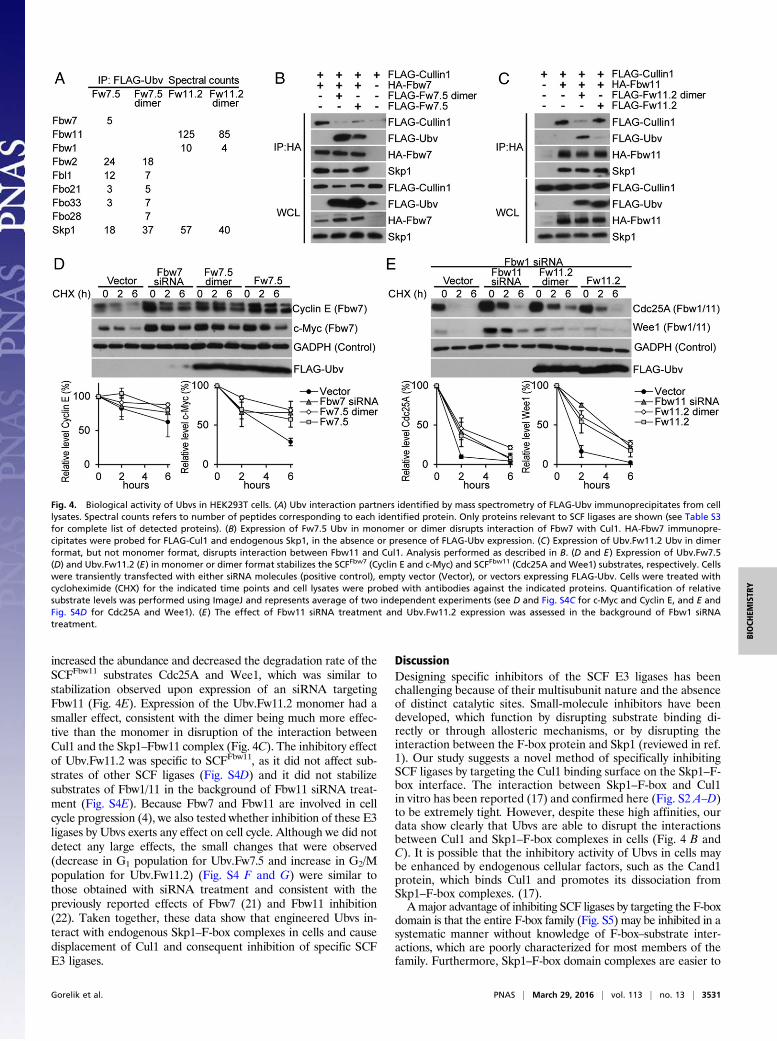
Intracellular Activity of Ubvs Targeting Fbw7 and Fbw11 Complexes.We transiently expressed Ubv.Fw7.5 or Ubv.Fw11.2 in HEK293Tcells to ascertain whether these Ubvs were able to exert effects inlive cells. Because Fbw7 and Fbw11 protein complexes function asdimers (18, 19), expression vectors were designed to express Ubvseither as monomers or as dimers held together by a homodimericGCN4 leucine zipper to enhance effective affinities throughavidity (Table S1) (20). To examine the interactions of Ubvs withendogenous proteins, Ubvs were immunoprecipitated, and copre-cipitated proteins were identified by mass spectrometry (Fig. 4A).Consistent with the in vitro specificity profiles (Fig. 3B), Ubv.Fw7.5coimmunoprecipitated Fbw7 and Skp1, and also several otherF-box proteins, including Fbw2 and Fbl1. Fbw7 was detected withthe lowest spectral counts among the F-box proteins, but this islikely a result of low expression levels of endogenous Fbw7. Insupport of this finding, a significant amount of Fbw7, but not Fbl1,coimmunoprecipitated with Ubv.Fw7.5 in cells overexpressingFbw7 or Fbl1 (Fig. S4A). In contrast, Ubv.Fw11.2 was very specificfor Fbw11, coimmunoprecipitating only Skp1, Fbw11, and smallamounts of Fbw1. Similar levels of interacting proteins were de-tected, whether Ubvs were expressed as monomers or dimers, butUbv dimers coimmunoprecipitated more nonspecific proteins in-volved in cell housekeeping functions (Table S3).To determine whether Ubvs are able to disrupt interactions be-
tween Cul1 and Skp1–F-box complexes in cells, exogenously ex-pressed Fbw7 or Fbw11 was immunoprecipitated in the absenceor presence of Ubv. Expression of Ubv.Fw7.5 monomer or dimersignificantly reduced or completely abrogated the coimmunopreci-pitation of Cul1 with Fbw7, respectively, but did not affect coim-munoprecipitation of Skp1 (Fig. 4B). In the case of Ubv.Fw11.2,expression of the dimer, but not the monomer, caused significantreduction in the amount of Cul1 (but not Skp1) that coimmuno-precipitated with Fbw11, and this was consistent with the factthat the dimer but not the monomer coimmunoprecipitatedwith Fbw11 (Fig. 4C). Thus, coimmunoprecipitation assays show thatboth Ubv.Fw7.5 and Ubv.Fw11.2 interfere with the interactions be-tween Skp1–F-box complexes and Cul1 in cells but do not affectinteractions between Skp1 and F-box proteins, although dimerizationis required to observe this effect in the case of Ubv.Fw11.2.To determine whether cellular expression of Ubv.Fw7.5 or
Ubv.Fw11.2 led to inhibition of their corresponding ligases, weanalyzed the stability of ligase substrates. Expression of Ubv.Fw7.5in either monomeric or dimeric format increased protein levels anddecreased degradation rate of the SCFFbw7 substrates Cyclin Eand c-Myc to levels comparable with those observed upon ex-pression of an siRNA targeting Fbw7 but had no effect on substratesof other SCF ligases, demonstrating that the observed inhibition wasspecific (Fig. 4D and Fig. S4C). In the case of Ubv.Fw11.2, assayswere performed in the presence of an siRNA targeting Fbw1 toreduce levels of SCFFbw1 (Fig. S4B), which shares substrateswith SCFFbw11. Expression of the Ubv.Fw11.2 dimer and monomer
Fig. 3. Ubvs selected for binding to the F-boxFbw7 or F-boxFbw11 domain incomplex with full-length Skp1. (A) Ubvs selected from Library 2 for binding toSkp1–F-boxFbw7. Positions that were soft-randomized in the library are shownand residues conserved as Ubv.Fw7.1 sequence are indicated by dashes. Posi-tions that diverge from the Ubv.Fw7.1 sequence but show consensus amongthe selected sequences are boxed and conserved residues at these positions areshaded gray. (B) Affinities of Ubv.Fw7.5, Ubv.Fw11.1, and Ubv.Fw11.2 fordifferent Skp1–F-box complexes. “NB” indicates no detectable binding and“WB” indicates weak binding for which IC50 values were >5,000 nM. Sequenceof Ubv binding region (F-box residues located within 10 Å of Ubv in thestructure of Skp1tr–F-boxFbw7
–Ubv.Fw7.1 complex) is shown for each F-boxprotein. Conserved positions are shaded gray and Fbw7 residues important forbinding to Ubv.Fw7.1 (Fig. 2F) are boxed. (C) The sequences and affinities ofUbv.Fw11.1 and its derivatives selected for binding to Skp1–F-boxFbw11. Onlythe sequence in region 1 that differs from Ubv.Fw7.5 is shown, and residuesconserved as Ubv.Fw11.1 sequence are indicated by dashes.
3530 | www.pnas.org/cgi/doi/10.1073/pnas.1519389113 Gorelik et al.
increased the abundance and decreased the degradation rate of theSCFFbw11 substrates Cdc25A and Wee1, which was similar tostabilization observed upon expression of an siRNA targetingFbw11 (Fig. 4E). Expression of the Ubv.Fw11.2 monomer had asmaller effect, consistent with the dimer being much more effec-tive than the monomer in disruption of the interaction betweenCul1 and the Skp1–Fbw11 complex (Fig. 4C). The inhibitory effectof Ubv.Fw11.2 was specific to SCFFbw11, as it did not affect sub-strates of other SCF ligases (Fig. S4D) and it did not stabilizesubstrates of Fbw1/11 in the background of Fbw11 siRNA treat-ment (Fig. S4E). Because Fbw7 and Fbw11 are involved in cellcycle progression (4), we also tested whether inhibition of these E3ligases by Ubvs exerts any effect on cell cycle. Although we did notdetect any large effects, the small changes that were observed(decrease in G1 population for Ubv.Fw7.5 and increase in G2/Mpopulation for Ubv.Fw11.2) (Fig. S4 F and G) were similar tothose obtained with siRNA treatment and consistent with thepreviously reported effects of Fbw7 (21) and Fbw11 inhibition(22). Taken together, these data show that engineered Ubvs in-teract with endogenous Skp1–F-box complexes in cells and causedisplacement of Cul1 and consequent inhibition of specific SCFE3 ligases.
DiscussionDesigning specific inhibitors of the SCF E3 ligases has beenchallenging because of their multisubunit nature and the absenceof distinct catalytic sites. Small-molecule inhibitors have beendeveloped, which function by disrupting substrate binding di-rectly or through allosteric mechanisms, or by disrupting theinteraction between the F-box protein and Skp1 (reviewed in ref.1). Our study suggests a novel method of specifically inhibitingSCF ligases by targeting the Cul1 binding surface on the Skp1–F-box interface. The interaction between Skp1–F-box and Cul1in vitro has been reported (17) and confirmed here (Fig. S2 A–D)to be extremely tight. However, despite these high affinities, ourdata show clearly that Ubvs are able to disrupt the interactionsbetween Cul1 and Skp1–F-box complexes in cells (Fig. 4 B andC). It is possible that the inhibitory activity of Ubvs in cells maybe enhanced by endogenous cellular factors, such as the Cand1protein, which binds Cul1 and promotes its dissociation fromSkp1–F-box complexes. (17).Amajor advantage of inhibiting SCF ligases by targeting the F-box
domain is that the entire F-box family (Fig. S5) may be inhibited in asystematic manner without knowledge of F-box–substrate inter-actions, which are poorly characterized for most members of thefamily. Furthermore, Skp1–F-box domain complexes are easier to
Fig. 4. Biological activity of Ubvs in HEK293T cells. (A) Ubv interaction partners identified by mass spectrometry of FLAG-Ubv immunoprecipitates from celllysates. Spectral counts refers to number of peptides corresponding to each identified protein. Only proteins relevant to SCF ligases are shown (see Table S3for complete list of detected proteins). (B) Expression of Fw7.5 Ubv in monomer or dimer disrupts interaction of Fbw7 with Cul1. HA-Fbw7 immunopre-cipitates were probed for FLAG-Cul1 and endogenous Skp1, in the absence or presence of FLAG-Ubv expression. (C) Expression of Ubv.Fw11.2 Ubv in dimerformat, but not monomer format, disrupts interaction between Fbw11 and Cul1. Analysis performed as described in B. (D and E) Expression of Ubv.Fw7.5(D) and Ubv.Fw11.2 (E) in monomer or dimer format stabilizes the SCFFbw7 (Cyclin E and c-Myc) and SCFFbw11 (Cdc25A and Wee1) substrates, respectively. Cellswere transiently transfected with either siRNA molecules (positive control), empty vector (Vector), or vectors expressing FLAG-Ubv. Cells were treated withcycloheximide (CHX) for the indicated time points and cell lysates were probed with antibodies against the indicated proteins. Quantification of relativesubstrate levels was performed using ImageJ and represents average of two independent experiments (see D and Fig. S4C for c-Myc and Cyclin E, and E andFig. S4D for Cdc25A and Wee1). (E) The effect of Fbw11 siRNA treatment and Ubv.Fw11.2 expression was assessed in the background of Fbw1 siRNAtreatment.
Gorelik et al. PNAS | March 29, 2016 | vol. 113 | no. 13 | 3531
purify and are more amenable to structure determination thantheir full-length counterparts, and this should facilitate the searchfor inhibitors with our approach. We have shown that Ubvsselected for binding to Skp1–F-box domain complexes are bi-ologically active as inhibitors of SCF function that act by dis-rupting binding of Cul1 (Fig. 4). This opens avenues for the useof these Ubv inhibitors as tools to validate potential drug tar-gets and to aid the development of small-molecule inhibitors.It is intriguing to speculate that Ubvs described in this study
target a natural Ub binding site [as observed for Ubvs targetingdeubiquitinases (6)], which is relevant to a natural mechanism forregulation of SCF function. In particular, it is striking that only sixmutations were sufficient to generate a high affinity binder to theSkp1tr–F-boxFbw7 complex (Fig. 2C), suggesting that this surfacemay be predisposed for binding to Ub. To explore this possibility,we looked for binding of monomeric Ub to the Skp1–F-boxFbw7
complex using NMR spectroscopy, but we did not find any evi-dence of interaction (Fig. S6). However, it is possible that theSkp1–F-box interface is involved in a regulatory interaction withmore complex Ub structures such as covalently attached Ubchains. For example, inhibition of Cul1 binding by growing Ubchains on the substrate might signal the termination of the ubiq-uitination reaction or Ub chains attached to the F-box proteinitself might function to accelerate the exchange of F-box sub-units in the SCF complex, in a manner analogous to the effectsof Cand1 binding to Cul1 (17). Further experiments aimed atstudying the interaction of more complex Ub structures withthe binding surface identified in this study might uncover newmechanisms regulating SCF function.In summary, we have discovered a previously unidentified
mechanism for inhibition of SCF ligases using engineered Ubvsthat target the Skp1–F-box interface and inhibit Cul1 binding. Wedemonstrate that high specificity is attainable by this method, asexemplified by Fbw11 inhibitors that can discriminate against eventhe close homolog Fbw1. However, the ability to engineer inhib-itors with broader specificities could also be useful, as it could beexploited to inhibit groups of SCF ligases containing similar F-boxproteins. We anticipate that the Ubv inhibitors described here will
be useful as tools for studying the function of SCF enzymes andfor facilitating the discovery of small-molecule inhibitors of theseenzymes through target validation, displacement screens, andstructure-based design.
Materials and MethodsProtein Purification and Structure Determination. His-tagged proteins wereexpressed in Escherichia coli BL21 and purified by Ni-NTA chromatographyusing standard techniques. See Table S1 for detailed list of all expressionconstructs. Refer to SI Materials and Methods for further details. Thestructure of the Skp1tr–F-boxFbw7
–Ubv.Fw7.1 complex was deposited in theProtein Data Bank with PDB ID code 5IBK.
Phage-Displayed Ubv Library Construction, Binding Selections, and in VitroBinding Assays. Previously described methods were used for the construc-tion of phage-displayed Ubv libraries, for binding selections, for the isolationof individual binding Ubv-phage clones, and for phage and protein ELISAs toestimate affinities (6). Refer to SI Materials and Methods for specific detailsdescribing library construction (Table S4), phage selections, ELISAs, andSPR analysis.
Cell-Based Assays. Genes encoding for FLAG-tagged Ubvs were cloned intopcDNA3.1/nFLAG-Dest vector for monomer expression or into the samevector modified to encode a GCN4 leucine zipper dimerization sequence(RMKQLEDKIEELLSKIYHLENEIARLKKLIGER) inserted in place of vector nu-cleotides 944–976 for dimer expression. Cul1, Fbw11, Fbw1, and Fbw7 wereexpressed from pcDNA3.1 based-vectors (see Table S1 for additional de-tails). See SI Materials and Methods for additional details on mass spec-trometry analysis, flow cytometry analysis, coimmunoprecipitation, andfunctional assays.
ACKNOWLEDGMENTS. This work was supported by the Canadian Institutesof Health Research Operating Grants MOP-136956 (to S.S.S.), MOP-126129(to M.T. and F.S.), Foundation Grant (to F.S.), and postdoctoral Fellowship(to M.G.); and an innovation grant from the Canadian Cancer Society ResearchInstitute (to M.T. and F.S.). This work is based upon research conducted at theAdvanced Photon Source on the Northeastern Collaborative Access Teambeamlines, which are supported by Award GM103403 from the NationalCenter for Research Resources at the National Institutes of Health. Use of theAdvanced Photon Source is supported by the US Department of Energy, Officeof Basic Energy Sciences, under Contract DE-AC02-06CH11357.
1. Weathington NM, Mallampalli RK (2014) Emerging therapies targeting the ubiquitinproteasome system in cancer. J Clin Invest 124(1):6–12.
2. Bhowmick P, Pancsa R, Guharoy M, Tompa P (2013) Functional diversity and structuraldisorder in the human ubiquitination pathway. PLoS One 8(5):e65443.
3. Jin J, et al. (2004) Systematic analysis and nomenclature of mammalian F-box proteins.Genes Dev 18(21):2573–2580.
4. Wang Z, Liu P, Inuzuka H, Wei W (2014) Roles of F-box proteins in cancer. Nat RevCancer 14(4):233–247.
5. Liu J, et al. (2015) Targeting the ubiquitin pathway for cancer treatment. BiochimBiophys Acta 1855(1):50–60.
6. Ernst A, et al. (2013) A strategy for modulation of enzymes in the ubiquitin system.Science 339(6119):590–595.
7. Pashkova N, et al. (2010) WD40 repeat propellers define a ubiquitin-binding domainthat regulates turnover of F box proteins. Mol Cell 40(3):433–443.
8. Davis RJ, Welcker M, Clurman BE (2014) Tumor suppression by the Fbw7 ubiquitinligase: Mechanisms and opportunities. Cancer Cell 26(4):455–464.
9. Busino L, et al. (2012) Fbxw7α- and GSK3-mediated degradation of p100 is a pro-survival mechanism in multiple myeloma. Nat Cell Biol 14(4):375–385.
10. Takeishi S, et al. (2013) Ablation of Fbxw7 eliminates leukemia-initiating cells bypreventing quiescence. Cancer Cell 23(3):347–361.
11. Lau AW, Fukushima H, Wei W (2012) The Fbw7 and betaTRCP E3 ubiquitin ligases andtheir roles in tumorigenesis. Front Biosci (Landmark Ed) 17:2197–2212.
12. Hao B, Oehlmann S, Sowa ME, Harper JW, Pavletich NP (2007) Structure of a Fbw7-Skp1-cyclin E complex: Multisite-phosphorylated substrate recognition by SCF ubiq-uitin ligases. Mol Cell 26(1):131–143.
13. Schulman BA, et al. (2000) Insights into SCF ubiquitin ligases from the structure of theSkp1-Skp2 complex. Nature 408(6810):381–386.
14. Orlicky S, et al. (2010) An allosteric inhibitor of substrate recognition by the SCF(Cdc4)ubiquitin ligase. Nat Biotechnol 28(7):733–737.
15. Lee CV, et al. (2004) High-affinity human antibodies from phage-displayed syntheticFab libraries with a single framework scaffold. J Mol Biol 340(5):1073–1093.
16. Zheng N, et al. (2002) Structure of the Cul1-Rbx1-Skp1-F boxSkp2 SCF ubiquitin ligasecomplex. Nature 416(6882):703–709.
17. Pierce NW, et al. (2013) Cand1 promotes assembly of new SCF complexes throughdynamic exchange of F box proteins. Cell 153(1):206–215.
18. Suzuki H, et al. (2000) Homodimer of two F-box proteins betaTrCP1 or betaTrCP2binds to IkappaBalpha for signal-dependent ubiquitination. J Biol Chem 275(4):2877–2884.
19. Welcker M, et al. (2013) Fbw7 dimerization determines the specificity and robustnessof substrate degradation. Genes Dev 27(23):2531–2536.
20. Harbury PB, Zhang T, Kim PS, Alber T (1993) A switch between two-, three-, and four-stranded coiled coils in GCN4 leucine zipper mutants. Science 262(5138):1401–1407.
21. Wu XZ, et al. (2015) MiR-27a-3p promotes esophageal cancer cell proliferation viaF-box and WD repeat domain-containing 7 (FBXW7) suppression. Int J Clin Exp Med8(9):15556–15562.
22. Guardavaccaro D, et al. (2003) Control of meiotic and mitotic progression by the F boxprotein beta-Trcp1 in vivo. Dev Cell 4(6):799–812.
23. Fellouse FA, Sidhu SS (2007) Making antibodies in bacteria. Making and UsingAntibodies, eds Howard GC, Kaser MS (CRC Press, Boca Raton, FL), pp 157–180.
24. Sidhu SS, Lowman HB, Cunningham BC, Wells JA (2000) Phage display for selection ofnovel binding peptides. Methods Enzymol 328:333–363.
25. Otwinowski Z, Minor W (1997) Processing of X-ray diffraction data collected in os-cillation mode. Methods Enzymol 276:307–326.
26. McCoy AJ, et al. (2007) Phaser crystallographic software. J Appl Cryst 40(Pt 4):658–674.27. Emsley P, Cowtan K (2004) Coot: Model-building tools for molecular graphics. Acta
Crystallogr D Biol Crystallogr 60(Pt 12 Pt 1):2126–2132.28. Adams PD, et al. (2010) PHENIX: A comprehensive Python-based system for macro-
molecular structure solution. Acta Crystallogr D Biol Crystallogr 66(Pt 2):213–221.29. Marcon E, et al. (2014) Human-chromatin-related protein interactions identify a de-
methylase complex required for chromosome segregation. Cell Reports 8(1):297–310.30. Peschiaroli A, Skaar JR, Pagano M, Melino G (2010) The ubiquitin-specific protease
USP47 is a novel beta-TRCP interactor regulating cell survival. Oncogene 29(9):1384–1393.
3532 | www.pnas.org/cgi/doi/10.1073/pnas.1519389113 Gorelik et al.


![Ubc13/Rnf8 ubiquitin ligases control foci formation of the ... · Abraxas interaction domain [AIR (Abraxas-interacting region)], re-quired for association of Rap80 with Abraxas, Brca1,](https://static.documents.pub/doc/80x56/5e77d5957e7d2f032f71b8e3/ubc13rnf8-ubiquitin-ligases-control-foci-formation-of-the-abraxas-interaction.jpg)


