Inhibitory Effects of D-Amino Acids on Staphylococcus aureusBiofilm Development�†
Allon I. Hochbaum,1,2§ Ilana Kolodkin-Gal,3§ Lucy Foulston,3 Roberto Kolter,4Joanna Aizenberg,1,2 and Richard Losick3*
Department of Chemistry and Chemical Biology, Harvard University, Cambridge, Massachusetts 021381; School ofEngineering and Applied Sciences, Harvard University, Cambridge, Massachusetts 021382; Department of
Molecular and Cellular Biology, Harvard University, Cambridge, Massachusetts 021383; andDepartment of Microbiology and Molecular Genetics, Harvard Medical School,
Boston, Massachusetts 021154
Received 14 June 2011/Accepted 10 August 2011
Biofilms are communities of cells held together by a self-produced extracellular matrix typically consistingof protein, exopolysaccharide, and often DNA. A natural signal for biofilm disassembly in Bacillus subtilis iscertain D-amino acids, which are incorporated into the peptidoglycan and trigger the release of the proteincomponent of the matrix. D-Amino acids also prevent biofilm formation by the related Gram-positive bacteriumStaphylococcus aureus. Here we employed fluorescence microscopy and confocal laser scanning microscopy toinvestigate how D-amino acids prevent biofilm formation by S. aureus. We report that biofilm formation takesplace in two stages, initial attachment to surfaces, resulting in small foci, and the subsequent growth of the fociinto large aggregates. D-Amino acids did not prevent the initial surface attachment of cells but blocked thesubsequent growth of the foci into larger assemblies of cells. Using protein- and polysaccharide-specific stains,we have shown that D-amino acids inhibited the accumulation of the protein component of the matrix but hadlittle effect on exopolysaccharide production and localization within the biofilm. We conclude that D-aminoacids act in an analogous manner to prevent biofilm development in B. subtilis and S. aureus. Finally, toinvestigate the potential utility of D-amino acids in preventing device-related infections, we have shown thatsurfaces impregnated with D-amino acids were effective in preventing biofilm growth.
Most bacteria form matrix-enclosed communities, or bio-films, when growing on surfaces. In clinical settings, biofilmsare particularly problematic since they tend to form on indwell-ing devices and cause persistent infections and sepsis. Biofilm-associated bacteria are much less sensitive to antibiotics, mak-ing biofilm-related infections especially difficult to cure (7, 13).Often the only solution for a biofilm-infected catheter is com-plete replacement, a procedure that can range from uncom-fortable and inconvenient to painful, expensive, and life threat-ening. Consequently, the development of methods to preventbiofilm formation may be just as important for treating hospi-tal-acquired infections as the development of new antibiotics.
Bacterial signaling molecules that trigger the dispersal of oldbiofilms hold promise as possible therapeutic agents. Recentwork has demonstrated that D-amino acids may be an exem-plary class of such compounds (10, 19). Certain D-amino acidsisolated from the supernatants of disassembled Bacillus subtilisbiofilms were shown to prevent biofilm formation in freshcultures by disrupting the connection between an extracellularmatrix protein and the cell. Similar inhibitory effects for Staph-ylococcus aureus and Pseudomonas aeruginosa biofilms sug-
gested that D-amino acids might constitute a general strategyfor inhibiting biofilm formation in opportunistic pathogens(10). Genes whose products are involved in biofilm formationare not orthologous across these species, however, and themechanism of action of D-amino acids against biofilms formedby these dissimilar pathogens remains unknown.
Here we describe investigations of the mechanism by whichD-amino acids inhibit biofilm formation by S. aureus usingfluorescence and confocal scanning laser microscopy. Thesetechniques provide a more detailed picture of biofilm devel-opment on surfaces than visual inspection and bulk stainingalone. Using dyes for specific components of the biofilm, suchas cells, proteins, and polysaccharides, we have found thatD-amino acids inhibit biofilm formation in S. aureus in muchthe same way as in B. subtilis: by preventing protein localizationat the cell surface. Since S. aureus employs cell surface-asso-ciated proteins to connect neighboring cells in large aggregates(9), D-amino acids could prove effective at preventing maturebiofilm development.
MATERIALS AND METHODS
Bacterial growth. Staphylococcus aureus wild-type (WT) strain SC01 (2) wasobtained from the Kolter lab collection. Tryptic soy broth (TSB) medium and D
and L isomers of proline, tyrosine, phenylalanine, tryptophan, and leucine wereobtained from Sigma-Aldrich (Atlanta). Cells were cultured in a shaking LBmedium overnight and diluted 1:100 in TSB medium supplemented with NaCl(3%), glucose (0.5%), and the appropriate concentrations of L- or D-amino acids(or lack thereof). Cells were grown for the specified period of time in the bottomof 6- or 12-well polystyrene plates or in 6-well plates with submerged substrateswithout shaking at 37°C. Planktonic cells were removed by gentle rinsing withphosphate-buffered saline (PBS) before further treatment.
* Corresponding author. Mailing address: Department of Molecularand Cellular Biology, Harvard University, Cambridge, MA 02138-1903. Phone: 617) 495-4905. Fax: (617) 496-4642. E-mail: [email protected].
§ These authors contributed equally to this work.† Supplemental material for this article may be found at http://jb
.asm.org/.� Published ahead of print on 19 August 2011.
Crystal violet staining. Cells were grown in 12-well plates for 24 h, and afterrinsing in PBS, adhered cells were stained with crystal violet (CV) as describedpreviously (16). Wells were stained with 500 �l of 0.1% crystal violet dye, rinsedtwice with 2 ml double-distilled water, and thoroughly dried. Images were takenwith an AMT 2k charge-coupled-device (CCD) camera. For the quantification ofbiofilm growth, 0.5 ml of ethanol (95%) was added to the CV-stained wells andincubated with shaking for 2 h. The resulting solutions of dissolved CV werediluted by a factor of 20, and their optical density was measured at 595 nm. Eachdata point is composed of three independent samples.
Polymer substrate fabrication. Polymer substrates used for these studies werefabricated by UV curing an epoxy (UVO-114; Epoxy Technology) or a polyure-thane (Norland optical adhesive 61) in a polydimethylsiloxane mold as previouslyreported (1).
Assessment of D-amino acid effects on biofilms adhered to different surfaces.Cells were grown as described above for 24 h with an either D or L mixture ofequal ratios of Pro, Phe, and Tyr in a final concentration of 100 �M. Cells weregrown either on 6-well polystyrene plates or on 18- by 18-mm glass cover slides(VWR) or on epoxy surfaces submerged in a 6-well plate. Glass and epoxysurfaces were submerged in 6 ml of the growth medium. After rinsing, adheredcells were fixed using a 5% glutaraldehyde solution for 1 h with 0.05 M surfactin(Sigma-Aldrich). Cells were washed twice in PBS. Images were taken with anAMT 2k CCD camera against a black background.
Fluorescence microscopy. All dyes (Sytox green, fluorescein isothiocyanate[FITC], Syto 63, and Texas Red-concanavalin A [ConA]) were obtained fromInvitrogen-Molecular Probes (Oregon). Fluorescence microscopy images wereobtained on a Leica DMRX compound microscope fitted with a 63� waterimmersion lens. Cells were grown in 6-well polystyrene plates in duplicate, asdescribed above. Cells were fixed as described above after 2, 12, and 24 h for 1 hat 50°C. Cells were then rinsed with PBS, and their membranes were madepermeable by soaking the cell growth substrates in Triton X-100 (VWR), 0.1%(vol/vol) in PBS (PBST), for 15 min. The cells were labeled by replacing thePBST with Sytox green nucleic acid stain, 0.5 �M in PBST, for 15 min to an hour.The substrates were rinsed again with PBS and then imaged with the waterimmersion lens using a Leica FITC (K3) filter cube.
Confocal microscopy. Confocal microscopy images were obtained on a LeicaTCS SP5 spectral confocal inverted microscope. S. aureus cells were grownwithout shaking in 37°C in TSB medium with 3% NaCl and 0.5% glucose for24 h, applied with either a D or an L mixture of equal ratios of Pro, Phe, and Tyrin a final concentration of 100 �M. Cells were grown on a glass cover slide of 18by 18 mm submerged in 6 ml of TSB with NaCl and glucose. Planktonic cellswere removed. Adhered cells were fixed using a 5% gluteraldehyde solution for1 h in 50°C, applied with surfactin (0.05 M). Cells were washed in 2 ml PBS. FITC(0.001%) and Syto 63 (100 �M) were added to the well, and the plate wasincubated with shaking for 5 min and for an additional hour without shaking atroom temperature. Cells were washed twice in PBS. Samples were then removedfrom the PBS rinse solution and inverted onto a cover glass (no. 1; VWR).Biofilms or adhered cells were imaged through the cover glass using the followingexcitation and emission wavelengths (11): 488 nm excitation and 505 to 530 nmemission detection range for FITC, 543 nm and 560 to 800 nm for TexasRed-ConA, and 633 nm and 650 to 800 nm for Syto 63.
Controlled release of D-amino acids from polymer substrates. Epoxy andpolyurethane substrates were soaked overnight in 1 mM solution of either L or D
isomers of Pro, Phe, and Tyr in equal molar ratios. The substrates were rinsedthoroughly with PBS, and then cells were grown on them in 6-well plates for 24 has described above. Fluorescence microscopy images of the adhered cells wereobtained as described above.
RESULTS
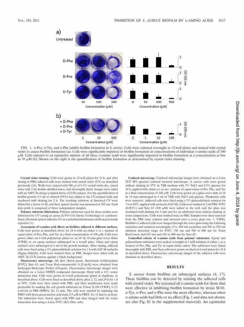
S. aureus forms biofilms on submerged surfaces (4, 17).These biofilms can be detected by staining the adhered cellswith crystal violet. We screened all D-amino acids for those thatwere effective in inhibiting biofilm formation by strain SC01.D-Tyr, D-Pro, and D-Phe were the most effective, whereas otherD-amino acids had little or no effect (Fig. 1 and data not shown;see also Fig. S1 in the supplemental material). An equimolar
FIG. 1. D-Pro, D-Tyr, and D-Phe inhibit biofilm formation in S. aureus. Cells were cultured overnight in 12-well plates and stained with crystalviolet to assess biofilm formation (a). Cells were significantly impaired in biofilm formation at concentrations of individual D-amino acids of 500�M. Cells exposed to an equimolar mixture of all three D-amino acids were significantly impaired in biofilm formation at a concentration as lowas 10 �M (b). Shown on the right is the quantification of biofilm formation as determined by crystal violet staining.
VOL. 193, 2011 INHIBITION OF S. AUREUS BIOFILM BY D-AMINO ACIDS 5617
mixture of D-Tyr, D-Pro, and D-Phe was more effective in pre-venting biofilm formation than any of the individual aminoacids (Fig. 1). In contrast, L-Tyr, L-Pro, L-Phe, and a mixture ofthe three had little or no inhibitory effect (data not shown).The D-Tyr, D-Pro, and D-Phe mixture was also more effectivethan the previously described mixture of D-Trp, D-Met, D-Leu,and D-Tyr that was optimal for inhibiting biofilm formation byB. subtilis (10) (see Fig. S2). It should be noted that D-aminoacids did not significantly impair the growth of S. aureus cells,even at millimolar concentrations (see Fig. S3), and therefore,as observed for B. subtilis, the inhibitory effect on biofilmformation was not a result of growth inhibition. The inhibitoryeffect of the D-amino acid mixture was not restricted to strainSC01 and was also seen with three other S. aureus strains (seeFig. S4 and S5). Finally, the D-amino acid mixture could alsodisassemble an existing biofilm, but this effect required muchhigher concentrations (10 mM) than that needed for inhibition(�100 �M) (see Fig. S6).
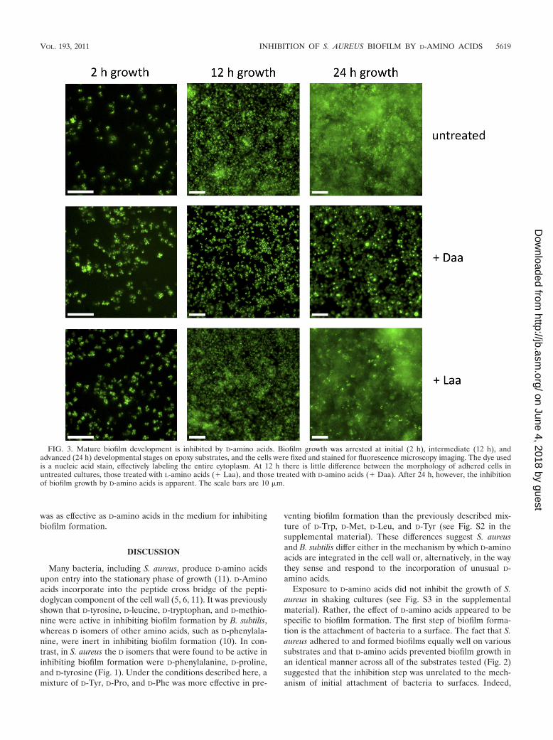
The adhesion of cells to a surface is an essential first step ofbiofilm formation and can occur by specific and nonspecificcell-surface interactions. To study whether D-amino acids dis-rupted the attachment stage of biofilm growth, we screened arepresentative sample of substrates. As shown in Fig. 2, D-amino acids but not L-amino acids inhibited biofilm formationequivalently on polystyrene, epoxy, and glass surfaces, suggest-ing that the surface properties of these different substrateswere irrelevant to the mode of action of D-amino acids. Thisresult led us to use fluorescence microscopy to investigate theeffect of D-amino acids on biofilm development and morphol-ogy. As shown in Fig. 3, S. aureus cells adhered to epoxysurfaces and continued to attach and divide up to 12 h ofgrowth. This initial attachment step (seen at 2 and 12 h), whichappears as foci in the figure, was not affected by exposure toD-amino acids. Under our growth conditions, S. aureus subse-quently formed large aggregates characteristic of mature bio-films on submerged surfaces within 24 h. The formation ofthese aggregates was blocked when cells were grown in the
presence of D- but not L-amino acids (Fig. 3). We conclude thatD-amino acids prevent biofilm development at a stage subse-quent to the initial attachment to surfaces and continue toinhibit biofilm growth for at least 48 h.
We hypothesized that aggregate formation during S. aureusbiofilm development is blocked by D-amino acids as a result ofa change in the properties of the extracellular matrix, as wasshown in the case of D-amino acid inhibition of B. subtilisbiofilms (10). To investigate this possibility, we characterizedbiofilms formed on glass coverslips with and without D-aminoacids, focusing on the protein and exopolysaccharide (EPS)components of the extracellular matrix. We stained the EPSwith a fluorescent concanavalin A (ConA) conjugate, and pro-teins were stained with the amine-reactive fluorescein isothio-cyanate (FITC) (14). These dyes may also bind to proteins andcarbohydrates inside the cells if the cell membrane has beencompromised. Therefore, we also stained with Syto 63, a DNA-reactive dye, to distinguish binding sites in the extracellularmatrix from those inside cells. Figures 4 and 5 show stackedsections of S. aureus biofilms imaged using confocal laser scan-ning microscopy (CLSM). Image slices were obtained at 2-�mincrements moving from the top of the biofilm toward thesubstrate. In this way we determined the minimum thickness ofthe biofilm. This thickness determination was limited by thediffusion of dye into the biofilm and therefore is given as aminimum value for mature biofilms. As can be seen in Fig. 4aand 4b, cells in mature biofilms displayed large amounts ofproteins at their surface. These cell surface-associated proteinswere also present when biofilms were grown in the presence ofL-amino acids. However, such proteins were largely absentwhen cells were grown in the presence of D-amino acids (Fig.4c; see also Fig. S7A in the supplemental material). In contrast,the localization of cell wall sugars and EPS exhibited littledifference between cells grown in the presence of D-aminoacids and those grown with L-amino acids (Fig. 5; see also Fig.S7B and S8). It should be noted that biofilms grown in thepresence of D-amino acids were significantly thinner (2 to 4�m) than the biofilms grown in the presence of L-amino acids(10 to 24 �m). Also, the large cell aggregates found in biofilmsgrown with L-amino acids were absent in surface-associatedcells grown in the presence of D-amino acids.
The ability of D-amino acids to disrupt biofilm formationmakes them potentially useful for addressing the problemsassociated with device-related infections. To investigate thispossibility, we asked whether the slow release of D-amino acidsfrom the substrate, rather than supplying them in the growthmedium, would be sufficient to impede biofilm formation. Ac-cordingly, we soaked substrates, including epoxy- and polyure-thane-based polymers, in medium containing D-amino acids orL-amino acids. The substrates were washed and placed in freshgrowth medium lacking supplemented D-amino acids or L-amino acids. The results shown in Fig. 6 demonstrate that S.aureus formed robust biofilms when cultured on immersedsurfaces impregnated with L-amino acids. In contrast, biofilmformation was inhibited on surfaces impregnated with D-aminoacids. As a negative control, glass surfaces, which do not ab-sorb the D-amino acid mixture, were used (see Fig. S9 in thesupplemental material). On these surfaces, S. aureus biofilmgrowth was unaffected. Therefore, diffusion of D-amino acids
FIG. 2. The inhibitory effect of D-amino acids on biofilm growth issimilar on different substrates. Biofilms formed equally on polystyrene,epoxy, and glass substrates after 24 h of growth and did not form in thepresence of D-amino acids (� D-aa), suggesting the mechanism ofaction of D-amino acids is unrelated to initial surface attachment.Biofilm attached to the surface appears white in these unstained sam-ples. For the glass and epoxy substrates, cover glass and molded epoxywere submerged in medium in 6-well plates. For growth on polysty-rene, the bottom of the well was used as the substrate.
was as effective as D-amino acids in the medium for inhibitingbiofilm formation.
DISCUSSION
Many bacteria, including S. aureus, produce D-amino acidsupon entry into the stationary phase of growth (11). D-Aminoacids incorporate into the peptide cross bridge of the pepti-doglycan component of the cell wall (5, 6, 11). It was previouslyshown that D-tyrosine, D-leucine, D-tryptophan, and D-methio-nine were active in inhibiting biofilm formation by B. subtilis,whereas D isomers of other amino acids, such as D-phenylala-nine, were inert in inhibiting biofilm formation (10). In con-trast, in S. aureus the D isomers that were found to be active ininhibiting biofilm formation were D-phenylalanine, D-proline,and D-tyrosine (Fig. 1). Under the conditions described here, amixture of D-Tyr, D-Pro, and D-Phe was more effective in pre-
venting biofilm formation than the previously described mix-ture of D-Trp, D-Met, D-Leu, and D-Tyr (see Fig. S2 in thesupplemental material). These differences suggest S. aureusand B. subtilis differ either in the mechanism by which D-aminoacids are integrated in the cell wall or, alternatively, in the waythey sense and respond to the incorporation of unusual D-amino acids.
Exposure to D-amino acids did not inhibit the growth of S.aureus in shaking cultures (see Fig. S3 in the supplementalmaterial). Rather, the effect of D-amino acids appeared to bespecific to biofilm formation. The first step of biofilm forma-tion is the attachment of bacteria to a surface. The fact that S.aureus adhered to and formed biofilms equally well on varioussubstrates and that D-amino acids prevented biofilm growth inan identical manner across all of the substrates tested (Fig. 2)suggested that the inhibition step was unrelated to the mech-anism of initial attachment of bacteria to surfaces. Indeed,
FIG. 3. Mature biofilm development is inhibited by D-amino acids. Biofilm growth was arrested at initial (2 h), intermediate (12 h), andadvanced (24 h) developmental stages on epoxy substrates, and the cells were fixed and stained for fluorescence microscopy imaging. The dye usedis a nucleic acid stain, effectively labeling the entire cytoplasm. At 12 h there is little difference between the morphology of adhered cells inuntreated cultures, those treated with L-amino acids (� Laa), and those treated with D-amino acids (� Daa). After 24 h, however, the inhibitionof biofilm growth by D-amino acids is apparent. The scale bars are 10 �m.
VOL. 193, 2011 INHIBITION OF S. AUREUS BIOFILM BY D-AMINO ACIDS 5619
fluorescence microscopy (Fig. 3) revealed that initial surfaceattachment was unaffected by treatment with D-amino acids.Instead, these images showed that bacteria adhered to thesubmerged surface but did not proceed to form fully developed
biofilms. The initial surface attachment of bacteria was similarirrespective of treatment with L- or D-amino acids. By 24 h ofgrowth, however, the untreated and L-amino acid-treated cul-tures had developed mature biofilms with large cell aggregates.
FIG. 4. Confocal microscopy images of S. aureus biofilms grown with or without D-amino acids reveal a difference in the presence ofextracellular protein. Biofilms were stained with protein-specific (green) and DNA-specific (red) dyes to image the localization of the peptidecomponent of the biofilm matrix. (a) CLSM imaging of wild-type biofilms shows a clear localization of extracellular proteins to the cell wall in animage slice taken from the interior of a large cell aggregate. The higher-magnification image on the bottom shows this localization clearly, witheven the septa between dividing cells stained. (b) Images were taken as lateral cross sections of the biofilm, with the height of each section indicatedfrom the top of the biofilm. The presence of protein at the cell surface is clearly seen throughout the aggregates. (c) In D-amino acid-treatedcultures, there was no biofilm present on the substrate surface and there was a distinct lack of protein localization on the cell surfaces, in contrastto the images in panel b. The dispersed cells and clusters seen in panel c were adhered to the substrate surface, and the two images shown aretypical of the morphology and staining across the entire substrate as seen in multiple additional images. The scale bars are 5 �m except for thatin the lower frame of panel a, which is 1 �m.
FIG. 5. Confocal laser scanning microscopy images show no difference in the localization of EPS in S. aureus aggregates from cultures grown withD- or L-amino acids. These cells were stained with EPS-specific (blue) and DNA-specific (red) dyes, and the imaging sections were obtained in the samemanner as those in Fig. 4. (a) Cultures treated with L-amino acids exhibited robust biofilms with localization of EPS around the cells in the aggregates.(b) Those treated with D-amino acids retained the EPS surrounding the cells but adhered as a submonolayer film, lacking the thickness and structure ofa mature biofilm. As in Fig. 4c, the cells shown in panel b were attached only at the surface of the substrate. The scale bars are 5 �m.
In contrast, biofilm development in cultures exposed to D-amino acids was arrested at the attachment stage. It is likely,therefore, that the effects of D-amino acids are restricted to theaccumulation step in biofilm development.
Confocal microscopy gave a more detailed picture of biofilmdevelopment during exposure to D-amino acids. Using FITCand Syto 63 as a cell body counterstain, protein could be seento localize at or around the cell wall of the constituent cells ofS. aureus biofilms (Fig. 4a). Whereas cells in biofilm aggregatesthat had formed in L-amino acid-treated cultures were clearlydecorated with protein (Fig. 4b), the lack of these large supra-cellular structures and of protein surrounding the cells in theD-amino acid cultures (Fig. 4c; see also Fig. S7A in the sup-plemental material) suggests a functional relationship betweenthe effect of D-amino acids and a protein component of thematrix. Several surface proteins play an important role in bio-film aggregates for S. aureus (8, 9, 15, 20). Thus, an appealinghypothesis is that D-amino acids prevent the localization ofcell-cell adhesion proteins, thereby inhibiting the aggregateformation necessary for biofilm development. If so, then therole of D-amino acids in S. aureus would be analogous to thatobserved in B. subtilis, for which D-amino acid treatments havebeen shown to trigger the release from cells of the matrixprotein TasA (3, 18). In B. subtilis, a recently characterizedaccessory protein, TapA (TasA anchoring/assembly protein),serves to anchor the TasA amyloid fibers to the cell wall and ismislocalized in response to D-amino acids (10, 19). Interest-ingly, however, TapA has no apparent ortholog in S. aureus.The fact that D-amino acids are effective at blocking thesedifferent and unrelated adhesion proteins from localizing at
the cell wall suggests that they may comprise a general biofilmdispersal strategy.
The polysaccharide component of the S. aureus biofilms, onthe other hand, was present in attached cells irrespective ofamino acid exposure. These polysaccharides were seen tobe concentrated in the large aggregates along with protein.Whereas biofilms did not form in D-amino acid-treated cul-tures, polysaccharides were still seen associated with the cellsurface, confirming that the principal effect of the D-aminoacids is to disrupt cell surface protein localization. These re-sults suggest that the mechanism of action of D-amino acidsmay be similar across dissimilar bacterial species.
S. aureus is a leading cause of hospital-acquired infections(12, 17). Many of the problems associated with this pathogenstem from its capacity to form biofilms. The apparently ubiq-uitous nature of the effects of D-amino acid exposure is prom-ising for applications in preventing biofilms in medical andindustrial settings. Specifically, the ability to control the releaseof D-amino acids from device surfaces, such as implant coatingsor catheter walls, to inhibit the formation of biofilms wouldsignificantly reduce the difficulty and cost associated withkeeping these devices in place. The experimental procedureto evaluate the effectiveness of surfaces impregnated withD-amino acids is illustrated in Fig. 6A. The effects of D-aminoacids on biofilm growth from impregnated substrates wereidentical to those on biofilms grown from cultures exposed toD-amino acids in the growth medium. Biofilms formed nor-mally on glass surfaces, which should not absorb D-amino acids(see Fig. S9 in the supplemental material), demonstrating thatthe effect of the D-amino acids was a result of controlled re-
FIG. 6. The diffusive release of D-amino acids from polymer surfaces inhibits biofilm growth. (A) Polymer substrates were soaked in L- orD-amino acids overnight and rinsed before use as growth substrates in medium lacking supplemented amino acids of either kind. (B) Both epoxy-and polyurethane-based growth substrates resisted biofilm accumulation when soaked with D-amino acids but not L-amino acids, recapitulating theresults of cultures grown in the presence of amino acids of either chirality. The images were obtained by fluorescence microscopy using Sytox greento label the cytoplasm. The scale bars are 10 �m.
VOL. 193, 2011 INHIBITION OF S. AUREUS BIOFILM BY D-AMINO ACIDS 5621
lease from the polymer substrate. Lacking the protective prop-erties of a mature biofilm, these adhered cells are likely moresusceptible to antimicrobial agents such as antibiotics, whichcan be used to treat bacterial infection more easily.
ACKNOWLEDGMENTS
We thank D. A. Weitz and the Harvard Materials Research andEngineering Center (DMR-0213805) for the use of their confocalimaging facilities and M. Gilmore for strains MN8, NCTC 10833, andRN4220.
I.K.-G. is a postdoctoral fellow of the Human Frontier ScienceProgram. This work was supported by NIH grants to R.K. (GM58213)and R.L. (GM18546), as well as grants from the BASF AdvancedResearch Initiative at Harvard University to J.A., R.K., and R.L.
REFERENCES
1. Aizenberg, J., B. Pokroy, A. K. Epstein, and M. C. M. Persson-Gulda. 2009.Fabrication of bioinspired actuated nanostructures with arbitrary geometryand stiffness. Adv. Mater. 21:463–469.
2. Blevins, J. S., et al. 2003. Role of sarA in the pathogenesis of Staphylococcusaureus musculoskeletal infection. Infect. Immun. 71:516–523.
3. Branda, S. S., F. Chu, D. B. Kearns, R. Losick, and R. Kolter. 2006. A majorprotein component of the Bacillus subtilis biofilm matrix. Mol. Microbiol.59:1229–1238.
4. Bryers, J. D. 2008. Medical biofilms. Biotechnol. Bioeng. 100:1–18.5. Cava, F., M. A. de Pedro, H. Lam, B. M. Davis, and M. K. Waldor. 2011.
Distinct pathways for modification of the bacterial cell wall by non-canonicalD-amino acids. EMBO J. 30:3442–3453.
6. Cava, F., H. Lam, M. A. de Pedro, and M. K. Waldor. 2011. Emergingknowledge of regulatory roles of D-amino acids in bacteria. Cell Mol. LifeSci. 68:817–831.
7. Costerton, J. W., P. S. Stewart, and E. P. Greenberg. 1999. Bacterial biofilms:a common cause of persistent infections. Science 284:1318–1322.
8. Cucarella, C., et al. 2001. Bap, a Staphylococcus aureus surface proteininvolved in biofilm formation. J. Bacteriol. 183:2888–2896.
9. Geoghegan, J. A., et al. 2010. Role of surface protein SasG in biofilmformation by Staphylococcus aureus. J. Bacteriol. 192:5663–5673.
10. Kolodkin-Gal, I., et al. 2010. D-Amino acids trigger biofilm disassembly.Science 328:627–629.
11. Lam, H., et al. 2009. D-Amino acids govern stationary phase cell wall re-modeling in bacteria. Science 325:1552–1555.
12. Lopez, D., H. Vlamakis, and R. Kolter. 2010. Biofilms. Cold Spring Harb.Perspect. Biol. 2:a000398.
13. Mah, T. F., and G. A. O’Toole. 2001. Mechanisms of biofilm resistance toantimicrobial agents. Trends Microbiol. 9:34–39.
14. McSwain, B. S., R. L. Irvine, M. Hausner, and P. A. Wilderer. 2005. Com-position and distribution of extracellular polymeric substances in aerobicflocs and granular sludge. Appl. Environ. Microbiol. 71:1051–1057.
15. O’Neill, E., et al. 2008. A novel Staphylococcus aureus biofilm phenotypemediated by the fibronectin-binding proteins, FnBPA and FnBPB. J. Bacte-riol. 190:3835–3850.
16. O’Toole, G. A., and R. Kolter. 1998. Flagellar and twitching motility arenecessary for Pseudomonas aeruginosa biofilm development. Mol. Micro-biol. 30:295–304.
17. Otto, M. 2008. Staphylococcal biofilms. Curr. Top. Microbiol. Immunol.322:207–228.
18. Romero, D., C. Aguilar, R. Losick, and R. Kolter. 2010. Amyloid fibersprovide structural integrity to Bacillus subtilis biofilms. Proc. Natl. Acad. Sci.U. S. A. 107:2230–2234.
19. Romero, D., H. Vlamakis, R. Losick, and R. Kolter. 2011. An accessoryprotein required for anchoring and assembly of amyloid fibers in B. subtilisbiofilms. Mol. Microbiol. 80:1155–1168.
20. Schroeder, K., et al. 2009. Molecular characterization of a novel Staphylo-coccus aureus surface protein (SasC) involved in cell aggregation and biofilmaccumulation. PLoS One 4:e7567.