Initiation and progression of mechanical damage in the intervertebral disc under cyclic loading using continuum damage mechanics methodology: A finite element study Muhammad Qasim a , Raghu N. Natarajan a,b,n , Howard S. An a , Gunnar B.J. Andersson a a Department of Bioengineering, University of Illinois at Chicago, Chicago, IL, USA b Department of Orthopedic Surgery, Rush University Medical Center, Chicago, IL, USA article info Article history: Accepted 13 May 2012 Keywords: Lumbar spine Disc degeneration Fatigue failure Finite element modelling Continuum damage mechanics abstract It is difficult to study the breakdown of disc tissue over several years of exposure to bending and lifting by experimental methods. There is also no finite element model that elucidates the failure mechanism due to repetitive loading of the lumbar motion segment. The aim of this study was to refine an already validated poro-elastic finite element model of lumbar motion segment to investigate the initiation and progression of mechanical damage in the disc under simple and complex cyclic loading conditions. Continuum damage mechanics methodology was incorporated into the finite element model to track the damage accumulation in the annulus in response to the repetitive loading. The analyses showed that the damage initiated at the posterior inner annulus adjacent to the endplates and propagated outwards towards its periphery under all loading conditions simulated. The damage accumulated preferentially in the posterior region of the annulus. The analyses also showed that the disc failure is unlikely to happen with repetitive bending in the absence of compressive load. Compressive cyclic loading with low peak load magnitude also did not create the failure of the disc. The finite element model results were consistent with the experimental and clinical observations in terms of the region of failure, magnitude of applied loads and the number of load cycles survived. & 2012 Elsevier Ltd. All rights reserved. 1. Introduction Low back pain is a major health condition affecting every population worldwide (Andersson, 1999). It can lead to decreased quality of life, diminished physical activity and psychological distress (Deyo and Tsui-Wu, 1987; Deyo et al., 2011). Interver- tebral disc degeneration is associated with low back pain (Cheung et al., 2009; Luoma et al., 2000; Samartzis et al., 2011; Savage et al., 1997). Appearance of annular lesions has been suggested (Osti et al., 1992; Sharma et al., 2009a, 2009b; Vernon-Roberts et al., 2007a, b) as the first sign of the disc degeneration process. Epidemiological studies have identified frequent bending and lifting as a major risk for disc prolapse (Kelsey et al., 1984; KUMAR, 1990). Damage to disc structure has been reported in response to cyclic loading of the motion segment by a number of studies involving human cadavers and animal models (Adams and Hutton, 1983; Adams and Hutton, 1985; Adams et al., 2000; Goel et al., 1988a, b; Hansson et al., 1987; Liu et al., 1983; Liu et al., 1985; Yoganandan et al., 1994). Yu et al. (2003) reported presence of irregular fibres, buckling and bleeding in the porcine annulus in response to compressive cyclic loading. Gordon et al. (1991) reported disc herniation in 14 cadaveric lumbar motion segments, subjected to combination of flexion, axial rotation and compres- sion for an average duration of 36,750 cycles. Liu et al. (1983) subjected cadaveric lumbar motion segments to cyclic axial loads ranging from 37%–80% of their failure load limit for up to 10,000 cycles. Disc injury was reported in 2 of 11 specimens while all the specimens experienced endplate or vertebral bone cracking. Parkinson and Callaghan (2009) conducted a series of in-vitro fatigue testing on porcine motion segments to understand the failure mechanism. They concluded that cyclic flexion/extension bending results in the failure of the disc while large cyclic compressive loading fractures the vertebral body. Average num- bers of load cycles for disc injury were reported to be 9000 as compared to 930 for vertebral bone fracture. Marshall and McGill (2010) showed that cyclic flexion/extension bending of porcine motion segments caused nucleus tracking through the posterior annulus, while cyclic axial rotation resulted in the radial delami- nation of the annulus. In case of the human cadaver studies, it is difficult to obtain a large number of specimens without disc degeneration or pre-existing annular disruptions. With current Contents lists available at SciVerse ScienceDirect journal homepage: www.elsevier.com/locate/jbiomech www.JBiomech.com Journal of Biomechanics 0021-9290/$ - see front matter & 2012 Elsevier Ltd. All rights reserved. http://dx.doi.org/10.1016/j.jbiomech.2012.05.022 n Corresponding author at: Department of Orthopedic Surgery, Rush University Medical Center, 1611 West Harrison Street, Suite 204, Chicago, IL 60612, USA. Tel.: þ1 312 942 5367; fax: þ1 312 942 2101. E-mail address: [email protected] (R.N. Natarajan). Journal of Biomechanics 45 (2012) 1934–1940
Transcript
Journal of Biomechanics 45 (2012) 1934–1940
Contents lists available at SciVerse ScienceDirect
Initiation and progression of mechanical damage in the intervertebral discunder cyclic loading using continuum damage mechanics methodology:A finite element study
Muhammad Qasim a, Raghu N. Natarajan a,b,n, Howard S. An a, Gunnar B.J. Andersson a
a Department of Bioengineering, University of Illinois at Chicago, Chicago, IL, USAb Department of Orthopedic Surgery, Rush University Medical Center, Chicago, IL, USA
a r t i c l e i n f o
Article history:
Accepted 13 May 2012It is difficult to study the breakdown of disc tissue over several years of exposure to bending and lifting
by experimental methods. There is also no finite element model that elucidates the failure mechanism
due to repetitive loading of the lumbar motion segment. The aim of this study was to refine an already
validated poro-elastic finite element model of lumbar motion segment to investigate the initiation and
progression of mechanical damage in the disc under simple and complex cyclic loading conditions.
Continuum damage mechanics methodology was incorporated into the finite element model to track
the damage accumulation in the annulus in response to the repetitive loading. The analyses showed
that the damage initiated at the posterior inner annulus adjacent to the endplates and propagated
outwards towards its periphery under all loading conditions simulated. The damage accumulated
preferentially in the posterior region of the annulus. The analyses also showed that the disc failure is
unlikely to happen with repetitive bending in the absence of compressive load. Compressive cyclic
loading with low peak load magnitude also did not create the failure of the disc. The finite element
model results were consistent with the experimental and clinical observations in terms of the region of
failure, magnitude of applied loads and the number of load cycles survived.
& 2012 Elsevier Ltd. All rights reserved.
1. Introduction
Low back pain is a major health condition affecting everypopulation worldwide (Andersson, 1999). It can lead to decreasedquality of life, diminished physical activity and psychologicaldistress (Deyo and Tsui-Wu, 1987; Deyo et al., 2011). Interver-tebral disc degeneration is associated with low back pain (Cheunget al., 2009; Luoma et al., 2000; Samartzis et al., 2011; Savageet al., 1997). Appearance of annular lesions has been suggested(Osti et al., 1992; Sharma et al., 2009a, 2009b; Vernon-Robertset al., 2007a, b) as the first sign of the disc degeneration process.Epidemiological studies have identified frequent bending andlifting as a major risk for disc prolapse (Kelsey et al., 1984;KUMAR, 1990). Damage to disc structure has been reported inresponse to cyclic loading of the motion segment by a number ofstudies involving human cadavers and animal models (Adams andHutton, 1983; Adams and Hutton, 1985; Adams et al., 2000; Goelet al., 1988a, b; Hansson et al., 1987; Liu et al., 1983; Liu et al.,
ll rights reserved.
dic Surgery, Rush University
04, Chicago, IL 60612, USA.
Natarajan).
1985; Yoganandan et al., 1994). Yu et al. (2003) reported presenceof irregular fibres, buckling and bleeding in the porcine annulus inresponse to compressive cyclic loading. Gordon et al. (1991)reported disc herniation in 14 cadaveric lumbar motion segments,subjected to combination of flexion, axial rotation and compres-sion for an average duration of 36,750 cycles. Liu et al. (1983)subjected cadaveric lumbar motion segments to cyclic axial loadsranging from 37%–80% of their failure load limit for up to 10,000cycles. Disc injury was reported in 2 of 11 specimens while allthe specimens experienced endplate or vertebral bone cracking.Parkinson and Callaghan (2009) conducted a series of in-vitrofatigue testing on porcine motion segments to understand thefailure mechanism. They concluded that cyclic flexion/extensionbending results in the failure of the disc while large cycliccompressive loading fractures the vertebral body. Average num-bers of load cycles for disc injury were reported to be 9000 ascompared to 930 for vertebral bone fracture. Marshall and McGill(2010) showed that cyclic flexion/extension bending of porcinemotion segments caused nucleus tracking through the posteriorannulus, while cyclic axial rotation resulted in the radial delami-nation of the annulus. In case of the human cadaver studies, itis difficult to obtain a large number of specimens without discdegeneration or pre-existing annular disruptions. With current
M. Qasim et al. / Journal of Biomechanics 45 (2012) 1934–1940 1935
imaging techniques it is not possible to identify the location andextent of damage during different stages of testing withoutinterruptions. It is difficult if not impossible to apply complexloadings that are representative of daily life activities in thecadaver testing setup. These limitations make it hard to trackthe initiation and progression of structural damage in the inter-vertebral disc under complex loading conditions in the experi-mental setup.
Finite element (FE) modelling has been used extensively toexplore the spine biomechanics. However most of the FE modelsof the spine are employed to elucidate the spine kinematics undersingle load cycle (Goel et al., 1995; Argoubi and Shirazi-Adl, 1996;Rohlmann et al., 2006; Little et al., 2007; Schmidt et al., 2007;Galbusera et al., 2011). Damage to disc structure had been studiedusing FE models but there is no FE study for lumbar spine thatinvestigates the degradation of the disc due to cyclic loading tothe best of the authors’ knowledge (Shirazi-Adl, 1989; Natarajanet al., 1994; Schimdt et al., 2009). Initiation and progression ofstructural damage can be tracked in a motion segment byemploying user written codes in conjunction with the FE model.The purpose of this study was to employ continuum damagemechanics methodology to predict damage initiation and pro-gression in the disc under cyclic loading using a poro-elastic FEmodel of a lumbar motion segment. The current analysis wasrestricted to the damage analyses in the annulus only. The FEmodel considered annulus as a single continuum body reinforcedby collagen fibres instead of multilayered structure. It washypothesised that the (a) number of load cycles to disc failurewill decrease as the motion segment is subjected to complexloading rather than uni-axial compressive loading and (b) damagewill initiate and progress preferentially in the posterior region ofthe disc under all loading conditions.
2. Materials and method
2.1. 3D poro-elastic finite element model of L4/L5 lumbar motion segment
A previously validated (Natarajan et al., 2006; Natarajan et al., 2008; Williams
et al., 2007; Tyrrell et al., 1985) three dimensional non-linear poro-elastic FE
model of a healthy lumbar L4–L5 motion segment was modified for the current
study. It included parameters such as porosity, osmotic pressure and the strain
dependent permeability. Element and material model information for the FE
model are listed in the Table 1 and detailed information is included in the
appendix. FE analyses were carried out using a commercially available software
Kachanov (1999) introduced a concept of damage being continuously dis-
tributed throughout the solid and proposed a damage variable as an internal state
variable describing the state of degradation of the material. A computational
methodology (Verdonschot and Huiskes, 1997) for the prediction of degradation of
Table 1Element and material model information for L4L5 finite element model. (Ebara et al., 19
Panjabi et al., 1984; Sanjeevi et al., 1982; Sharma et al., 1995).
Structure Drained elastic modulus Poisson’s ratio
Cortical bone 12 GPa 0.30
Cancellous bone 100 MPa 0.20
Posterior elements 3.5 GPa 0.25
Endplate 20 MPa 0.40
Nucleus 1.0 MPa 0.40
Annulus 4.2 MPa 0.10
Annular fibres – –
Ligaments – –
Facet cartilage 11 MPa 0.4
Facet contacts – –
materials under cyclic loading based on Kachanov’s concept was employed in the
current study to investigate the failure progression in the annulus. Continuum
damage mechanics formulation along with the FE modelling was employed to
simulate the fatigue behaviour of the human cortical bone (Taylor et al., 1999a, b).
Jeffers et al. (2007) and Lennon et al. (2007) also used it to investigate the cement
mantle failure and loosening of femoral components in total hip arthroplasty
respectively.
2.3. Application of continuum damage methodology to lumbar spine FE model
In the FE model, annulus was divided into 1920 elements. Element properties
were calculated at eight integration points distributed within the element. At the
beginning of the analysis each integration point in the elements representing
annulus was assigned a value of zero for the damage variable d representing its
healthy state (Fig. 1). The loading was applied to the FE model in incremental
steps. At the maximum load step, principal tensile stress was calculated at each
integration point in the annulus elements. The number of load cycles to failure
(N) was calculated at each integration point in the annulus using a Stress–Failure
(S–N) curve. The lowest number of cycles to failure (Nmin) corresponded to
the integration point with the highest tensile stress value. Damage d at each
integration point was incremented as
ðdiÞt ¼ ðdiÞt�1þðNmin=NiÞt
where i represents the integration point and t represent the iteration number.
When damage d for an integration point reached a predefined limit, the
corresponding integration point in the element was assumed unable to share any
load. The elastic modulus at the damaged integration points was reduced to a
predetermined value thus introducing the degradation of the material at that
location in the annulus. Even though the damage to the tissue occur only in the
direction of tensile principal stress, the algorithm assumes damage equally occurs
in all the three principal directions at each integrating point within an element.
The number of load cycles required to cause the given damage in the annulus was
equal to Nmin.. The stiffness matrix was then updated. The same loading was again
applied to the motion segment and damage was incremented for each integration
point following the above procedure. The damage initiation and progression was
tracked by recording the damaged integration points. The procedure was imple-
mented by introducing a FORTRAN code in the ADINA subroutine (‘‘User Supplied
Material’’) that allowed changing elastic modulus at each integration point of the
annulus.
2.4. Stress–failure (S–N) curve for annulus
The S–N curve for the annulus was developed by using the data from a cyclic
cadaver study carried out by Green et al. (1993). They tested 22 annulus slices
from the anterior and posterior regions of the lumbar discs (age range 19–71
years) under different magnitudes of tensile stress for up to 10,000 cycles. In situ
tensile strength of the annulus was then estimated based on the size of the
specimens. The numbers of cycles to failure at different magnitudes of stress for
individual specimens were plotted. A curve fit based on the power–law represents
the S–N curve for the annulus (Fig. 2). The logic behind using power–law model
rather than a linear model as reported for other biological tissues (Schechtman
and Bader, 1997; Wang et al., 1995) was to include the effect of endurance limit
observed during cyclic testing of annulus fibrosus (Green et al., 1993).
2.5. Effect of magnitudes of elastic modulus and damage parameter at damaged
integrating points on the damage progression
Analyses were carried out to investigate the effect of elastic modulus and
damage parameter value at the damaged integration points on the damage
accumulation in the annulus. For this the motion segment was subjected to
96; Elliott and Setton, 2001; Goel et al., 1988; Gu et al., 1999; Koeller et al., 1986;
Type of element No. of elements Material model
3-D Solid (8 node) 1759 Linear elastic
3-D Solid (8 node) 3112 Linear elastic
3-D Solid (8 node) 2112 Linear elastic
3-D Solid (8 node) 264 Linear elastic
3-D Solid (8 node) 720 Linear elastic
3-D Solid (8 node) 1920 Linear elastic
Rebar Elements 1760 Non-linear elastic
Truss 32 Non-linear elastic
3-D Solid (8 node) 192 Linear elastic
Contact 24 –
M. Qasim et al. / Journal of Biomechanics 45 (2012) 1934–19401936
a compressive cyclic loading with a peak load of 800 N. Analyses were conducted
by reducing the elastic modulus at the damaged integration points to one tenth,
one hundredth and one thousandth of its original value at three different values
of damage parameter (0.99, 0.90, and 0.80). In total nine simulations were
Fig. 1. Numerical algorithm based on continuum damage mechanics methodology
incorporated into finite element model to investigate the initiation and progres-
sion of structural damage in the annulus.
Fig. 2. Stress–Failure (S–N) curve for annulus fibrosus. Annulus specimens from differ
(1993). Stress level and the numbers of load cycles to failure are plotted for each specim
annulus ground material.
performed; three different values of damage parameter paired with three different
values of elastic modulus at damaged integration points.
A much faster failure progression was observed when the elastic modulus was
reduced to one hundredth than if it was reduced to one tenth of its original value
(Fig. 3). However damage progression rate did not change appreciably when
elastic modulus was reduced to one thousandth rather than one hundredth of its
original value (Fig. 3). Thus the magnitude of the elastic modulus at the damaged
integration point had a considerable effect on the rate of damage accumulation.
The damage accumulation was faster with a decreasing value of d and became
slower with an increasing value of d (Fig. 4). However, the difference between the
three cases was not appreciable. Thus the damage parameter value at which the
integration point was considered degraded did not have a considerable effect on
the damage progression rate.
Same conclusions were reached from all the nine combinations. Based on the
above findings it was decided to reduce the elastic modulus of the integration
point to one hundredth of its original value, if its damage parameter reached a
value of 0.90 for subsequent analyses.
2.6. Validation of the FE model incorporated with damage accumulation formulation
The FE model incorporated with the continuum damage mechanics metho-
dology was validated by comparing the results with the human cadaver study
carried out by Gordon et al. (1991). They studied the disc rupture mechanism
using 14 human lumbar motion segments (age range 18–65 years) under complex
cyclic loading. 12 motion segments were from L1L2, L3L4 and L4L5 levels with
four specimens at each level and two specimens were from L2L3 level. Testing was
carried out under displacement control. Motion segments were subjected to 71
flexion, 0.9370.56 mm compression and 1.970.61 axial rotation simultaneously.
The testing was stopped when a sharp decrease in the forces was observed and the
motion segment was considered failed at that load cycle. They reported mean
failure cycles of 36,750 with a standard deviation of 12,612. Ten discs showed
annular protrusion and four showed nuclear extrusion in the posterior region.
Annular tears were found in all specimens in the posterolateral region. The current
FE model was subjected to 7.161 flexion accompanied by 1.09 mm axial compres-
sion and 1.671 axial rotation simultaneously in order to compare the results with
the in vitro study.
2.7. Loading conditions
In order to investigate the effect of different modes of loading on damage
accumulation in a lumbar disc, simple and complex loadings were applied to the
motion segment. Simple loading conditions involve the application of either the
axial compressive load or the bending moments in one of the three principal
directions (Table 2, Load cases 1–5). Complex loading scenarios were simulated by
the application of the bending moments in single or multiple directions along with
the compressive load (Load cases 6–10). Compressive load was simulated by
ent discs were cyclically loaded in tension for up to 10,000 cycles by Green et al.
en. A trend line based on power function represents the fatigue behaviour of the
Fig. 3. Failure progression for different values of elastic modulus for failed integrating point. Elastic modulus of failed integrating points was reduced by one tenth, one
hundredth and one thousandth of its normal value.
Fig. 4. Failure progression for different values of damage parameter d at which integrating point is declared failed. A value of zero for d represents the normal state of the
annulus with no damage. Analyses was carried out for three values of d i.e. 0.80, 0.90, 0.99.
M. Qasim et al. / Journal of Biomechanics 45 (2012) 1934–1940 1937
applying a uniform pressure on the top of the superior endplate. Bending
moments were simulated by applying equal and opposite forces at appropriate
points on the top surface of L4 vertebra.
3. Results
The FE model subjected to the loading conditions similar to thein vitro study predicted that the motion segment will require31,855 cycles to fail for the given loading. The FE model identifieddamage initiation and progression in the posterior region of theannulus. The current FE study results thus matched well with thecadaver study observations in terms of number of load cycles tofailure and location of damage accumulation (Gordon et al., 1991).
Damage accumulation in the annulus with increasing numberof load cycles was plotted under different simple and complexloading conditions (Fig. 5). The damaged annulus volumeincreased almost linearly with increasing number of load cyclesuntil the point of failure under all loading modes. At the failurepoint an exponential increase in the damaged annulus volumewas observed against a very small increase in the number of loadcycles. The number of load cycles to failure decreased as themotion segment was subjected to bending moments in additionto the compressive load. The failure load cycle was identified bysharp increase in failure volume against a very small increase innumber of load cycles.
Application of 6 Nm moments in the three principal directionswithout any compressive load (Load cases 3–5) did not create the
M. Qasim et al. / Journal of Biomechanics 45 (2012) 1934–19401938
failure of the disc, regardless of the number of applied load cycles.Similarly, cyclic compressive loading with a peak load of 400 N(Load case 1) did not fail the disc. The FE model predicted thefailure of the disc in 50,798 load cycles under cyclic compressiveloading with a peak load of 800 N (Load case 2). Introductionof 6 Nm moments in the three principal directions in concertwith the cyclic compressive load (Load cases 6–8) decreased thenumber of load cycles to failure by 50%(flexion), 32%(lateralbending) and 18%(axial rotation) as compared to the uni-axialcyclic compressive loading (Load case 2). Application of 6 Nmmoments in flexion, lateral bending and axial rotation simulta-neously along with the compressive cyclic load (Load case 10),reduced the number of load cycles to failure by 71% as comparedto the cyclic compressive loading (Load case 2).
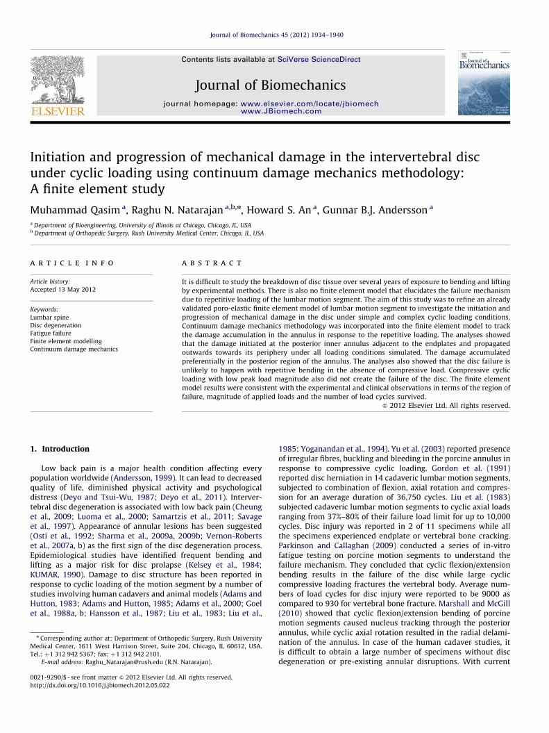
FE model predicted the initiation of damage at the posteriorregion of the inner annulus next to the inferior endplate andprogressed towards outer periphery under all loading conditionsconsidered (Fig. 6). Damage was also identified at the mid discheight in the posterior annulus which did not propagate beyondfew inner annulus layers. Introduction of bending momentscaused the damage to progress preferentially in the posteriolat-eral region of the annulus.
Even though the failure of annular fibres based on maximumstrain value (for assumed fibre failure strain of 15%) was also
Table 2Failure initiation and progression was analysed under simple and complex loading
modes. Loading conditions consisted of different combinations of bending
moments and axial compressive load.
Load case Peak compressive load Peak bending moment (6 Nm)
400 N 800 N Flexion Torsion Lateral
bending
1 x
2 x
3 x
4 x
5 x
6 x x
7 x x
8 x x
9 x x x
10 x x x x
Fig. 5. Damage accumulation in the annulus fibrosus under different loading condition
increment in the number of load cycles.
included in the analyses (Shirazi-Adl et al., 1986), no fibre damagewas observed in any of the simulations.
4. Discussion
A poro-elastic FE model of L4/L5 lumbar motion segmentincorporated with continuum damage mechanics methodologywas presented that predicted structural damage in the disc undercyclic loading. Results of the validation study were consistentwith the experimental observations in terms of the region offailure, magnitude of applied loads and the number of load cycles.In vitro and in vivo human studies have reported occurrence ofradial fissures in the posterior annulus (Haefeli et al., 2006; Ostiet al., 1992; Sharma et al., 2009a, 2009b; Vernon-Roberts et al.,2007a, b). Cyclic testing of porcine discs also reported failure inthe posterior annulus (Marshall and McGill, 2010;Callaghan andMcGill,2001). Thus damage accumulation in the posterior annulusas predicted by the current FE model matched well with clinicaland experimental observations. The numbers of load cycles tofailure predicted for complex loading modes were considerablysmaller than those for uni-axial loading supporting the firsthypothesis. Damage initiated and progressed preferentially inthe posterior annulus under all loading conditions simulated inthis study validating the other hypothesis.
Goel et al. (1988) subjected 11 cadaveric human lumbarspines T12-S1 to 3Nm flexion moment for up to 9600 cycles.They reported structural failure in none of the discs, which isconsistent with the results from the current study. The FEmodel results showed that the repetitive bending withoutcompressive load and cyclic compressive loading with low peakload magnitude did not create a failure in the disc irrespectiveof the number of load cycles. These findings compare well withthe results presented by Callaghan and McGill (2001) whoobserved just an initiation of a fissure in the posterior regionin only 1 out of 5 discs subjected to low compressive load.Marshall and McGill (2010) reported increased annulus damagein porcine discs subjected to combination of axial rotation andflexion/extension than those tested under flexion/extensionalone. This result supports the less number of load cycles tofailure predicted under complex loading modes than single axisbending in the current study.
s. Failure cycle is identified by sharp increase in the failure volume against a small
Superior Inferior
Fig. 6. Damage accumulated in the annulus up to the failure cycle under cyclic compressive loading with a peak load of 800 N. White colour shows the volume of the
annulus that has degraded while black colour represents the normal annulus.
M. Qasim et al. / Journal of Biomechanics 45 (2012) 1934–1940 1939
One of the major limitations of the algorithm used here isthat even though the damage to the tissue occurs only in thedirection of tensile principal stress, the algorithm assumesdamage equally occurs in all the three principal directions ateach integrating point within an element. The current analysesalso did not take into account the changes in the viscoelasticcharacteristics of the annulus due to the fluid flowing in an outof the disc with increasing number of load cycles which isanother major limitation of the analyses. Instead, the damageaccumulation methodology was employed as it enabled thesimulation of large number of load cycles without having to runevery load cycle, thus dramatically reducing the computationalexpense. Further, the change in permeability resulting frominstantaneous disc volume change due to loading was approxi-mated by the change in axial strain in the tissue rather thanchange in void ratio in the tissue. It should also be noted thatthe modulus values assumed for facet cartilage and AF matrixin the current analyses were based on tensile tests and muchsmaller values should be considered in compression.
Damage accumulation was designed as a linear process which is afair assumption in absence of any experimentally derived data ondamage propagation in the annulus. The analyses further assumed norest periods between the loading cycles as well as the tissue healingprocess. Shearing between the annulus layers has been suggested tocause the delamination of the annulus layers (Iatridis and ap Gwynn,2004; Marshall and McGill, 2010; Schmidt et al., 2009). However, lackof stress–failure curve based on shear stress made it impossible to
include the damage mechanism due to shearing.
Conflict of interest statement
None of the authors has any conflict of interest to report.
Acknowledgement
NIH AR48152-02.
Appendix A. Supplementary material
Supplementary data associated with this article can be foundin the online version at http://dx.doi.org/10.1016/j.jbiomech.2012.05.022.
Adams, M.A., Hutton, W.C., 1983. The effect of fatigue on the lumbar intervertebraldisc. The journal of bone and joint surgery. British Volume 65, 199–203.
Andersson, G.B., 1999. Epidemiological features of chronic low-back pain. Lancet354, 581–585.
Argoubi, M., Shirazi-Adl, A., 1996. Poroelastic creep response analysis of a lumbarmotion segment in compression. Journal of Biomechanics 29, 1331–1339.
Callaghan, J.P., McGill, S.M., 2001. Intervertebral disc herniation: studies on aporcine model exposed to highly repetitive flexion/extension motion withcompressive force. Clinical Biomechanics (Bristol, Avon) 16, 28–37.
Cheung, K.M., Karppinen, J., Chan, D., Ho, D.W., Song, Y.Q., Sham, P., Cheah, K.S.,Leong, J.C., Luk, K.D., 2009. Prevalence and pattern of lumbar magneticresonance imaging changes in a population study of one thousand forty-threeindividuals. Spine 34, 934–940.
Deyo, R.A., Mirza, S.K., Martin, B.I., 2011. Error in trends, major medical complica-tions, and charges associated with surgery for lumbar spinal stenosis in olderadults. JAMA: The Journal of the American Medical Association 306, 1088.
Deyo, R.A., Tsui-Wu, Y.J., 1987. Descriptive epidemiology of low-back pain and itsrelated medical care in the United States. Spine 12, 264–268.
Ebara, S., Iatridis, J.C., Setton, L.A., Foster, R.J., Mow, V.C., Weidenbaum, M., 1996.Tensile properties of nondegenerate human lumbar anulus fibrosus. Spine 21,452–461.
Elliott, D.M., Setton, L.A., 2001. Anisotropic and inhomogeneous tensile behavior ofthe human anulus fibrosus: experimental measurement and material modelpredictions. Journal of Biomechanical Engineering 123, 256–263.
Galbusera, F., Schmidt, H., Neidlinger-Wilke, C., Gottschalk, A., Wilke, H.J., 2011.The mechanical response of the lumbar spine to different combinations of discdegenerative changes investigated using randomized poroelastic finite ele-ment models. European Spine Journal: Official Publication of the EuropeanSpine Society, the European Spinal Deformity Society, and the EuropeanSection of the Cervical Spine Research Society 20, 563–571.
Goel, V.K., Kim, Y.E., Lim, T.H., Weinstein, J.N., 1988a. An analytical investigation ofthe mechanics of spinal instrumentation. Spine 13, 1003–1011.
Goel, V.K., Monroe, B.T., Gilbertson, L.G., Brinckmann, P., 1995. Interlaminar shearstresses and laminae separation in a disc. Finite element analysis of the L3-L4motion segment subjected to axial compressive loads. Spine 20, 689–698.
Green, T.P., Adams, M.A., Dolan, P., 1993. Tensile properties of the annulus fibrosusII. Ultimate tensile strength and fatigue life. European Spine Journal: OfficialPublication of the European Spine Society, the European Spinal DeformitySociety, and the European Section of the Cervical Spine Research Society 2,209–214.
Gu, W.Y., Mao, X.G., Foster, R.J., Weidenbaum, M., Mow, V.C., Rawlins, B.A., 1999.The anisotropic hydraulic permeability of human lumbar anulus fibrosus.Influence of age, degeneration, direction, and water content. Spine 24,2449–2455.
Haefeli, M., Kalberer, F., Saegesser, D., Nerlich, A.G., Boos, N., Paesold, G., 2006. Thecourse of macroscopic degeneration in the human lumbar intervertebral disc.Spine 31, 1522–1531.
Hansson, T.H., Keller, T.S., Spengler, D.M., 1987. Mechanical behavior of the humanlumbar spine. II. Fatigue strength during dynamic compressive loading.Journal of Orthopaedic Research: Official Publication of the OrthopaedicResearch Society 5, 479–487.
Iatridis, J.C., ap Gwynn, I., 2004. Mechanisms for mechanical damage in theintervertebral disc annulus fibrosus. Journal of Biomechanics 37, 1165–1175.
Jeffers, J.R., Browne, M., Lennon, A.B., Prendergast, P.J., Taylor, M., 2007. Cementmantle fatigue failure in total hip replacement: experimental and computa-tional testing. Journal of Biomechanics 40, 1525–1533.
Kachanov, L., 1999. Rupture time under creep conditions. International Journal ofFracture 97, 11–18.
M. Qasim et al. / Journal of Biomechanics 45 (2012) 1934–19401940
Kelsey, J.L., Githens, P.B., White 3rd, A.A., Holford, T.R., Walter, S.D., O’Connor, T.,Ostfeld, A.M., Weil, U., Southwick, W.O., Calogero, J.A., 1984. An epidemiologicstudy of lifting and twisting on the job and risk for acute prolapsed lumbarintervertebral disc. Journal of Orthopaedic Research: Official Publication of theOrthopaedic Research Society 2, 61–66.
Koeller, W., Muehlhaus, S., Meier, W., Hartmann, F., 1986. Biomechanical properties ofhuman intervertebral discs subjected to axial dynamic compression—influence ofage and degeneration. Journal of Biomechanics 19, 807–816.
Kumar, S., 1990. Cumulative load as a risk factor for back pain. Spine 15, 1311.Lennon, A.B., Britton, J.R., MacNiocaill, R.F., Byrne, D.P., Kenny, P.J., Prendergast, P.J.,
2007. Predicting revision risk for aseptic loosening of femoral components intotal hip arthroplasty in individual patients—a finite element study. Journal ofOrthopaedic Research: Official Publication of the Orthopaedic Research Society25, 779–788.
Little, J.P., Adam, C.J., Evans, J.H., Pettet, G.J., Pearcy, M.J., 2007. Nonlinear finiteelement analysis of anular lesions in the L4/5 intervertebral disc. Journal ofBiomechanics 40, 2744–2751.
Liu, Y.K., Goel, V.K., Dejong, A., Njus, G., Nishiyama, K., Buckwalter, J., 1985.Torsional fatigue of the lumbar intervertebral joints. Spine 10, 894–900.
Liu, Y.K., Njus, G., Buckwalter, J., Wakano, K., 1983. Fatigue response of lumbarintervertebral joints under axial cyclic loading. Spine 8, 857–865.
Luoma, K., Riihimaki, H., Luukkonen, R., Raininko, R., Viikari-Juntura, E., Lamminen,A., 2000. Low back pain in relation to lumbar disc degeneration. Spine 25,487–492.
Marshall, L.W., McGill, S.M., 2010. The role of axial torque in disc herniation.Clinical Biomechanics (Bristol, Avon) 25, 6–9.
Natarajan, R.N., Ke, J.H., Andersson, G.B., 1994. A model to study the discdegeneration process. Spine 19, 259–265.
Natarajan, R.N., Lavender, S.A., An, H.A., Andersson, G.B., 2008. Biomechanicalresponse of a lumbar intervertebral disc to manual lifting activities: aporoelastic finite element model study. Spine 33, 1958–1965.
Natarajan, R.N., Williams, J.R., Andersson, G.B., 2006. Modeling changes in inter-vertebral disc mechanics with degeneration. The Journal of Bone and JointSurgery.American 88 (2), 36–40.
Osti, O.L., Vernon-Roberts, B., Moore, R., Fraser, R.D., 1992. Annular tears and discdegeneration in the lumbar spine. A post-mortem study of 135 discs. TheJournal of Bone and Joint Surgery. British 74, 678–682.
Panjabi, M.M., Krag, M.H., Chung, T.Q., 1984. Effects of disc injury on mechanicalbehavior of the human spine. Spine 9, 707–713.
Parkinson, R.J., Callaghan, J.P., 2009. The role of dynamic flexion in spine injury isaltered by increasing dynamic load magnitude. Clinical Biomechanics (Bristol,Avon) 24, 148–154.
Rohlmann, A., Zander, T., Schmidt, H., Wilke, H.J., Bergmann, G., 2006. Analysis ofthe influence of disc degeneration on the mechanical behaviour of a lumbarmotion segment using the finite element method. Journal of Biomechanics 39,2484–2490.
Samartzis, D., Karppinen, J., Mok, F., Fong, D.Y., Luk, K.D., Cheung, K.M., 2011. Apopulation-based study of juvenile disc degeneration and its association withoverweight and obesity, low back pain, and diminished functional status. TheJournal of Bone and Joint Surgery. American 93, 662–670.
Sanjeevi, R., Somanathan, N., Ramaswamy, D., 1982. A viscoelastic model forcollagen fibres. Journal of Biomechanics 15, 181–183.
Savage, R.A., Whitehouse, G.H., Roberts, N., 1997. The relationship between themagnetic resonance imaging appearance of the lumbar spine and low back
pain, age and occupation in males. European Spine Journal: Official Publicationof the European Spine Society, the European Spinal Deformity Society, and theEuropean Section of the Cervical Spine Research Society 6, 106–114.
Schechtman, H., Bader, D.L., 1997. In vitro fatigue of human tendons. Journal ofBiomechanics 30, 829–835.
Schmidt, H., Kettler, A., Heuer, F., Simon, U., Claes, L., Wilke, H.J., 2007. Intradiscalpressure, shear strain, and fiber strain in the intervertebral disc undercombined loading. Spine 32, 748.
Schmidt, H., Heuer, F., Wilke, H.J., 2009. Dependency of disc degeneration on shearand tensile strains between annular fiber layers for complex loads. MedicalEngineering and Physics 31, 642–649.
Sharma, A., Parsons, M.S., Pilgram, T.K., 2009a. Temporal association of annulartears and nuclear degeneration: lessons from the pediatric population. Amer-ican Journal of Neuroradiology 30, 1541–1545.
Sharma, A., Pilgram, T., Wippold 2nd, F.J., 2009b. Association between annulartears and disk degeneration: a longitudinal study. American Journal ofNeuroradiology 30, 500–506.
Sharma, M., Langrana, N.A., Rodriguez, J., 1995. Role of ligaments and facets inlumbar spinal stability. Spine 20, 887–900.
Shirazi-Adl, A., 1989. Strain in fibers of a lumbar disc. Analysis of the role of liftingin producing disc prolapse. Spine 14, 96–103.
Shirazi-Adl, A., Ahmed, A.M., Shrivastava, S.C., 1986. A finite element study of alumbar motion segment subjected to pure sagittal plane moments. Journal ofBiomechanics 19 (4), 331–350.
Taylor, M., Verdonschot, N., Huiskes, R., Zioupos, P., 1999a. A combined finiteelement method and continuum damage mechanics approach to simulate thein vitro fatigue behavior of human cortical bone. Journal of Materials Science.Materials in Medicine 10, 841–846.
Taylor, M., Verdonschot, N., Huiskes, R., Zioupos, P., 1999b. A combined finiteelement method and continuum damage mechanics approach to simulate thein vitro fatigue behavior of human cortical bone. Journal of Materials Science.Materials in Medicine 10, 841–846.
Tyrrell, A.R., Reilly, T., Troup, J.D., 1985. Circadian variation in stature and theeffects of spinal loading. Spine 10, 161–164.
Verdonschot, N., Huiskes, R., 1997. The effects of cement-stem debonding in THAon the long-term failure probability of cement. Journal of Biomechanics 30,795–802.
Vernon-Roberts, B., Moore, R.J., Fraser, R.D., 2007a. The natural history of age-related disc degeneration: the pathology and sequelae of tears. Spine 32,2797–2804.
Vernon-Roberts, B., Moore, R.J., Fraser, R.D., 2007b. The natural history of age-related disc degeneration: the pathology and sequelae of tears. Spine 32,2797–2804.
Wang, X.T., Ker, R.F., Alexander, R.M., 1995. Fatigue rupture of wallaby tailtendons. The Journal of Experimental Biology 198, 847–852.
Williams, J.R., Natarajan, R.N., Andersson, G.B., 2007. Inclusion of regional poroelasticmaterial properties better predicts biomechanical behavior of lumbar discssubjected to dynamic loading. Journal of Biomechanics 40, 1981–1987.
Yoganandan, N., Cusick, J.F., Pintar, F.A., Droese, K., Reinartz, J., 1994. Cycliccompression-flexion loading of the human lumbar spine. Spine 19, 784–90;discussion 791.
Yu, C.Y., Tsai, K.H., Hu, W.P., Lin, R.M., Song, H.W., Chang, G.L., 2003. Geometric andmorphological changes of the intervertebral disc under fatigue testing. ClinicalBiomechanics (Bristol, Avon) 18, S3–9.