Editor’s Note T his newsleer issue focuses on diagnosc procedures for insects, diseases, disorders and other plant problems which are crical concerns for the whole- sale nursery. The perishable nature of nursery commodies and the market de- mand for high-quality, defect-free plants place great pressure on growers, nurse- ry managers, pest control advisers, and other personnel to minimize damage caused by these problems. In addion, the introducon of new invasive exoc species in the nursery can result in quaranne restricons and significant eco- nomic damage if these pests aren’t managed in a mely fashion. The feature ar- cle provides detailed informaon about the steps involved in the plant diagnosis process with ps for solving specific types of plant problems. Supplementary in- formaon on diagnosis and tools for idenfying plant problems is provided in Steve Tjosvold’s and Jim Bethke’s regional reports. We hope this informaon will help you to make accurate diagnoses of your plant problems so that appropriate measures can be implemented. Julie Newman and Steve Tjosvold Strategies for Diagnosing Abiotic and Biotic Problems by Steve Tjosvold and Steve Koike Spring 2015 · Volume 19, issue 1 Inside Strategies for Diagnosing Abioc and Bioc Problems……………………………. 1 Science to The Grower: No maer how you spell edema, it’s an excrescent intumescence on the plant leaf …….……………………………………. 8 Get Cultured: Horcultural pracces to conserve water and migate salt precipitaon in container producon……………………….. 12 Insect Hot Topics: Lobate lac scale…………….….. 15 Regional Report: Santa Cruz/Monterey Ramorum blight and how to keep it out of your nursery, Pathogen field test kits…….. 17 Regional Report: Ventura Bot fungi wreak havoc during drought………………………….... 20 Regional Report: San Diego/Riverside Three unusual diagnoses, Field Observaons- Ficus eye-spot midge………………………..…..… 22 New Publicaons …………….. 24 D iseases, disorders and other plant problems are crical concerns for the wholesale nursery. These include bioc problems — caused by living organisms such as pathogens, nematodes, and insects and other arthropods — as well as abioc problems — caused by factors such as temperature and moisture extremes, mechanical damage, chemicals, nu- trient deficiencies or excesses, salt damage and other environmental fac- tors. Many plant problems, especially bioc problems, if not recognized and controlled early in their development, can result in significant eco- nomic damage for the producer. Therefore, mely and accurate diagno- ses are required so that appropriate pest and disease management op- ons and other correcve measures can be implemented. Definion of Plant Diagnosis and Steps Diagnosis is the science and art of idenfying the agent or cause of the problem under invesgaon. When one renders a diagnosis, one has col- lected all available informaon, clues and observaons and then arrives at an informed conclusion as to the causal factor(s). Hence, plant problem
Transcript
Editor’s Note
T his newsletter issue focuses on diagnostic procedures for insects, diseases, disorders and other plant problems which are critical concerns for the whole-
sale nursery. The perishable nature of nursery commodities and the market de-mand for high-quality, defect-free plants place great pressure on growers, nurse-ry managers, pest control advisers, and other personnel to minimize damage caused by these problems. In addition, the introduction of new invasive exotic species in the nursery can result in quarantine restrictions and significant eco-nomic damage if these pests aren’t managed in a timely fashion. The feature arti-cle provides detailed information about the steps involved in the plant diagnosis process with tips for solving specific types of plant problems. Supplementary in-formation on diagnosis and tools for identifying plant problems is provided in Steve Tjosvold’s and Jim Bethke’s regional reports. We hope this information will help you to make accurate diagnoses of your plant problems so that appropriate measures can be implemented.
Julie Newman and Steve Tjosvold
Strategies for Diagnosing Abiotic
and Biotic Problems
by Steve Tjosvold and Steve Koike
Spring 2015 · Volume 19, issue 1
Inside
Strategies for Diagnosing Abiotic and Biotic Problems……………………………. 1 Science to The Grower: No matter how you spell edema, it’s an excrescent intumescence on the plant leaf …….……………………………………. 8 Get Cultured: Horticultural practices to conserve water and mitigate salt precipitation in container production……………………….. 12 Insect Hot Topics: Lobate lac scale…………….….. 15 Regional Report: Santa Cruz/Monterey Ramorum blight and how to keep it out of your nursery, Pathogen field test kits…….. 17 Regional Report: Ventura Bot fungi wreak havoc during drought………………………….... 20 Regional Report: San Diego/Riverside Three unusual diagnoses, Field Observations- Ficus eye-spot midge………………………..…..… 22 New Publications …………….. 24
D iseases, disorders and other plant problems are critical concerns for the wholesale nursery. These include biotic problems — caused by
living organisms such as pathogens, nematodes, and insects and other arthropods — as well as abiotic problems — caused by factors such as temperature and moisture extremes, mechanical damage, chemicals, nu-trient deficiencies or excesses, salt damage and other environmental fac-tors. Many plant problems, especially biotic problems, if not recognized and controlled early in their development, can result in significant eco-nomic damage for the producer. Therefore, timely and accurate diagno-ses are required so that appropriate pest and disease management op-tions and other corrective measures can be implemented. Definition of Plant Diagnosis and Steps Diagnosis is the science and art of identifying the agent or cause of the problem under investigation. When one renders a diagnosis, one has col-lected all available information, clues and observations and then arrives at an informed conclusion as to the causal factor(s). Hence, plant problem
2 · UCNFA News · Spring 2015 · Volume 19, issue 1
STRATEGIES FOR DIAGNOSING ABIOTIC AND BIOTIC
PROBLEMS
continued from page 1
diagnosis is an investigative, problem-solving pro-cess that involves the following steps: 1. Ask and answer the appropriate questions to de-
fine the problem and obtain information that is relevant to the case under investigation.
2. Conduct a detailed, thorough examination of the plants and production areas.
3. Use appropriate field diagnostic kits and lab tests to obtain clinical information on possible causal agents and factors.
4. Compile all the collected information and consult additional resources and references.
5. Finally, make an informed diagnosis. Throughout this process compile all notes, observa-tions, maps, laboratory results, photographs and other information. This compilation will be the infor-mation base for the present diagnosis and can also be a useful resource for future diagnostic cases. Keep an open mind as the information is analyzed
Table 1 DISTINGUISHING ABIOTIC AND BIOTIC PROBLEMS
Characteristics Abiotic Biotic
Hosts often affects several species or plants of
various ages
often affects one species or cultivar of
the same age
Pattern of plant symptoms often related to environmental or physi-
cal factors or cultural practices; may be
regular or uniform
often initially observed in random or
irregular locations
Rate of symptom development relatively uniform, extent of damage
appears similar among plants
relatively uneven, time of appearance
and damage severity varies among
affected plants
Signs no evidence of the kinds of pests or
pathogens known to cause the current
symptoms
presence of insects, mites, fungal myce-
lium and spore clusters, bacterial ooze,
mollusks; products produced by pests
such as honeydew/sooty mold, cast
skins, frass, or mollusk slime.
Spread is not infectious, is not progressive,
commonly caused by one incident and
does not spread
infectious, spreads on host over time if
environmental conditions are suitable
Recurrence possibly previously associated with cur-
rent or prior environmental conditions
or cultural practices
possibly caused by pests that have
affected this crop during previous grow-
ing seasons or are known to commonly
affect this crop species or cultivar
Adapted from Table 18, ANR Pub 3420
and do not make unwarranted assumptions. Distinguishing Abiotic and Biotic Problems The first step is to determine whether the problem is caused by an infectious agent, and this can be diffi-cult. Plant symptoms caused by biotic factors such as infectious diseases and arthropod pests are often similar to damage caused by other factors. Leaf spots, chlorosis, blights, deformities, defoliation, wilting, stunting and plant death can be common symptoms of both biotic and abiotic problems; therefore, the presence of these symptoms does not necessarily mean the problem is a disease. Some general guidelines for distinguishing abiotic and bio-tic problems follow and are summarized in table 1. Biotic problems. Identifying biotic problems is some-times facilitated if signs of a pathogen, primarily the growth of a fungus, are present. The most obvious examples of such signs are the mycelium and spores
3 · UCNFA News · Spring 2015 · Volume 19, issue 1
STRATEGIES FOR DIAGNOSING ABIOTIC AND BIOTIC
PROBLEMS
continued from page 2
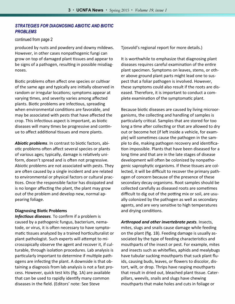
produced by rusts and powdery and downy mildews. However, in other cases nonpathogenic fungi can grow on top of damaged plant tissues and appear to be signs of a pathogen, resulting in possible misdiag-noses. Biotic problems often affect one species or cultivar of the same age and typically are initially observed in random or irregular locations; symptoms appear at varying times, and severity varies among affected plants. Biotic problems are infectious, spreading when environmental conditions are favorable, and may be associated with pests that have affected the crop. This infectious aspect is important, as biotic diseases will many times be progressive and contin-ue to affect additional tissues and more plants. Abiotic problems. In contrast to biotic factors, abi-otic problems often affect several species or plants of various ages; typically, damage is relatively uni-form, doesn’t spread and is often not progressive. Abiotic problems are not associated with pests. They are often caused by a single incident and are related to environmental or physical factors or cultural prac-tices. Once the responsible factor has dissipated and is no longer affecting the plant, the plant may grow out of the problem and develop new, normal ap-pearing foliage. Diagnosing Biotic Problems Infectious diseases. To confirm if a problem is caused by a pathogenic fungus, bacterium, nema-tode, or virus, it is often necessary to have sympto-matic tissues analyzed by a trained horticulturalist or plant pathologist. Such experts will attempt to mi-croscopically observe the agent and recover it, if cul-turable, through isolation procedures. Lab analysis is particularly important to determine if multiple path-ogens are infecting the plant. A downside is that ob-taining a diagnosis from lab analysis is not a fast pro-cess. However, quick test kits (fig. 1A) are available that can be used to rapidly identify many common diseases in the field. (Editors’ note: See Steve
Tjosvold’s regional report for more details.) It is worthwhile to emphasize that diagnosing plant diseases requires careful examination of the entire plant specimen. Symptoms on leaves, stems, or oth-er above ground plant parts might lead one to sus-pect that a foliar pathogen is involved. However, these symptoms could also result if the roots are dis-eased. Therefore, it is important to conduct a com-plete examination of the symptomatic plant. Because biotic diseases are caused by living microor-ganisms, the collecting and handling of samples is particularly critical. Samples that are stored for too long a time after collecting or that are allowed to dry out or become hot (if left inside a vehicle, for exam-ple) will sometimes cause the pathogen in the sam-ple to die, making pathogen recovery and identifica-tion impossible. Plants that have been diseased for a long time and that are in the late stages of disease development will often be colonized by nonpatho-genic saprophytic organisms. If these tissues are col-lected, it will be difficult to recover the primary path-ogen of concern because of the presence of these secondary decay organisms. Root samples should be collected carefully as diseased roots are sometimes difficult to dig out of the potting mix or soil, are usu-ally colonized by the pathogen as well as secondary agents, and are very sensitive to high temperatures and drying conditions.
Arthropod and other invertebrate pests. Insects, mites, slugs and snails cause damage while feeding on the plant (fig. 1B). Feeding damage is usually as-sociated by the type of feeding characteristics and mouthparts of the insect or pest. For example, mites and insects such as whiteflies, aphids and mealybugs have tubular sucking mouthparts that suck plant flu-ids, causing buds, leaves, or flowers to discolor, dis-tort, wilt, or drop. Thrips have rasping mouthparts that result in dried out, bleached plant tissue. Cater-pillars, weevils, snails and slugs have chewing mouthparts that make holes and cuts in foliage or
4 · UCNFA News · Spring 2015 · Volume 19, issue 1
STRATEGIES FOR DIAGNOSING ABIOTIC AND BIOTIC
PROBLEMS
continued from page 3
flowers. They can also prune plant parts and sometimes consume entire plants. If present, these pests are visible with the naked eye, a 10 X hand lens, or stereomicroscope, all depending up-on their size. An assessment of whether the identified arthropod or invertebrate matches the plant dam-age it is associated with must be de-termined. Sometimes the identified arthropod or invertebrate may not be the sole problem or could, in fact, be a beneficial organism or insignificant pest. Aphids, whiteflies, thrips, leafhoppers and some other insects that suck plant juices may vector pathogens such as viruses and phytoplasmas (and to a lesser extent fungi and bac-teria). They can feed on infected plants, acquire the pathogen, feed on healthy host plants and transmit the pathogen to the new host. The insects do not necessarily have to be present in large numbers to cause a significant disease outbreak. The insect vec-tors are not always present at the same time the disease symptoms are being expressed. The excrement and byproducts from these pests can also provide clues that the pests have been or are actively present. Caterpillars and other chewing pests produce dark excrement or droppings. Green-house thrips and plant bugs produce dark, watery, or varnish-like droppings on foliage. Aphids, white-flies, soft scales, and some other sap-sucking insects excrete excess plant fluids as honeydew, a sticky sap, which provides a medium for the growth of sooty mold.
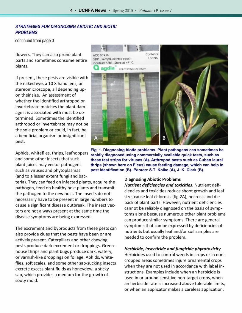
Diagnosing Abiotic Problems Nutrient deficiencies and toxicities. Nutrient defi-ciencies and toxicities reduce shoot growth and leaf size, cause leaf chlorosis (fig.2A), necrosis and die-back of plant parts. However, nutrient deficiencies cannot be reliably diagnosed on the basis of symp-toms alone because numerous other plant problems can produce similar symptoms. There are general symptoms that can be expressed by deficiencies of nutrients but usually leaf and/or soil samples are needed to confirm the problem. Herbicide, insecticide and fungicide phytotoxicity. Herbicides used to control weeds in crops or in non-cropped areas sometimes injure ornamental crops when they are not used in accordance with label in-structions. Examples include when an herbicide is used in or around sensitive non-target crops, when an herbicide rate is increased above tolerable limits, or when an applicator makes a careless application.
Fig. 1. Diagnosing biotic problems. Plant pathogens can sometimes be
rapidly diagnosed using commercially available quick tests, such as
these test strips for viruses (A). Arthropod pests such as Cuban laurel
thrips (shown here on Ficus) cause feeding damage, which can help in
pest identification (B). Photos: S.T. Koike (A), J. K. Clark (B).
A B
Fig. 2. Examples of abiotic problems. Iron deficiency on sweet gum (Liquidambar styracifolia) showing inter-
veinal chlorosis (A). Chorotic spots on Hedera caused by a miticide application at a higher dosage rate than
specified on the pesticide label (B). Photos: E. Martin (A), S. A. Tjosvold (B).
5 · UCNFA News · Spring 2015 · Volume 19, issue 1
STRATEGIES FOR DIAGNOSING ABIOTIC AND BIOTIC
PROBLEMS
continued from page 4
By understanding the mode of action of the herbi-cide, one can determine if the symptom fits an herb-icide application. Herbicide detection in affected plants is possible with the help of a specialized la-boratory but the analysis can be expensive. To mini-mize the cost of testing, the laboratory will need to know the suspected herbicide or its chemical group to narrow the analysis. Insecticides and fungicides occasionally cause obvi-ous plant damage. Symptoms can vary widely. Gen-erally, flower petals are more susceptible to damage from pesticide applications than are leaves. The younger and more tender the leaves the more sus-ceptible they are to pesticide applications. Hot weather can exacerbate the damage the chemicals cause. Pesticides that have systemic action can have a more profound effect. Some active ingredients can adversely affect the photosynthetic mechanism or other physiological processes and can result in a general leaf chlorosis, interveinal chlorosis, leaf curl-ing and stunting. Emulsifiable concentrate (EC) for-mulations, soaps and oils can adversely affect the waxy surface layer that protects the leaf from desic-
cation. Applications with these products can result in the loss of the shiny appearance of a leaf, leaf spotting and necrosis. Pesticides applied as soil drenches can cause poor germination, seedling death, or distorted plant growth. Check label precautions against use on certain spe-cies. Make sure the pesticide is not applied more frequently or at a higher rate (fig. 2B) than recom-mended, or that the pesticide is not mixed with in-compatible pesticides. When in doubt as to whether the plant species is sensitive to the pesticide, spray a few plants and observe them for several days to a week for any signs of damage before spraying any more of the plants. Physiological and Genetic Disorders There are numerous disorders that can occur be-cause of environmental extremes — too much or too little of an environmental element such as light, temperature, water, or wind. Sunburn is damage to foliage and other herbaceous plant parts caused by a combination of too much light and heat and insuffi-cient moisture. A yellow or brown area develops on
A B
6 · UCNFA News · Spring 2015 · Volume 19, issue 1
Fig. 3. Poor air quality can lead to physiological disorders. Shattering (petal drop) on geranium was caused by
plant exposure to low levels of ethylene in the greenhouse or during postharvest storage (A). Yellowish and
brownish patches on Japanese maple leaves are damage caused by ozone (B), an outdoor air pollutant. Photos:
J. K. Clark.
STRATEGIES FOR DIAGNOSING ABIOTIC AND BIOTIC
PROBLEMS
continued from page 5
foliage, which then dies beginning in areas between the veins. Sunscald is damage to bark caused by ex-cessive light or heat. Damaged bark becomes cracked and sunken. Frost damage causes shoots, buds and flowers to curl, turn brown or black and die. Hailstones injure leaves, twigs, and in serious cases even the bark. Chilling damage in sensitive plants can cause wilting of foliage and flowers and development of dark water-soaked spots on leaves that can eventually turn light brown or bleached, and die. Physical and mechanical injury can occur when plants are mishandled during transport or rou-tine cultural practices. Wounds might serve as entry sites for plant pathogens and can attract boring in-sects to woody stems. In closed environments such as greenhouses and nursery storage areas, plants can be exposed to toxic levels of ethylene gas. Sources of ethylene include improperly functioning or unvented greenhouse heaters; exhaust from engines of forklifts and vehi-cles; cigarette smoke; damaged, decaying, or dying plants; and ripe or decaying fruit. Toxic levels of eth-
ylene gas can cause premature abscission of flower buds, petals (fig. 3A) and leaves. Other symptoms include wilted flowers, chlorosis, twisted growth or downward bending of stems and leaves and under-sized or narrow leaves. Outdoors, exposure of nursery plants to air pollutant gases such as ozone (fig. 3B), carbon monoxide, ni-trous oxides and sulfur dioxide can cause damage. Typical symptoms vary widely, but include slow growth and discolored, dying, or prematurely drop-ping foliage. Damage is often found where plants are located near sources of polluted air such as near freeways or industries or where weather and topog-raphy concentrate the pollutants. Sometimes plants or plant shoots exhibit an unusual and sudden change of color producing discrete markings of variegation. For example, a plant with entirely green leaves suddenly produces a shoot that has leaves with edges lacking green pigment, stripes, or blotches. A new shoot such as this is probably a chimera (fig. 4). It is produced when a genetic muta-
A B
7 · UCNFA News · Spring 2015 · Volume 19, issue 1
Fig. 4. Genetic disorder. Growing
points with variegated leaves can
sometimes arise spontaneously
from some species such as this
Origanum. Genetic variants such as
this are sometimes confused with
plants with virus disease or nutri-
ent deficiency symptoms. Photo: S.
A. Tjosvold.
STRATEGIES FOR DIAGNOSING ABIOTIC AND BIOTIC
PROBLEMS
continued from page 6
tion occurs in a specific region of the growing tip re-sulting in a section with genetically different cells. The ostensible result of the genetic change is de-pendent on the arrangement of the genetically different cells in the shoot tip and their expression. This can lead to sometimes bizarre variegation forms or sometimes forms that are quite desirable. Some-times variegation can be caused by viruses. Viruses usually cause non-uniform chlorosis, such as mosa-ics, while chimeras usually produce patterned forms such as variegation of color on leaf margins, stripes, or complete loss of pigment. Some viroids may also cause bleaching of pigments in leaves; such symp-toms, however, are generally produced throughout the plant and are not restricted to a single shoot.
Some nutrient disorders can cause variegation but these disorders usually do not arise from a specific shoot as with chimeras. Steve Tjosvold is Environmental Horticulture Advi-sor and Steve Koike is Plant Pathology Farm Advi-sor, UC Cooperative Extension, Santa Cruz and Monterey counties. This article was condensed from: Diagnosing Plant Problems, Chapter 11. In Newman, J. (ed) Contain-er Nursery Production and Business Management. Univ. of Calif. Agric. and Nat. Resources. Publica-tion 3540. Richmond, CA.
References
Boger P, Sandmann G. 1989. Target sites of herbicide action. Boca Raton, FL: CRC Press.
Costello L, Perry E, Matheny N, Henry M, Geisel P. 2003. Abiotic disorders of landscape plants: A diagnostic guide. Oak-land: University of California Division of Agriculture and Natural Resources Publication 3420.
Derr JF, Appleton BL. 1988. Herbicide injury to trees and shrubs: A pictorial guide to symptom diagnosis. Virginia Beach, VA: Blue Crab Press.
Dreistadt SH. 2001. Integrated pest management for floriculture and nurseries. Oakland: University of California Divi-sion of Agriculture and Natural Resources Publication 3402.
8 · UCNFA News · Spring 2015 · Volume 19, issue 1
SCIENCE TO THE GROWER: No matter how you
spell edema, it’s an excrescent intumescence on the
plant leaf by Richard Evans
STRATEGIES FOR DIAGNOSING ABIOTIC AND BIOTIC
PROBLEMS
continued from page 7
References (continued)
Eagle, DJ. 1981. Diagnosis of herbicide damage to crops. New York, NY: Chemical Publishing Co.
Grogan RG. 1981. The science and art of plant disease diagnosis. Annual Review of Phytopathology 19:333–351.
Retzinger EJ, Mallory-Smith C. 1997. Classification of herbicides by the site of action for weed resistance management strategies. Weed Technology 11:384–393.
Schubert TS, Breman LL. 1988. Basic concepts of plant disease and how to collect a sample for disease diagnosis. Plant Pathology Circular No. 307. Florida Department ofAgriculture and Consumer Services, Plant Pathology Circular No. 307.
Sharma MP. 1986. Recognizing herbicide action and injury. Alberta Environmental Centre, Alberta Agriculture. Agdex 641–647.
Shurtleff MC, Averre CW. 1997. The plant disease clinic and field diagnosis of abiotic diseases. St. Paul, MN: American Phytopathological Society Press.
Stewart TM, Galea VJ. 2006. Approaches to training practitioners in the art and science of plant disease diagnosis. Plant Disease 90:539–547.
Tickes B, Cudney D, and Elmore C. 1996. Herbicide injury symptoms. Tucson, AZ: University of Arizona Cooperative Ex-tension Publication No. 195021.
I f you see abnormal, translucent, warty growths bulging out of the leaves of one of your crop
plants and you ask your local farm advisor to identi-fy the problem, you may be told that your plant has edema. Or your farm advisor may identify it as oe-dema. Or an intumescence. Or an oedemata, neo-plasm, enation, excrescence, leaf lesion, genetic tu-mor, or gall. This profusion of names illustrates the unclear status of a problem that has been attributed to physical injury, chemical injury, insect injury, fun-gal infection, plant nutrition, air quality, light quality or quantity, soil temperature, air temperature, hu-midity, excess soil water, plant growth substances, plant genetics and — for those who can imagine the existence of other causes but can’t imagine what
they are — “unspecified factors.” Something about this disorder’s warty appearance and muddled sta-tus reminds the UCNFA News editors of me, and they have asked me (as the God of Agriculture) to explain what we know, and don’t know, about these curious lesions. Intumescence May Be the Best Name First, let’s settle on a name for them. The first pub-lished scientific description of the disorder was made in 1886 by Paul Sorauer, a German plant pathologist, who observed it on the Tasmanian snow gum (Eucalyptus coccifera). Sorauer called it an intu-mescence (enlarged or swollen plant part). In 1893, George Atkinson, assistant professor of cryptogamic
9 · UCNFA News · Spring 2015 · Volume 19, issue 1
SCIENCE TO THE GROWER
continued from page 8
botany at Cornell University, used the word oe-dema to describe a leaf disorder on tomato plants. He observed that the “veinlets as well as the midrib, petioles and the surface of the stem presented numerous elevated areas of a frosty aspect.” Then, in a scientific paper published in 1900, a Cambridge student, Elizabeth Dale, called the outgrowths on rose mallow (Hibiscus vitifolius) intumescences and noted that Ameri-cans favor the term oedemata. The other names listed above didn’t appear until twenty or more years later. Since intumescence came first, and since edema (or its older spelling, oedema) is defined by the Oxford English Dictionary as “a fluid-filled tumour or swelling,” which doesn’t always occur as a symptom of this disorder, I rule in favor of calling it an intumescence. Cause of Intumescence Development Remains Unclear Naming the disorder isn’t the only problem. Carl La Rue, one of the scientists who has studied intumescences, said, “There seems to be no law as to the development of intumescences within groups of plants.” Some scientists have report-ed that intumescences occur when the outer layer of cells in leaves (the epidermis) is rup-tured because of swelling of underlying water-soaked cells (the palisade parenchyma). Other scientists have attributed intumescences to ab-normal cell division, and still others have said
Fig. 1A, B, C. Tibouchina urvelleana (princess flower)
grown in a greenhouse (Feb 2015) show symptoms of
what has conventionally been called edema. These
symptoms are also seen under some magnification
showing the warty appearance of the affected areas.
These affected areas began to become necrotic a few
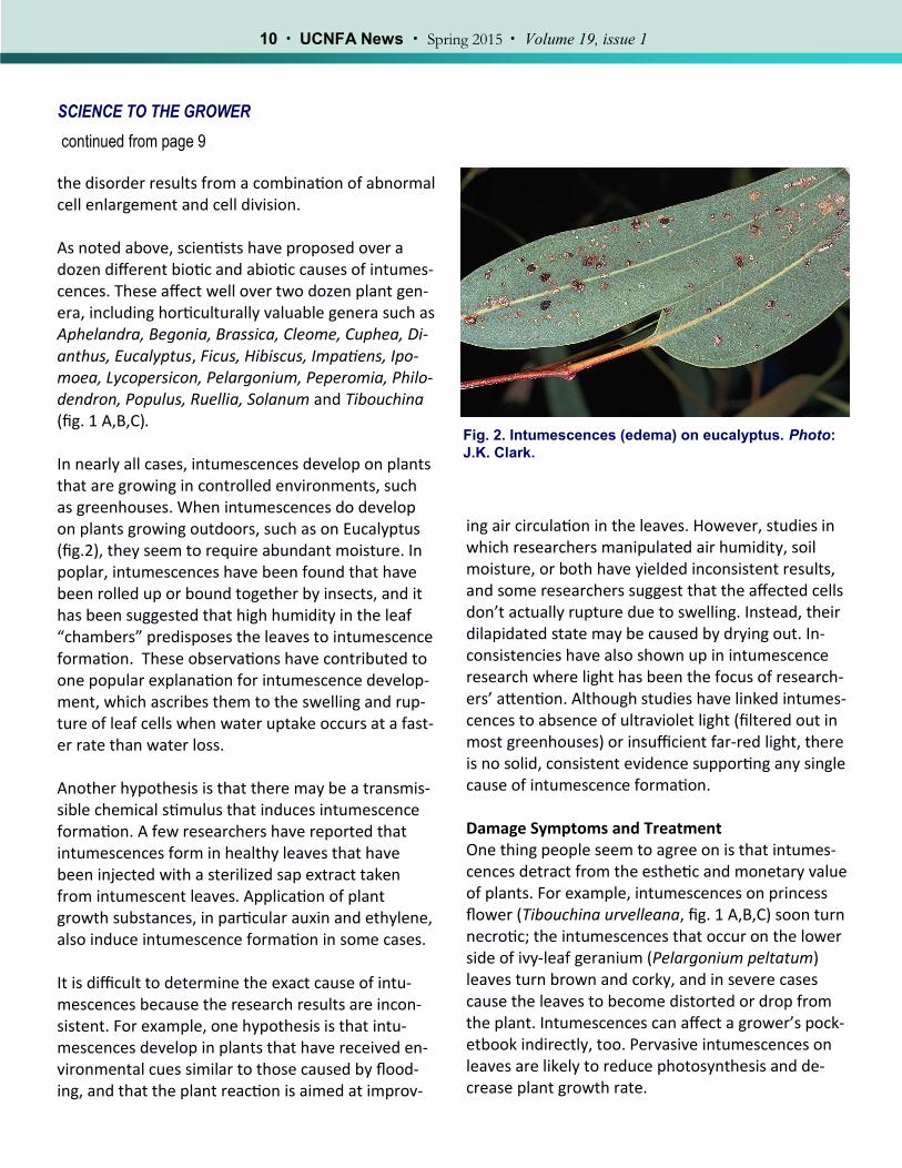
the disorder results from a combination of abnormal cell enlargement and cell division. As noted above, scientists have proposed over a dozen different biotic and abiotic causes of intumes-cences. These affect well over two dozen plant gen-era, including horticulturally valuable genera such as Aphelandra, Begonia, Brassica, Cleome, Cuphea, Di-anthus, Eucalyptus, Ficus, Hibiscus, Impatiens, Ipo-moea, Lycopersicon, Pelargonium, Peperomia, Philo-dendron, Populus, Ruellia, Solanum and Tibouchina (fig. 1 A,B,C). In nearly all cases, intumescences develop on plants that are growing in controlled environments, such as greenhouses. When intumescences do develop on plants growing outdoors, such as on Eucalyptus (fig.2), they seem to require abundant moisture. In poplar, intumescences have been found that have been rolled up or bound together by insects, and it has been suggested that high humidity in the leaf “chambers” predisposes the leaves to intumescence formation. These observations have contributed to one popular explanation for intumescence develop-ment, which ascribes them to the swelling and rup-ture of leaf cells when water uptake occurs at a fast-er rate than water loss. Another hypothesis is that there may be a transmis-sible chemical stimulus that induces intumescence formation. A few researchers have reported that intumescences form in healthy leaves that have been injected with a sterilized sap extract taken from intumescent leaves. Application of plant growth substances, in particular auxin and ethylene, also induce intumescence formation in some cases. It is difficult to determine the exact cause of intu-mescences because the research results are incon-sistent. For example, one hypothesis is that intu-mescences develop in plants that have received en-vironmental cues similar to those caused by flood-ing, and that the plant reaction is aimed at improv-
Fig. 2. Intumescences (edema) on eucalyptus. Photo:
J.K. Clark.
ing air circulation in the leaves. However, studies in which researchers manipulated air humidity, soil moisture, or both have yielded inconsistent results, and some researchers suggest that the affected cells don’t actually rupture due to swelling. Instead, their dilapidated state may be caused by drying out. In-consistencies have also shown up in intumescence research where light has been the focus of research-ers’ attention. Although studies have linked intumes-cences to absence of ultraviolet light (filtered out in most greenhouses) or insufficient far-red light, there is no solid, consistent evidence supporting any single cause of intumescence formation. Damage Symptoms and Treatment One thing people seem to agree on is that intumes-cences detract from the esthetic and monetary value of plants. For example, intumescences on princess flower (Tibouchina urvelleana, fig. 1 A,B,C) soon turn necrotic; the intumescences that occur on the lower side of ivy-leaf geranium (Pelargonium peltatum) leaves turn brown and corky, and in severe cases cause the leaves to become distorted or drop from the plant. Intumescences can affect a grower’s pock-etbook indirectly, too. Pervasive intumescences on leaves are likely to reduce photosynthesis and de-crease plant growth rate.
References Atkinson GF. 1893. Oedema of the tomato. Cornell University Agricultural Experiment Station Bulletin 53. Craver JK, Miller CT, Williams KA, Boyle DL. 2014. Characterization and comparison of lesions on ornamental sweetpotato ‘Blackie’, tomato ‘Maxifort’, interspecific geranium ‘Caliente Coral’ and bat-faced cuphea ‘Tiny Mice’. Journal of the American Society for Horticultural Science 139: 603-615. Dale E. 1900. On certain outgrowths (intumescences) on the green parts of Hibiscus vitifolius Linn. Proceed-ings of the Cambridge Philosophical Society 10: 192-209. Lang SP, Struckmeyer BE, Tibbitts TW. 1983. Morphology and anatomy of intumescence development on to-mato plants. Journal of the American Society for Horticultural Science 108: 266-271. La Rue CD. 1933. Intumescences on poplar leaves. I. Structure and development. American Journal of Botany 20: 1-17. Morrow RC, Tibbitts TW. 1988. Evidence for involvement of phytochrome in tumor development on plants. Plant Physiology 88: 1110-1114. Pinkard E, Gill W, Mohammed C. 2006. Physiology and anatomy of lenticel-like structures on leaves of Euca-lyptus nitens and Eucalyptus globulus seedlings. Tree Physiology 26: 989-999. Rangarajan A, Tibbitts TW. 1994. Exposure with far-red radiation for control of oedema injury on ‘Yale’ ivy geranium. HortScience 29: 38-40. Sorauer P. 1886. Handbuch der Pflanzenkrankheiten, 2nd edition. Parey, Berlin.
So what can be done? Unfortunately, not much. The presence of an array of potential causes has led to a mixed bag of recommended practices, but none has been shown to work consistently. Perhaps continuing study of intumescence development will lead to a better understanding of this scourge, as well as some solu-tions for growers. Richard Evans is UC Cooperative Extension Environmental Horticulturist, Department of Plant Sciences, UC Davis.
water and mitigate salt precipitation in container
production by Donald J. Merhaut
This is the second in a series of “Get Cultured” articles on reclaimed water in nursery production. In the previ-ous newsletter, we discussed various chemical traits that render challenge in using reclaimed water in nursery production. In this article, we will elaborate on horticultural practices and cultural care options to minimize the negative impact of poor quality irrigation water on plant production. Even though water treatments such as filtration techniques and blending water are viable options, this article will focus only on horticultural and
cultural methods. Fortunately, the recommendations that follow will also help reduce water usage.
I rrigation of containerized plants serves four pri-mary functions: (1) provides water directly for
plant uptake and transpiration, (2) provides dis-solved fertilizers in the water to supply plants with essential nutrients, (3) facilitates the application of some pesticides via the irrigation system, and (4) maintains relatively high humidity for propagation facilities.
Horticultural practices and other cultural recommen-dations are often based on the assumption that the water being used is fairly good for plant production. These “good” traits include ideal pH, low alkalinity, low electrical conductivity (EC) and nonharmful lev-els of salts such as sodium, chloride and heavy met-als. Unfortunately, the quality of reclaimed water is often poor and requires treatment prior to use or the implementation of cultural practices to ensure that irrigation is effective in carrying out the func-tions listed above. Options include:
Removing harmful impurities. The quality of re-claimed water can be improved by cleaning the water source of the harmful impurities that are present using filtration techniques such as re-verse osmosis. Filtration has been addressed in previous issues, and we may need to address this again in future issues. Filtration may be the only option for propagation facilities, where misters and foggers are used and the propagative mate-rial is often very sensitive to these impurities.
Calcium salts can build up on foliage and clog the tiny orifices on irrigation emitters.
Blending. Another option for improving water quality is to blend the lower-quality water (reclaimed or other secondary water sources) with a clean water source in a ratio that reduces the harmful salts to non-toxic concentrations. This method is ideal if better water sources are available. However, in some areas of California, only reclaimed water is available for some facili-ties.
Adjusting cultural practices. A variety of cultural management practices can be implemented to reduce the negative effects of poor quality re-claimed water when good quality water is not available for blending. These practices are the primary focus of this article and are described below.
Reduce Water-Holding Capacity
Since some reclaimed water sources have a high EC, that is, high concentrations of dissolved essential
and non-essential salts, it will be important to have a media that is hydrophilic (“water-loving”), yet very well drained. Traditionally, media containing peat-moss or coir, which have high water-holding capacity (WHC), have been favored since this allows for ex-tended periods between irrigation episodes. How-ever, in the case of water sources with high salinity, the objective of irrigation is to provide water to the
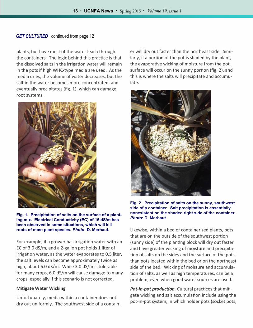
plants, but have most of the water leach through the containers. The logic behind this practice is that the dissolved salts in the irrigation water will remain in the pots if high WHC-type media are used. As the media dries, the volume of water decreases, but the salt in the water becomes more concentrated, and eventually precipitates (fig. 1), which can damage root systems.
Fig. 1. Precipitation of salts on the surface of a plant-
ing mix. Electrical Conductivity (EC) of 16 dS/m has
been observed in some situations, which will kill
roots of most plant species. Photo: D. Merhaut.
For example, if a grower has irrigation water with an EC of 3.0 dS/m, and a 2-gallon pot holds 1 liter of irrigation water, as the water evaporates to 0.5 liter,
the salt levels can become approximately twice as high, about 6.0 dS/m. While 3.0 dS/m is tolerable for many crops, 6.0 dS/m will cause damage to many crops, especially if this scenario is not corrected.
Mitigate Water Wicking
Unfortunately, media within a container does not dry out uniformly. The southwest side of a contain-
er will dry out faster than the northeast side. Simi-larly, if a portion of the pot is shaded by the plant, the evaporative wicking of moisture from the pot surface will occur on the sunny portion (fig. 2), and this is where the salts will precipitate and accumu-late.
Fig. 2. Precipitation of salts on the sunny, southwest
side of a container. Salt precipitation is essentially
nonexistent on the shaded right side of the container.
Photo: D. Merhaut.
Likewise, within a bed of containerized plants, pots that are on the outside of the southwest portion (sunny side) of the planting block will dry out faster and have greater wicking of moisture and precipita-tion of salts on the sides and the surface of the pots
than pots located within the bed or on the northeast side of the bed. Wicking of moisture and accumula-tion of salts, as well as high temperatures, can be a problem, even when good water sources are used.
Pot-in-pot production. Cultural practices that miti-gate wicking and salt accumulation include using the pot-in-pot system, in which holder pots (socket pots,
moat pots) are buried in the ground and the contain-erized plants are placed inside these pots (fig. 3). This system reduces the heating of the containers by the sun since they are shaded by the larger holder pots. Additionally, temperatures in the root zone are moderated because of the insulation from the soil. Also, once the plant material grows and shades the pot surfaces, the high loss of moisture at the me-dium surface will be reduced. Depending on the se-verity of the problem, the pot-in-pot method may
only be necessary on the southwest side of a pro-duction bed.
Fig. 3. Trees in a pot-in-pot system in which contain-
erized plants are placed in larger holder pots installed
in the ground. Temperatures in the root zone are
moderated because the holder pots shade the smaller
containers and the soil around the containers pro-
vides insulation. Photo: U.K. Schuch.
Shading. Another method that maintains tempera-ture uniformity and mitigates wicking in the contain-er is shading. This is a good cultural practice, since it will also avoid excessive moisture loss, reducing heat and water stress of newly planted crops which have tender young shoot growth and have not established an extensive root system. Once the plants have de-
veloped and have “hardened off,” they can be tran-sitioned to their permanent production site. In hotter climates of the interior valleys, some degree of shade may be ideal, especially during the summer months.
Windbreaks. Windbreaks can be provided in the form of fencing, trees, hedges or even larger plants, such as 24-inch boxed trees, with the smaller con-tainers in between. In addition to reducing water
loss, windbreaks provide shade, where trees are large enough, that can help mitigate wicking. These types of cultural practices need to be carefully thought out, since irrigation systems, pest manage-ment and other cultural practices will need to be met for the two different plant types in the same growing area.
Drip vs. Overhead Irrigation
The proper use of drip irrigation does improve water use efficiency in the crop and eliminates the poten-tial of water marks forming on the leaves, which can occur with overhead irrigation. However, there is increased risk of salt accumulation in the containers since water is slowly released into the container. Another concern with drip irrigation is that the small openings of the emitters are more likely to clog than the larger openings present in overhead irrigation systems. Careful selection and maintenance of irri-gation nozzles will be important to optimize efficien-cy of irrigation programs.
Record Keeping
As with any crop production program, keeping track
of salt accumulation in containers should be regular-ly monitored, especially during the summer. Meth-ods of measurement include pour-through and soil-water extraction methods. However, more im-portant than the method used, is that the same method is used so that results from past tests can be compared to future tests.
In the next issue, we will address fertilizer selection, use, storage and other aspects of plant nutrition which may be affected by secondary water sources such as reclaimed water.
Don Merhaut is a UC Cooperative Extension Specialist for Nursery and Floriculture Crops, Department of Botany and Plant Sciences, UC Riverside.
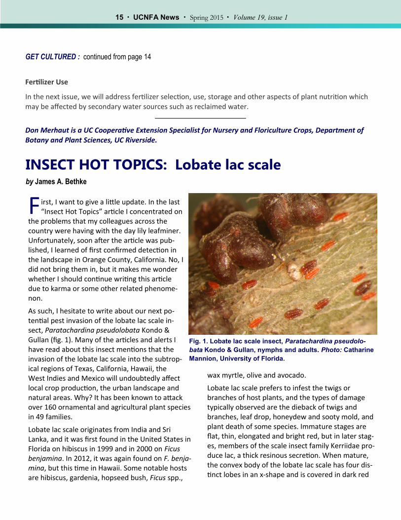
INSECT HOT TOPICS: Lobate lac scale by James A. Bethke
F irst, I want to give a little update. In the last “Insect Hot Topics” article I concentrated on
the problems that my colleagues across the country were having with the day lily leafminer. Unfortunately, soon after the article was pub-lished, I learned of first confirmed detection in the landscape in Orange County, California. No, I did not bring them in, but it makes me wonder whether I should continue writing this article due to karma or some other related phenome-non.
As such, I hesitate to write about our next po-tential pest invasion of the lobate lac scale in-sect, Paratachardina pseudolobata Kondo & Gullan (fig. 1). Many of the articles and alerts I have read about this insect mentions that the invasion of the lobate lac scale into the subtrop-ical regions of Texas, California, Hawaii, the West Indies and Mexico will undoubtedly affect local crop production, the urban landscape and natural areas. Why? It has been known to attack over 160 ornamental and agricultural plant species in 49 families.
Lobate lac scale originates from India and Sri Lanka, and it was first found in the United States in Florida on hibiscus in 1999 and in 2000 on Ficus benjamina. In 2012, it was again found on F. benja-mina, but this time in Hawaii. Some notable hosts are hibiscus, gardenia, hopseed bush, Ficus spp.,
bata Kondo & Gullan, nymphs and adults. Photo: Catharine
Mannion, University of Florida.
wax myrtle, olive and avocado.
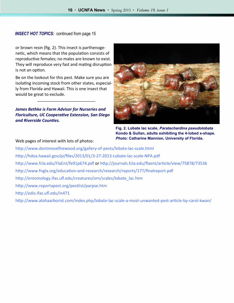
Lobate lac scale prefers to infest the twigs or branches of host plants, and the types of damage typically observed are the dieback of twigs and branches, leaf drop, honeydew and sooty mold, and plant death of some species. Immature stages are flat, thin, elongated and bright red, but in later stag-es, members of the scale insect family Kerriidae pro-duce lac, a thick resinous secretion. When mature, the convex body of the lobate lac scale has four dis-tinct lobes in an x-shape and is covered in dark red
or brown resin (fig. 2). This insect is parthenoge-netic, which means that the population consists of reproductive females; no males are known to exist. They will reproduce very fast and mating disruption is not an option.
Be on the lookout for this pest. Make sure you are isolating incoming stock from other states, especial-ly from Florida and Hawaii. This is one insect that would be great to exclude.
James Bethke is Farm Advisor for Nurseries and Floriculture, UC Cooperative Extension, San Diego and Riverside Counties.
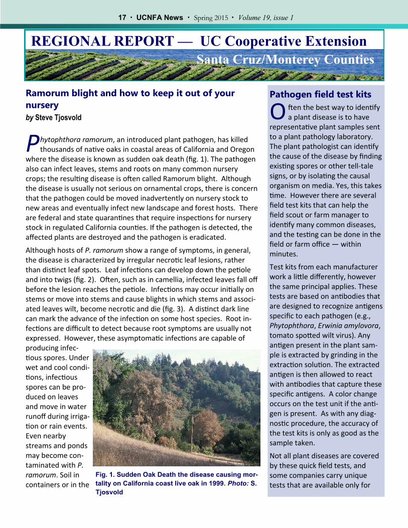
P hytophthora ramorum, an introduced plant pathogen, has killed thousands of native oaks in coastal areas of California and Oregon
where the disease is known as sudden oak death (fig. 1). The pathogen also can infect leaves, stems and roots on many common nursery crops; the resulting disease is often called Ramorum blight. Although the disease is usually not serious on ornamental crops, there is concern that the pathogen could be moved inadvertently on nursery stock to new areas and eventually infect new landscape and forest hosts. There are federal and state quarantines that require inspections for nursery stock in regulated California counties. If the pathogen is detected, the affected plants are destroyed and the pathogen is eradicated.
Although hosts of P. ramorum show a range of symptoms, in general, the disease is characterized by irregular necrotic leaf lesions, rather than distinct leaf spots. Leaf infections can develop down the petiole and into twigs (fig. 2). Often, such as in camellia, infected leaves fall off before the lesion reaches the petiole. Infections may occur initially on stems or move into stems and cause blights in which stems and associ-ated leaves wilt, become necrotic and die (fig. 3). A distinct dark line can mark the advance of the infection on some host species. Root in-fections are difficult to detect because root symptoms are usually not expressed. However, these asymptomatic infections are capable of producing infec-tious spores. Under wet and cool condi-tions, infectious spores can be pro-duced on leaves and move in water runoff during irriga-tion or rain events. Even nearby streams and ponds may become con-taminated with P. ramorum. Soil in containers or in the
Pathogen field test kits
O ften the best way to identify a plant disease is to have
representative plant samples sent to a plant pathology laboratory. The plant pathologist can identify the cause of the disease by finding existing spores or other tell-tale signs, or by isolating the causal organism on media. Yes, this takes time. However there are several field test kits that can help the field scout or farm manager to identify many common diseases, and the testing can be done in the field or farm office — within minutes.
Test kits from each manufacturer work a little differently, however the same principal applies. These tests are based on antibodies that are designed to recognize antigens specific to each pathogen (e.g., Phytophthora, Erwinia amylovora, tomato spotted wilt virus). Any antigen present in the plant sam-ple is extracted by grinding in the extraction solution. The extracted antigen is then allowed to react with antibodies that capture these specific antigens. A color change occurs on the test unit if the anti-gen is present. As with any diag-nostic procedure, the accuracy of the test kits is only as good as the sample taken.
Not all plant diseases are covered by these quick field tests, and some companies carry unique tests that are available only for
Fig. 1. Sudden Oak Death the disease causing mor-
tality on California coast live oak in 1999. Photo: S.
field can be infested with long-lived chlamydospores and mycelium in plant de-bris and fallen leaves.
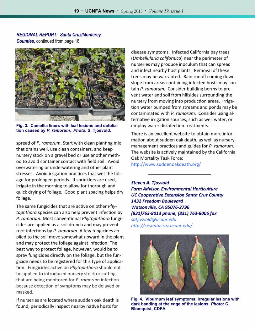
Five genera of ornamental hosts have accounted for most of the P. ramorum official detections in nurseries: Rhododendron, Camellia, Pieris, Kalmia and Viburnum (fig. 4). However, P. ramorum has a wide host range, and other hosts could be im-portant carriers of the pathogen in the nursery trade. For the complete list of known hosts, see the currently identified hosts by USDA APHIS: (http://www.aphis.usda.gov/plant_health/plant_pest_info/pram/downloads/pdf_files/DA-2013-41.pdf).
Management of this disease begins with preventing it from being in-troduced into the nursery. First, make sure you are buying propaga-tion plant material or other plants from trusted suppliers. The Califor-nia Department of Food and Agriculture maintains lists of California suppliers that are inspected and approved to ship nursery stock. See: http://www.cdfa.ca.gov/plant/PE/InteriorExclusion/SuddenOakDeath/index.html. For high-risk incoming shipments, unload nursery stock in an area of the nursery that can be cleaned of leafy debris. Sweep de-bris from the receiving area and delivery truck and bag for disposal. Loading and delivery areas should be isolated from production areas. Infected leafy debris can be blown into production areas by the wind.
Symptoms on plants introduced to the nursery are not always readily apparent at first. These plants can be isolated away from normal pro-duction areas and systematically monitored for symptoms about once a week for several weeks. Symptoms of many nursery hosts can be found at the website given below. Field diagnostic kits from AgDia (Elkart, IN) or Neogen (Lansing, MI) can aid in detecting Phytophthora species. These Phytophthora kits do not detect specific species, but can detect numerous Phytophthora species including P. ramorum. See side bar, “Pathogen Field Kits.”
In general, the same cultural management practices that help control other Phytophthora species will also help control the introduction and
REGIONAL REPORT: Santa Cruz/Monterey
Counties, continued from page 17
Pathogen field test kits (cont’d)
processing large numbers of sam-ples.
ImmunoStrips from Agdia Inc. They carry field test strips for Phy-tophthora (fig. 1), and many bac-teria and viruses. Contact: 1-800-622-4342 http://www.agdia.com. Recent tests by CDFA Plant Pathologist Susan Lathram indi-cate that these test strips will also detect root infections of Phy-tophthora tentaculata, a new root pathogen of concern to the nurse-ry industry, especially those that produce plants for restoration in natural systems. See:
http://ucanr.edu/p/50127 Alert Kits from Neogen Company. They carry field test kits for the common root pathogens: Phy-tophthora, Pythium and Rhi-zoctonia. Also they carry several other bacteria and virus test kits. Contact: 800/477-8201 http://www.neogen.com/PlantDiagnostics/index.html.
Fig. 3. Camellia liners with leaf lesions and defolia-tion caused by P. ramorum. Photo: S. Tjosvold.
spread of P. ramorum. Start with clean planting mix that drains well, use clean containers, and keep nursery stock on a gravel bed or use another meth-od to avoid container contact with field soil. Avoid overwatering or underwatering and other plant stresses. Avoid irrigation practices that wet the foli-age for prolonged periods. If sprinklers are used, irrigate in the morning to allow for thorough and quick drying of foliage. Good plant spacing helps dry foliage.
The same fungicides that are active on other Phy-tophthora species can also help prevent infection by P. ramorum. Most conventional Phytophthora fungi-cides are applied as a soil drench and may prevent root infections by P. ramorum. A few fungicides ap-plied to the soil move somewhat upward in the plant and may protect the foliage against infection. The best way to protect foliage, however, would be to spray fungicides directly on the foliage, but the fun-gicide needs to be registered for this type of applica-tion. Fungicides active on Phytophthora should not be applied to introduced nursery stock or cuttings that are being monitored for P. ramorum infection because detection of symptoms may be delayed or masked.
If nurseries are located where sudden oak death is found, periodically inspect nearby native hosts for
Fig. 4. Viburnum leaf symptoms. Irregular lesions with dark banding at the edge of the lesions. Photo: C. Blomquist, CDFA.
disease symptoms. Infected California bay trees (Umbellularia californica) near the perimeter of nurseries may produce inoculum that can spread and infect nearby host plants. Removal of these trees may be warranted. Rain runoff coming down slope from areas containing infected hosts may con-tain P. ramorum. Consider building berms to pre-vent water and soil from hillsides surrounding the nursery from moving into production areas. Irriga-tion water pumped from streams and ponds may be contaminated with P. ramorum. Consider using al-ternative irrigation sources, such as well water, or employ water disinfection treatments.
There is an excellent website to obtain more infor-mation about sudden oak death, as well as nursery management practices and guides for P. ramorum. The website is actively maintained by the California Oak Mortality Task Force: http://www.suddenoakdeath.org/ Steven A. Tjosvold Farm Advisor, Environmental Horticulture UC Cooperative Extension Santa Cruz County 1432 Freedom Boulevard Watsonville, CA 95076-2796 (831)763-8013 phone, (831) 763-8006 fax [email protected] http://cesantacruz.ucanr.edu/
REGIONAL REPORT — UC Cooperative Extension Ventura County
Bot fungi wreak havoc during drought
by Jim Downer
N urseries growing woody plants often suffer with disease caused by Botryosphaeria and
affiliated asexual stages of Botryosphaeria such as Fussicoccum, Neofussicoccum and Dothiorella (Bot fungi). These fungi are common in landscapes and wildlands and take advantage of drought-stressed plants. Bot fungi produce cankers and infect stems and branches with diameters from pencil sized up to many inches. Bot fungi infect either as conidia from the asexual stages of the fungus (fig. 1) or as ascospores from the Botryosphaeria stages. These spores are univer-sally produced in dead or dying tissues on diseased plants. Spores are splashed in water, wind borne, or moved in brush, clipping or trimmings of diseased plants. Ascospores and conidia germinate readily at 28 to 32oC (82 to 90oF) (Sutton 1990), suggesting that current warm conditions in California could sig-nal the onset of a difficult year with this pathogen.
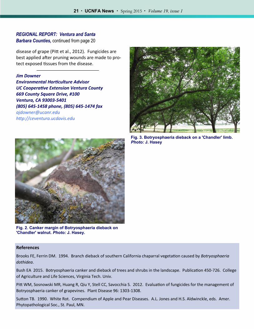
Germinating spores can enter directly into stems through lenticels or through wounds made during pruning or other injuries. The various Bot fungi cause cankers (fig. 2) and stem lesions on susceptible woody plants, and eventually girdle stems, resulting in foliage symptoms of yel-lowing to browning leaves and flagging or dead branches in shrubs and trees (fig. 3). Sometimes the tips of plants are affected, especially if plants are hedged or wounded repeatedly. Bot fungi seem to attack drought-stressed plants, producing large amounts of inoculum under drought conditions in the dead and dying portions of affected plants. Bot-ryosphaeria dothidea is the most commonly ob-served species but there are about 200 species worldwide affecting thousands of hosts. Native shrubs such as ceanothus, mountain mahogany, and manzanita are preferred hosts, especially following or during drought (Brooks and Ferrin, 1994). Bot fungi also affect trees such as oak, alder, redwood, avocado, maple and apple, both in production and in landscapes, though this disease is typically more se-rious in landscape situations where trees are not irri-gated. In nursery production, preventing drought stress by consistent irrigation helps to preclude infection by bot fungi but does not totally prevent it in very sus-ceptible species. Rogueing out infested plant mate-rial or pruning out infected branches helps to reduce inoculum. On larger specimens, pruning out dead-wood is essential to controlling the disease (Bush, 2015), as the fungus usually sporulates in dead-wood. Fungicides are available for control of Bot fungi but labeling must be checked before applica-tion. Fludioxonil, carbendazim, fluazinam, tebucona-zole, flusilazole, penconazole, procymidone, iprodi-one, myclobutanil, and pyraclostrobin have all been shown to be effective in controlling Botryosphaeria
Fig. 1. Black and white fruiting bodies of Bot fungi that produce asexual spores (conidia) on walnut. Pho-to: T. Michailides
Brooks FE, Ferrin DM. 1994. Branch dieback of southern California chaparral vegetation caused by Botryosphaeria
dothidea.
Bush EA 2015. Botryosphaeria canker and dieback of trees and shrubs in the landscape. Publication 450-726. College
of Agriculture and Life Sciences, Virginia Tech. Univ.
Pitt WM, Sosnowski MR, Huang R, Qiu Y, Stell CC, Savocchia S. 2012. Evaluation of fungicides for the management of
Botryosphaeria canker of grapevines. Plant Disease 96: 1303-1308.
Sutton TB. 1990. White Rot. Compendium of Apple and Pear Diseases. A.L. Jones and H.S. Aldwinckle, eds. Amer.
Phytopathological Soc., St. Paul, MN.
disease of grape (Pitt et al., 2012). Fungicides are best applied after pruning wounds are made to pro-tect exposed tissues from the disease. Jim Downer Environmental Horticulture Advisor UC Cooperative Extension Ventura County 669 County Square Drive, #100 Ventura, CA 93003-5401 (805) 645-1458 phone, (805) 645-1474 fax [email protected] http://ceventura.ucdavis.edu
REGIONAL REPORT: Ventura and Santa
Barbara Counties, continued from page 20
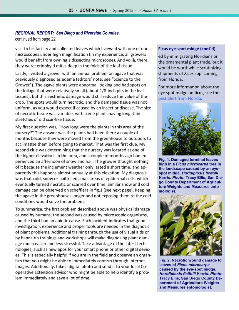
Fig. 2. Canker margin of Botryosphaeria dieback on 'Chandler' walnut. Photo: J. Hasey.
Fig. 3. Botryosphaeria dieback on a 'Chandler' limb. Photo: J. Hasey
I ’m sure you’ve heard of the “bad things always come in threes” rule. Seems like every time a famous actor or actress dies, everyone starts
looking for two other celebrity deaths to keep this superstition alive. If you search the Internet, you will find lots of information related to this popular belief and the general subject of things that occur in threes, such as the blog “45 wonderful things that come in threes,” written in honor of the publishing date (12/12/12) with the same number repeat-ed thrice. In this short article, I want to provide you with my own ver-sion of occurrences in threes with three recent stories about plant damage diagnoses that emphasize the need and the importance for good observation skills, good diagnostic tools and good resources.
First, my favorite story begins with my son. We live in a rural area with a large backyard where he likes to shoot his airsoft rifle at paper tar-gets and cans. Watching my son has taught me a lot about airsoft, and I obviously have had to clean up thousands of pellets from our patio and landscape. Keep this in mind as a preface to this ornamental plant pro-duction problem that I diagnosed in San Diego County.
I received a call from a bird-of-paradise cut flower grower who lives in a rural area, and he grows many of his plants along his lengthy drive-way. He called about what he thought were “big insect eggs” embed-ded in the plant stalks and leaf petioles. I asked if he would send me a digital photo because I can usually identify things like that relatively easily. Sure enough, I saw what indeed looked like white eggs laid in rows up the plant from the base to the top of the petioles in the photo he sent me. However, my experience with my son taught me better. The objects that looked like eggs shoved into the tissues were actually airsoft pellets, which had penetrated the stringy, vertical plant tissues and spread them open side to side. After discussing this with the grow-er, he confirmed that the pests in question were Homo sapiens Linnae-us, the neighborhood children, not big insects.
The second story is about a grower who was concerned that he had a disease on his young queen palms. Many of the palm leaves were bent in half, near the middle of the leaf, and did not fully unfold. He thought it was a disease because he couldn’t see a living organism there, even with a hand lens. No matter what he did to change the environmental conditions or what he sprayed, he couldn’t solve the problem. I made a
Field Observations
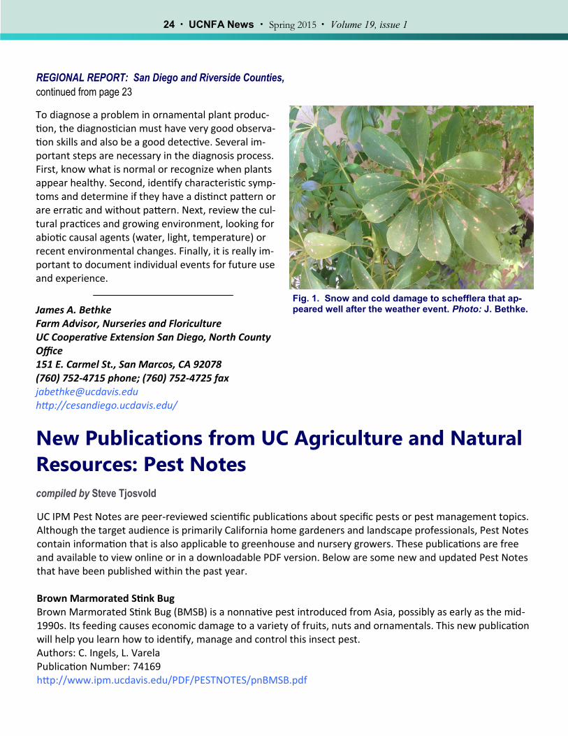
Ficus eye-spot midge
R ecently, a new established pest has been confirmed and
observed in the cities of San Die-go, Rancho Santa Fe, Del Mar and LaJolla. Dr. Tracy Ellis, San Diego County Department of Agriculture Weights and Measures entomolo-gist, sent samples of Ficus micro-carpa from the landscape (fig. 1) and a local nursery to the Califor-nia Department of Food and Agri-culture Plant Pest Diagnostics Lab, and the pest was identified as an eye-spot midge (fly species, family Cecidomyiidae), Horidiplosis ficifo-lii Harris. This is the first report of this pest in California, but it was found in Florida in 2008. Inter-estingly, it was found on F. micro-carpa in Florida and not on F. ben- jamina growing directly below the F. microcarpa. It was first de-scribed in 2003, and originates from China, Taiwan and Japan.
The tiny fly maggot lives inside the blister-like gall (fig. 2), eventu-ally causing a necrotic spot that in some cases resembles an eyespot, a dark center with a light-colored ring around it. Each spot contains one midge larva. As the damage progresses, leaves begin to drop from the trees.
It appears that a couple of ficus pests moved here from Florida in the last few years. It’s hard to say whether they are being transport-
REGIONAL REPORT: San Diego and Riverside Counties,
continued from page 22
Ficus eye-spot midge (cont’d)
ed by immigrating Floridians or the ornamental plant trade, but it would be worthwhile scrutinizing shipments of Ficus spp. coming from Florida.
For more information about the eye spot midge on ficus, see the pest alert from Florida.
Fig. 2. Necrotic wound damage to leaves of Ficus microcarpa caused by the eye-spot midge, Horidiplosis ficifolii Harris. Photo: Tracy Ellis, San Diego County De-partment of Agriculture Weights and Measures entomologist.
visit to his facility and collected leaves which I viewed with one of our microscopes under high magnification (in my experience, all growers would benefit from owning a dissecting microscope). And voilà, there they were: eriophyid mites deep in the folds of the leaf tissue.
Lastly, I visited a grower with an annual problem on agave that was previously diagnosed as edema (editors’ note: see “Science to the Grower”). The agave plants were abnormal looking and had spots on the foliage that were relatively small (about 1/8-inch pits in the leaf tissues), but this aesthetic damage would still reduce the value of the crop. The spots would turn necrotic, and the damaged tissue was not uniform, as you would expect if caused by an insect or disease. The size of necrotic tissue was variable, with some plants having long, thin stretches of old scar-like tissue.
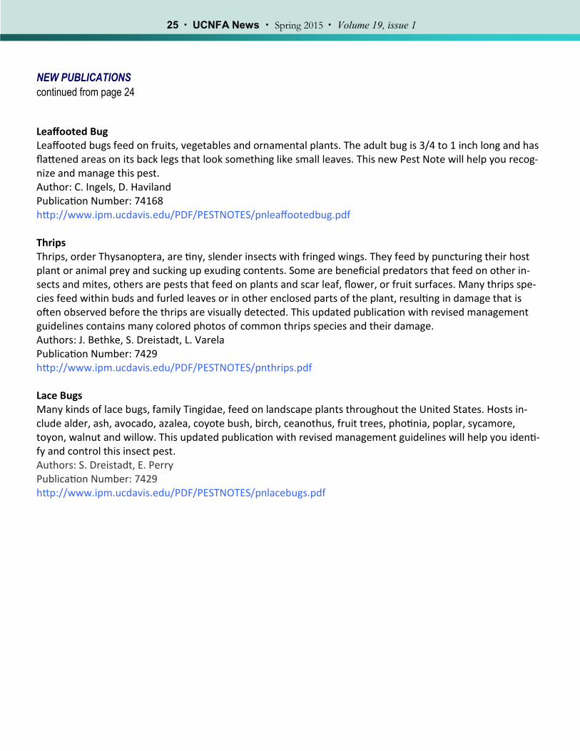
My first question was, “How long were the plants in this area of the nursery?” The answer was the plants had been there a couple of months because they were moved from the greenhouse to outdoors to acclimatize them before going to market. That was the first clue. My second clue was determining that the nursery was located at one of the higher elevations in the area, and a couple of months ago had ex-perienced an afternoon of snow and hail. The grower thought nothing of it because the inclement weather only lasted a short time, and ap-parently this happens almost annually at this elevation. My diagnosis was that cold, snow or hail killed small areas of epidermal cells, which eventually turned necrotic or scarred over time. Similar snow and cold damage can be observed on schefflera in fig.1 (see next page). Keeping the agave in the greenhouses longer and not exposing them to the cold conditions would solve the problem.
To summarize, the first problem described above was physical damage caused by humans, the second was caused by microscopic organisms, and the third had an abiotic cause. Each incident indicates that good investigation, experience and proper tools are needed in the diagnosis of plant problems. Additional training through the use of visual aids or by hands-on trainings and workshops will make diagnosing plant dam-age much easier and less stressful. Take advantage of the latest tech-nologies, such as new apps for your smart phone or other digital devic-es. This is especially helpful if you are in the field and observe an organ-ism that you might be able to immediately confirm through Internet images. Additionally, take a digital photo and send it to your local Co-operative Extension advisor who might be able to help identify a prob-lem immediately and save a lot of time.
Fig. 1. Damaged terminal leaves high in a Ficus microcarpa tree in the landscape caused by an eye-spot midge, Horidiplosis ficifolii Harris. Photo: Tracy Ellis, San Die-go County Department of Agricul-ture Weights and Measures ento-mologist.
REGIONAL REPORT: San Diego and Riverside Counties,
continued from page 23
To diagnose a problem in ornamental plant produc-tion, the diagnostician must have very good observa-tion skills and also be a good detective. Several im-portant steps are necessary in the diagnosis process. First, know what is normal or recognize when plants appear healthy. Second, identify characteristic symp-toms and determine if they have a distinct pattern or are erratic and without pattern. Next, review the cul-tural practices and growing environment, looking for abiotic causal agents (water, light, temperature) or recent environmental changes. Finally, it is really im-portant to document individual events for future use and experience.
James A. Bethke Farm Advisor, Nurseries and Floriculture UC Cooperative Extension San Diego, North County Office 151 E. Carmel St., San Marcos, CA 92078 (760) 752-4715 phone; (760) 752-4725 fax [email protected] http://cesandiego.ucdavis.edu/
UC IPM Pest Notes are peer-reviewed scientific publications about specific pests or pest management topics. Although the target audience is primarily California home gardeners and landscape professionals, Pest Notes contain information that is also applicable to greenhouse and nursery growers. These publications are free and available to view online or in a downloadable PDF version. Below are some new and updated Pest Notes that have been published within the past year. Brown Marmorated Stink Bug Brown Marmorated Stink Bug (BMSB) is a nonnative pest introduced from Asia, possibly as early as the mid-1990s. Its feeding causes economic damage to a variety of fruits, nuts and ornamentals. This new publication will help you learn how to identify, manage and control this insect pest. Authors: C. Ingels, L. Varela Publication Number: 74169 http://www.ipm.ucdavis.edu/PDF/PESTNOTES/pnBMSB.pdf
New Publications from UC Agriculture and Natural
Resources: Pest Notes
compiled by Steve Tjosvold
Fig. 1. Snow and cold damage to schefflera that ap-peared well after the weather event. Photo: J. Bethke.
Leaffooted Bug Leaffooted bugs feed on fruits, vegetables and ornamental plants. The adult bug is 3/4 to 1 inch long and has flattened areas on its back legs that look something like small leaves. This new Pest Note will help you recog-nize and manage this pest. Author: C. Ingels, D. Haviland Publication Number: 74168 http://www.ipm.ucdavis.edu/PDF/PESTNOTES/pnleaffootedbug.pdf Thrips Thrips, order Thysanoptera, are tiny, slender insects with fringed wings. They feed by puncturing their host plant or animal prey and sucking up exuding contents. Some are beneficial predators that feed on other in-sects and mites, others are pests that feed on plants and scar leaf, flower, or fruit surfaces. Many thrips spe-cies feed within buds and furled leaves or in other enclosed parts of the plant, resulting in damage that is often observed before the thrips are visually detected. This updated publication with revised management guidelines contains many colored photos of common thrips species and their damage. Authors: J. Bethke, S. Dreistadt, L. Varela Publication Number: 7429 http://www.ipm.ucdavis.edu/PDF/PESTNOTES/pnthrips.pdf Lace Bugs Many kinds of lace bugs, family Tingidae, feed on landscape plants throughout the United States. Hosts in-clude alder, ash, avocado, azalea, coyote bush, birch, ceanothus, fruit trees, photinia, poplar, sycamore, toyon, walnut and willow. This updated publication with revised management guidelines will help you identi-fy and control this insect pest. Authors: S. Dreistadt, E. Perry Publication Number: 7429 http://www.ipm.ucdavis.edu/PDF/PESTNOTES/pnlacebugs.pdf
UCNFA News is published by the University of California Nursery and Floriculture Alliance, a statewide partnership of researchers and educators,
growers, floriculture associations and allied industry.
UCNFA Directors:
Loren Oki, UC Cooperative Extension Specialist for Landscape Horticulture, UC Davis
David Fujino, Executive Director, California Center for Urban Horticulture (CCUH)
Website - http://ucanr.edu/sites/UCNFA
Reproducing and distributing material from this newsletter is encouraged provided credit is given to the
author and UCNFA
Managing Editor: Steve Tjosvold, UC Cooperative Extension Monterey & Santa Cruz counties
Co-Editor: Julie Newman, UC Cooperative Extension Ventura and Santa Barbara counties
Layout and Design:
Linda Dodge, Plant Sciences Dept., UC Davis
Editorial Committee:
James Bethke, UC Cooperative Extension San Diego County
Maria de la Fuente, UC Cooperative Extension Santa Clara & San Benito counties
Don Merhaut, UC Cooperative Extension Specialist for Nursery and Floriculture Crops, UC Riverside
A. James Downer, UC Cooperative Extension Ventura County
To simplify information, trade names of products may have been used in this publication. No endorsement of named or illustrated products is intended, nor is criticism implied of similar products that are not mentioned or illustrated. ANR NONDISCRIMINATION AND AFFIRMATIVE ACTION POLICY STATEMENT
The University of California prohibits discrimination or harassment of any person in any of its programs or activities. The complete nondiscrimina-tion policy statement can be found at http://ucanr.edu/sites/anrstaff/files/1258.pdf. Inquiries regarding the University’s equal employment op-portunity policies may be directed to Linda Marie Manton, Affirmative Action Contact, University of California, Davis, Agriculture and Natural Re-sources, One Shields Avenue, Davis, CA 95616, (530) 752-0495.