31
Diabetes and the ocular surface Insight into the systemic disease Maria Markoulli MOptom PhD FBCLA FAAO GradCertOcTher NZAO: 9 th October 2015 Dunedin, NZ No Commercial Relationships

Diabetes and the ocular surface Insight into the systemic disease
Maria Markoulli MOptom PhD FBCLA FAAO GradCertOcTher
NZAO: 9th October 2015 Dunedin, NZ
No Commercial Relationships

Rupert Myers Building, UNSW
Sydney, Australia UNSW Australia

Vision hub at UNSW Australia

The South Island
Diabetes and the Ocular Surface
1. Diabetes: the systemic disease
2. Neuropathy
3. Corneal neuropathy
4. Ocular surface integrity

Diabetes is a chronic disease that occurs when the pancreas does not produce enough insulin, or when the body cannot effectively use the insulin it produces. Hyperglycaemia, or raised blood sugar, is a common effect of uncontrolled diabetes and over time leads to serious damage to many of the body's systems, especially the nerves and blood vessels.
- World Health Organisation
Diabetes

Diabetes
Diagnosis • Fasting venous plasma
glucose: ≥7.0mmol/l • 2 hours after ingestion of
75g oral glucose load: ≥11.1mmol/l
Prevalence • NZ prevalence: 257,7761
• Worldwide: 173 million people2 and continues to be on the rise3
1. Ministry of Health, 2014 2. WHO, 2002 3. Seidell JC.. Br J Nutr 2000;83 Suppl 1:S5-8
www.clipartsheep.com
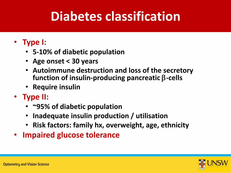
Diabetes classification
• Type I: • 5-10% of diabetic population • Age onset < 30 years • Autoimmune destruction and loss of the secretory
function of insulin-producing pancreatic b-cells • Require insulin
• Type II: • ~95% of diabetic population • Inadequate insulin production / utilisation • Risk factors: family hx, overweight, age, ethnicity
• Impaired glucose tolerance

• Multi-system disease
• Premature mortality
• Macrovascular complications
• Microvascular complications
oRetinopathy
- Retinal capillary damage
- Progressive capillary occlusion retinal ischaemia new vessels
oNephropathy
oNeuropathy
Systemic impact of diabetes
1. Charnogursky G, Lee H, Lopez N. Diabetic neuropathy. Handbook of clinical neurology 2014; 120: 773-785

Diabetic Peripheral Neuropathy
• 60-70% of people with diabetes
• “chronic, symmetrical, length-dependent diabetic sensorimotor polyneuropathy”1
• Foot ulceration-> 7% of patients 2
– lower limb amputation, severe pain
– Significant quality of life costs and financial burden (28K USD at 2 years!)3
• Early and accurate detection
The Glenn A. Fry Award Lecture 2010: Ophthalmic Markers of Diabetic Neuropathy. Efron, Nathan Optometry & Vision Science. 88(6):661-683, June 2011.
1. Toronto Diabetic Neuropathy Expert Group
2. Tavakoli M, Mitu-Pretorian M, Petropoulos IN et al. Diabetes 2013.
3. Ramsey SD, Newton K, Blough D et al. Diabetes care 1999.
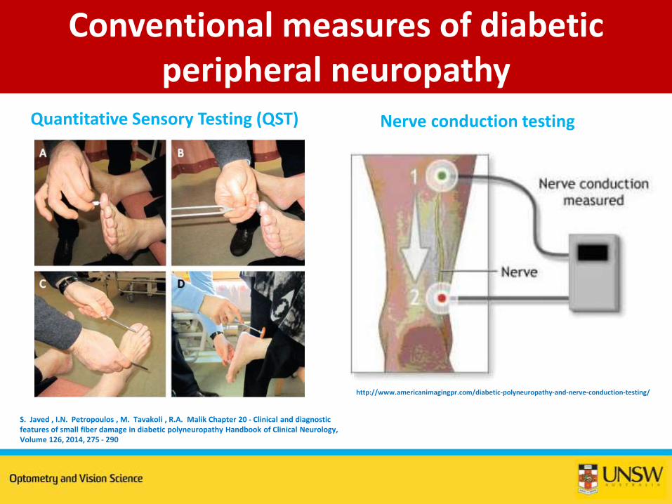
Conventional measures of diabetic peripheral neuropathy
http://www.americanimagingpr.com/diabetic-polyneuropathy-and-nerve-conduction-testing/
Quantitative Sensory Testing (QST) Nerve conduction testing
S. Javed , I.N. Petropoulos , M. Tavakoli , R.A. Malik Chapter 20 - Clinical and diagnostic features of small fiber damage in diabetic polyneuropathy Handbook of Clinical Neurology, Volume 126, 2014, 275 - 290
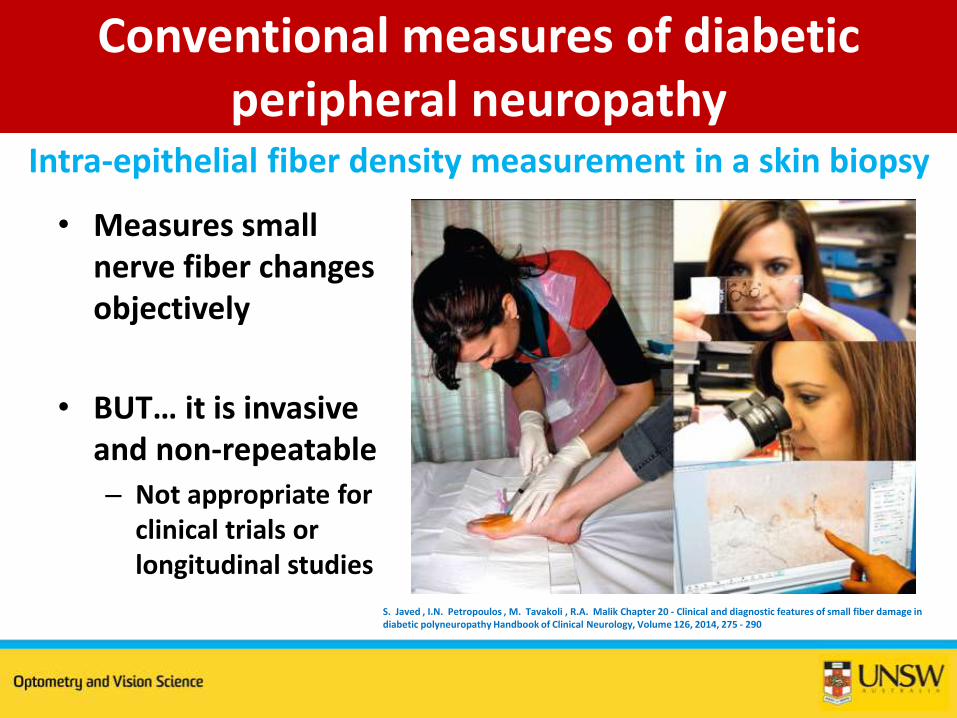
Conventional measures of diabetic peripheral neuropathy
• Measures small nerve fiber changes objectively
• BUT… it is invasive and non-repeatable
– Not appropriate for clinical trials or longitudinal studies
Intra-epithelial fiber density measurement in a skin biopsy
S. Javed , I.N. Petropoulos , M. Tavakoli , R.A. Malik Chapter 20 - Clinical and diagnostic features of small fiber damage in diabetic polyneuropathy Handbook of Clinical Neurology, Volume 126, 2014, 275 - 290

Corneal confocal microscopy
• Only organ where we can directly visualise nerves in vivo
• Single points of tissue simultaneously illuminated & imaged in the same plane
• Resulting image very high in resolution1
• 500× magnification • 400 x 400 mm dimension
1. Efron N. The Glenn A. Fry award lecture 2010: Ophthalmic markers of diabetic neuropathy. Optometry and vision science : official publication of the American Academy of Optometry 2011; 88: 661-683.
Ph
oto
co
urt
esy
: Dr
Vin
od
Mas
ee
du
pal
ly

• Most densely innervated organ in the body1
• 100x more sensitive than the conjunctiva1
• Derived from the ophthalmic division of the trigeminal nerve
Corneal nerve structure and function
1. Cruzat A, Pavan-Langston D, Hamrah P. In vivo confocal microscopy of corneal nerves: analysis and clinical correlation. Seminars in ophthalmology 2010; 25: 171-177.
http://www.edoctoronline.com/medical-atlas.asp?c=4&id=21956

Corneal nerve structure and function
• Nerve bundles enter the cornea at the limbus parallel to the corneal surface
• Penetrate Bowman’s layer to form the corneal subbasal nerve plexus
The Glenn A. Fry Award Lecture 2010: Ophthalmic Markers of Diabetic Neuropathy. Efron, Nathan Optometry & Vision Science. 88(6):661-683, June 2011.
Corneal subbasal
nerve plexus
The Glenn A. Fry Award Lecture 2010: Ophthalmic Markers of Diabetic Neuropathy. Efron, Nathan Optometry & Vision Science. 88(6):661-683, June 2011.

Corneal confocal microscopy
• Nerve fibre bundle density • Nerve branch density • Nerve fibre length • Nerve fibre width • Nerve fibre tortuosity


Corneal montage
1. Edwards K, Pritchard N, Gosschallk et al, Cornea, 2012;31:1078–1082
Diabetes WITH neuropathy Diabetes WITHOUT neuropathy
Nerve mapping technique originally developed by Patel and McGhee

Corneal montage
1. Edwards K, Pritchard N, Gosschallk et al, Cornea, 2012;31:1078–1082
Diabetes WITH neuropathy Diabetes WITHOUT neuropathy

Corneal confocal microscopy
Diabetes with neuropathy:
Corneal nerve density
corneal nerve length
corneal nerve branch density1,2
Corneal nerve parameters correlate well with nerve fibre loss in skin biopsy1
Can be used to detect and stratify the severity of diabetic peripheral neuropathy
The Glenn A. Fry Award Lecture 2010: Ophthalmic Markers of
Diabetic Neuropathy. Efron, Nathan Optometry & Vision Science.
88(6):661-683, June 2011.
1. Edwards K, Pritchard N, Vagenas D et al. Clinical & Experimental Optometry, 2012; 95: 348-354.
2. Dehgani C, Pritchard N, Edwards K et al. Cornea, 2014; 55: 7982-7990
Normal
Severe
diabetic
neuropathy

• Corneal nerve length can predict peripheral neuropathy1
o 90 px with type 1 diabetes and no DPN
o Corneal nerve length could predict DPN incidence with 63% sensitivity and 74% specificity
Predicting diabetic peripheral neuropathy
1. Pritchard N, Edwards K, Russell AW et al. Diabetes care 2015.
2. Dehghani C, Pritchard N, Edwards K et al. Cornea, 2014.
Healthy normals
Diabetes – NO neuropathy
Diabetes – WITH neuropathy

Corneal nerve regeneration - After pancreas and kidney transplantation-
1. Tavakoli M, Mitu-Pretorian M, Petropoulos IN et al. Corneal
confocal microscopy detects early nerve regeneration in
diabetic neuropathy after simultaneous pancreas and kidney
transplantation. Diabetes 2013; 62: 254-260.
1. Corneal nerve markers are more sensitive
2. Corneal nerves can regenerate

Corneal nerve function
Corneal nerves • Trophic support to epithelial cells, lacrimal gland, and goblet
cells • Stimulate cell growth, mitosis, differentiation and migration Epithelial cells • Trophic support to neurons • Secrete growth factors • Promote neurite extension In diabetes • Reduction in these mediators • Disruption in epithelial integrity - risk of corneal erosions • Neurotrophic keratopathy
1. Muller LJ, Marfurt CF, Kruse F et al. Corneal nerves: structure, contents and function.
Experimental eye research 2003; 76: 521-542.
2. Lambiase A, Micera A, Sacchetti M et al. Alterations of tear neuromediators in dry eye
disease. Archives of ophthalmology 2011; 129: 981-986.

• dry eye signs & symptoms
• severity with diabetes severity
• Tear production
• Tear film stability
• goblet cell density
Dry eye in diabetes
Yin et al., Invest Ophthalmol Vis Sci. 2011;52:6589–6596

Neurotrophic keratopathy
Impaired corneal sensitivity
Epithelial breakdown
Delayed wound healing
Corneal ulceration
Vision loss1
These signs increase with neuropathic severity, and severity of diabetes2
1. Alves Mde C, Carvalheira JB, Modulo CM et al. Tear film and ocular surface changes in diabetes mellitus. Arquivos brasileiros de oftalmologia 2008; 71: 96-103.
2. O'Donnell C, Efron N. Diabetes and contact lens wear. Clinical & experimental optometry : journal of the Australian Optometrical Association 2012; 95: 328-337.

Neurotrophic keratopathy
Yin et al., Invest Ophthalmol Vis Sci. 2011;52:6589–6596

Standard treatment
• Preservative free lubricants
• Minimising evaporation – punctal plugs
• Topical antibiotics
• Bandage CL
• Patching
• Tarsorrhaphy / induced ptosis

• Insulin-like growth factor-1 (IGF-1)
– Mediates proliferation and differentiation
• Substance P
– Neurotransmitter
– Reduced in eyes with hypoesthesia
• IGF-1 and Substance P
– In vitro: stimulate epithelial migration
– In vivo: effective in treating neurotrophic ulcers1 and superficial punctate keratitis2
Growth Factors
1. Abdelkader, Clinical and Experimental Ophthalmology 2011; 39: 259–270
2. Chikamoto et al, Jpn J Ophthalmol 2009;53:464–469

• Nerve Growth Factor
– Modulates ocular inflammation
– Corneal epithelial proliferation and differentiation
– Wound healing promoted1
– No relapse1
Growth Factors
Before NGF After NGF
Bonini et al, Ophthalmology 2000;107:1347–1352
1. Bonini et al, Ophthalmology 2000;107:1347–1352
2. Abdelkader, Clinical and Experimental Ophthalmology 2011; 39: 259–270

Summary
• Expect greater application of the confocal microscopy in both research and clinical settings
• Beware of diabetic ocular surface disease
• Future treatments:
– NGF
– IGF & Substance P
• Expect expanded role of optometry in the management of diabetic peripheral neuropathy

Acknowledgements
Jenny Wu Canberra


