Tumour markers Tumour markers Marta Kalousová Institute of Medical Biochemistry and Laboratory Diagnostics, 1 st Faculty of Medicine and General University Hospital, Charles University, Prague
Transcript
Tumour markersTumour markers
Marta KalousováInstitute of Medical Biochemistry and Laboratory
Diagnostics,
1st Faculty of Medicine and General University Hospital, Charles University, Prague
Laboratory examination in Laboratory examination in
• Tissue and organ specific antigens• Tissue and organ specific antigens
• Non-specific antigens
Oncofetal antigensOncofetal antigens
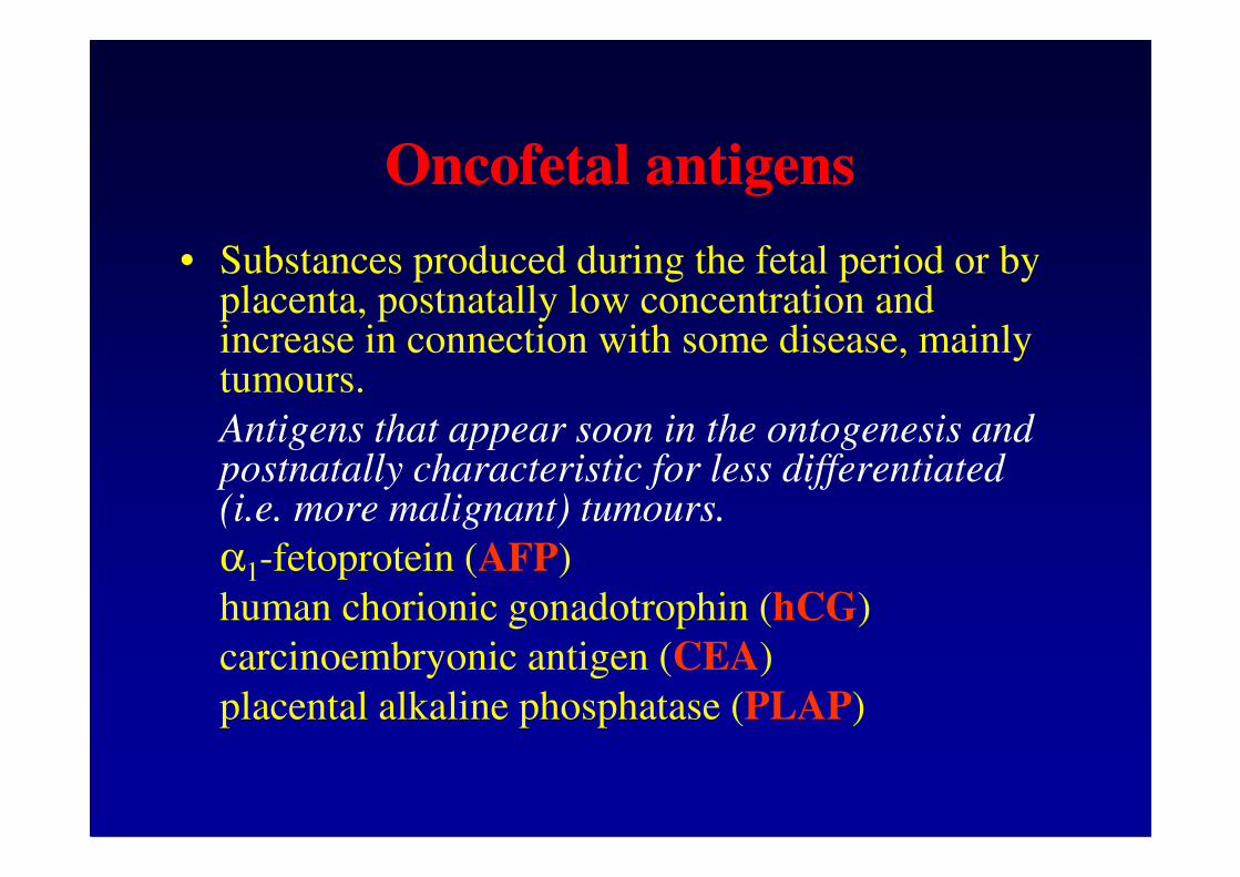
• Substances produced during the fetal period or by placenta, postnatally low concentration and increase in connection with some disease, mainly tumours.
Antigens that appear soon in the ontogenesis and Antigens that appear soon in the ontogenesis and postnatally characteristic for less differentiated (i.e. more malignant) tumours.
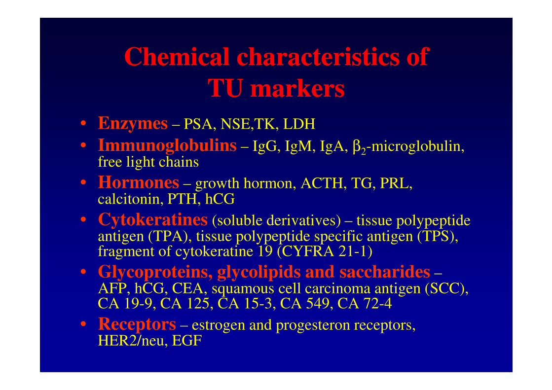
α1-fetoprotein (AFP)
human chorionic gonadotrophin (hCG)
carcinoembryonic antigen (CEA)
placental alkaline phosphatase (PLAP)
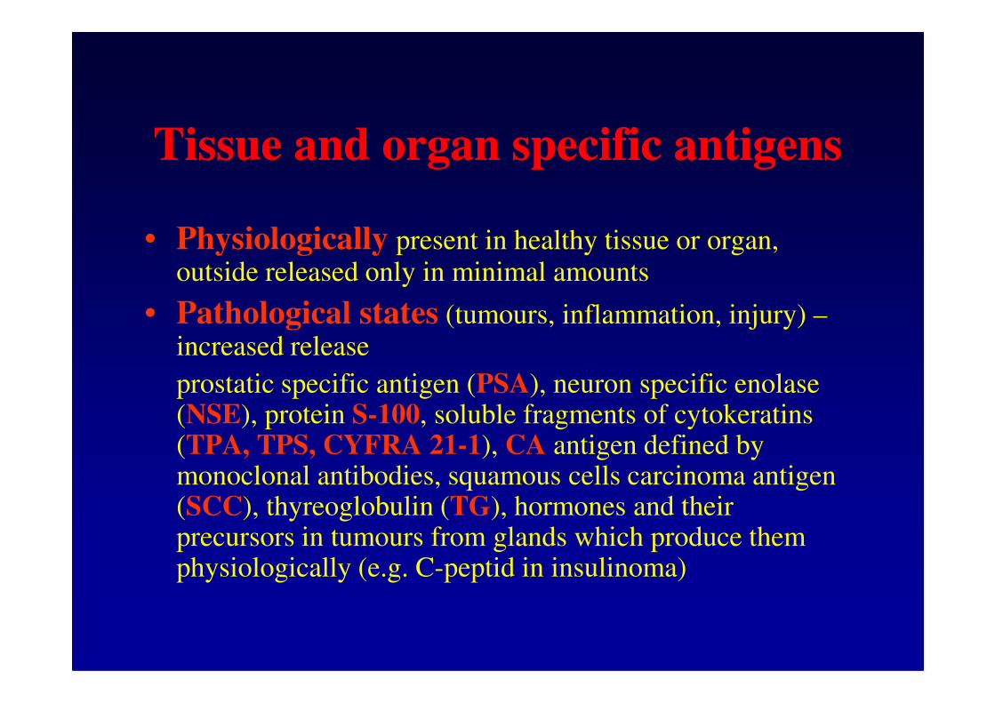
Tissue and organ specific antigensTissue and organ specific antigens
• Physiologically present in healthy tissue or organ, outside released only in minimal amounts
• Pathological states (tumours, inflammation, injury) –increased release increased release
prostatic specific antigen (PSA), neuron specific enolase (NSE), protein S-100, soluble fragments of cytokeratins (TPA, TPS, CYFRA 21-1), CA antigen defined by monoclonal antibodies, squamous cells carcinoma antigen (SCC), thyreoglobulin (TG), hormones and their precursors in tumours from glands which produce them physiologically (e.g. C-peptid in insulinoma)
NonNon--specific antigensspecific antigens
• enzymes and hormones produced by tumours (tumours from organs which do not produce them physiologically – paraneoplastic production), reaction to the presence of tumourreaction to the presence of tumour
•• AFP (AFP (αααααααα11--fetoprotein)fetoprotein) – glycoprotein structurally similar to albumin, physiologically produced by yolk sack, later by fetal liver. Used for dg and monitoring of hepatocellular carcinoma and germ cells testicular and ovarian, also in prenatal screening of Down syndrome in the 2nd trimester of pregnancy.
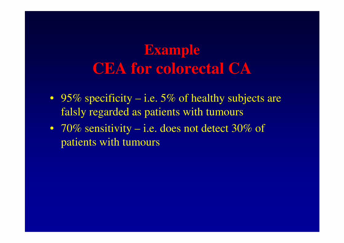
•• CEACEA – glycoproteins with high saccharides content, MW 180 kDa, present in fetal intestine, used for monitoring of colorectal CA, event. other CA (breast, lung), higher levels colorectal CA, event. other CA (breast, lung), higher levels in smokers.
•• Human chorionic gonadotrophin (hCG)Human chorionic gonadotrophin (hCG) –glycoprotein, α and β subunits non-covalently bound, αsubunit identical with LH, FSH and TSH.
Indication of examination: dg of pregnancy (hCG), prenatal screening of Down syndrome (free β hCG), monitoring and prognosis of germ cell tumours, trophoblastic disease (β hCG – specific hCG)
•• CA 125CA 125 – monitoring of ovarian CA
•• CA 15CA 15--33 – monitoring of breast CA
•• CA 72CA 72--44 – monitoring of gastric CA
•• CA 19CA 19--99 – glycolipid, determinant of blood group Lewis a (5% of population does not produce it), for monitoring of pancreas CA (and bile ducts), CAVE – contamination by salivasaliva
•• CYFRA 21CYFRA 21--11 – soluble fragment of cytokeratine 19, for lung CA (non-small cell) and urinary bladder
•• NSENSE – for monitoring of small cell lung cancer, neuroblastoma, apudoma, CAVE – hemolysis
•• PSAPSA – serin protease, glycoprotein, monitoring of prostata CA, CAVE – preanalytical phase
ratio fPSA/PSA, velocity, density
•• SCCSCC – squamous cell carcinoma antigen, monitoring of head and neck tumours, genital tumours and oesophagus tumour
•• TPATPA – tissue polypeptide antigen, mixture of soluble cytokeratines 8, 18 and 19, monitoring of CA of urinary bladder
•• TPSTPS – tissue polypeptide specific antigen, soluble fragment of cytokeratine 18, monitoring of metastasing breast CAbreast CA
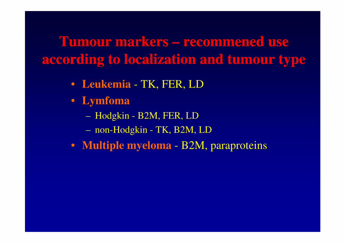
•• TKTK – thymidinkinase, marker of proliferation, leukemias
•• ββββββββ22--microglobulinmicroglobulin – hematological malignancies (NHL), influenced by renal function
•• FerritinFerritin – hematological malignancies
•• Paraprotein, free light chains Paraprotein, free light chains – monoclonal gamapathy (urine – Bence-Jones protein, not determined by the dip stick test)
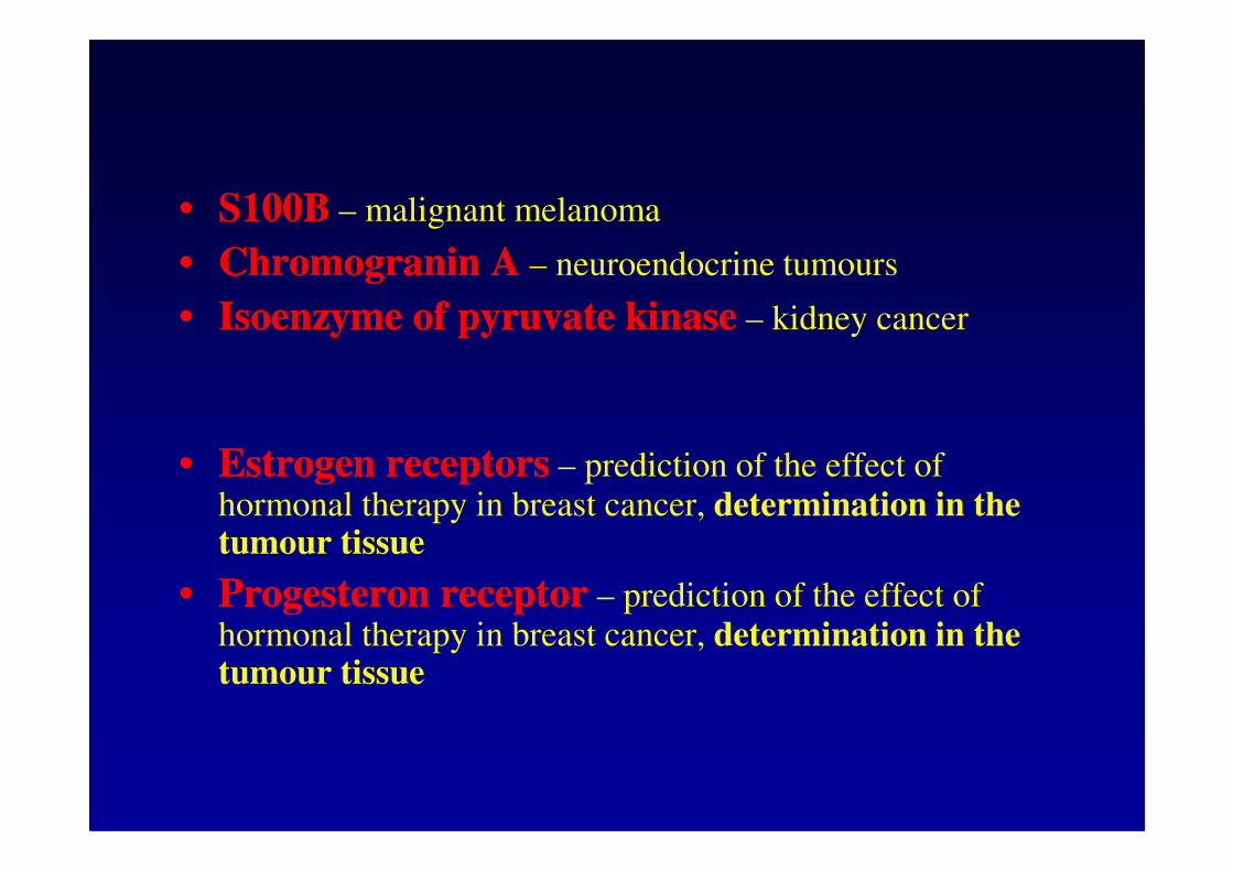
•• S100BS100B – malignant melanoma
•• Chromogranin AChromogranin A – neuroendocrine tumours
•• Isoenzyme of pyruvate kinaseIsoenzyme of pyruvate kinase – kidney cancer
•• Estrogen receptorsEstrogen receptors – prediction of the effect of hormonal therapy in breast cancer, determination in the tumour tissue
•• Progesteron receptorProgesteron receptor – prediction of the effect of hormonal therapy in breast cancer, determination in the tumour tissue
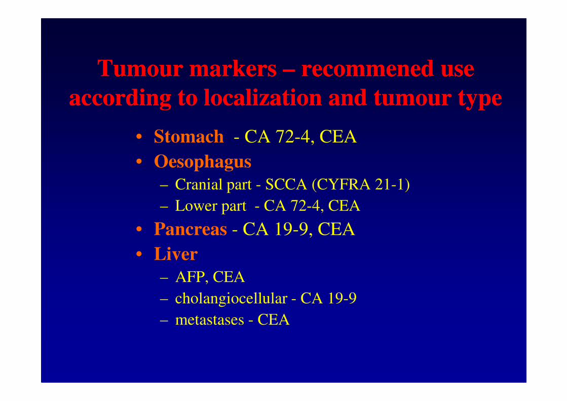
Tumour markers Tumour markers –– recommened use recommened use
according to localization and tumour typeaccording to localization and tumour type
• Stomach - CA 72-4, CEA
• Oesophagus
– Cranial part - SCCA (CYFRA 21-1)
– Lower part - CA 72-4, CEA– Lower part - CA 72-4, CEA
• Pancreas - CA 19-9, CEA
• Liver
– AFP, CEA
– cholangiocellular - CA 19-9
– metastases - CEA
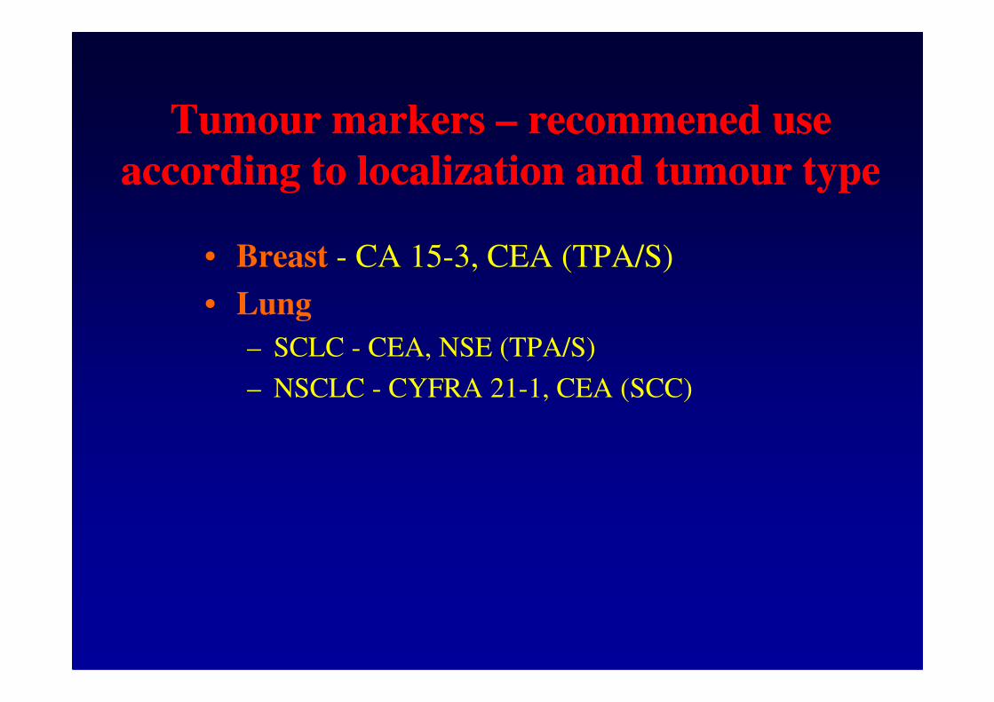
• Breast - CA 15-3, CEA (TPA/S)
• Lung
– SCLC - CEA, NSE (TPA/S)
Tumour markers Tumour markers –– recommened use recommened use
according to localization and tumour typeaccording to localization and tumour type
– SCLC - CEA, NSE (TPA/S)
– NSCLC - CYFRA 21-1, CEA (SCC)
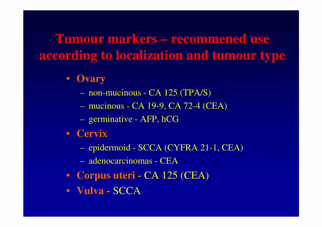
• Ovary
– non-mucinous - CA 125 (TPA/S)
– mucinous - CA 19-9, CA 72-4 (CEA)
Tumour markers Tumour markers –– recommened use recommened use
according to localization and tumour typeaccording to localization and tumour type
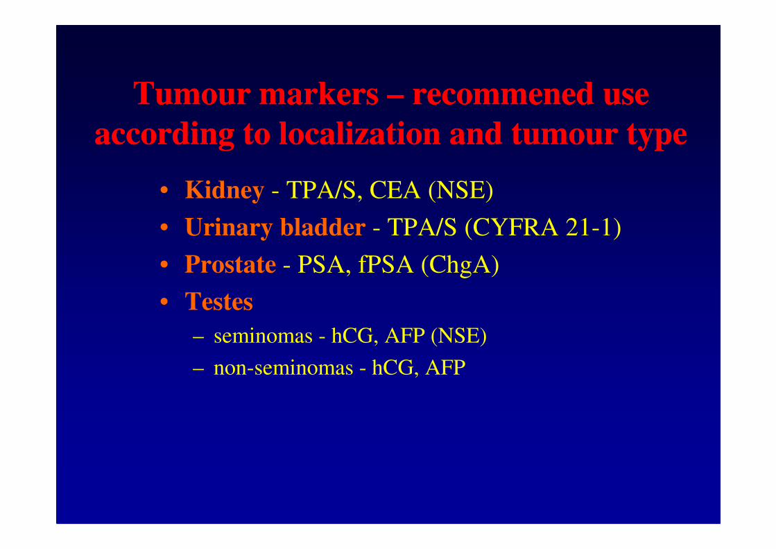
– germinative - AFP, hCG
• Cervix
– epidermoid - SCCA (CYFRA 21-1, CEA)
– adenocarcinomas - CEA
• Corpus uteri - CA 125 (CEA)
• Vulva - SCCA
• Kidney - TPA/S, CEA (NSE)
• Urinary bladder - TPA/S (CYFRA 21-1)
• Prostate - PSA, fPSA (ChgA)
Tumour markers Tumour markers –– recommened use recommened use
according to localization and tumour typeaccording to localization and tumour type
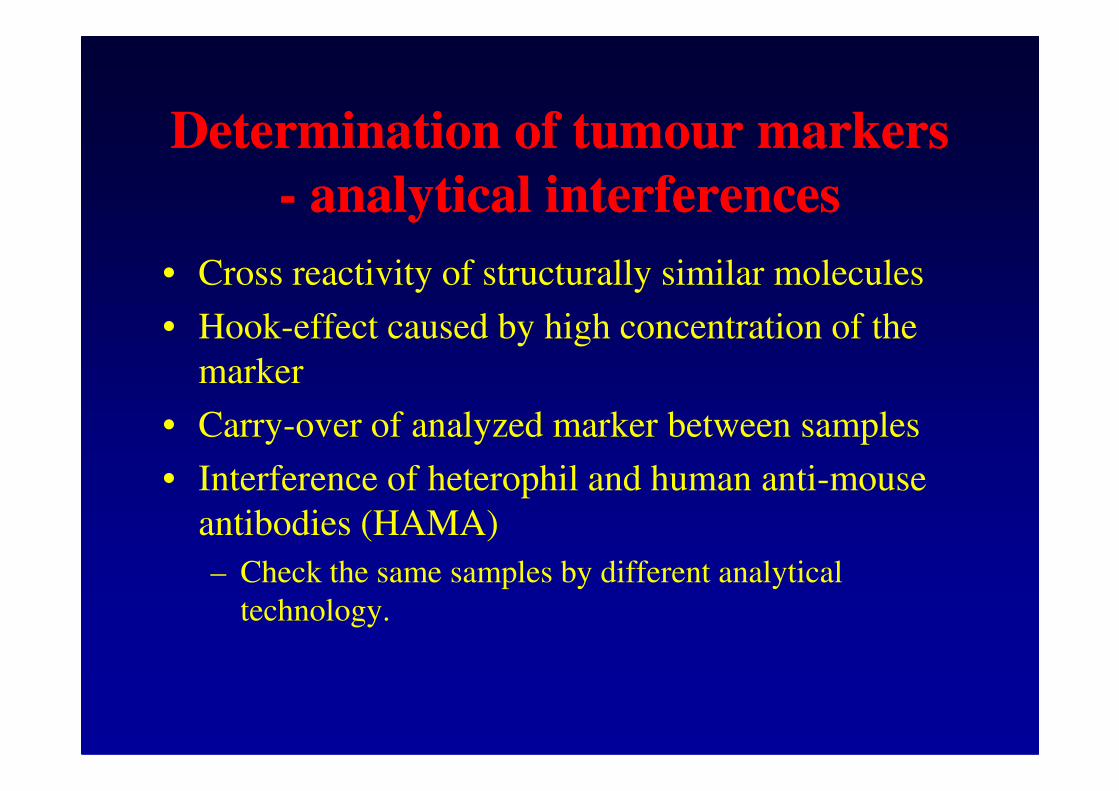
• Cross reactivity of structurally similar molecules
• Hook-effect caused by high concentration of the
marker
• Carry-over of analyzed marker between samples
• Interference of heterophil and human anti-mouse
antibodies (HAMA)
– Check the same samples by different analytical
technology.
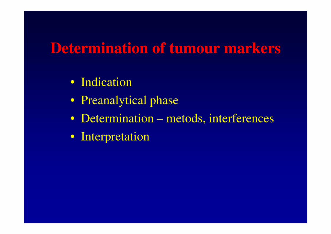
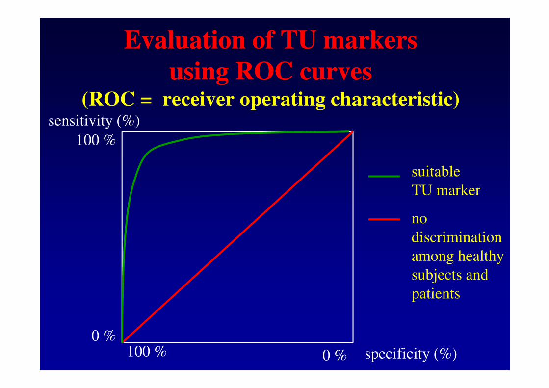
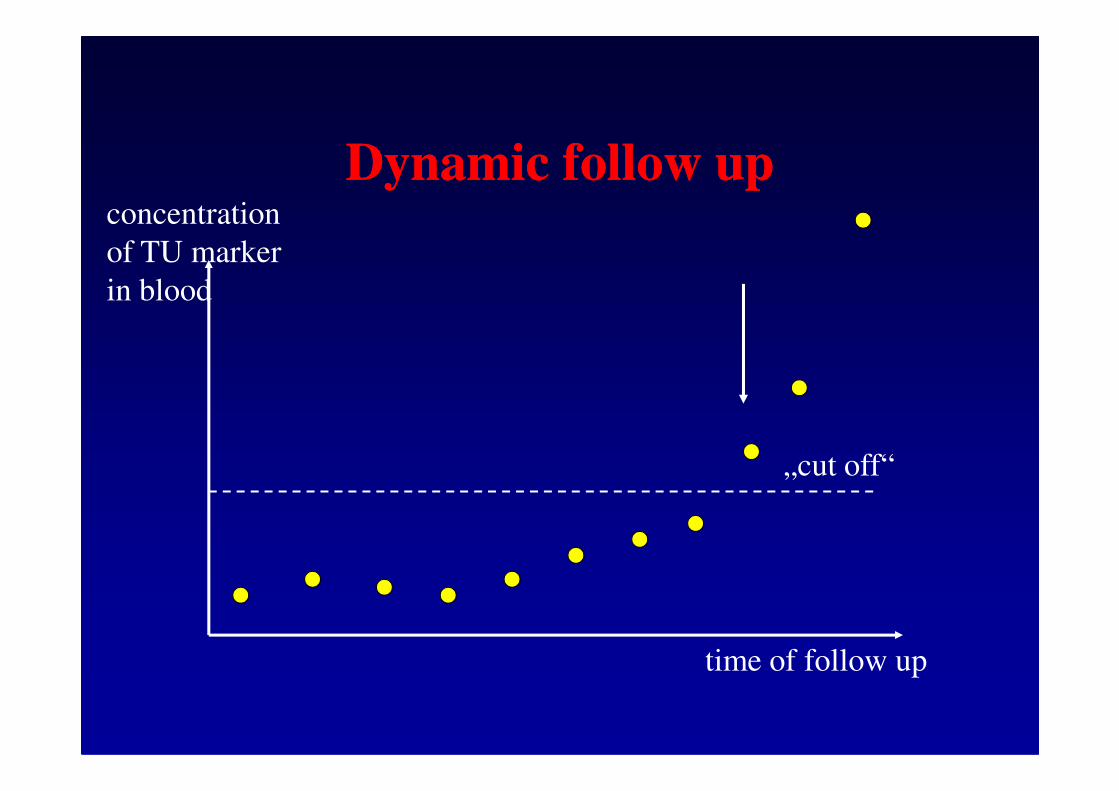
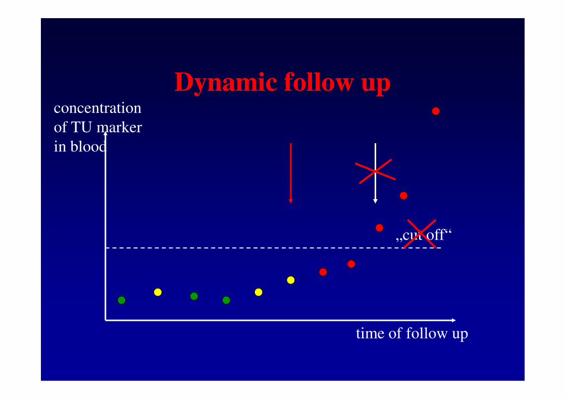
Indication and interpretation Indication and interpretation
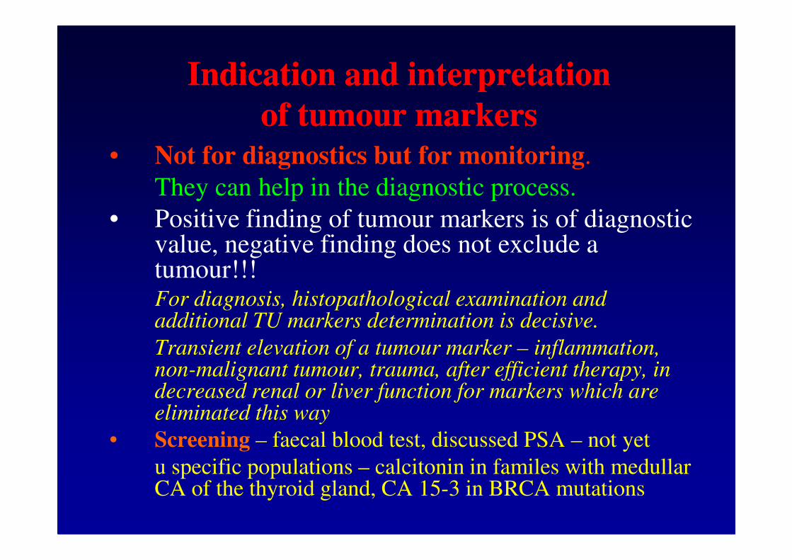
of tumour markersof tumour markers• Not for diagnostics but for monitoring.
They can help in the diagnostic process.
• Positive finding of tumour markers is of diagnostic value, negative finding does not exclude a tumour!!!tumour!!!For diagnosis, histopathological examination and additional TU markers determination is decisive.
Transient elevation of a tumour marker – inflammation, non-malignant tumour, trauma, after efficient therapy, in decreased renal or liver function for markers which are eliminated this way
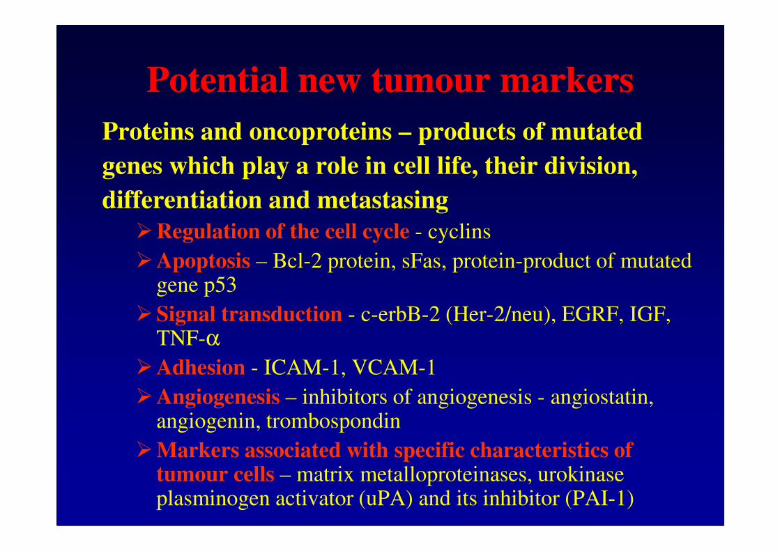
�Angiogenesis – inhibitors of angiogenesis - angiostatin, angiogenin, trombospondin
�Markers associated with specific characteristics of tumour cells – matrix metalloproteinases, urokinase plasminogen activator (uPA) and its inhibitor (PAI-1)
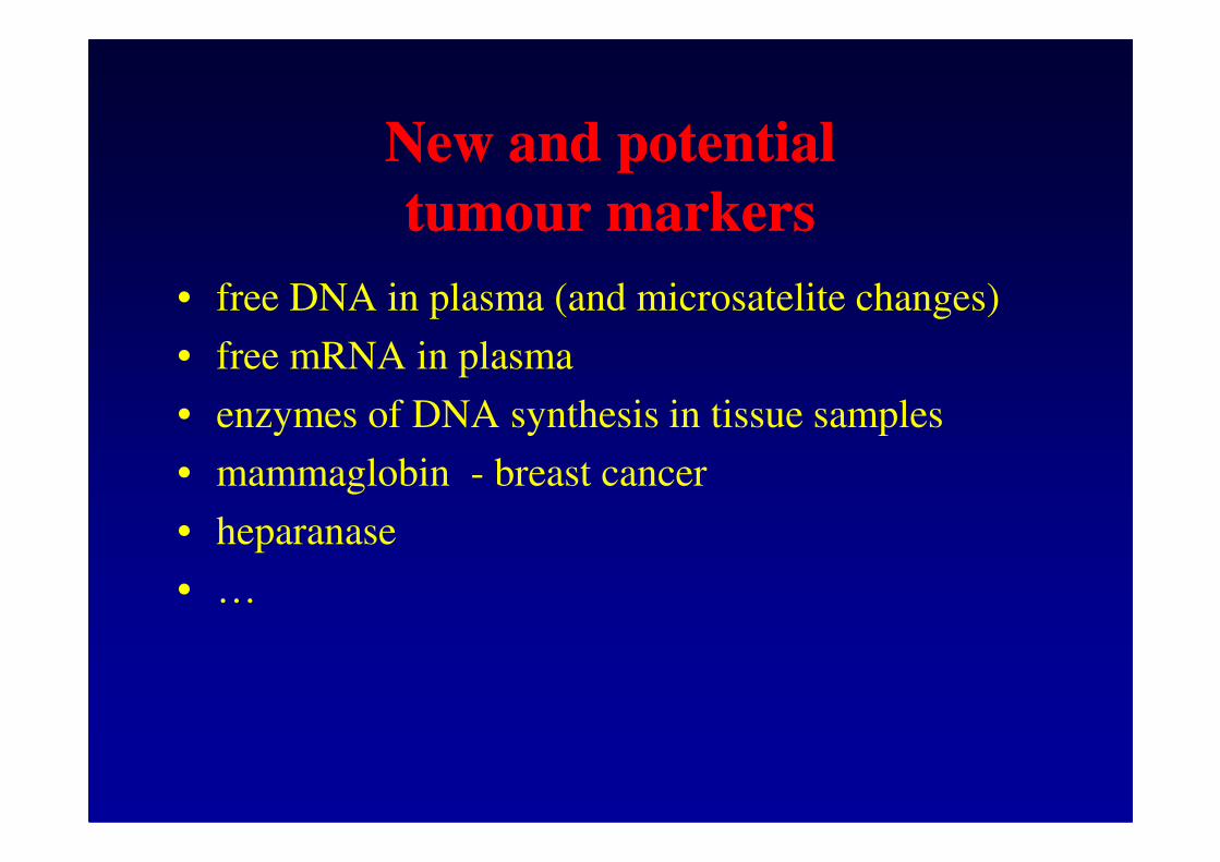
New and potential New and potential
tumour markerstumour markers
• free DNA in plasma (and microsatelite changes)
• free mRNA in plasma
• enzymes of DNA synthesis in tissue samples• enzymes of DNA synthesis in tissue samples
• mammaglobin - breast cancer
• heparanase
• …
LiteratureLiterature
• Guidelines of the Czech Society of Clinical Chemistry – www.cskb.cz
• Guidelines of the European Group for Tumour • Guidelines of the European Group for Tumour Markers (EGTM) – www.egtm.eu