68
Institute Report 2011–2013 Max Planck Institute of Immunobiology and Epigenetics

Institute Report 2011–2013
Max Planck Institute of Immunobiology and Epigenetics

2 INSTITUTE REPORT 2011–2013 | Max Planck InstItute of IMMunobIology and ePIgenetIcs

3
Preface 4
About usHistory of the Institute 6Previous Directors 7Institute Highlights 8Organization of the MPI-IE 10Scientific Advisory Board and Board of Trustees 11Facts and Collaborations 12Administration and Service 13Focus Immunobiology 14Focus Epigenetics 15The International Max Planck Research School 16
Research Groups Senior Group Asifa Akhtar 20Senior Group Thomas Boehm 22Group Taro Fukao 24Senior Group Rudolf Grosschedl 26Group Patrick Heun 28Group Ana Izcue 30Senior Group Thomas Jenuwein 32University/MPI-IE Group Hassan Jumaa 34Group Andrea Pichler 36Group J. Andrew Pospisilik 38University/MPI-IE Senior Group Michael Reth 40Group Simona Saccani 42Guest Scientist Ritwick Sawarkar 44Group Eirini Trompouki 46Emeritus Group Rolf Kemler 48Past Research Groups 49
Research Facilities
Laboratory Animal Facility 52Transgenic Mouse Facility 53
Fish Facility 54
Fly Facility 55
Flow Cytometry and DNA Sequencing
Facility 56 Proteomics Facility 57Imaging Facility 58Deep Sequencing Facility 59Bioinformatics Facility 60
Around the Institute
Life at the Institute 62Life in and around Freiburg 63Special Guest Seminar Series 64Directions to the Institute 66Imprint 67
01
02
03
04
Content

4 INSTITUTE REPORT 2011–2013 | Max Planck InstItute of IMMunobIology and ePIgenetIcs
AbOUT US
Preface
evaluation through the Scientific Ad-visory Board (SAB) in May 2013 that highlighted the productivity, innova-tion and scientific excellence of the Institute. To proceed even further, we continuously adopt measures to build a united vision of the Institute, to in-crease interactions among departments and to liaise with all peer groups of the Institute. There are also long-standing co-operations with the University of Freiburg that are manifested by a joint University/Max Planck department, several innovative research consortia and an international PhD program. This successful IMPRS-MCB PhD pro-gram ensures that the Institute can at-tract academic talent from all over the world.
The success of the recent develop-ments is also demonstrated by the awards of two ERC Starting Grants to Andrew Pospisilik and Patrick Heun and of two ERC Advanced Grants to Thomas Boehm and Michael Reth. With the promotion of Asifa Akhtar to full directorship in April 2013, a major milestone for the consolidation of the Epigenetics focus was accomplished.
Initiated in December 2010, the Max Planck Freiburg Epigenetics Meeting was held again in December 2012, and has rapidly gained international reputation with lectures from many renowned scientists and poster presen-tations by Postdocs and PhD students. This successful meeting will be contin-ued on a bi-annual basis.
The past three years have also seen some changes within the faculty of the Institute: with the retirement of Mari-na Freudenberg and the Emeritus tran-sition of Rolf Kemler, two colleagues
Consolidation and Change: what seems contradictory at first has been sensibly combined in our Institute dur-ing the last years. On the occasion of the 50th anniversary of the Institute in December 2011, Peter Gruss, President of the Max Planck Society, emphasized that “Future needs Ancestry” – both is lived at our Institute.
Since its foundation in 1961, the Insti-tute has undergone substantial chang-es, while the continuing interest in the function of B and T cells provided a natural bridge for studies into host-pathogen interactions, signaling path-ways, self vs. non-self discrimination, transcriptional control and lineage plasticity. During the last years, the Institute has adopted the strategy to focus on two key areas of modern biol-ogy: Immunobiology and Epigenetics. This strategy rests on but also expands the proven strengths of the Institute and aims at dissecting the molecu-lar mechanisms that govern cell type identity and chromatin-dependent re-sponses of the epigenome to changes in the environment.
The definition of these research areas was also recognized by the external
ThOMAS JEnUwEInMAnAGInG DIRECTOR(2012–2014)
who have shaped the research profile of the Institute for many years have com-pleted the active part of their scientific careers. Several group leaders (Tilman Borggrefe, Wolfgang Schamel, Robert Schneider) have been rewarded for their excellent work and were appoint-ed to senior positions at other institu-tions both in Germany and abroad. At the same time, new group leaders (Rit-wick Sawarkar, Eirini Trompouki) have joined the Institute, and more will fol-low soon. This testifies to the attractive career building opportunities for junior faculty of the Institute.
The success and attractiveness of our Institute would not be possible without the support of state-of-the-art research facilities and service units and the help of the administration. This enables us to focus on the research and to address the big questions with the most recent and advanced technologies. I would like to thank all colleagues, scientific and non-scientific staff for their com-mitment and dedication to build a co-herent structure and to develop the full potential of the Max Planck Institute of Immunobiology and Epigenetics.
This Institute Report covers a three year period (2011–2013), is a continuation of the previous ‘Annual Report’ (2008–2010) and supplements the newly re-vised website (www.ie-freiburg.mpg.de) that is regularly curated and updated.
Enjoy reading our Institute Report 2011–2013.
Prof. Dr. Thomas JenuweinManaging Director, November 2013

5
01 About us01 About us

6 INSTITUTE REPORT 2011–2013 | Max Planck InstItute of IMMunobIology and ePIgenetIcs
history of the Institute
1961
The Max Planck Institute of Immuno-biology (MPI-IB) was founded in 1961 on the premises of the former research institute of the pharmaceutical com-pany Wander AG in Freiburg. Until the end of the 1970’s, under the director-ship of Otto Westphal, Herbert Fischer and Otto Lüderitz, the institute was primarily engaged in studying the in-teractions between infectious agents and the immune system, with special emphasis on the bacterial substance endotoxin.
1980
With the recruitment of Klaus Eich-mann (1981) and Georges Köhler (1984), the thematic focus of the insti-tute expanded to cellular and molecu-lar mechanisms of B and T cells. Klaus Eichmann and colleagues were the first to describe the development of func-tional lymphoid tissue from embryonic stem cell lineages. In 1984, Niels Jerne, Georges Köhler and Cesar Milstein were awarded the Nobel Prize for their pioneering work on monoclonal anti-bodies using the hybridoma technique.
1990 Through a special funding by the State of Baden-Württemberg, Developmen-tal Biology was added as another sci-entific focus, resulting in the recruit-ment of Davor Solter (1991) and Rolf Kemler (1992). Davor Solter was one of the first to identify genomic imprint-ing and his research focused on genetic and epigenetic mechanisms regulating mouse pre-implantation development. Rolf Kemler identified the first cell-cell adhesion molecule (E-cadherin) in
mouse development and significantly advanced the understanding of mouse embryogenesis.
1998With the appointment of Thomas Boehm (1998) as successor of George Köhler, Developmental Immunology was added as a new research focus. Ef-forts towards a stronger cooperation between MPI-IB and the Faculty of Bi-ology at the University of Freiburg led to the establishment of the University Department of Molecular Immunol-ogy at the MPI-IB and recruitment of Michael Reth as its head (1998). In ad-dition, the Spemann Laboratory, con-sisting of three independent junior re-search groups, was established with the aim of promoting early independence of junior scientists. With the appoint-ment of Rudolf Grosschedl as successor of Klaus Eichmann (2004), the themat-ic connection between Immunology and Developmental Biology was fur-ther strengthened and the molecular mechanisms of lymphoid cell differen-tiation and the regulation of genes via extracellular signals were added as new research areas.
2006In 2006, the International Max Planck Research School for Molecular and Cel-lular Biology (IMPRS-MCB) was intiti-ated by Rudolf Grosschedl, in collabo-ration with colleagues of the University of Freiburg. At the beginning of 2006, the President of the Max Planck Soci-ety launched a competition between all institutes of the Society to establish a new department with an innovative research theme. Among all propos-als, “Epigenetics” was selected and
Thomas Jenuwein (2008) accepted an offer of the Max Planck Society to di-rect the new department “Epigenetics”. To make a relevant impact in the field of epigenetic research, the Kollegium decided to additionally appoint an epigenetics researcher as successor of Davor Solter.
At the end of 2009, Asifa Akhtar was appointed as a Max Planck Investigator focusing on “Chromatin Regulation” and was promoted to Director in April 2013.
2010In December 2010, the institute was renamed to “Max Planck Institute of Immunobiology and Epigenetics” (MPI-IE), reflecting the two key areas of modern biology being conducted at the institute. With the establishment of the “Epigenetic Focus” at the MPI-IE, an international biennial meeting on the broad area of epigenetics and chro-matin was founded. In December 2011, more than 200 guests celebrated the 50th anniversary of the MPI-IE. “Fu-ture needs ancestry“ emphasized Peter Gruss, President of the Max Planck So-ciety, and honoured the achievements of the MPI-IE.
In February 2013, Rolf Kemler retired from the director position. He will con-tinue research in an emeritus group until 2015. The search for a successor of the director position in the field of immunobiology is currently underway. As in the past, new junior group leader positions are continuously being estab-lished at the MPI-IE to ensure new in-put for exciting fields of research.
AbOUT US
1961 1976 2010

7
AbOUT US
Previous Directors of the Institute
Otto Westphal(Director from 1961–1982)
Herbert Fischer(Director from 1964–1981)
Otto Lüderitz(Director from 1965–1988)
Klaus Eichmann(Director from 1981–2004)
Georges Köhler(Director from 1984–1995)
Davor Solter(Director from 1991–2006)
Rolf Kemler(Director from 1992–2013)
Otto Westphal founded the Max Planck Institute of Immunobiology in 1961 and established it as a leading research facility. His scientific achievements include the determination of the primary structure of E. coli lipid A, an endotoxic lipopolysac-charide. He was the founder of the European Journal of Immunology and found-ing President of the German Society for Immunology.
Herbert Fischer had an ardent interest in macrophages at a time when the interest in the field of immunology was universally focused on lymphocytes. His group studied the role of phospholipid metabolism in the activation of macrophages and lymphocytes and its subsequent effects on the activation of the innate and adaptive immune systems.
Otto Lüderitz and his group showed that lipopolysaccharides (LPS) of Gram-neg-ative bacteria are built up according to a common architecture, consisting of the O-polysaccharide chain, the core and lipid A. In chemical and biological studies they brought the final evidence that lipid A is the toxic and biologically active part of LPS which led to the total chemical synthesis of biologically active lipid A.
Klaus Eichmann and coworkers were involved in research on T cell development, T cell activation, and antigen processing in cell-mediated immunity. They discov-ered the autonomous signaling function of the pre-T cell receptor in the develop-ment of the alpha/beta T cell lineage. They were the first to describe the develop-ment of functional lymphoid tissue from ES cell lines.
In the year Georges Köhler joined the Max Planck Institute of Immunobiology he was awarded the Nobel prize in Physiology or Medicine, together with Cesar Milstein and Niels Jerne for their pioneering work on the immune system and the generation of monoclonal antibodies using the hybridoma technique. His un-timely death in 1995 was a great loss to the institute and the scientific community.
In seminal experiments, Davor Solter studied the developmental potential of ma-ternal and paternal genomes by nuclear transplantation. He was one of the first t o identify genomic imprinting. His research focused on genetic and epigenetic mechanisms regulating mouse pre-implantation development. Solter made sig-nificant contributions to mammalian development, including differentiation of germ layers, biology and genetics of teratocarcinoma, biology of embryonic stem cells, cloning, and reprogramming.
Rolf Kemler identified the first cell-cell adhesion molecule in mouse development, E-cadherin. He discovered catenins as cytoplasmic anchorage proteins. Particu-larly β-catenin is well-known because of its dual function in cell adhesion and Wnt signaling. Kemler was the first to establish mouse embryonic stem (ES) cells in Germany. He studied their differentiation potential and used gene targeting to investigate the function of cadherin and catenins in development, genomic main-tenance and stem cell vs. oncogenic potential.

8 INSTITUTE REPORT 2011–2013 | Max Planck InstItute of IMMunobIology and ePIgenetIcs
AbOUT US
Institutehighlights
DECEMbER
2012
The 2nd Max Planck Epigenetics Meeting takes place. Talks are giv-en by 20 invited speakers plus 15 speakers that are selected from the abstracts. About 120 scientists par-ticipate in the meeting and about 60 posters are presented.
OCTObER
AUGUST
On the occasion of the 60th birthday of Rudolf Grosschedl, a one-day sym-posium takes place with 20 scientists, presenting their research within four sessions: gene control, long-range chromatin interactions, B lympho-poiesis, signaling and development.
Patrick Heun is awarded an ERC Start-ing Grant for “Dissection of centro-meric chromatin and components: A biosynthetic approach”
J. Andrew Pospisilik is awarded an ERC Starting Grant for “Metabolic Polycombics”.
The scientific members of the MPI-IE attend the institute retreat in Saint Hippolyte, Alsace, France. The event aims at strengthening scientific inter-actions and supporting the collabora-tive atmosphere at the institute.
With a ground-breaking ceremony the construction of the new child care facility is initiated. From December 2013 on, it will provide space for chil-dren at the age of three months to six years in close vicinity to the institute.
2011nOvEMbER
The first Max Planck Day, initiated by the Max Planck Society and hosted by the individual institutes, takes place. The event at the MPI-IE aims at bring-ing school classes in contact with re-search, work and life at the institute.
DECEMbER
More than 200 guests celebrate the 50th Anniversary of the MPI-IE with festivities including commemorative speeches and a scientific symposium with notable speakers from all over the world. Peter Gruss, President of the Max Planck Society honours the accomplishments of the institute.
October 2012: Ground-breaking ceremony for the child care facility.
nOvEMbER
Michael Reth is awarded an Advanced ERC Grant for his project “Nanoscale analysis of protein islands on lym-phocytes”.
Thomas Boehm is awarded an Ad-vanced ERC Grant for the project “Thymopoiesis: From evolutionary origins to future therapies”.
Peter Gruss
9
JUnE
MAy
2013
SEPTEMbER
JUly
The Max Planck – The University of Tokyo Center of Integrative In-flammology is established under co-directorships of Rudolf Grosschedl from the MPI-IE and Tadatsugu Tani-guchi from the University of Tokyo. It aims at bundling research projects of both institutions and facilitating the exchange of knowledge and ex-perience.
Kickoff meeting for the construction of a new animal service building. It will house core infrastructure, isola-tor units and personnel rooms, en-suring highest scientific and animal care standards.
The Scientific Advisory Board (SAB) conducts the external evaluation of the MPI-IE. In its feedback, the SAB highlights the excellence and innova-tion of the institute and encourages towards continuous growth and the development of a strong identity to exploit the full potential of the insti-tute.
EMBO announces Asifa Akhtar as a new member.
Andrew Pospisilik is awarded the Rising Star Award of the EASD for a new concept about the emergence of diabetes.
Eirini Trompouki starts as Group Leader in the Department of Cellular and Molecular Immunology.
Ritwick Sawarkar starts as a Guest Scientist in the Epigenetic focus.
Building component BT IV is re-opened after extensive renovation. It provides room for the new caf-eteria, offices, seminar rooms and a centralized IT and server area.
MPI-IE contributes to the Freiburg Science Fair, an event for the general public.
APRIl
Asifa Akhtar is promoted to full Max Planck director position after four years of highly successful research at the MPI-IE. She leads the department of Chromatin Regulation.
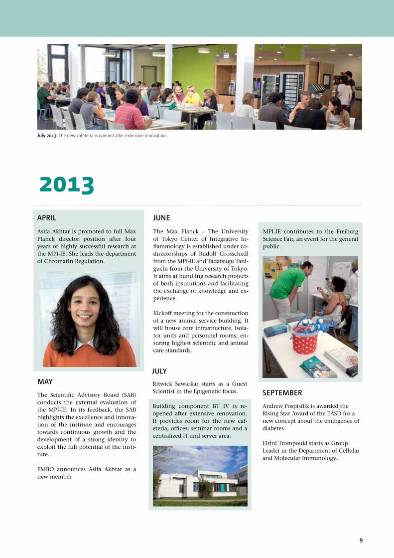
July 2013: The new cafeteria is opened after extensive renovation.

10 INSTITUTE REPORT 2011–2013 | Max Planck InstItute of IMMunobIology and ePIgenetIcs
Organization of the MPI-IE
The Max Planck Institute of Im-munobiology and Epigenetics (MPI-IE) is organized in five departments, plus one joint appointment between the University of Freiburg and the MPI-IE. Each department of the MPI-IE is head-ed by a director, also named ‘senior group leader’ (at present one director position vacant).In addition, currently nine junior group leaders conduct research at the MPI-IE. They are either department-associated or department-independent. All junior groups are considered equivalent. They have their own budget and pursue – within the framework of the MPI-IE – their research fully independently. Junior group leader positions are established for five years (with possibility of exten-sion) and are attractive career-building appointments. Central scientific infra-structure units and the administration complement the MPI-IE.The central decision-making body of the MPI-IE is the management board
(“Kollegium”), comprising the direc-tors and the senior executive manager. It meets on a bi-weekly basis. The man-aging directorship rotates every three years among the department heads of the MPI-IE. The management board establishes the general scientific and administrative policies and promotes long-term developments of the MPI-IE. Both the management board and the administration interact closely with the Max Planck Society in Munich regarding budgetary, personnel, and policy issues.In coordination with all group leaders and heads of infrastructure, the man-agement board initiates the establish-ment of new scientific facilities, and ensures a collaborative atmosphere at the institute. Monthly team leader meetings (‘Faculty lunches‘) facilitate internal communication, identifica-tion of solutions, and dissemination of information.
The Management Board of the MPI-IE: (from left) T. Boehm, T. Jenuwein, A. Akhtar, B. Tarkan, R. Grosschedl.
Managing Director (2012-2014)
– Thomas Jenuwein
Scientific Members of the Institute
– Asifa Akhtar – Thomas Boehm – Rudolf Grosschedl – Thomas Jenuwein
Senior Executive Manager – Bülent Tarkan
Adjunct External Scientific Member
– Michael Reth: since 2002 Department of Molecular Immunol-ogy, University of Freiburg / MPI-IE, Freiburg, Germany
External Scientific Members
– Michael Sela: since 1967 Department of Immunology, Weiz-mann Institute of Science Rehovot, Israel
– Barbara B. Knowles: since 2002 Institute of Medical Biology, Singapore
– Paolo Sassone-Corsi: since 2011 Center for Epigenetics and Metabolism, University of California, Irvine, USA
T. boehm Developmental Immunology
A. Akhtar Chromatin Regulation
Management board Asifa Akhtar, Thomas Boehm, Rudolf Grosschedl, Thomas Jenuwein, Bülent Tarkan
represented by Thomas Jenuwein, Managing Director (2012–2014)
R. Grosschedl Cell. & Mol.
Immunology
Department-independent
Research Groups
R. Kemler* Molecular
Embryology
T. Jenuwein Epigenetics
b. Tarkan Senior Execu-tive Manager
J. Faber Public Relations
C. Gartmann IT Services
b. lippok Safety
C. Johner Mouse Facility
D. Moll Human Resources
M. Enderlein Finances
M. Fieber Purchasing
R. volz Workshop
P. Georgiev Fly Facility
b. Kanzler Transgenesis
M. Schorpp Fish Facility
A. Izcue P. heun P. Kindle ImagingA. Pichler T. Fukao h. Jumaa
A. würch FACS
E. Trompouki
S. Saccani A. Pospisilik R. Sawarkar Guest Scientist
Graduate Education
M. baer IMPRS
R. black Library
G. Mittler Proteomics
T. Manke Bioinformatics
U. bönisch Deep Sequencing
DEPARTMEnT HEAD AnD SEnIoR GRouP LEADER
JunIoR GRouP LEADER
SCIEnTIFIC InFRASTRuCTuRE
JoInT unIvERSITy/MPI-IE GRouPS
SERvICE unITS
ADMInISTRATIon
AbOUT US
*emeritus since 3/2013
M. Reth Molecular
Immunology

11
AbOUT US
Scientific Advisory board and board of Trustees
To ensure the high quality and productivity of research, the Max Planck Institute of Immunobiology and Epigenetics (MPI-IE) routinely undergoes evaluations by independent scientific advisors – the ‘Scientific Advisory Board‘ (SAB).
Members of the MPI-IE SAB are internationally renowned scientists who are appointed by the president of the Max Planck Society for a period of six years and who are not affili-ated with the Max Planck Society.
The Board of Trustees provides the institute with valuable advice in social and science-political issues and supports fur-ther developments of the institute. The board meets once
Scientific Advisory board
board of Trustees
The SAB reviews the activities of the institute every three years and issues a report to the President of the Max Planck Society. This evaluation serves as an important basis for the planning of further scientific developments as well as for the distribution of resources by the Max Planck Society. The SAB also supports the MPI-IE in recruiting new directors and group leaders.
Prof. Dr. Susan GasserFriedrich Miescher Institute, Basel, Switzerland
Prof. Dr. Dr. h.c. mult.hubert E. blum Dean of the Medical Faculty, university Medical Center Freiburg, Germany
Michael KleinerMinisterialdirigentMinistry of Science, Research and ArtStuttgart, Germany
Dr. nicola von lutterotti (Chair person)Free Journalist Zurich, Switzerland
Prof. Dr. Gunther neuhausvice Rector for Research university of FreiburgFreiburg, Germany
Prof. Dr. Markus Affolteruniversität Basel Basel, Switzerland
Dr. Dieter Salomon Lord Mayor of the City of FreiburgFreiburg, Germany
Prof. Dr. Philip AvnerEMBL Montero-tondo Monterotondo, Italy
Prof. Dr. Max D. CooperEmory university School of Medi-cine, Atlanta, uSA
Prof. Dr. Adrian haydayKing’s College London London, uK
Dr. Axel Glatz Managing DirectorPfizer Deutschland GmbHFreiburg, Germany
Prof. Dr. Amanda Fisher(Chair person)Imperial College LondonLondon, uK
Prof. Dr. Dan littmanLangone Medical Center, Skirball Inst. new york, uSA
Prof. Dr. Martin haagMayor of the City of FreiburgFreiburg, Germany
Dr. Christian hodeige Publisher and Man-aging DirectorBadische Zeitung GroupFreiburg, Germany
a year (November) to interact with the management board of the MPI-IE and with other members of the Max Planck Society.
12 InSTITUTE REPORT 2011–2013 | MAx PLAnCK InSTITuTE oF IMMunoBIoLoGy AnD EPIGEnETICS
AbOUT US
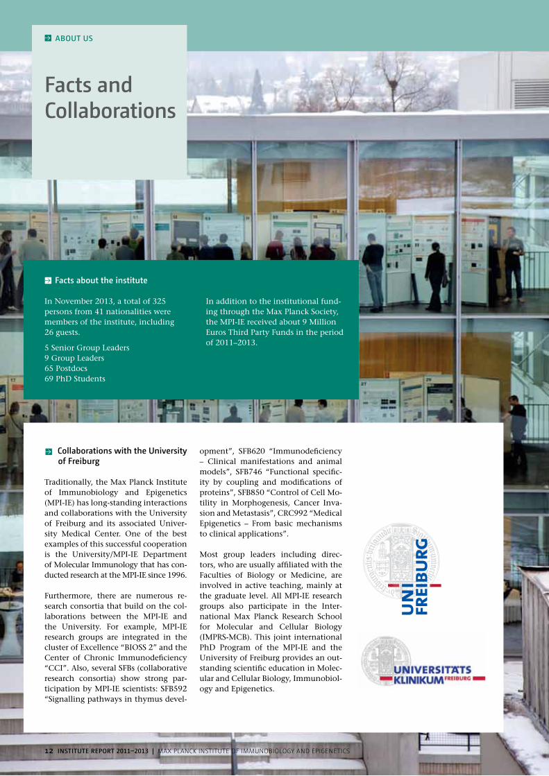
Facts and Collaborations
Facts about the institute
In November 2013, a total of 325 persons from 41 nationalities were members of the institute, including 26 guests.
Collaborations with the University of Freiburg
Traditionally, the Max Planck Institute of Immunobiology and Epigenetics (MPI-IE) has long-standing interactions and collaborations with the University of Freiburg and its associated Univer-sity Medical Center. One of the best examples of this successful cooperation is the University/MPI-IE Department of Molecular Immunology that has con-ducted research at the MPI-IE since 1996.
Furthermore, there are numerous re-search consortia that build on the col-laborations between the MPI-IE and the University. For example, MPI-IE research groups are integrated in the cluster of Excellence “BIOSS 2” and the Center of Chronic Immunodeficiency “CCI”. Also, several SFBs (collaborative research consortia) show strong par-ticipation by MPI-IE scientists: SFB592 “Signalling pathways in thymus devel-
opment”, SFB620 “Immunodeficiency – Clinical manifestations and animal models”, SFB746 “Functional specific-ity by coupling and modifications of proteins”, SFB850 “Control of Cell Mo-tility in Morphogenesis, Cancer Inva-sion and Metastasis”, CRC992 “Medical Epigenetics – From basic mechanisms to clinical applications”.
Most group leaders including direc-tors, who are usually affiliated with the Faculties of Biology or Medicine, are involved in active teaching, mainly at the graduate level. All MPI-IE research groups also participate in the Inter-national Max Planck Research School for Molecular and Cellular Biology (IMPRS-MCB). This joint international PhD Program of the MPI-IE and the University of Freiburg provides an out-standing scientific education in Molec-ular and Cellular Biology, Immunobiol-ogy and Epigenetics.
5 Senior Group Leaders9 Group Leaders65 Postdocs69 PhD Students
In addition to the institutional fund-ing through the Max Planck Society, the MPI-IE received about 9 Million Euros Third Party Funds in the period of 2011–2013.
13
AbOUT US
Adminis-tration and Service
FinancesHead: Martina Enderlein – Jenny Barabasch – Manuela Mattmüller – Saskia Moos – Birgit Poitz
human ResourcesHead: Daniela Moll – Susanne Demme – Corinna Kanisch – Veronika Klank – Sabine Stallone
Purchasing Head: Michele Fieber – Regina Burger – Claudia Höferlin – Sven Mußmann
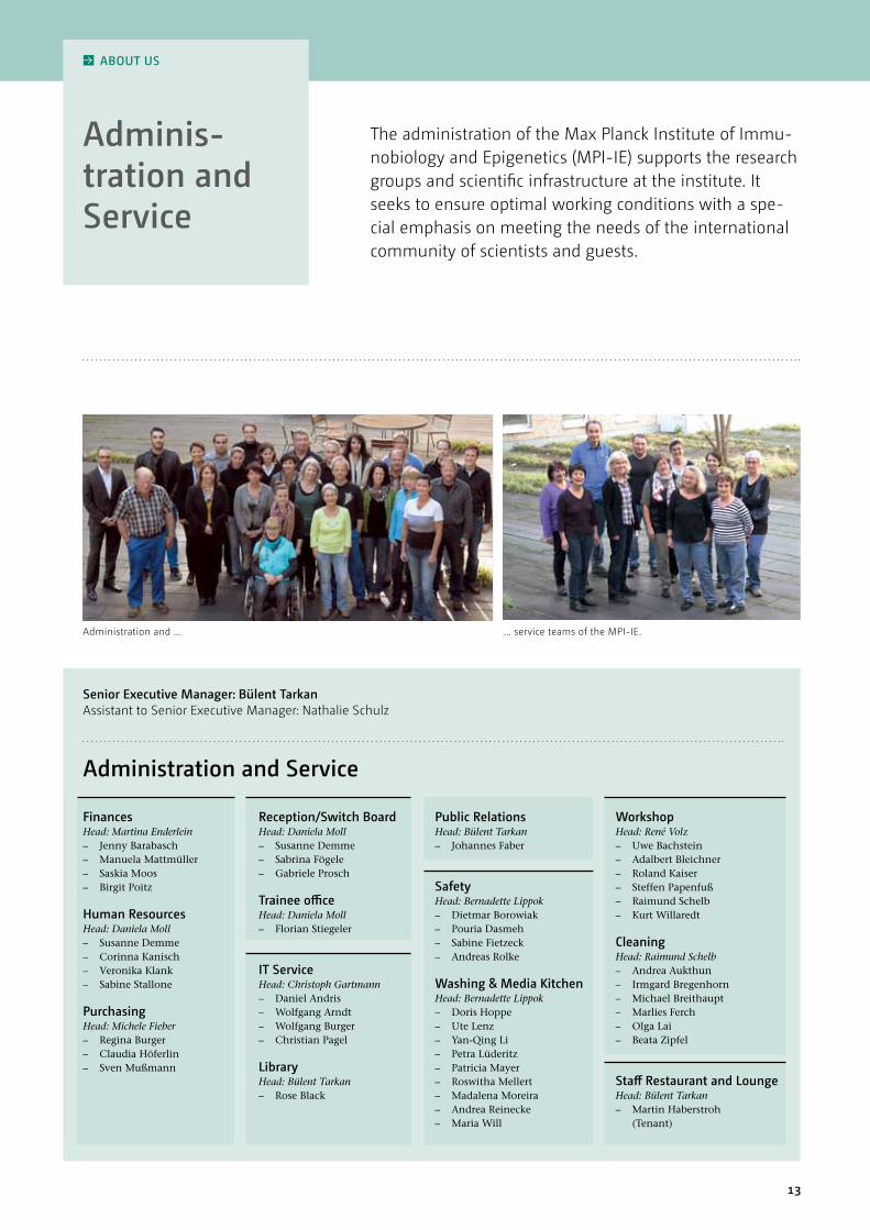
The administration of the Max Planck Institute of Immu-nobiology and Epigenetics (MPI-IE) supports the research groups and scientific infrastructure at the institute. It seeks to ensure optimal working conditions with a spe-cial emphasis on meeting the needs of the international community of scientists and guests.
Administration and ... ... service teams of the MPI-IE.
Senior Executive Manager: bülent TarkanAssistant to Senior Executive Manager: nathalie Schulz
Reception/Switch board Head: Daniela Moll – Susanne Demme – Sabrina Fögele – Gabriele Prosch
Trainee office Head: Daniela Moll – Florian Stiegeler
IT ServiceHead: Christoph Gartmann – Daniel Andris – Wolfgang Arndt – Wolfgang Burger – Christian Pagel
libraryHead: Bülent Tarkan – Rose Black
Public RelationsHead: Bülent Tarkan – Johannes Faber
SafetyHead: Bernadette Lippok – Dietmar Borowiak – Pouria Dasmeh – Sabine Fietzeck – Andreas Rolke
washing & Media Kitchen Head: Bernadette Lippok – Doris Hoppe – Ute Lenz – Yan-Qing Li – Petra Lüderitz – Patricia Mayer – Roswitha Mellert – Madalena Moreira – Andrea Reinecke – Maria Will
workshop Head: René Volz – Uwe Bachstein – Adalbert Bleichner – Roland Kaiser – Steffen Papenfuß – Raimund Schelb – Kurt Willaredt
CleaningHead: Raimund Schelb – Andrea Aukthun – Irmgard Bregenhorn – Michael Breithaupt – Marlies Ferch – Olga Lai – Beata Zipfel
Staff Restaurant and loungeHead: Bülent Tarkan – Martin Haberstroh
(Tenant)
Administration and Service

14 INSTITUTE REPORT 2011–2013 | Max Planck InstItute of IMMunobIology and ePIgenetIcs
RESEARCh OvERvIEw
Focus Immuno-biology
Immunobiology is concerned with the ways multicellular organisms de-fend themselves against the onslaught of pathogens. They have evolved a plethora of strategies to guard their bodily integrity, and to promote sur-vival and reproduction.Also for humans, a properly function-ing immune system is of central im-portance. Indeed, of all branches of medicine, the translation of results from immunological research to medi-cal treatments over the last two centu-ries has probably had the most signifi-cant impact on human life expectancy.
Many devastating infectious diseases have lost their grip on humankind, thanks to preventive strategies such as vaccination and general hygiene. Furthermore, immunology provides us with critical information for the treat-ment of inflammatory diseases and cancer that can afflict many organ sys-tems and are a substantial burden to patients and modern health care sys-tems.Our current research focuses on the molecular mechanisms underlying lymphocyte generation from hemato-poietic stem cells. Of exceptional in-
terest are factors within lymphocytes and those emanating from the micro-environment in lymphoid organs that foster the emergence of mature effector cells. Apart from sophisticated in vitro systems, we use a wide range of animal model systems to study various aspects of the immune system in a physiologi-cal context.Additionally, immunobiology serves as a paradigmatic research field for key questions in modern biology, such as cellular identity, cell-cell interactions, the structure of protein complexes and signal transduction in cells.
owing to immunological research many infec-tious diseases have lost their grip on humankind.
The evolutionary and life-time development of hematopoetic cells can serve as a model system for fundamen-tal questions such as cellular identity, cell-cell interactions, the structure of protein complexes and signal transduction in cells. At the same time, it allows a better understanding of many diseases.
Stem Cell
Multipotent-Progenitor Cell
Pro-b Pre-b
b Cell
T Cell
nK Cell
basophil
Eosinophil
neutrophil
Macrophage
Dendritic Cell
Erythrocyte
Platelets
Pro-T
Committed Progenitor
Mature Cell
Megakaryocyte

15
RESEARCh OvERvIEw
Focus Epigenetics
“Are we more than the sum of our genes and how can environmental cues alter gene expression?” While al-most all cell types (ca. 200) within a human body share an identical DNA sequence, its utilization will differ sig-nificantly according to the designated function of a cell. The DNA template within the cell nucleus is wrapped and protected by specific proteins (his-tones). This DNA-histone polymer is called chromatin. Stable chromatin alterations that do not affect the DNA sequence, are summarized under the term ‘Epigenetics‘.
Due to the plasticity of chromatin states a genome has a variety of epi-genetic variants (epigenomes). Estab-lishment and maintenance of these epigenomes is critical for embryonic development, cell type identity and cell differentiation. Although many diseases (e.g. cancer, neurodegenerative and metabolic dis-orders) are primarily caused by DNA mutations, epigenetic disregulation can significantly contribute to disease progression. Thus, epigenetic research promises far-reaching implications for new forms of therapy and diagnosis. Epigenetic changes also allow respons-
es to environmental influences such as nutrition, stress and hormones. In-triguingly, there is growing evidence that epigenetic alterations might even be inheritable over a few generations.The research groups of the Epigenetic Focus combine topics addressing dos-age compensation, heterochromatin formation, and posttranslational modi-fications of histones. A variety of model organisms and experimental approach-es (biochemistry, cell biology, Drosoph-ila and mouse genetics, genome-wide profiling) are used to dissect the epig-enome of distinct cell types.
Epigenetics describes the inheritance of ac-quired traits that are not based on alterations of DnA sequence.
The DnA template within the cell nucleus is not naked, but wrapped and protected by specific proteins (histones). This DnA-histone poly-mer is called chromatin. Histone modifications and other chromatin alterations are important elements of epigenetic gene control. This is critical for embryonic development, cell type identity and cell differentiation.
Me

16 INSTITUTE REPORT 2011–2013 | Max Planck InstItute of IMMunobIology and ePIgenetIcs
The International Max Planck Research School
AbOUT US
The International Max Planck Re-search School for Molecular and Cel-lular Biology (IMPRS-MCB) was started in 2006 upon an initiative of scientists from the Max Planck Institute of Im-munobiology and Epigenetics (MPI-IE) and the University of Freiburg. The idea of the research school is to provide a broad scientific education to young researchers interested in molecular and cellular biology, immunobiology and epigenetics.Since its initiation, the PhD program has been growing steadily and cur-
From the beginning, I have been positively surprised by the trust and support I received from ev-eryone – from fellow PhD stu-dents to group leaders and the IMPRS. Even though I did not have any formal training and very little experience, I was al-lowed to dive into the bioinfor-matic analysis of high-through-
put sequencing data. I am ever so grateful for this chance as I am now involved in a very young, very dynamic, quickly develop-ing field that is directly at the interface between cutting edge molecular biology and state of the art analysis methods.Against my initial skepticism, Freiburg, as a city, has also completely won me over. Despite its modest size, it is a very friendly, even international city with a lively arts and music scene and its surroundings are amazingly beautiful. The prox-imity to France and Switzerland is an additional plus - not only regarding culinary aspects: it is also the basis for an ac-tive exchange between the MPI-IE in Freiburg, the FMI and ETH in Basel and the IGBMC in Strasbourg.
rently has 63 students, coming from 31 countries, and 23 faculty members (15 from MPI-IE and 8 from the University of Freiburg).
Our PhD students are given an oppor-tunity to carry out research on specific projects of their choice. In addition they participate in the education pro-gram covering: – experimental methods in molecular
and cellular biology; – theoretical knowledge and in-depth
analysis of scientific literature;
– complementary skills in presenting scientific data in oral and written form and in applying for research funding.
Besides this mandatory program, we organize additional scientific courses (e.g. statistics, bioinformatics) and support participation of students in external workshops and scientific conferences. Students also have the possibility to learn German and to obtain advice in planning their future career.
FRIEDERIKE DünDAR from Germany IMPRS Student since 2011, joint PhD at bioinformatics Facility and Senior Group Akhtar
After being an IMPRS as-sociated PhD student for three years, I can hon-estly say that moving to Freiburg was one of the best decisions I have made in my career so far. From the beginning I have felt very welcomed by ev-eryone, particularly the
IMPRS office. After a three months‘ rotation period in dif-ferent laboratories, a strong relationship with other research groups was established, which is essential for constructive feedback and advice during your studies. I believe having experts in different fields next door is one of the hallmarks of MPI-IE, leading us to conduct cutting-edge research. I am very grateful for being an IMPRS associated student and have gotten the chance to work in a young and dynamic group, where we investigate epigenetic regulation of complex meta-bolic diseases.
KEvIn DAlGAARD
from DenmarkIMPRS Student since 2010, Group Pospisilik

17
I decided to apply to the IMPRS programme in Freiburg mainly because I was interested in the research topics on offer. I also liked the fact that there was a ro-tation period that gave me more confidence in choosing my final lab and supervisor. Another plus point is the size of the institute: it is not a big impersonal environ-ment and this provides an oppor-tunity to have discussions with
people from different fields and get a different perspective on your work which is highly beneficial.
After acclimatizing to life in the institute, I really started to appreciate the scientific environment we work in. The institute in-vites amazing guest speak-ers, has highly equipped labs and secure funding, and has the advantage of great collaborations be-
tween different labs and departments. I personally also like the feeling of flat hierarchies. You can sit next to a PI in the lounge and start asking questions, and the group leaders’ doors are always open to you. All these factors allow you to perform cutting-edge science and to become an expert in your field quickly. The IMPRS coordinators are also very helpful and friendly, and helped us to settle down in Freiburg and feel at home faster.
Magdalena baer Rademacher
More information can be found at www.imprs-mcb.mpg.de
TAnyA KAPOORfrom IndiaIMPRS Student since 2011, Senior Group Grosschedl
OSKAR SChnAPPAUF from GermanyIMPRS Student since 2010, Group Saccani
IMPRS COORDInATOR
Schedule for IMPRS students (SMLC: Scientific Method & Logic Course)
One of the distinctive aspects of the program is a rotation period. Students spend three months in three differ-ent laboratories before starting the PhD project. In this way, they become familiar with research themes and methodology of different groups and concurrently can identify which labo-ratory is the most suitable for their PhD work. The rotations are also a chance for group leaders to verify which stu-dent will be the best fit for their groups. In addition, this period enhances com-
munication and networking within the institute.Carrying out their PhD studies at IMPRS-MCB enables students to work on exciting projects in a first class re-search environment, providing them with broad training in different areas and support for their scientific devel-opment.
1st year 2nd year 3rd year 4th yearPre-PhD Phase
Thesis Committee Meeting
Institute Seminar
obligatory Courses
Retreat
Course of Choice
Conferences
Teaching Activities
Final Phase
lab Rotations
Introduction week
Scientific Presentation SMlC
Regional International Thesis writing & submission
Defence

18 INSTITUTE REPORT 2011–2013 | Max Planck InstItute of IMMunobIology and ePIgenetIcs
RESEARCh
Minerva
Minerva is the Roman goddess of science and wisdom and the emblem of the Max Planck Society. This bust was a gift of Peter Gruss, President of the Max Planck Society, on the occasion of the 50th anniversary of the Institute.

02 Research groups

20 INSTITUTE REPORT 2011–2013 | Max Planck InstItute of IMMunobIology and ePIgenetIcs20
Figure 1: The MSL complex in Drosophila, consisting of two non-coding RnAs and five proteins, is a key factor in male dosage compensation.
MSl2
MSl1
MSl3
MSl Complex MlE
MOF
AbOUT US
Dosage compensation as a paradigm to study transcriptional complexity
SEnIOR GROUP ASIFA AKhTAR
Epigenetic mechanisms under- lying X chromosomal regulation
Figure 2:a) In male Drosophila, gene expression of the single x chromosome is upregulated to get equal levels with female individuals.b) In female mammals, one of the two x chromosomes is silenced to compensate for differences in x chromosome dosage between male and female.
a) b)
Drosophila Mammals
X
XyXy XXXX
XXX XX
DNA tightly packed together with histones into nucleosomes is not eas-ily accessible to the enzymes that use it as a template for transcription or replication. Consequently, remodel-ling of chromatin structure may play an essential role in the regulation of gene expression. Structural changes in chromatin may also form the basis for dosage compensation mechanisms that have evolved to equalise levels of X-linked gene products between males and females. In humans, one of the two X chromosomes in females is randomly inactivated by condensation of the chromosome into a Barr body, a process known as X-inactivation. In contrast, in Drosophila this is achieved by a twofold hyper-transcription of the genes on the male X chromosome. Ge-netic studies have identified a number
of factors that are important for dosage compensation in Drosophila, including five proteins [MSL1, MSL2, MSL3, MLE, MOF] and two non-coding RNAs [roX1 and roX2], known as the Male-Specific-Lethal (MSL) complex. The hyperactive X is also specifically hyper-acetylated at histone H4, which is achieved by the MOF histone acetyl transferase.
Our major goal is to study the epigen-etic mechanisms underlying X-chro-mosome specific gene regulation using Drosophila dosage compensation as a model system. More specifically, we are interested in addressing how the dos-age compensation complex, composed of RNA and proteins [the MSL com-plex], gets targeted to the X chromo-some. In addition, we are studying the mechanism by which the MSL com-plex modulates X chromosomal tran-scriptional output.
The role of non-coding RnA in dosage-compensation
Long non-coding RNAs (lncRNAs) are emerging as important regulators of chromatin state and transcription in eukaryotic cells. They can contribute to the regulation of single genes or whole chromosomes and can influence the 3-D structure of large genomic re-gions. Due to their length, which typi-cally is in the range of kilobases, it has been difficult to understand their exact contributions to transcriptional regu-
lation. Interestingly, the dosage com-pensation complex includes two long non-coding roX RNAs. However, the mechanism by which these RNAs func-tion is poorly understood. Our recent work has shown that roX RNAs harbor several binding sites for MSL complex members, thus providing a platform for complex assembly. One of our fu-ture aims will be to elucidate how these RNA-protein interactions in the MSL complex influence transcription activa-tion of the male X chromosome.
Chromosome dynamics and gene ex-pression
It is becoming increasingly clear that
Figure 3: The protein MLE grabs the RnA strand like a monkey grabs a liana. one site serves as a simple anchor (feet), while the other is able to mould the strand. This consumes energy (banana). The moulded RnA allows other proteins to bind and thus to activate the x chromosome in male flies. (Highlight from Ilik et al, Mol Cell 2013)
roX
MlE
ATP

21
From left: nguyen, nhuong (PhD Student), Chlamydas, Sarantis (Postdoctoral Fellow), Karpiuk, Oleksandra (Postdoctoral Fellow), Georgiev, Plamen (Postdoctoral Fellow), Semplicio, Giuseppe (PhD Student), Chatterjee Aindrila (PhD Student), Aktas Ilik, Tugce (Postdoctoral Fellow), lingg, Thomas (PhD Student), Shvedunova, Maria (PhD Student), hallacli, Erinc (Postdoctoral Fellow), lucci, Jacopo (Postdoctoral Fellow), Schmidl, linda (Administrative Assistant), holz, herbert (Technician), Toscano, Sarah (Postdoctoral Fellow), Ilik, Ibrahim Avsar (Postdoctoral Fellow), herquel, benjamin (Postdoctoral Fellow), Khanam, Tasneem (Postdoctoral Fellow), Stehle, Thomas (Technician), Chelmicki, Tomasz (PhD Student), lam, Ken (PhD Student), Dasmeh, Pouriah (Postdoctoral Fellow), Akhtar, Asifa (Group Leader). Not present: Gaub, Aline (PhD Student), Panhale, Amol (PhD Student), Turley, Matthew (PhD Student)
SElECTED PUblICATIOnS
Ilik IA, Quinn JJ, Georgiev P, Tavares-Cadete F, Maticzka D, Toscano S, Wan Y, Spitale RC, Luscombe N, Backofen R, Chang HY, Akhtar A. (2013) Tandem stem-loops in rox RnAs act together to mediate x chromosome dosage compensa-tion in Drosophila. Mol Cell. 51(2): 156–173.
Hallacli E, Lipp M, Georgiev P, Spielman C, Cusack S, Akhtar A*, Kadlec J*. (2012) MSL1 mediated dimerization of the dosage compensation complex is essential for male x chromosome regulation in Drosophila. Mol Cell. 48(4): 587–600.* co-corresponding authors.
Conrad T, Cavalli FMG, Vaquerizas JM, Luscombe NM*, Akhtar A.* (2012) Drosophila dosage compensation involves enhanced Pol II recruitment to male x-linked promoters. Science. 337(6095): 742–746.*co-corresponding authors.
Kadlec J, Hallacli E, Lipp M, Holz H, Weatherby JS, Cusack S*, Akhtar A*. (2010) The structural basis for the recruitment of MoF and MSL3 into the dosage compensation complex by MSL1. nat Struct Mol biol. 18(2): 142–149.*co-corresponding authors
Raja SJ, Charapitsa I, Gebhardt P, Holz H, Conrad T, Fraterman S, Vaquerizas JM, Luscombe NM, Akhtar A. (2010) nSL complex is a novel transcription regulator in Drosophila. Mol Cell. 38(6): 827–841.
1971 Born in Karachi, Pakistan, under-graduate studies in Biology at university College London, uK
1994–1997 PhD stud-ies at Imperial Cancer Research Fund, London, uK
1998–2001 Postdoc-toral Fellow at EMBL, Heidelberg and the Adolf-Butenandt-Institut, Munich, Germany
2001–2009 Group Leader at EMBL, Heidelberg, Germany
Since 2009 Senior Group Leader and Max Planck Investigator, Max Planck Institute of Immunobiology and Epigenetics, Freiburg, Germany
Since 2013 Senior Group Leader and Director at the Max Planck Institute of Immunobiology and Epigenetics, Freiburg, Germany
lAb MEMbERS ASIFA AKhTAR
Figure 4: The nSL complex binds to all chro-mosomes. It is enriched on promoter regions and is involved in the regulation of many housekeeping genes in Drosophila.
nSl2
nSl1
MCRS2MbDR2
nSl3
MOF
chromosomal organization as well as gene positioning has the potential to influence gene expression. The X chro-mosome provides a nice example of a chromosome that is decorated with a ribonucleoprotein complex and is tran-scriptionally upregulated. We are inter-ested in understanding how X chro-mosomal genes are organized within the X chromosomal territory but also within the nucleus with respect to the nuclear periphery to study whether and how this influences X-linked gene expression. We employ a multifaceted approach combining cell biology, bio-chemistry and genetics to gain novel insights into the role of genome orga-nization and gene expression.
The role of the nSl complex in gene regulation
Our earlier work identified that in ad-dition to the MSL complex the MOF histone acetyltransferase is part of an evolutionary conserved Non-Specific Lethal (NSL) complex in Drosophila and mammals. Members of this complex are essential for male and female Drosoph-ila. We have subsequently shown that the NSL complex is a chromatin bound complex that is enriched on promoters of target genes. Furthermore, it appears to be a major regulator of expression of housekeeping genes in Drosophila. We are currently exploring how this com-plex alone or in association with MOF regulates gene expression. We are also interested in studying how MOF activ-ity is regulated in the NSL complex.
The function of the mammalian MSl and nSl complexes
There is a remarkable evolutionary con-servation of the Drosophila and mam-malian MSL and NSL complexes at the biochemical level, implying a functional role for these proteins in gene regula-tion. Interestingly, loss of MOF leads to early embryonic lethality indicating that this protein is essential during mouse development. Furthermore, MOF and H4K16ac are frequently mis-regulated in cancer suggesting that it is critical for cellular homeostasis of mammalian cells to maintain appropriate levels of this histone modification. We are interested in exploring what aspect of MOF medi-ated regulation is conserved in mam-mals and how is the division of labor be-tween the MSL and the NSL complexes achieved in mammalian cells.

22 INSTITUTE REPORT 2011–2013 | Max Planck InstItute of IMMunobIology and ePIgenetIcs22
Thymopoiesis: From evolutionary origins to future therapies
SEnIOR GROUP ThOMAS bOEhM
dedicated epithelial microenvironment that attracts, maintains and specifies T cell progenitors and supports their differentiation into mature, self-toler-ant T cells. We are interested in the mo-lecular basis of thymic epithelial devel-opment and the characterization of the epithelial progenitor cell. To this end, we interfere with the function of vari-ous signalling pathways, such as BMP, Wnt, Fgf, etc. in thymic epithelial cells (TEC) in vivo in order to study their roles in the regulation of TEC specifica-tion, proliferation and differentiation. We have previously shown that the function of the stromal niche required for the attraction and specification of lymphoid progenitor cells depends on the Foxn1 transcription factor. We have rebuilt this niche function in vivo in
Our goal is to contribute to the un-derstanding of the genetic basis of im-mune system function with a view to explaining human disease and to de-veloping targeted therapies for correct-ing failing immune function. Studying animals as diverse as lam-preys and mice, we aim to understand the mechanism(s) by which adaptive immune systems achieve an effec-tive quality control to eliminate and/or control the function of potentially self-reactive receptors that are gener-ated by a somatic and essentially ran-dom assembly process. Because this selection process takes place in primary lymphoid organs such as the thymus, we are investigating the genetic ba-sis of the development and function of organs. In an iterative process, we
combine forward genetic screens and methods of precise genetic interference in two model systems – zebrafish and mouse – to examine the role of single genes or combinations thereof in the formation of the thymic anlage and the development of T cells. Our aim is to use this information to reconstruct ancient forms of thymopoietic tissue and to build artificial equivalents for potential therapeutic use.
Thymus and T cell development in the mouse
The thymus is a primary lymphoid or-gan whose function is to provide ma-ture and self-tolerant T lymphocytes that are required to fight infection and maintain tissue integrity. Thymo-poiesis depends on the provision of a
Evolution of adaptive immunity in vertebrates
Figure 1: A histological section of thymus tissue of a young shark shows that the organ structure has remained essentially unchanged for about 500 million years.
Figure 3: Macroscopic view of an embryonic mouse thymus. The fluorescence originates from the epithelial compartment.
Figure 2: Thymocyte development in wild-type (left panel) and mutant (right panel) fish, visualized by RnA in situ hybridization. The thymus is marked with red arrowheads.

23
1956 Born in Geln-hausen, Germany; Study of Medicine at Goethe university Frankfurt, Germany
1982–1986 Fellow in Biological Chemis-try and Pediatrics, university of Frankfurt Medical School
1987–1991 Staff Sci-entist at the Laboratory of Molecular Biology, Cambridge, uK
1992–1994 Professor at the university of Freiburg Medical School, Germany
1995–1997 Professor at the German Cancer Research Center, Heidelberg, Germany
Since 1998 Senior Group Leader and Director at the Max Planck Institute of Immunobiology and Epigenetics, Freiburg, Germany
SElECTED PUblICATIOnS
Calderon L, Boehm T. (2012)Synergistic, context-dependent and hierarchi-cal functions of epithelial components in thymic microenvironments. Cell. 149(1): 159–172.
Hess I, Boehm T. (2012)Intra-vital imaging of thymopoiesis reveals dynamic lympho-epithelial interactions. Immunity. 36(2): 298–309.
Bajoghli B, Guo P, Aghaallaei N, Hirano M, Strohmeier C, McCurley N, Bockman DE, Schorpp M, Cooper MD, Boehm T. (2011)A thymus candidate in lampreys.nature. 470(7332): 90–94.
Boehm. T (2011)Design principles of adaptive immune systems. nat Rev Immunol. 11(5): 307–317.
Bajoghli B, Aghaallaei N, Hess I, Rode I, Netuschil N, Tay B-H, Venkatesh B, Yu J-K, Kaltenbach SL, Holland ND, Diekhoff D, Happe C, Schorpp M, Boehm T. (2009)Evolution of genetic networks underlying the emergence of thymopoiesis in vertebrates. Cell. 138(1): 186–197.
From left: Fidler, Ingrid (Technician), Kramps, laura (Trainee Technician), birmelin, Stefanie (Technician), Schorpp, Michael (Project Leader), Strohmeier, Christine (Technician), Simons, laura (PhD Student), Swann, Jeremy (Postdoctoral Fellow), Diekhoff, Dagmar (Technician), Kijima-Iwanami, Mika (Postdoctoral Fellow), heinzmann, Uwe (Animal Caretaker), holland, Stephen (Postdoctoral Fellow), Sommer, nadine (Technician), Guerri, lucia (Postdoctoral Fellow), Mateos, Fernando (Technician), Iwanami, norimasa (Postdoctoral Fellow), happe, Christiane (Technician), boehm, Thomas (Group Leader), hirakawa, Mayumi (PhD Student), Krauth, brigitte (Technician), Kirk, helen (Administrative Assistant), lawir, Divine Fondzenyuy (PhD Student), Franz, Tanna (Technician), haas-Assenbaum, Annette (Laboratory Manager), nagakubo, Daisuke (Postdoctoral Fellow, until 09/2013) Not present: Garbers, Beate (Student Assistant), Held, Monika (Technician), Heß, Isabell (Postdoctoral Fellow), Julier, Patricia (Student Assistant)
lAb MEMbERS ThOMAS bOEhM
transgenic mice nullizygous for Foxn1 by re-expression of individual target genes of the Foxn1 transcription fac-tor, singly or in combination. To date, we have achieved the reconstitution of T cell development until the CD4+CD8+-double-positive stage of αβ T cells using just two factors, Cxcl12 and Dll4. Ulti-mately, we wish to use this information to engineer artificial thymus stroma at ectopic sites as a potential means of countering the ill-effects of diseased thymic tissue.
Genetics of thymopoiesis and T cell development in vertebrates
A forward genetic screen in zebrafishwas undertaken in order to establish the genetic basis of thymopoiesis and T cell development in vertebrates and about 40 mutant lines have been es-tablished. The mutant genes so far identified by positional cloning show that the zebrafish model is an excellent tool to define novel genetic pathways important for T cell development. For instance, we have identified an evo-lutionarily conserved function of the ikaros transcription factor in zebrafish lymphopoiesis and defined the key re-quirement of the c-myb transcription factor for definitive hematopoiesis. The molecular nature of other genes iden-tified in this screen also support the notion that the overall mechanism of thymopoiesis is well conserved in ver-tebrates, and we are working towards the application of these findings to explain previously uncharacterized
immunodeficiency syndromes in hu-mans. We also use long-term live imag-ing analysis with our mutants and nov-el transgenic fish lines to examine the genetic basis of essential steps during thymopoiesis, i.e. migration and speci-fication, and to establish their spatial and temporal characteristics. Here, we are exploiting the unique possibility in fish of interfering in vivo with single and multiple gene functions through sequence-specific genetic interference in order to examine the structure of genetic networks controlling key steps in the thymopoietic process, such as homing and T lineage specification.
Evolution of adaptive immune systems
Most species in the animal kingdom lack an adaptive immune system and instead rely on innate immune func-tions for immune defence. By contrast, vertebrates additionally employ an adaptive immune system. Based on a broad-ranging analysis of chordate spe-cies, we are examining the structure, function and evolutionary trajectories of genetic networks underlying the emergence of mechanistic and morpho-logical features of adaptive immune sys-tems. This work encompasses the devel-opment of gene inventories for species occupying key phylogenetic positions, such as cephalochordates, lamprey and shark, and the functional probing of gene functions in genetically tractable animals such as teleosts and mouse, with a particular focus on the thymus.

24 INSTITUTE REPORT 2011–2013 | Max Planck InstItute of IMMunobIology and ePIgenetIcs24
The role of micro-RnAs in hemato-poietic and immune system
GROUP TARO FUKAO
system integrity. Furthermore, the knowledge of microRNAs in immunol-ogy would provide clues to elucidate the molecular pathogenesis of infec-tion and immune diseases such as au-toimmune and inflammatory diseases. In our lab, we are studying the roles of microRNAs in the hematopoietic-im-mune system by using various reverse genetics approaches (Knock-out and Knock-in) and transgenic technolo-gies in the mouse system with high-throughput multi-(prote-/gen-/etc.)Omics-based bioinformatic analyses.
The aim of our research group is to understand the biology of functional RNAs in the hematopoietic-immune system. Currently, we are focusing on the role of microRNAs in the mam-malian hematopoietic-immune sys-tem. MicroRNAs are a class of non-coding RNAs that bind to the complementary mRNAs and thereby regulate their ex-pression. They are found in animals, plants and viruses with their sequenc-es conserved even among relatively distant species. Various reports have
shown the involvement of microR-NAs in a broad range of physiological events such as development, differen-tiation, proliferation, morphogenesis, apoptosis and metabolism.At present, little is known about the role of microRNAs in hematopoiesis and immunity. However, microRNAs are considered to be critical regulators for development and functions of im-mune cells. Studying the biology of mi-cro-RNAs in the immune system may directly contribute to understanding the molecular mechanism of immune
Functional genomics and reverse genetic approaches to uncover mechanisms of malignant hematopoiesis
Figure 1: Background: Heat map of miRnA microarray. Expression level of each miRnA is compared from mouse tissue samples. Front: ChIP-Seq result of wild-type and miRnA knockout mouse tissue samples.
Figure 3: Conventional dendritic cells (cDC) and plasmacytoid DC (pDC) were generated and subjected to transcriptomic analysis (on the Agilent micro array platform) and miRnome analysis (on miRxplorer platform).
Figure 2: Ligand (Flt3L) stimulation induces the differentiation of both conventional dendritic cells (cDC) and plasmacytoid DC (pDC). The term cDC summarizes all DC subsets with “professional antigen presenting” function, while pDC represent the “professional interferon-alpha producers”.
Trend analysis of miR-transcriptome dynamics
cDC pDC
Trend analysis of the target genes for specific microRnAs
Transcriptome, microRibo-nucleome analyses
(mRnS/miRnA)
Comparativetranscriptomics/microRibonucleomics
Current model for Flt3l-dependent steady-state DC production
Common DendriticCell progenitor (CDP)
Flt3L
maturation
conventional DC (cDC)
PlasmacytoidDC (pDC)
Read depth
1
1
300
300100
100
(sense)
(sense)
(antisense)
(antisense)
74960000 74970000 74980000 74990000 75000000

25
1976 Born in Tokyo, Japan
2002 MD degree from Keio university School of Medicine, Tokyo, Japan
2002–2007 Postdoc-toral fellow at CIML Marseille, France and the university of Tokyo, Japan
2008–2013 Max Planck Research Group Leader at the Max Planck Institute of Immunobiology and Epigenetics, Freiburg, Germany
From left: yada, Erika (Postdoctoral Fellow), Absalyamova, nailya (Student Assistant), Kiga, Kotaro (Postdoc-toral Fellow), Kayo, hiroyuki (Postdoctoral Fellow)
lAb MEMbERS TARO FUKAO
SElECTED PUblICATIOnS
Kayo H, Kiga K, Fukuda Y, Hedlund S, Murakami K, De La Rosa-Velazquez IA, Kimura T, Shimoda K, Tanabe M & Fukao T. (2013)miR-212 and miR-132 are dispensable for mouse mammary gland development.nat Genet, in press.
Iwasaki YW, Kiga K, Kayo H, Fukuda-Yuzawa Y, Weise J, Inada T, Tomita M, Ishihama Y, and Fukao T. (2013)Global MicroRnA Elevation by Inducible Expor-tin 5 Regulates Cell Cycle Entry.RnA. doi: 10.1261/rna.036608.112 [Epub ahead of Print].
Mechanisms of malignant hemato-poiesis through functional genomics and reverse genetic approaches
In this project my group is focusing on the biology of functional RNAs in the hematopoietic-immune system. Taking advantage of various reverse genetics approaches and transgenic technologies in mouse system with high-throughput multi-(prote-/gen-/etc.) Omics-based bioinformatics, we inves-tigate the roles of microRNAs in the he-matopoietic and immune system.
MicroRnA profiling in hematopoietic cells
Profiling of microRNA expression pat-terns in different tumor tissues has been extensively performed to under-stand the role of microRNAs in ma-lignant hematopoiesis. As we have substantial information on microRNA genes and strong bioinformatics tools to analyse the microRNA functional genomics, we would like to perform microRNA functional genomics in multiple myeloid leukemia types. This challenge would give insights into pathogenesis of many types of myeloid leukemia. Furthermore, characterizing the microRNome with the functional profile in each myeloid leukemia type might provide clues to establish novel therapeutic and diagnostic approaches.
Exploiting a bioinformatics-based ap-proach, we recently demonstrated that some microRNAs are expressed in hematopoietic cells and regulated by well-known hematopoietic transcrip-tion factors such as PU.1 and C/EBPs. Furthermore, our preliminary study predicted that such microRNAs might
target several molecules involved in hematopoiesis. These observations suggest a role for microRNAs in hema-topoiesis. Based on the genomic infor-mation, we developed several mouse models carrying genetically modified microRNA genes. Using these mouse models, we would like to uncover the role of microRNAs in nomal hemato-poiesis. We expect that these genetical-ly modified mice might show some ab-normal hematopoietic phenotypes and give insights to understand the role of microRNAs in normal hematopoiesis. We would like to understand how the microRNA system is influencing other molecular systems (transcriptome, proteome, etc.) during hematopoietic lineage commitment. Our preliminary results showed that the microRNA ex-pression pattern (microRNome) is dif-ferent between distinct hematopoietic lineages.
Identification of microRnAs dif-ferentially regulated during DC differentiation
Multiple subsets have been identified so far in the dendritic cell (DC) lineage and shown to play distinct roles in var-ious physiological circumstances. The identity of each DC subset should be defined by the unique gene expression pattern, associated with the special-ized functions. We explored the role of microRNAs in the control of gene ex-pression in DC subsets by exploiting a comprehensive systems biology ap-proach. Concomitant profiling of the microRNome and transcriptome in plasmacytoid DCs, convectional DCs and their common progenitor (com-mon DC progenitor) revealed a dynam-ic change of microRNA expression sig-
natures during differentiation into each subset. The alteration in transcriptome patterns during the subset specification was significantly correlated with such dynamic regulation of microRNAs. Thus, we identified several microRNAs differentially regulated during DC dif-ferentiation and observed intriguing trends of the target gene expression consistent with the level of corre-sponding microRNAs in DC subsets. In-deed, aberrant transcriptome patterns were observed in DC subsets isolated from several knockout mice lacking some of those differentially regulated microRNAs, indicating that dynamic regulation of microRNA expression during DC differentiation would give significant impacts on DC functions.On the other hand, it is now well ac-cepted that microRNAs shape multiple -Omes patterns and thus confer the cel-lular identity to specific cell lineages. Given the distinct microRNome pat-terns, it is expected that cellular iden-tities of different blood cells would be also maintained by microRNAs through modulation of other molecu-lar systems.

26 INSTITUTE REPORT 2011–2013 | Max Planck InstItute of IMMunobIology and ePIgenetIcs26
hlh
nA
Regulatory circuitries of b lymphopoiesis
SEnIOR GROUP RUDOlF GROSSChEDl
poise the genes for expression at sub-sequent stages of differentiation. These and other data on B cell-specific EBF1 targets suggested that EBF1 can act as a “pioneer” factor in a hematopoietic chromatin context.
Stem cell pluripotency and higher-order chromatin structure
We found that the nuclear proteins Satb1 and Satb2, which function as determinants of higher-order chro-matin structure, have opposing roles in the regulation of the pluripotency gene Nanog. In particular, Satb1 re-presses Nanog, whereas the closely
Lymphocytes are generated from multipotential hematopoietic stem cells in an ordered process of terminal differentiation. Several transcription factors regulate distinct steps of lym-phocyte differentiation, including the specification and commitment of pro-genitor cells to a particular cell lineage and the maturation of these cells into functionally distinct subsets. We ad-dress questions of which genes regulate functional differences between con-ventional B cells and innate-like B cells, which signals from stromal niches reg-ulate the expression of transcriptional determinants of lymphopoiesis, and how specific signals and transcription factors coordinate the complex differ-entiation process. Many regulators of specific cell lineages are also expressed in other cell types and therefore, it is important to understand the combi-natorial code of these proteins and to elucidate the regulatory network of transcription factors and cis-regulatory sequences. Other questions include the role of epigenetic modifications in cell differentiation, the influence of higher-order chromatin structure and the function of transcription networks in the regulation of stem cell pluripo-tency.
Regulatory circuitries of b lympho-poiesis
B lymphopoiesis depends on the in-tegration of extracellular signals by
transcription factors that specify he-matopoietic progenitors and allow for differentiation into highly-specialized effector cells. We are interested in un-derstanding the molecular basis of B cell differentiation by dissecting the regulatory circuitries in which cell-type-specific transcription factors oper-ate. Toward this goal, we are studying the function of Early B cell Factor-1, EBF1, which is expressed in the early stages of the B cell lineage and in the stromal cells of the bone marrow. Loss- and gain-of-function experiments indi-cated that EBF1 functions in a complex regulatory network with other tran-scription factors, in which positive feedback loops and cross-antagonism sta-bilize the establishment of the B cell program. Genome-wide analysis of EBF1-bound regions by functional targets of EBF1 chromatin immu-noprecipitations with anti-EBF1 antibodies and deep sequencing (ChIP-seq analysis) allowed for the identification of genes that are bound and tran-scriptionally regulated by EBF1. Among the targets, we also identified genes at which EBF1 induces chro-matin changes, defined by H3K4 dimethylation, that
Role of transcription factors in signal integra-tion and higher-order chromatin structure
Figure 1: Structure of the Ebf1 : DnA complex reveals structural simi-larities with nF-κB and nFAT proteins (DBD; DnA-binding domain; TIG transcription factor/immunoglobulin domain; HLH; helix-loop-helix domain).
T cell program
M-CSFR Notch
Myeloid cell program
B cell program
Id2
Pax5 E2A Ebf1
Tcf1
Innate lymphoid cell program
IL-7Raα
CEBPα
Surface Receptor
Transcription factor
Figure 2: Schematic representation of the regulatory network in which the transcription factor EBF1 acts.
TIG
DbD
nAnb
nb
DbD
DnA

27
1952 Born in Salzburg, Austria; un-dergraduate studies in Biology in Freiburg, Germany
1978–1982 PhD studies at university Zurich, Switzerland
1982–1986 Postdoc-toral Fellow at MIT, Cambridge, MA, uSA
1986–1999 Professor at the university of California, San Francisco and Investigator of Howard Hughes Medical Institute
1999–2004 Professor and Director of Gene Center, university of Munich, Germany
Since 2004 Senior Group Leader and Director at the Max Planck Institute of Immunobiology and Epigenetics, Freiburg, Germany
SElECTED PUblICATIOnS
Nechanitzky R, Akbas D, Scherer S, Györy I, Hoy-ler T, Ramamoorthy S, Diefenbach A andGrosschedl R. (2013)Ebf1 is essential to maintain B cell identity and to prevent alternative cell fates in committed cells.nat Immunol. 14(8): 867–875.
Györy I, Boller S, Nechanitzky R, Mandel EM, Pott S, Liu ET, Grosschedl R. (2012)Transcription factor Ebf1 regulates differentia-tion stage-specific signaling, proliferation, and survival of B cells.Genes Dev. 26(7): 668–682.
Flach H, Rosenbaum M, Duchniewicz M, Kim S, Zhang SL, Cahalan MD, Mittler G, Grosschedl R. (2010)Mzb1 protein regulates calcium homeostasis, antibody secretion, and integrin activation in innate-like B cells.Immunity. 33(5): 723–735.
Kieslinger M, Hiechinger S, Dobreva G, Consalez GG, Grosschedl R. (2010)Early B cell factor 2 regulates hematopoietic stem cell homeostasis in a cell-nonautono-mous manner.Cell Stem Cell. 7(4): 496–507.
Savarese F, Dávila A, Nechanitzky R, de la Rosa-Velazquez IA, Pereira CF, Engelke R, Takahashi K, Jenuwein T, Kohwi-Shigematsu T, Fisher AG, and Grosschedl R. (2009)Satb1 and Satb2 regulate embryonic stem cell differentiation and nanog expression.Genes Dev. 23(22): 2625–2638.
From left: Korniychuk, Ganna (Postdoctoral Fellow), Andreani, virginia (Postdoctoral Fellow), yang, Cheng-yuan (PhD Student), boller, Sören (Postdoctoral Fellow), Derecka, Marta (Postdoctoral Fellow), Kapoor, Tanya (PhD Student), Antonio Urrutia, Gustavo (PhD Student), Rosenbaum, Marc (PhD Student), Müllerke, Stefanie (Technician), Akbas, Duygu (PhD Student), Phongbunchoo, yutthaphong (PhD Student), Falk, Ingrid (Technician), Ramamoorthy, Senthilkumar (Postdoctoral Fellow), nechanitzky, Robert (PhD Student), Düring, Franziska (Technician), Grosschedl, Rudolf (Group Leader), Rott, Marika (Administrative Assistant)
lAb MEMbERS RUDOlF GROSSChEDl
related Satb2 protein activates Nanog. Moreover, both Satb1-deficient ES cells and wild-type ES cells in which Satb2 is overexpressed are more efficient in reprogramming human B lymphocytes in heterokaryon fusion experiments. Currently, we are examining whether or not the balance of Satb1 and Satb2 expression contributes to the hetero-geneity of ES cells in the expression of pluripotency genes. Satb 2 also plays a role in B lymphocytes by determining the higher-order chromatin structure of the immunoglobulin heavy chain (IgH) locus. We found that Satb2 is bound in vivo to AT-rich sequences that flank the intronic IgH enhancer. By studying the subnuclear localization and higher-order chromatin structure of the 3.2 Mb IgH locus in Satb2-defi-cient pro-B cells, we anticipate to un-ravel the molecular basis of Satb2 func-tion in the regulation of higher-order
chromatin structure. By combining biochemical, imaging and genetic ap-proaches, we are interested in elucidat-ing how Satb proteins functionally or-ganize chromatin via looping and how these proteins contribute to changes in epigenetic marks during stem cell dif-ferentiation.
Role of Mzb1 in peripheral b cell subsets
Peripheral B lymphocytes consist of multiple cell populations that differ in their phenotype, functional properties and anatomic locations. In addition to the vast majority of conventional B cells, also termed follicular B cells, which resides in lymph nodes and fol-licles of the spleen, marginal zone B cells occupy the marginal sinus of the spleen, and B1 cells are predominantly found in the peritoneal pleural cavi-ties. B1 cells and marginal zone B cells have been termed ‘innate-like B cells’ because these cells quickly differentiate into antibody-secreting cells that pro-duce ‘natural’, polyreactive antibodies. Therefore, these cells are considered to bridge the innate and adaptive im-mune responses. To gain insight into the regulation of functional differences between innate-like and conventional B cells, we have previously identi-fied and cloned a gene, termed Mzb1, which is abundantly expressed in marginal zone B cells and B1 cells. We have shown that Mzb1 protein is an endoplasmic reticulum-localized pro-tein that regulates antibody secretion, calcium homeostasis and integrin-me-diated cell adhesion. Mzb1 interacts with a substrate-specific chaperone Grp94 and current efforts focus on the mechanism by which Mzb1 regulates functions specific to innate-like B cells.
Figure 3: Subnuclear localization of Satb2, a regulator of higher-order chromatin structure
Figure 4: Regulatory circuitry of Satb2 in ES cells.
Merge DAPI a-SATb2
pluripotencyself renewal
differentiation
Nestin
Satb1
Klf4 Oct4
Satb2
Bcl2
Nanog

28 INSTITUTE REPORT 2011–2013 | Max Planck InstItute of IMMunobIology and ePIgenetIcs28
Epigenetic regula-tion of chromosome and nuclear organization
GROUP PATRICK hEUn
cently shown that overexpression of dCENP-AcenH3 (CID in Drosophila) re-sults in its incorporation into chromo-somes arms to form functional ectopic kinetochores in tissue culture and the fly. As a consequence mitotic arrest and chromosome fragmentation are observed. Following cells after a pulse-chase induction of ectopic dCENP-AcenH3 expression, we could identify “dCENP-AcenH3 islands”, representing functional kinetochores. Importantly, these sites are not randomly distributed along the chromosome, but are prefer-entially localized to hotspot regions at telomeres and pericentric heterochro-matin. This suggests that heterochro-matin boundaries contribute to the choice of ectopic kinetochore position and contribute to centromere identity.Independently of the approach out-lined above the possibility remained
All inheritable chromosome con-ditions not encoded by the DNA se-quence itself are called epigenetic, in-cluding gene expression and for most eukaryotes also centromere and telo-mere identity. The epigenetic transmis-sion of these states through many cell generations is highly relevant for prop-er genome regulation and when per-turbed can lead to genome instability and cellular malfunction. The goal of my lab is to understand how chromo-some and nuclear organization affects genome function.
Centromeres are found at the primary constriction of chromosomes in mito-sis where they remain connected be-fore cell division. This structure is es-sential for an equivalent chromosomes distribution to the daughter cells. Us-ing the fruit fly Drosophila melanogas-
ter as a model organism my lab is par-ticularly interested in the epigenetic regulation of centromere identity with a focus on neocentromere formation in flies and human tissue culture. We are further interested in the nuclear or-ganization of centromeres and a third project investigates the role of the pro-tein SUMO-E3 ligase dPIAS in chroma-tin organization and gene expression. In addition to genetic, developmental, biochemical, and cytological analysis, the lab uses live imaging and time-lapse microscopy as major tools in these studies.
Epigenetic regulation of centromere identity
The human centromere specific his-tone H3-variant CENP-A is essen-tial for kinetochore formation and centromere function. We have re-
unravelling centromere identityFigure 1: Centromeres cluster at the periphery of the nucleolus. Anti-dCEnP-AcenH3 antibodies reveal that the 13 centromeres of Drosophila Schneider S2 cells cluster in about 4–5 foci around the nucleolus marked by anti-fibrillarin antibody.
Figure 3: Scheme how PIAS might effect hetero-chromatin organization by SuMoylating one of its components or regulators.
Centrmoere targeting
Inheritance / Self-propagation PIASX
X
Kinetochore formation
Stable Plasmid segregation
nucleus
Epigenetic inheritance of centromere function
Segregation &Inheritance
removal ofCEnP-A-lacl
CEnP-A
+ lacO plasmid
Figure 2: dCEnP-AcenH3-LacI is efficiently targeted to endogenous centromeres and laco plasmids and induces centromeres on artificial chromosomes. The LacI/Laco approach is used to dissect dCEnP-AcenH3 function and optimize the stability of artificial chromosomes.
Centromeres
nucleus
nucleolus
S
S

29
1968 born in Frank-furt a.M., Germany; undergraduate Stud-ies in Biochemistry at Goethe university, Frankfurt, Germany
1996–2000 PhD studies at the ISREC, Epalinges, Switzer-land
2001–2005 Post-doctoral Fellow at the Salk Institute, La Jolla and Lawrence Berkeley national Laboratory, Berkeley, California, uSA
Since 2005 Group Leader at the Max Planck Institute of Immunobiology and Epigenetics, Freiburg, Germany
SElECTED PUblICATIOnS
Padeken J, Mendiburo M J, Chlamydas S, Schwarz H-J, Kremmer E, and Heun P. (2013)The nucleoplasmin Homolog nLP Mediates Centromere Clustering and Anchoring to the nucleolus. Mol Cell. 50(2): 236–249.
Mendiburo MJ, Padeken J, Fulöp S, Schepers A, and Heun P. (2011)Drosophila CEnH3 is sufficient for centromere formation.Science. 334(6056): 686–690.
Olszak A, van Essen D, Pereira AL, Diehl S, Manke T, Maiato H, Saccani S, and Heun P. (2011)Heterochromatin boundaries are hotspots for de novo kinetochore formation.nat Cell biol. 13(7): 799–808.
From left: heun, Patrick (Group Leader), Schade, Georg (PhD Student), Padeken, Jan (PhD Student, until 8/2013), Roure, virginie (Postdoctoral Fellow), Schwarz, hans-Jürgen (Technician), Kyriacou, Eftychia (PhD Student). Not present: Anselm, Eduard (PhD Student), Barrey, Evelyne (PhD Student)
lAb MEMbERS PATRICK hEUn
that dCENP-AcenH3 alone may be suf-ficient for centromere formation. To address this question we have estab-lished a biosynthetic approach to target dCENP-AcenH3 to specific non-centromeric sequences such as the Lac Operator and follow the formation of functional neocentromeres. Using this approach we were able to directly demonstrate that a dCENP-AcenH3 -LacI fusion can induce centromere forma-tion at Lac Operator sites as well as self-propagation and inheritance of the epigenetic centromere mark. Us-ing our LacO/LacI tethering system, we are now interested in exploring the formation of human artificial chromo-somes (HACs) in human cells. In addi-tion we are dissecting the function of dCENP-AcenH3 in Drosophila and human cells for its centromere targeting, kine-tochore formation and self-propaga-tion properties.
nuclear organization of centromeres
The compartmentalization of the eu-karyotic cell helps regulating proper genome function and gene expres-sion. Using live imaging of stable cell lines as well as fluorescence microsco-py in fixed cells we can show that in D. melanogaster centromeres and the pericentric heterochromatin are not randomly positioned in the nucleus, but are tightly tethered to the hetero-chromatin surrounding the nucleo-lus. There they form only 3 to 5 big-ger clusters throughout interphase, although 13 centromeres are present in these cells in G1 (figure 1). We are currently investigating the factors in-volved in this particular organization. Recent findings have shown that the
protein NLP (Nucleoplasmin Like Pro-tein) a member of the Nucleophosmin protein family, plays a major role for centromere positioning. It binds specif-ically to the centromere region of the chromosomes and causes their cluster-ing near the nucleolus. The nucleolus protein Modulo anchors the complex consisting of the centromere and NLP to the nucleolus, and the protein CTCF supports NLP in the clustering of the centromeres. We now investigate whether this effect can also be observed in mammalian cells and its potential contribution to mis-regulated gene expression in can-cer.
The role of PIAS in heterochromatin organization
In Drosophila, PIAS (Protein Inhibi-tor of Activated STAT) has previously been identified as a suppressor of Po-sition Effect Variegation (PEV). PEV is observed when a normally active gene is artificially placed close to silent het-erochromatin leading to variegated gene expression. Homozygous mu-tants in dPIAS die as 3rd instar larvae and display melanotic tumors, hypo-condensed chromosomes in mitosis, and disorganized chromosomes and telomeres in polytene chromosomes, suggesting roles also in chromosome function and nuclear organization. The diverse roles of dPIAS are consis-tent with the demonstration that PIAS proteins encode a SUMO E3 Ligase. The small ubiquitin-like modifier (SUMO) is a versatile modification, which serves a variety of functions, including tran-scriptional regulation, protein stabil-ity, nuclear transport and chromosome
and nuclear organization. SUMO E3 li-gases are part of the SUMO conjugating system and together with the SUMO conjugating enzyme Ubc9 provide tar-get specificity for SUMOylation of sub-strate proteins. The aim of this project is to find out how PIAS mediated SUMOylation is involved in regulating heterochro-matin organization. Currently we are determining SUMOylation targets in Drosophila tissue culture and whether these are SUMOylated in a PIAS depen-dent manner. The target proteins are being verified by SUMOylation assays in vitro and in bacteria.

30 INSTITUTE REPORT 2011–2013 | Max Planck InstItute of IMMunobIology and ePIgenetIcs30
Mechanisms of gut immune tolerance
GROUP AnA IzCUE
Role of the intestinal epithelium in shaping the immune response
The complex intestinal microbiota is separated from the immune system by a single layer of epithelial cells. In the thymus, epithelial cells play a crucial role in T cell development through antigen presentation and provision of survival, apoptosis and differentiation signals. Intestinal epithelial cells can also produce signals that modulate lymphoid activity; however, the role of the epithelium in intestinal immune responses is not yet completely under-stood.We are trying to identify in which way communication between epithelial cells and the immune system shapes inflammation and tolerance in the gut.
Intestinal epithelial cells can produce signals that modulate lymphoid activ-ity, for example IL-7 and TSLP. On the other hand, the immune system har-bours receptors capable of recognizing molecules on epithelial cells, and it also produces cytokines that affect epithe-lial activity. The outcome of immune responses depends on this crosstalk. We want to identify how intestinal epi-thelial cells interact with the immune system to establish and abrogate toler-ance. By analysing systemic and intes-tinal immune populations and their activity, our lab aims to understand how tolerance is tailored to the differ-ent needs of each environment.
The immune system has evolved to fight pathogens while remaining tolerant to self and harmless antigens. The need for simultaneous immune vigilance and tolerance is particularly evident in the intestine, which is con-fronted with antigens coming not only from pathogens, but also from food and harmless commensals. Since the intestine is the largest barrier surface of the body with the outside world, it needs to be able to mount quick, effi-cient immune responses against poten-tial pathogens. However, most of the intestinal antigens come from the food and the commensal flora, which need to be tolerated. Hence, the intestinal immune system combines a high num-ber of inflammatory cells with a strong population of immune regulators that prevent detrimental reactions against harmless antigens. How the balance
between inflammation and tolerance is achieved is still not well understood.Our group focuses on the induction and maintenance of immune toler-ance, especially through the generation and control of CD4+ Foxp3+ regulatory T cells (Treg). Treg can be produced in the thymus and the periphery, and pre-vent misdirected immune responses. They are key to avoid autoimmunity and inflammatory responses against exogenous antigens as the ones from food or the commensal microbiota, as shown in mouse models and immuno-deficient patients. We want to dissect the factors that control the activity of antigen-specific Treg. Due to the spe-cific requirements for tolerance in the gut, we are especially interested in the interactions of intestinal epithelial cells with the immune system.
CD4+ Foxp3+ Treg and tolerance to self- and exogenous antigens
Figure 1: Foxp3+ T cells (Treg) in a section of mouse colon. Blue: cell nuclei, red: E-cadherin as marker for epithelial cells, green: Foxp3 as marker for Treg cells
Figure 2: Flow cytometry analysis of CD4+ splenic lympho-cytes showing Foxp3 expression together with the receptors for E-cadherin CD103 and KLRG1.
105 105
104 104
103
Foxp
3
Foxp
3
KlRG1CD103
103
102
102 102103 103104 104105 105
102
0
0 0
0

31
1974 Born in San Sebastian, Spain; undergraduate stud-ies in Biology at EHu-uPv, Bilbao, Spain
1997–2003 PhD studies in Molecular Biology at the Centro de Biologia Molecular Severo ochoa, Ma-drid, Spain
2004–2009 Postdoctoral fellow at the Sir Wil-liam Dunn School of Pathology, oxford, uK
Since 2009 Group Leader at the Max Planck Institute of Immunobiology and Epigenetics and Centre of Chronic Immunodeficiency (uni-versity Hospital Freiburg), Freiburg, Germany
SElECTED PUblICATIOnS
Izcue A & Powrie F. (2012)Immunology: Malnutrition promotes rogue bacteria.nature. 487(7408): 437–439.
Barnes MJ, Griseri T, Johnson A, Young W, Powrie F, and Izcue A. (2012)CTLA-4 promotes Foxp3 induction and T regulatory cell accumulation in the intestinal lamina propria. Mucosal Immunolgy. 6(2): 324–34.
Yue X, Izcue A, and Borggrefe T. (2011)Essential role of Mediator subunit Med1 in invariant natural killer T-cell development. Proc natl Acad Sci USA. 108(41): 17105-17110.
Izcue A, Coombes JL, and Powrie F. (2009)Regulatory lymphocytes and intestinal inflammation Annu Rev Immunol. 27: 313–338.
From left: Desouki, Mahmoud (Summer Student, until 8/2013), lupar, Ekaterina (PhD Student), Rauf, Philipp (PhD Student), bremser, Anna (PhD Student), hirth, vanessa (Trainee Technician, until 10/2013), Izcue, Ana (Group Leader), brack, Maria (Technical Assistant)
lAb MEMbERS AnA IzCUE
CD4+ T cell subsets in the regulation of immune responses
The intestine has a strong require-ment for specific immune tolerance. One population playing an important role in intestinal immune homeostasis are CD4+ Foxp3+ T cells (Treg). Treg are found not only in the lymphoid organs, but also in tissues such as the intestinal lamina propria, where they account for a larger proportion of the T cells than in the spleen or lymph nodes. There are two different pathways for Treg generation. On one hand, Foxp3 expression and regulatory activity can be induced in the thymus during T cell development. On the other hand, Foxp3 can be induced in naive T cells in the periphery after antigen stimu-
lation, through a mechanism relying on the cytokine TGF-ββand modulated by positive and negative factors such as IL-2 and IL-6. We and others have shown that Foxp3 induction in the in-testine can be sufficient to inhibit local inflammation. We want to define the factors controlling Treg induction and activity in the periphery, and to find out if intestinal Tregs have specific re-quirements in this respect. We are espe-cially interested in Treg subsets express-ing specific receptors for molecules on intestinal epithelial cells and their role in local tolerance. The analysis of fac-tors controlling local tolerance in the tissues could lead to new therapeutical treatments for intestinal inflammatory diseases such as Crohn’s disease and ul-cerative colitis.
Small intestine large intestine
Dietary antigen
LUMEN
LUMEN
Mcells
Peyer´s Patch
Foxp3-
IL-10+TregFoxp3+
IL-10+Treg
Foxp3+
IL-10+Treg
Foxp3+
IL-10+Treg
Foxp3 CCR9α4β7
RATGF-β
CD1dhl
IL-10+B cell
neutrophil
B cell
Isolated lymphoid
follicle
Mesentericlymph node
LAMINAPROPRIA
nK cell nKT cell
EffectorTcell
EffectorTcell
Macrophage
DC
TCRγδ+
IEL
TCRαβ+
IELEpithelium
bacterial antigen
GALT structures
Regulatory cells
Proinflammatory cells
Figure 3: Intestinal immune environ-ment. The immune homeostasis relies on the integration of various signals from intestinal immune and non-immune cells.
Figure reproduced from Izcue A, Coombes J, and Powrie F. (2009) Regulatory lympho-cytes and intestinal inflammation. Annu Rev Immunol 27, 313-338.

32 INSTITUTE REPORT 2011–2013 | Max Planck InstItute of IMMunobIology and ePIgenetIcs32
SEnIOR GROUP ThOMAS JEnUwEIn
Investigating the basic composition of mammalian heterochromatin
Figure 1: Impaired Prdm3/Prdm16 function disintegrates hetero-chromatic foci. DnA-FISH for major satellite repeats in wild-type iMEF cells upon control and Prdm3/Prdm16 double knock-down. DnA is shown in blue and DnA FISH signals in green.
Epigenetic control by histone lysine methylation
Epigenetic mechanisms control eu-karyotic development beyond DNA-stored information. There are many pathways, such as DNA methylation, nucleosome remodelling, histone modifications, exchange of histone variants and non-coding RNAs that to-gether contribute to differences of the chromatin template. In particular, the great diversity of covalent histone tail modifications has been proposed to reflect an index (‚histone code‘) that can stabilize distinct proliferative and developmental options. We discovered the first histone lysine methyltransfer-ase and then showed that histone ly-sine methylation represents a central epigenetic modification in eukaryotic chromatin. We are continuing with our analyses on histone lysine methylation to further dissect epigenetic gene regu-lation and to identify molecular path-ways that initiate and maintain het-erochromatic domains in mammalian chromatin.
how to make heterochromatin
Heterochromatin is important to pro-tect genome integrity and to stabilize gene expression programs. Mammalian heterochromatin is characterized by its underlying repetitive DNA sequence, several epigenetic hallmarks such as H3K9me3 and H4K20me3, DNA meth-ylation and non-coding heterochro-matic RNAs and the localization of at least 16 core components (Fodor et al., 2010). Despite the identification of
these key players that ensure hetero-chromatic integrity, it is still unknown how heterochromatin is initiated and by which mechanism(s) it remains dis-criminated from euchromatin.We have recently identified the tran-scription factors Pax3 and Pax9 as re-dundant regulators of mouse hetero-chromatin, as they repress RNA output from major satellite repeats by associ-ating with DNA within pericentric het-erochromatin (Bulut-Karslioglu et al., 2012). Simultaneous depletion of Pax3 and Pax9 resulted in dramatic derepres-sion of major satellite transcripts, per-sistent impairment of heterochromatic marks and defects in chromosome segregation. Genome-wide analyses of H3K9me3 showed enrichment at inter-genic major satellite repeats only when these sequences retained intact bind-ing sites for Pax and other tran-scription factors. Additionally, bioinformatic interrogation of all histone methyltransferase Suv39h–dependent heterochro- matic repeat regions in the mouse genome revealed a high concordance with the presence of transcription factor binding sites. These data define a gen-eral model in which reiterated arrangement of transcription factor binding sites within re-peat sequences is an intrinsic mechanism of the formation of heterochromatin (see Figure 2). In extension of this study, we
are interested to identify additional transcription factors involved in het-erochromatin formation. Furthermore, we would like to test our model that predicts a distinct arrangement of tran-scription factor binding sites as deter-minant for heterochromatin in human cells.
how to break heterochromatin
Although the Suv39h methyltrans-ferases (KMTs) are known to ensure pericentric H3K9me3 methylation, the mechanisms that initiate and maintain mammalian heterochromatin organi-zation remain elusive. While Suv39h dn cells display dispersed H3K9me3, H3K9me1 accumulates at pericentric regions, indicating the action of a spe-cific H3K9me1 KMT. We developed a biochemical assay and used in vivo
Figure 2: A transcription factor-based model for mouse heterochromatin formation.Model for the distinction between euchromatin and heterochromatin that is based on a synergistic vs. a more random organization of transcription factor bind-ing sites. TF: transcription factor; KMT/Suv39h: histone methyltransferase; HP1: Heterochromatic Protein 1.
TFeuchromatin heterochromatin
KMT
TF TFTFMe
TF
TF
TF
mRNA
heterochromatin
ncRNA
MeMe
MeMeMe
Suv39h
hP1

33
1956 Born in Lohr am Main, undergraduate studies of Biology at university Erlangen, Germany
1983 PhD studies at EMBL, Heidelberg, Germany
1988–1993 Postdoc-toral Fellow at uCSF, San Francisco, uSA
1993–2001 Group Leader at the Institute of Molecular Pathology (IMP), vienna, Austria
2002–2008 Senior Scientist at IMP, vienna, Austria
Since 2008 Senior Group Leader and Director at the Max Planck Institute of Immunobiology and Epigenetics, Freiburg, Germany
From left: lachner, Monika (Scientific Coordinator), Pinheiro, Ines (Postdoctoral Fellow), bulut-Karslioglu, Aydan (Postdoctoral Fellow, until 8/2013), Mare, Marcela (Administrative Assistant), Galan, Carmen (Postdoc-toral Fellow), Koschorz, birgit (Technician), Shukeir, nicholas (Staff Scientist), Perrera, valentina (Postdoctoral Fellow), Engist, bettina (Technician), velazquez, Oscar (PhD Student), Jenuwein, Thomas (Group Leader), Ching, Reagan (Postdoctoral Fellow), Montavon, Thomas (Postdoctoral Fellow), De la Rosa-velazquez, Inti Alberto (Postdoctoral Fellow), Not present: Onishi-Seebacher, Megumi (Postdoctoral Fellow)
lAb MEMbERS ThOMAS JEnUwEIn
SElECTED PUblICATIOnS
Bulut-Karslioglu A, Perrera V, Scaranaro M,de la Rosa-Velazquez IA, van de Nobelen S, Shukeir N, Popow J, Gerle B, Opravil S,Pagani M, Meidhof S, Brabletz T, Manke T,Lachner M, Jenuwein T. (2012)A transcription factor-based mechanism for mouse heterochromatin formation.nat Struct Mol biol. 19(10): 1023–1030.
Pinheiro I, Margueron R, Shukeir N, Eisold M, Fritzsch C, Richter FM, Mittler G, Genoud C, Goyama S, Kurokawa M, Son J, Reinberg D, Lachner M, Jenuwein T. (2012)Prdm3 and Prdm16 are H3K9me1 methyltrans-ferases required for mammalian heterochro-matin integrity. Cell. 150(5): 948–60.
Fodor BD, Shukeir N, Reuter G and Jenuwein T. (2010)Mammalian Su(var) genes in chromatin control. Annu Rev Cell Dev biol. 26: 471–501.
Feng W, Yonezawa M, Ye J, Jenuwein T andGrummt I. (2010)PHF8 activates transcription of rRnA genes by binding to trimethylated H3K4 and demethyl-ating H3K9me1/2 .nat Struct Mol biol. 17(4): 445–450.
analyses in murine embryonic fibro-blast cells (MEFs) to identify Prdm3 and Prdm16 as redundant H3K9me1-specific KMTs that direct cytoplasmic H3K9me1 (Pinheiro et al., 2012). The H3K9me1 is converted in the nucleus to H3K9me3 by the Suv39h enzymes to reinforce heterochromatin. Simulta-neous depletion of Prdm3 and Prdm16 abrogates H3K9me1, prevents Suv39h-dependent H3K9me3 and derepresses major satellite transcription. Most strikingly, DNA-FISH and electron mi-croscopy reveal that combined impair-ment of Prdm3 and Prdm16 results in disintegration of heterochromatic foci and disruption of the nuclear lamina (see Figure 1). Our data identify Prdm3 and Prdm16 as H3K9me1 methyltrans-ferases and expose a functional frame-work in which anchoring to the nuclear periphery helps maintain the integrity of mammalian heterochromatin.We will now focus on the detailed mo-lecular mechanism, by which these enzymes exert their function. In par-ticular, we are interested in exploring a potential deposition related mecha-
nism for H3K9me1 and signalling pathways that may regulate the enzy-matic activity of Prdm3 and Prdm16.
Genome-wide signatures for hetero-chromatin
Interstitial heterochromatin is required to restrict aberrant expression of ret-rotransposons, but it remains poorly defined due to the underlying repeat-rich sequences. We dissected Suv39h-dependent H3K9me3 by genome-wide ChIP-sequencing in mouse embryonic stem cells (ESCs) (Bulut-Karslioglu et al., submitted). Refined bioinformatic analyses of repeat subfamilies indicat-ed selective accumulation of Suv39h-dependent H3K9me3 at interspersed repetitive elements that cover ~ 5% of the ESC epigenome. The majority of the ~ 8,150 intact long interspersed nuclear elements (LINEs) and endo-genous retroviruses (ERVs), but only a minor fraction of the > 1.8 million degenerate and truncated LINEs/ERVs, are enriched for Suv39h-dependent H3K9me3. Transcriptional repression of these intact LINEs and ERVs is dif-
Figure 3: Suv39h enzymes target retrotransposons in mouse ESCs.ChIP-seq analysis for H3K9me3 in wild-type and Suv39h dn ESCs and for Suv39h enzymes in Suv39h1- and Suv39h2-HA/FLAG knock-in ESCs. A representative region of 30 kb on chromosome 11, which contains several LInE and LTR elements, is shown.
ferentially regulated by Suv39h and other chromatin modifiers in ESCs but governed by DNA methylation in committed cells. These data provide a novel function for Suv39h-dependent H3K9me3 chromatin in the ESC epig-enome and reveal that interstitial heterochromatin is restricted to the intact fraction of retrotransposon ele-ments. We plan to extend our genome-wide analyses to other core components of heterochromatin and to identify non-coding RNA moieties that associate with these factors.
SINELINELTR
DNASimpleLow C.
wild-type H3K9me3
Input
Suv39h1-HA/FLAG
30 kb
chr11:30,201,292-30,232,206
Suv39h2-HA/FLAG
[0–100]
[0–100]
[0–100]
[0–100]

34 INSTITUTE REPORT 2011–2013 | Max Planck InstItute of IMMunobIology and ePIgenetIcs34
UnIvERSITy/MPI-IE GROUP hASSAn JUMAA
The B cell antigen receptor and its down-stream signaling in normal B cell develop-ment and B cell transformation
Figure 1: Inducible TEl-Syk (TS) expression leads to initial expansion of TS-expressing b cells in peripheral lymphoid organs.Photographs of spleens and lymph nodes from TS/iCre and iCre control mice at different time-points after tamoxifen (+tam) treatment.
Regulation of proliferation and differentiation of b cells
The aim of our research group is to obtain a global understanding of the signalling pathways that regulate the proliferation and differentiation of B cells. B cells play a key role in adaptive immune response and enable the body to attack pathogens highly efficient and persistent. They bind pathogenic substances at the B cell antigen recep-tor (BCR). Signals emanating from the BCR or its precursor (pre-BCR) lead to changes in gene expression and de-termine the fate of developing B cells. Malfunctions in the signal transduc-tion of B cells result in immunodefi-ciency, autoimmunity or leukaemia. Employing a multidisciplinary ap-proach, we aim to provide a molecular and quantitative understanding of the signalling networks acting in B cells. Studying signal transduction in B cell development allows multiple interac-tions and interdisciplinary collabora-tions with research groups in both ba-sic science and in the clinic.
Regulation of proliferation and differ-entiation of b cells and the develop-ment of lymphomas
The aim of our research is the charac-terization of molecular processes that regulate the normal development of B cells and the mechanisms that underlie B lymphocyte transformation. The BCR is a central regulator of B lymphocyte differentiation and proliferation. We are interested in how signalling from
BCRs may promote lymphoma devel-opment and whether there are struc-tural and/or functional features spe-cific for BCRs expressed on lymphoma cells. Based on our previous studies on autonomous signalling of BCRs from polyreactive B cells, we have character-ized BCRs from patients with Chronic Lymphocytic Leukemia (CLL). We could show that, in contrast to other B cell neoplasias, such as Multiple My-eloma, Mantle Cell Myeloma, Marginal Zone Myeloma and Follicular Lympho-ma, CLL-derived BCRs induce antigen-independent, cell-autonomous signal-
ling which is dependent on the heavy chain complementarity determining region 3 (HCDR3) and an internal epi-tope in framework region 2 of the BCR heavy chain (HC) (Figure 2). Transfer-ring the HCDR3 of CLL-derived BCRs confers autonomous signalling capac-ity to non-autonomously active BCRs, whereas mutations in the internal epi-
Figure 2: Cll-derived bCRs possess autono-mous signalling capacityFluorescence-activated cell sorting (FACS) analyses of Ca2+ flux after the addition of 4-hydroxytamoxifen (4-oHT) to TKo cells expressing the ERT2–SLP65 fusion protein and two representative CLL-derived BCRs or two representative BCRs from non-CLL lymphomas.
Figure 3: n46 glycosylation in µhC is essential for pre-bCR function. Ca2+ mobilization in SLP-65, λ5, RAG2 triple-deficient (TKo) cells expressing the ERT2-SLP65 fusion protein reconstituted with µHC or the n-glycosylation mutant n46Q µHC together with λ5 stimulated with either 4-hydroxytamoxifen (4-oHT) only or with 4-oHT together with anti-HC antibody. Ca2+ mobilization upon stimulation with 4-oHT indicates autonomous pre-BCR signalling. The empty vector pair served as a control.
non Cll1
non Cll2Cll225K
20K
15K
10K
5000
00 100 200 300 400
Ca2+
-re
leas
e Ca2+
-re
leas
e
Time (sec.)
Time (sec.)
Cll2
+OhT +OhT+α-hC
λ5+µhC
λ5+n46Q µhC
Control
+tam
iCre TS/Cre TS/Cre
d7 d7
d3 d3
d1 d1
d0 d0
iCre

35
1965 Born in Sanamein, Syria; undergraduate stud-ies in Biology at the university of Freiburg
1993–2000 PhD studies and postdoctoral research at the Max Planck Institute of Immuno-biology, Freiburg
Since 2001 university Group Leader in the Department of Molecular Immunology, university of Freiburg/Max Planck Institute of Immunobiology and Epigenetics, Freiburg, Germany
From left: Surova, Elena (PhD Student), Outaleb, Issam (PhD Student, until 4/2013), wossning, Thomas (Postdoctoral Fellow, until 9/2013), waheed, Oli (Master Student, until 4/2013), Krause, Arndt (Master Student, until 9/2013), Jumaa, hassan (Group Leader), Dühren- von Minden, Marcus (Postdoctoral Fellow), Schneider, Dunja (PhD Student), hug, Eva (Postdoctoral Fellow), übelhart, Rudolf (Postdoctoral Fellow), bach, Martina (Postdoctoral Fellow), werner, Markus (Postdoctoral Fellow). not present: Alkhatib, Alabbas (Postdoc-toral Fellow), Iype, Joseena (PhD Student), Sainz Rueda, Concepcion (Technician)
lAb MEMbERS hASSAn JUMAA
SElECTED PUblICATIOnS
Hug E, Hobeika E, Reth M, and Jumaa H. (2013)Inducible expression of hyperactive Syk in Bcells activates Blimp-1-dependent terminaldifferentiation.Oncogene, doi: 10.1038/onc.2013.326.
Duhren-von Minden M, Übelhart R, Schneider D, Wossning T, Surova E, Bach M, Hofman D, Köhler F, Wardemann H, Zirlik K, Veelken H, and Jumaa H. (2012)Chronic lymphocytic leukemia is driven by anti-gen-independent cell autonomous signaling.nature. 489(7415): 309–312.
Alkhatib A, Werner M, Hug E, Herzog S, Eschbach C, Faraidun F, Köhler F, and Jumaa H. (2012)Foxo1 induces Ikaros splicing to promote im-munoglobulin gene recombination.J Exp Med. 209(1): 395–406.
Duy C, Hurtz C, Shojaee S, Cerchietti L, Geng H, Swaminathan S, Klemm L, Kweon SM, Nahar R, Braig M, Park E, Kim YM, Hofmann WK, Herzog S, Jumaa H, Koeffler HP, Yu JJ, Heisterkamp N, Graeber TG, Wu H, Ye BH, Melnick A, Muschen M. (2011)BCL6 enables Ph+ acute lymphoblastic leukemia cells to survive BCR-ABL1 kinase inhibition.nature. 473(7347): 384–388.
Übelhart R, Bach MP, Eschbach C, Wossning T, Reth M, Jumaa H. (2010)n-linked glycosylation selectively regulates autonomous pre-BCR function.nat Immunol. 11(8): 759–65.
tope abolish this capacity. In this on-going project, we further characterize structural and functional features of BCRs from lymphoma patients and are in the process of identifying bio-mol-ecules suitable to specifically influence signalling from autonomously active receptors.
The role of n-glycosylation in bCRs of different classes and in precursor-bCRs
N-linked glycans are carbohydrates that are attached to multiple proteins that pass the secretory pathway and have an impact on e.g. protein stability and function. Similar to secreted anti-bodies, BCRs and their precursors are highly glycosylated. In this project, we aim at elucidating the impact of N-gly-cosylation on the function of BCRs at different developmental stages, of BCRs of different subtypes, and of BCRs from different lymphomas. We were able to show that the function of the pre-BCR is strictly dependent on the presence of an N-linked glycan in the µ-heavy chain constant region 1, where it pro-motes self-ligation between pre-BCRs on the very same cell. Self-ligation then leads to cell-autonomous signalling, proliferation and further development of the lymphocyte (Figure 3). During B cell development, the subtype of the HC of BCRs changes from a state where only the µ-type is expressed to a state where the µ-type is co-expressed with HCs of the δ-type. Since the variable region, important for antigen recogni-tion remains the same, the reason for this is still poorly understood. We are interested in how signalling from BCRs with HCs of the µ- and δ-type differs depending on the particular subtype
and are studying the structural basis and functional consequences of these differences. Here, we focus on differen-tial glycosylation, which we study e.g. by using a panel of glycan-binding pro-teins called lectins. Interestingly, the glycosylation pattern of BCRs derived from malignant B lymphocytes dif-fers from that of healthy B cells. It has been shown that, for instance, in BCRs from Follicular Lymphomas, additional N-glycosylation sites are frequently in-troduced during the process of somatic hypermutation. In this part of the proj-ect, we seek to find out whether and how these changes in glycosylation influence BCR signalling and may con-tribute to the onset and/or progression of the disease.
Inducible activation of oncogenic Syk isoforms
The non-receptor protein tyrosine ki-nase Syk is a key mediator of signal transduction in B cells. By acting down-stream of the BCR, Syk promotes sig-nalling pathways involved in prolifera-tion, differentiation and in the survival of transformed B cells. The goal of this project is to analyse the mechanisms and kinetics of Syk-induced prolifera-tion and malignant transformation. To this end, we generated a mouse model for the inducible expression of the leu-kemia-derived TS fusion protein using a tamoxifen-inducible Cre mouse line for B cell-specific expression of TEL-Syk in adult mice. This study shows that TS expression leads to a marked transient expansion of the B cell pool in the pe-riphery (Figure 1). However, our results suggest that inducible expression of TS in B cells is not sufficient for the trans-formation of B cells, as corresponding
cells react with escape mechanisms leading to expression of tumor sup-pressors and initiation of terminal dif-ferentiation that limit the survival and expansion of the activated B cell. In a related project, we analyse the effects of expression of ITK-Syk, another leu-kemia-derived Syk fusion protein.

36 INSTITUTE REPORT 2011–2013 | Max Planck InstItute of IMMunobIology and ePIgenetIcs36
SUMO and Ubi-quitin control at the level of E2 enzymes
GROUP AnDREA PIChlER
vating, one E2 conjugating and a few E3 ligating enzymes, whereas deconju-gation is performed by SUMO specific proteases.
In understanding how regulation is performed the focus in the field con-centrates on regulation via E3 ligases and proteases because these enzymes mainly ensure substrate specificity. Whether and how E2 enzymes contri-bute to regulation is poorly understood but is expected to have widespread con-sequences on all downstream events (E3 ligases and substrates). In line, dif-ferent studies correlate E2 upregulation with various types of cancer emphasiz-ing the importance to understand the
Posttranslational modifications are powerful tools to reversibly modulate protein function. They allow dynamic control of cellular processes like tran-scription, DNA repair, cell cycle pro-gression or meiosis without the need of de novo protein synthesis. Besides phosphorylation, methylation or acet-ylation, the attachment of ubiquitin and its relatives are amongst the most frequently used reversible modifica-tions.
Ubiquitin and SUMO (small ubiquitin related modifier) are small proteins involved in the dynamic regulation of protein function including stabil-ity, activity, intracellular localization etc.. Both modifications are essential for viability in most organisms. We investigate the regulation of covalent attachment of these modifiers to their substrates. In both cases, modification is performed by a hierarchical enzyme cascade involving E1, E2 and E3 en-zymes. We are interested in the con-sequences of regulation at the level of E2 enzymes in mammalian and yeast cells. As deregulation of the SUMO and ubiquitin system has been impli-cated in various diseases ranging from diverse types of cancer to several neu-ropathological diseases it is important to understand all levels of regulation. In our studies, we put strong emphasis on biochemical approaches in combi-nation with general cell biology to gain
novel mechanistic insights in the pow-erful complexity of such regulatory en-zymes.
Regulation of sumoylation by its sole E2 enzyme Ubc9
SUMO is an essential posttranslational modification with roles in most cellu-lar pathways. It is generally conjugated to its substrates as a single moiety al-though SUMO chain formation is in-volved in some functions. The conse-quences of sumoylation for a respective substrate are highly diverse including changes in interactions, localisation, stability and activity. SUMO conjuga-tion occurs via an ATP-dependent en-zymatic cascade involving one E1 acti-
upstream and downstream signals of E2 regulation
Figure 1: ubiquitin and SuMo, small proteins regulating protein functions.
Ubiquitin
Small Ubiquitin related MOdifier
S
Figure 2: SuMo conjugation via E1, E2 and E3 enzymes and deconjugation via SuMo specific proteases (Senps).
E1 activatingenzyme
(one)
ATP
AMP+PPi
S S
E2 conjugating enzyme
(one)
E3 ligating enzymes (a few)
S S S S
S
(hundreds)
SUMO Proteases
/////////////////////////Senps
/////////////////////////Uba2
/////////////////////////Aos1/////////////////////////Uba2
/////////////////////////Aos1/////////////////////////E3
S
S
S
S
S
TargetTarget
Target
Ubc9
Target

37
1966 Born in St. Pölten, Austria; undergraduate studies in Biology at university of vienna, Austria
1998 PhD studies at university of vienna, Austria
1998–2005 Postdoctoral fellow at the novartis Research Institute, vienna, Austria, at the Max Planck Institute of Biochemistry, Munich and at the university of Göttingen, Germany
2006–2009 Independent Project Leader at the Max F. Perutz Laboratories, vienna, Austria
Since 2010 Group Leader at the Max Planck Institute of Immunobiology and Epigenetics, Freiburg, Germany
SElECTED PUblICATIOnS
Klug H, Xaver M, Chaugule V, Mittler G, Koidl S, Klein F and Pichler A. (2013)ubc9 sumoylation controls SuMo chain formation and governs meiotic chromosome synapsis in Saccharomyces cerevisiae. Mol Cell. 50(5): 625–36.
Droescher M, Chaugule V and Pichler A. (2013)SuMo rules: regulatory concepts and their implication in neurologic function. neuroMol Med. 4: 639–60
Maderböck K and Pichler A. (2010)SuMo control. Chapter in “Conjugation and deconjugation of ubiquitin family modifiers”. landes biosciences. Chapter 13: 158–69.
Knipscheer P, Klug H, Sixma TK and Pichler A. (2009)Preparation of sumoylated substrates for biochemical analysis. Methods Mol biol. 497: 201–10.
From left: Platania, leonardo (Technician part-time), Talloji, Prabhavathi (Postdoctoral Fellow), Dogan, Esen (PhD Student), Droescher, Mathias (Postdoctoral Fellow), Pichler, Andrea (Group Leader), Chaugule, viduth (Postdoctoral Fellow), Koidl, Stefanie (Technician), Rettich, Jan (PhD Student)
lAb MEMbERS AnDREA PIChlER
molecular basis of E2 enzyme regula-tion. We aim to understand mecha-nisms of E2 regulation in E3 dependent and independent manner and its con-sequences on the respective substrates. We investigate the consequences of SUMO E2 deregulation in mammalian and yeast cells.
Regulation of ubiquitination by the E2-enzyme E2-25K
Ubiquitination regulates thousands of cellular proteins involved in multiple cellular functions requiring a tightly regulated system. Ubiquitin is attached to its substrate either as a single moiety or as a polymeric chain. How ubiqui-tin is linked to the substrate (mono- ubiquitin or differently linked ubiqui-tin chains) determines the fate of this protein and results in either protea-some dependent degradation or other
Figure 3: ubiquitin conjugation via E1, E2 and E3 enzymes and deconjugation via deubiquitinating enzymes (Dubs).
non-proteolytic regulatory conse-quences. Like sumoylation, ubiquiti-nation is performed by an energy de-pendent tri-enzyme cascade involving E1, E2 and E3 enzymes and deconju-gation depends on deubiquitinating enzymes. Deregulation of only one E3 enzyme can have severe conse-quences for the cell. It is not surpris-ing then that different studies have connected deregulation of ubiquitin E2 conjugating enzymes to distinct diseases as these enzymes cooperate with a set of E3 ligases. We aim to un-derstand mechanisms of ubiquitin E2 regulation and its consequences on cooperating E3 ligases and substrates. Currently, we focus on E2-25K, a mammalian ubiquitin E2 enzyme which upon deregulation is involved in neuropathological diseases such as Alzheimer´s disease.
E1 activatingenzyme
(two)
ATP
AMP+PPi
S S
S
E2 conjugating enzyme
(~38)
E3 ligating enzymes
(hundreds)
Ub Ub Ub
Ub
Ub
Ub
Ub
Ub
Ub
Ub
Ub
Ub
Ub
Ub
(thousands)
/////////////////////////Uba /////////////////////////Uba2 /////////////////////////RInG
Target
Target
Target
Ubc
/////////////////////////hECT
Deubiquitinating enzymes(hundred)
/////////////////////////Dub

38 INSTITUTE REPORT 2011–2013 | Max Planck InstItute of IMMunobIology and ePIgenetIcs38
Current estimates place the preva-lence of diabetes and obesity in the range of 300 million to beyond 1 bil-lion by the year 2030. As critical risk factors for heart disease, cancer and stroke, obesity and diabetes current-ly represent one of the world‘s chief economic and health care challenges. While studies have established elegant genetic frameworks for our current understanding of these complex disor-ders, the contribution of a number of critical regulatory layers, in particular developmental and epigenetic regula-tion, remains poorly understood.
Our lab is interested in understand-ing epigenetic regulatory systems that contribute to the susceptibility and development of complex disease. In particular, we are interested in signal-
Figure 1: Recent studies have shown beta-cell de-differentiation to be a hallmark of diabetes progression. We find that PcG disruption can induce normal islets (left) to de-differentiate (right), without altering key cellular processes such as proliferation or apoptosis.
ing to and signaling from chromatin, and how canonical signaling modules define for instance phenotypic varia-tion. These paradigms are broad and include, among others, signaling me-diated changes to post-translational modifications of histones, non-coding RNAs, and modifiers of chromatin sta-bility such as the Polycomb-Trithorax Groups. What is clear at present is that these epigenetic effectors play a criti-cal role in defining set-points for entire functional gene sets; the fundamental outstanding question we are interested in is how these epigenetic cues influ-ence the susceptibility and develop-ment of human disease. In addition to examining novel genetic determinants of metabolic disease control our chro-matin efforts are split into two avenues:
Epigenetic mapping of mammalian disease
The first comprehensive attempts to provide an epigenetic framework on a genome wide scale have now been re-alized (Bernstein et al. 2007). These ef-forts, primarily in stem cells, have pro-vided the seminal cataloguing system from which to organize and compare information of epigenetic marks un-der varying experimental conditions, including disease states. A significant body of evidence supports the exis-tence of a robust layer of epigenetic control in metabolic tuning and in complex disease. Our first long-term goal is to characterize using ChIP-seq and chromatin interaction mapping the plasticity of chromatin regulatory circuits in the contexts of metabolic disease (obesity, diabetes, and cancer). Our primary approach is to combine the rapidly evolving potential of the next generation sequencing approach-es with gene-targeting in mice and will be complemented with characterization of highly characterized human patient samples (together with H. Esterbauer, Vienna) as well as in the context of three larger-scale consortia: Epigenesys – a EU network of excellence on Sys-tems Biology and Epigenetics; Medep – a DFG funded collaborative research center focusing on Medical Epigenetics; and DEEP – the BMBF funded German Epigenome Project. The long-term goal will be to help build an international re-
Dosage compensation as a paradigm to analyse transcriptional complexity
Figure 2 left: Hedgehog mutant mice (Ko) are born normal and live healthy lives despite having virtually no fat tissue. These mice are the first completely healthy lipoatrophic mice generated to date.
Figure 2 right: The hedgehog pathway in white and brown fat stem cells was geneti-cally activated (green). As brown fat is often considered “good fat”, and white consid-ered “bad fat”, hedgehog plays a permissive role in “good” fat development.
Epigenetic control of complex disease
GROUP J. AnDREw POSPISIlIK
Chromatin plasticity in metabolic disease
whi
te
Control KO Control KO
bro
wn

39
SElECTED PUblICATIOnS
Teperino R, Amann S, Bayer M, McGee SL, Loipetzberger A, Connor T, Jaeger C, Kammerer B, Winter L, Wiche G, Dalgaard K, Selvaraj M, Gaster M, Lee-Young RS, Febbraio MA, Knauf C, Cani PD, Aberger F, Penninger JM, Pospisilik JA, Esterbauer H. (2012)Hedgehog partial agonism drives Warburg-like metabolism in muscle and brown fat.Cell. 151(2): 414–26.
Haschemi A, Kosma P, Gille L, Evans CR, Burant CF, Starkl P, Knapp B, Haas R, Schmid JA, Jandl C, Amir S, Lubec G, Park J, Esterbauer H, Bilban M, Brizuela L, Pospisilik JA, Otterbein LE, Wag-ner O. (2012)The sedoheptulose kinase CARKL directs macrophage polarization through control of glucose metabolism.Cell Metab. 15(6): 813–26.
Pospisilik JA, Schramek D, Schnidar H, Cronin SJ, Nehme NT, Zhang X, Knauf C, Cani PD, Aumayr K, Todoric J, Bayer M, Haschemi A, Puviindran V, Tar K, Orthofer M, Neely GG, Dietzl G, Manoukian A, Funovics M, Prager G, Wagner O, Ferrandon D, Aberger F, Hui CC, Esterbauer H, Penninger JM. (2010)Drosophila genome-wide obesity screen reveals hedgehog as a determinant of brown versus white adipose cell fate.Cell. 140(1): 148–60.
1976 Born in vancouver, Canadaundergraduate studies in Physiol-ogy at the university of British Columbia, vancouver, Canada
1998–2003 PhD stud-ies at the university of British Columbia, vanvouver, Canada
2004–2009 Post-doctoral fellow at the Institute of Molecular Biotechnology, vienna, Austria
Since 2010Group Leader at the Max Planck Institute of Immunobiology and Epigenetics, Freiburg, Germany
lAb MEMbERS J. AnDREw POSPISIlIK
Figure 3:A protein interac-tion map of ~100 previ-ously unknown obesity factors. using a novel high throughput method we were able to interrogate over 10,000 genes (~75% of Drosoph-ila protein coding genes) for their ability to influence fat levels in living adult Drosophila (fruit fly).
gene nodeProposed modulator of hedgehog signaling (nybakken et al., 2005)yeast 2-hybrid (high confidence)yeast 2-hybrid (medium confidence)genetic evidenceinterlogtext-mining / database
hedgehog signaling
hh
RfabgSu(Fu)
CKlalpha
S6KII
InR Tsc1 Pp2C1
ttv
Insulin signaling
source correlating chromatin plasticity with DNA accessibility, gene expression and disease state.
Functional translation of epigenetic cues
A compliment to the first strategy, our second long-term goal is to function-ally characterize disease-specific epigen-etic modifications in vivo. These studies, which will capitalize on the integration of targeted mouse genetics and systems biology approaches will address cau-sality and mechanism of action. This approach is being complimented by Drosophila genetic and RNAi-based in vivo screening approaches (Figure 3) as
well as collaborative ventures examin-ing epigenetic-targeted mouse muta-genesis systems. Using this approach we have already identified several novel epigenetic modulators of metabolism. Our ongoing projects currently span the characterization of a unique epi-genetically-sensitive stochastic obesity phenotype, an inducible cell fate “lock” in adipocyte stem-cells (Figure 2), un-derstanding beta-cell dedifferentiation (Figure 1) as well as a novel Warburg-like metabolic shift in non-transformed cells. These studies will help us define novel disease regulatory elements and broaden our understanding and thera-peutic arsenal for human disease.
proteasome
cell cycle
Chromatin remodellers
translation initiation & ribosome
development
nutrient transport
From left: villegas, Florian (MSc Student), Tiko, Theo (MSc Student, until 2012), Gossens, Klaus (Postdoctoral Fellow), Selvaraj, Madhan (PhD Student), Teperino, Raffaele (Postdoctoral Fellow), Öst, Anita (Postdoctoral Fellow), Pospisilik, John Andrew (Group Leader), lempradl, heidi (Postdoctoral Fellow), Ruf, Marius (Techni-cian), Esterbauer, harald (visiting Scientist 2012), Einwallner, Elisa (visiting Scientist 2012), Dalgaard, Kevin (PhD Student), Jayaramaiah Raja, Sunil (Postdoctoral Fellow), welz, Steffi (Technician Trainee), lu, Tsai-hsiu (PhD Student), Not present: Heyne, Steffen (Postdoc toral Fellow), Longinotto, John (PhD Student)

40 INSTITUTE REPORT 2011–2013 | Max Planck InstItute of IMMunobIology and ePIgenetIcs40
Signal processes in b cells: from analy-sis to synthesis
UnIvERSITy/MPI-IE SEnIOR GROUP MIChAEl RETh
ments and obtained insight into the working principles of these molecules. In this way, we discovered that a direct positive-(BCR/Syk)-feedback amplifies the signal of the BCR once it meets its cognate antigen. We then made the surprising discovery that Syk is a dual-specific kinase that can switch from ty-rosine to serine phosphorylation, thus reversing its role from activation to suppression of BCR signalling (our pa-per describing these results was chosen as a signalling breakthrough of the year 2011). We are currently using the same methods to study the diverse ways that the cytoskeleton is associated with the resting and activated BCR.
nano-scale organization of the bCR on resting and activated b cells
The exact organization of proteins in the membrane of living cells is still poorly understood, but a better know-ledge of this topic is of great impor-tance for biological research and medi-cal applications. We have recently showed with a quan-titative bimolecular fluorescence com-plementation assay (BiFC) that the B cell antigen receptor (BCR) forms au-to-inhibitory dimers on the surface of resting B cells. This discovery lead us to develop the dissociation activation model (DAM) whereby the dissociation and reorganization of BCR oligomers are proposed to be key events during B cell activation. To obtain direct evi-
Research in the Department of Mo-lecular Immunology seeks a better un-derstanding of the organisation and regulation of receptors and intracellu-lar signalling pathways in normal and diseased lymphocytes. Our research is largely focused on the development and function of B lymphocytes. We have proposed a new model for the structure and activation of the B cell antigen re-ceptor (BCR) and have discovered new signalling components in activated B cells. Several of our findings have con-tributed to a better understanding of human diseases such as leukemias and autoimmunity. Furthermore, we are among the first to adapt synthetic biol-ogy approaches for studying signalling in mammalian cells. In a collaboration between the University of Freiburg and the MPI-IE, the department also orga-nizes an advanced study program for molecular immunology. This popular
program started in 1997 and attracts students both from Freiburg and from other Universities.
Over the last two decades, more than 100 components have been discovered to be part of the diverse signalling path-ways connected to the BCR. How can we ever reach a comprehensive under-standing of such a complex signalling system that is essential for the normal life of B cells and whose malfunction is associated with many human dis-eases? The loss-of-function approaches (knock-out) prominently conducted in recent years gave important clues about the biological role of each signalling element but failed to reveal, in detail, the signalling mechanisms of these ele-ments. For this, the rebuilding of mini-mal functional signalling systems is a new powerful tool stemming from the field of synthetic biology.
We have devel-oped a method allowing the tran-sient and induc-ible co-expression of up to 12 genes in the S2 Dro-sophila cell line. With this syn-thetic biology ap-proach, we have rebuilt the BCR and its proximal signalling ele-
Synthetic and nanoscale biology to decipher structure and function of the B cell antigen receptor
Figure 1: Confocal microscopic image shows proximity of BCRs de-tected by Fab-PLA, nuclei are stained with DAPI and depicted in blue while the PLA signals are in red.
Figure 2: Schematic drawing of the dissociation activation model (DAM). According to this model the BCR has an auto-inhibitory oligomeric structure on resting B cells. In the presence of antigen, the equilibrium between closed BCR oligomers and monomers is shifted towards the open clustered monomers. The dissociation of BCR oligomers leads to B cell activation.
Plama membrane
Oligimer (Closed)
Monomer (Closed)
Monomer (open)
Cluster(open)
Antigen

41
1950 Born in Düssel-dorf, Germany; Studies in Biology, university of Cologne, Germany
1977–1981 PhD, university of Cologne
1982–1985 Post-doctoral studies Columbia university, new york, uSA
1985–1988 Assistant Professor, university of Cologne, Germany
1989–1996 Associate Professor Max Planck Insti-tute of Immunobiology, Freiburg, Germany
Since 1996 Professor at the university of Freiburg, Head of the Department of Molecular Immunobiology, at university of Freiburg and MPI-IE, Freiburg, Germany
Since 2007 Scientific director of the Center for Biological Signalling Studies (BIoSS) funded by the excellence program of the German government
From left: Reth, Michael (Group Leader), Dolezal, Elmar (PhD Student), yang, Jianying (Project Leader), Rajasundaram, nisha (Student Assistant), brenker, Kathrin (PhD Student), becker, Martin (PhD Student), levit zerdoun, Ella (PhD Student), Kalmbach-zuern, Christa (Technician), Maity, Palash (Postdoctoral Fellow), brings, naema (PhD Student), nielsen, Peter-Jess (Senior Scientist), Medgyesi, David (Postdoctoral Fellow), Keim, Sarah (Technician), volkmann, Christoph (PhD Student). Not present: Börsig, Theresa (Student Assistant), Hobeika, Elias (Project Leader), Jäger, Birgit (Administrative Assistant), Kläsener, Kathrin (PhD Student), Lindner, John (PhD Student), Mentzel, Jan (Student Assistant), Mitterer, Michael (Technical Assistant), Pohlmeyer, Roland (Research Assistant), Staniek, Julian (Student Assistant)
lAb MEMbERS MIChAEl RETh
dence for the proposed BCR dissocia-tion process, we have improved the in situ proximity ligation assay (PLA). In situ PLA detects the close proximity of two target proteins by amplifying a proximity signal using oligo-coupled secondary (2-PLA) or primary (1-PLA) antibodies. By conjugating oligos di-rectly to Fab fragments (Fab-PLA), we improved the detection limit of PLA down to 10-20 nm and could directly monitor the dissociation of BCR oligo-mers on the surface of both murine and human naive B cells. Currently, we are combining Fab-PLA studies with state-of-art super-resolu-tion microscopy techniques, to investi-gate the structure, organization and dy-namics of the BCR and its interaction with co-receptors. In contrast to the Singer-Nicolson fluid mosaic model, our studies suggest that many mem-brane proteins are not freely diffusing monomers but rather multicomponent protein complexes pre-organized in nano-size protein islands.
Studying b cell survival and function in vivo with the Cre/loxP technique
Our department was one of the first to
develop a tamoxifen-regulated Cre re-combinase system for regulating gene activity. To date, we have sent our vec-tors and B cell specific mice to more than 200 labs all over the world. Using our B-cell specific, tamoxifen-inducible Cre mice, we are able to activate or de-lete any floxed gene in the B cell linage.
Currently, we are using this technique to study the role of BCR components as well as the kinase Syk during B cell development and survival. The dele-tion of either the gene encoding the heavy chain (HC) or the BCR signal-ling subunit Ig-αβ results in a loss of BCR expression on the surface of ma-ture B cells. However, while HC defi-cient B cells die rapidly, Ig-αβdeficient B cells are long-lived (up to 200 days) in tamoxifen-treated mice, even though they lack surface BCR. In the absence of the kinase Syk, B cell development is arrested at the pre-B cell stage. How-ever, the deletion of the Syk gene in mature B cells, results in only a partial B cell deficiency and more than 30 % of the mature B cells survive for longer times without Syk. Our current studies suggest that in the absence of Syk, the
Figure 3: Size comparison between a textbook model of a B cell, an antibody molecule, and the human body. A: Illustration of a B cell with attached membrane-bound antibody molecules from Janeway’s Immunobiol-ogy (8th edition) and its apparent sizes. B: Structure and size of an IgG2 antibody molecule taken from PDB 1IGT. C: Illustration of an adult man and its average size.
BCR coreceptor CD19 and the BAFF-receptor can provide an essential pro-survival signal for Syk-negative mature B cells.Phosphatases clearly play an important role in suppressing hyperactive B cells associated with autoimmune diseases. We have generated mice deficient for the phosphatase PTP1B specifically in B cells and found that these mice de-velop an autoimmune disease. Further-more, we discovered that B cells from patients suffering from an important human autoimmune disease show a down-regulation of PTP1B.
SElECTED PUblICATIOnS
Reth M. (2013)Matching cellular dimensions with molecular sizes.nat Immunol. 14(8): 765–767.
Thedieck K, Holzwarth B, Prentzell MT, Boehlke C, Kläsener K, Ruf S, Sonntag AG, Maerz L, Grellscheid SN, Kremmer E, Nitschke R, Kuehn EW, Jonker JW, Groen AK, Reth M, Hall MN, Baumeister R. (2013)Inhibition of mTORC1 by Astrin and Stress Gran-ules Prevents Apoptosis in Cancer Cells.Cell. 154(4): 859–874.
Yang J and Reth M. (2010)oligomeric organization of the B-cell antigen receptor on resting cells.nature. 467(7314): 465–469.
Yang J and Reth M. (2010)The dissociation activation model of B cell antigen receptor triggering.FEbS lett. 584(24): 4872–4877.
Heizmann B, Reth M, Infantino S. (2010)Syk is a dual-specificity kinase that self-regu-lates the signal output from the B-cell antigen receptor.Proc natl Acad Sci USA. 107(43): 18563–18568.
BCR
B cell
~3µ
m~7
µm
3µm/10nm=300
10n
m
10-15 nm
2.5nm 180c
m
50-180cm
40cm
A b C

42 INSTITUTE REPORT 2011–2013 | Max Planck InstItute of IMMunobIology and ePIgenetIcs42
Molecular mechanisms of gene regulation
GROUP SIMOnA SACCAnI
which regulate gene expression, using the NF-κB family of transcription fac-tors as a model system. A major focus is to understand how the activities of transcription factors can be con-trolled in promoter-specific and cell type specific fashion. The NF-κB fam-ily of transcription factors is crucial for the expression of multiple genes involved in cell survival, proliferation, differentiation and inflammation. The molecular basis by which NF-κB activates endogenous promoters is largely unknown, but it seems likely that it should include the means to tailor transcriptional output to match the wide functional range of its target genes. To dissect NF-κB-driven tran-scription at native promoters, we dis-rupted the interaction between NF-κB p65 and the Mediator complex. We found that expression of many endo-genous NF-κB target genes depends on direct contact between p65 and Media-tor, and that this occurs through the Trap-80 subunit and the TA1 and TA2 regions of p65. Unexpectedly, how-ever, a subset of p65-dependent genes are transcribed normally even when the interaction of p65 with Mediator is abolished. Moreover, a mutant form of p65 lacking all transcription activa-tion domains previously identified in vitro can still activate such promoters in vivo. We found that without p65, native NF-κB target promoters cannot be bound by secondary transcription
The Nuclear Factor kappa B (NF-κB) is a family of transcription factors that play a crucial role in regulating a number of genes controlling the im-mune system, apoptosis, cell growth and tissue differentiation. NF-κB ex-ists in most vertebrate cell types as a combination of five structurally related Rel/NF-κB proteins which, in order to function, must associate in homo- and hetero-dimeric forms. NF-κBs dimers are retained in the cytoplasm by inter-action with IκBs. Upon activation, the IκB proteins are phosphorylated and
targeted for proteasomal degradation; the NF-κB dimers are then free to trans-locate into the nucleus, bind to target sequences (termed κB sites) in the pro-moters of several genes, and activate transcription. In vitro studies have shown that the different NF-κB dimers have almost the same ability to bind any given κB site; however in vivo there are cases in which the different κB sites are bound preferentially by specific NF-κB dimers and with higher affinity than others. While the basic mechanisms of NF-κB-mediated gene activation are
relatively well studied, very lit-tle is understood at the molecular level about how promoter and cellular specific-ity is achieved. Depending on the cell type and the stimulus, each NF-κB dimer has been shown to regulate spe-cific targets, even though some genes respond to several NF-κB species in a re-dundant fashion.Our lab stud-ies the molecu-lar mechanisms
nF-κB family of transcription factors as paradigm for the molecular basis of tran-scriptional specificity
Figure 1: Heatmap: image of genome-wide H3K9me3 around enhancers.
Figure 2: nF-κB p65 exhibits two modes of transcriptional activity.
nF-κb p65 exhibits 2 modes of activation at native promoters:
Direct contact between p65 and the Mediator complex enables pol-II recruitment
Mediatorcomplex
direct activation at promoter
80
p65
pol-II
1
p65
pol-II
p65 controls the binding of secondary transcription factors. once bound, these can drive transcriptional activation of some promoters.
activation via control of promoter accessibility for secondary transcription factors
2
van Essen et al., PLos Biol 2009.

43
1968 Born in Milan, Italy; undergraduate Studies in Biology at university of Milan, and Institute of Phar-macological Research “Mario negri“, Milan, Italy
1995–1997 PhD studies at the Insti-tute of Pharmacologi-cal Research “Mario negri“
1997–2000 Postdoctoral Fellow at the uMDnJ-Robert Wood Johnson Medical School, Piscata-way, new Jersey, uSA
2000–2005 Postdoctaral fellow at IRB, Bellinzona, Switzerland
Since 2005 Group Leader at the Max Planck Institute of Immunobiology and Epigenetics, Freiburg, Germany
SElECTED PUblICATIOnS
Zhu Y, van Essen D, Saccani S. (2012)Cell-type-specific control of enhancer activity by H3K9 trimethylation. Mol Cell. 46(4): 408–23.
van Essen D, Zhu Y, Saccani S. (2010)A feed-forward circuit controlling inducible nF-kappaB target gene activation by promoter histone demethylation Mol Cell. 39(5): 750–60.
van Essen D, Engist B, Natoli G, Saccani S. (2009)Two modes of transcriptional activation at na-tive promoters by nF-kappaB p65. PloS biol. 7(3): e73.
From left: Ramos-Pittol, Jose Miguel ( PhD Student), van Essen, Dominic (Postdoctoral Fellow), Oruba, Agata (PhD Student), zhu, yina (Postdoctoral Fellow, until 1/2012), Saccani, Simona (Group Leader)
lAb MEMbERS SIMOnA SACCAnI
factors. Artificial recruitment of a sec-ondary transcription factor was able to restore transcription of an other-wise NF-κB-dependent target gene in the absence of p65, showing that the control of promoter occupancy consti-tutes a second, independent mode of transcriptional activation by p65. This enables a subset of promoters to utilize a wide choice of transcription factors, with the potential to regulate their expression accordingly, whilst remain-ing dependent for their activation on NF-κB.
Determinants of specificity and redun-dancy are still poorly defined. We are interested in elucidating the molecular mechanisms which control how par-ticular promoters are able to recruit specific NF-κB dimers, and not others; and to understand how this specificity is further modulated by signals ema-nating from different cellular stimuli.We have also shown that in vivo some genes can recruit specifically only a
Figure 3: Promoter H3K9 dimethylation regulates the recruitment of activating transcription factors. Stimulus-induced H3K9me2 demethylation by Aof1 is required for activation of in ammatory genes in DCs. The c-Rel subunit of nF-κB functions as a targeting signal for Aof1.
van Essen et al., Mol Cell 2010
NF-κB
NF-κB
K9
Aof1
K9
me2K9
me
NF-
κB
K9
NF-κB
LPS stimulation
me2K9
me2K9me2K9
NF-κB
cRel
single dimer, whereas others can re-cruit more than one, either sequential-ly (showing a complete replacement of the different NF-κB subunits bound to the promoter over time) or simultane-ously. This promoter specificity can in turn be regulated in different cell types by differences in the chromatin struc-ture of the promoter itself, in particular by covalent modifications to histones. We recently identified a subset of tight-ly regulated inflammatory genes whose promoters, when inactive, are associ-ated with high levels of dimethylated H3K9 (van Essen et al., Mol Cell 2010). Upon stimulation of dendritic cells (DCs) with LPS, this methylation is erased, and transcription of these genes is induced. By initially analysing cells treated with amine-oxidase inhibitors, and subsequently by shRNAmediated knock-down of candidate enzymes, we cloned and characterized a novel H3K9 demethylase responsible for this effect. We found that without H3K9 de-methylation, stimulus-induced NF-κB
recruitment to these promoters is pre-vented, and transcription is blocked, indicating that H3K9me2 represses the activity of these promoters by limiting transcription factor access. Interest-ingly, we noticed that low-level, pre-stimulus binding of c-Rel to promoters was unaffected, and in further experi-ments we established that this binding acts as an essential targeting signal for stimulus-induced promoter derepres-sion, by directly guiding demethylase recruitment (van Essen et al., Mol Cell 2010). Current experiments are aimed at elucidating the signal-dependent up-stream steps which control this.

44 INSTITUTE REPORT 2011–2013 | Max Planck InstItute of IMMunobIology and ePIgenetIcs44
Chaperoning gene expression
GUEST SCIEnTIST RITwICK SAwARKAR
cells. In combination with global tran-scriptome and chromatin profiling, such a method will answer fundamen-
A typical metazoan cell expresses only a fraction of all the genes it in-herits from the mother cell. How does a cell decide which genes to express? Organisms have evolved sophisticated logical networks, which select genes for expression. An important element in these networks is chromatin that influences the activity of RNA poly-merase enzyme complex. Over the past few years, it has been shown that RNA polymerase II (pol II) gets recruited to promoters of several genes, but pauses shortly thereafter. This appears to be a critical rate-limiting step for gene regu-lation. The precise reasons and conse-quences of this event, especially in the context of chromatin, remain largely unknown. Furthermore, how misregu-lation of pol II pausing is instrumen-tal in diseases such as cancer is unex-plored. By employing systems biology, genetics and single cell approaches in Drosophila and mammalian systems, we will elucidate the basic principles of the crosstalk between chromatin and RNA pol II pausing.
Crosstalk between chromatin factors and RnA pol II pausing
RNA pol II pausing is a dynamic pro-cess regulated during development, which suggests that both epigenetic and genetic factors influence pausing. Conversely, paused RNA pol II complex
itself can be instructive in shaping the chromatin around promoters. We aim to carefully dissect the crosstalk be-tween core transcriptional machinery and chromatin modifying systems. In particular we will take a systems biol-ogy approach to decipher how DNA sequences and chromatin factors func-tion in concert to regulate pol II paus-ing.
DNA sequences at promoters are thought to influence pol II pausing, and also to recruit chromatin modify-ing machinery. The quantitative prin-ciples of this are yet to be learnt and we will approach this issue by integrating chromatin profiles with sequence- and pausing information. By systemati-cally changing DNA sequences in vivo, we will identify motifs that regulate chromatin modification and RNA pol II pausing.
Most studies rely on knock-down of chromatin modifying proteins to study how they influence pol II activity in cell populations. Given the dynamic nature of the pausing process, the slow depletion of factors by the knock-down approach is unlikely to lend itself for precise analyses. We will develop new genetic tools that rapidly degrade pro-teins in vivo such that pausing analyses can be performed in single or groups of
The interplay of DnA sequence, chromatin and chaperones in transcriptional control
Figure 1: Activity of individual genes can be visualized by staining the gi-ant chromosomes of salivary glands of Drosophila. In this picture, Cdc37 co-chaperone of heat-shock protein 90 (Hsp90) is shown to bind to specific loci on polytene chromosome. This co-chaperone helps Hsp90 in stabilizing protein kinases. Which kinases are substrates of Cdc37-Hsp90 complex is not known.
Figure 2: A cartoon depicting the process of RnA polymerase II pausing and elongation, analogous to traffic lights. Pol II pausing may influence promoter chromatin and vice versa [(ii)] in the figure], and the rules of this exciting communication are being unraveled. nELF: negative elongation factor, DSIF: DRB sensitivity inducing factor, P-TEFb: positive transcription elongation factor b. Picture adapted from Sawarkar and Paro (2013) Trends in Cell Biology.
DnA
CDC37
1.
RnA Pol II
Paused RnA Pol II
P-TEFb
Ful-length transcript
short RnA
DSIFnELFHsp90
Chromatinfactors
Transcription start site (TSS)
2.
3.
4.

45
1980 Born in Mumbai, India; un-dergraduate studies in Biology at Mumbai university, India2004–2010 PhD (Biology) at Indian Institute of Science, Bangalore, India2009–2013 Post-doctoral Research at ETH-Zurich, Basel, SwitzerlandSince 2013 Independent Guest Scientist at the Max Planck Institute of Immunobiology and Epigenetics, Freiburg, Germany
SElECTED PUblICATIOnS
Sawarkar R and Paro R. (2013)Hsp90 @ chromatin.nucleus: an emerging hub of a networker. Trends in Cell biol. 23(4): 193–201.
Sawarkar R, Sievers C and Paro R. (2012) Hsp90 globally targets paused RnA polymerase to regulate gene expression in response to stimuli. Cell. 149(4): 807–18.
Sievers C, Schlumf T, Sawarkar R, Comoglio, F and Paro R. (2012) Mixture models and wavelet transforms reveal high confidence RnA-protein interaction sites in Mov10 PAR-CLIP data. nucleic Acids Res. 40(20):e160.
Sawarkar R and Paro R. (2010) Interpretation of developmental signaling at chromatin: a Polycomb perspective. Dev Cell. 19(5): 651–661.
Sawarkar R and Paro R. (2010)Heat shock protein 90: a capacitor or a mutator? J biosci. 35(2): 163–165.
From left: yoveva, Anelia (PhD Student), Sawarkar, Ritwick (Guest Scientist), zamattia, Enrico (PhD Student)
lAb MEMbERS Ritwick Sawarkar
tal questions relating chromatin and transcription.
nuclear proteostasis in transcription-al dynamics
Proteins are folded and stabilized by chaperones and degraded by protea-somal machinery. Both these impor-tant arms of protein homeostasis or proteostasis function not only in cy-tosol but also at chromatin, as recent exciting studies have shown. Our long-term goal is to systematically quanti-tate proteostasis at chromatin in the context of transcription. How these processes are regulated when the cel-lular environment changes during de-velopment, stress, ageing and disease will be investigated. In particular the following two aspects of proteostasis will be studied in detail.
Chromatin-based chaperone network:
The chromatin proteins controlling RNA pol II activity are stabilized by chaperones such as heat-shock protein 90 (Hsp90). Inhibitors of Hsp90 are in advanced clinical trials for cancer treatment implicating the chaperone in cancer pathology. Using a combi-nation of proteomics, fly genetics and systems biology approaches, we are mapping the chromatin-based chaper-one network and its influence on DNA transactions such as transcription and replication. We will elucidate how this network is integrated with the extra-nuclear signaling and how this affects cancer initiation and progression.
Chromatin-based degradation control:
Cells respond to external environment by typically mounting a rapid tran-scriptional response. These processes require rapid removal of chromatin factors from specific loci, and hence are degraded at chromatin. How this process achieves the specificity and is regulated by external cues remains a mystery. We will utilize systematic ap-proaches to decipher the mechanisms and controls operating. By providing synthetic and specific degradation ma-chinery at chromatin, we will test the design principles of nuclear proteosta-sis.
Figure 3: A silver stained gel depicting the outcome of a tandem affinity purification of the molecular chaperone Hsp90 from chro-matin. nuclear-localisation signal containing red fluorescent protein (nLS-RFP) acts as a negative control.
ControlTAP-hsp90A
TAP-hsp90b
TAP-Hsp90
Interactors
NLS-RFP

46 INSTITUTE REPORT 2011–2013 | Max Planck InstItute of IMMunobIology and ePIgenetIcs46
adult HSCs. The identified pathways will be studied for their effects on tran-scriptional regulation. Genome-wide techniques like ChIP-seq, RNA-seq, Hi-C will be used to establish how in-flammation affects hematopoietic de-velopment. Our ultimate goal is to un-derstand how these pathways are used during development and thus be able to manipulate them during disease.
Crosstalk between inflammatory and developmental signaling pathways
Developmental signaling pathways, like the Wnt and the BMP pathways, and inflammatory pathways like the TNF, IFN, TLR, affect HSC quiescence and subsequently their ability to re-constitute irradiated recipients. Inhibi-tion of the Wnt signaling in the hemat-opoietic niche or activation of the IFN pathway both affect HSC proliferation. Interactions between these pathways have been observed during gene tran-scription, for example it was shown that signaling responsive transcription factors like TCF7L2 (formerly TCF4), SMAD1 and STAT1 bind to the same genomic regions. In addition ChIP-seq for the Wnt mediator TCF7L2 on progenitor hematopoietic cells shows binding on inflammatory genes and specifically on STAT1 regulatory ele-ments. We hypothesize that these path-ways may interact and control normal and stress hematopoiesis. We will use
Hematopoiesis is the process of gen-erating new cells of all the blood lin-eages. Exceedingly rare hematopoietic stem cells (HSCs) are responsible for maintaining the balance between dif-ferent blood lineages and replenishing the system in case of stress such as in-fection, trauma or irradiation. Signal transduction pathways continuously deliver information to the cells by me-diating the actions of signaling tran-scription factors. We are particularly interested in inflammatory pathways that have recently been implicated in HSC quiescence and regeneration. HSCs were thought to respond to in-flammatory signals indirectly, just to replenish immune hematopoietic cells. Only lately, it has been proven that HSCs can directly sense and respond to inflammatory signals, not only in cases of infection but also under steady state conditions. Acute inflammatory signal-ing affects HSC quiescence and chronic exposure can lead to HSC exhaustion and may cause hematopoietic malig-nancies. Using zebrafish and mice as model organisms we want to under-stand how these pathways control HSC quiescence, and affect hematopoietic differentiation and regeneration. We are mainly interested in studying how inflammatory signals formulate the transcriptional landscape of HSCs. The ultimate goal is to understand how we can manipulate HSCs and improve
hematopoietic outcomes during stress and disease.
Inflammatory signaling during he-matopoietic development
Like all cells, HSCs are influenced by a complex cascade of signaling path-ways that are either synergistic or an-tagonistic and ultimately determine all cell decisions. Inflammatory signaling pathways like TLR, TNF, IFN pathways were mostly associated with immune hematopoietic cells, but recently, it was shown that they have the capacity to directly stimulate HSCs both under steady-state and stress conditions. We reason that since organisms do not live in sterile environments inflammatory signals can possibly affect the develop-ment of HSCs. We will focus on under-standing how inflammatory signaling affects HSC formation during hemato- poietic development. We will undertake a reverse genetic and chemical screens to elucidate the role of inflammatory pathways in HSC for-mation during development. Zebrafish is an ideal system to study hematopoi-etic development since the hematopoi-etic system is conserved and the HSC formation adequately described. In vivo microscopy will be used to understand how inflammatory signals affect HSCs in the context of the whole organism and serial and competitive transplan-tation will determine their effect on
The role of inflammatory pathways in hematopoietic stem cell biology
Figure 1: Whole mount in situ hybridization of zebrafish hematopoi-etic stem and progenitor cell.
Zebrafish hematopoietic progenitor cells
cmyb 36 hpf
cmyb 3.5 dpf
runx1 36 hpf
Signals and hematopoietic stem cells
GROUP EIRInI TROMPOUKI

47
SElECTED PUblICATIOnS
Xu C, Fan ZP, Muller P, Fogley R, DiBiase A, Trompouki E, Unternaehrer J, Xiong F, Torre-groza I, Evans T, Megason SG, Daley GQ, Schier AF, Young RA, Zon LI. (2012)nanog-like regulates endoderm formation through the Mxtx2-nodal pathway. Dev Cell. 22(3): 625–38.
Trompouki E, Bowman TV, Lawton LN, Fan ZP, Wu DC, Dibiase A, Martin CS, Cech JN, Sessa AK, Leblanc JL, Li P, Durand EM, Mosimann C, Heffner GC, Daley GQ, Paulson RF, Young RA, Zon LI. (2011)Lineage regulators direct BMP and Wnt pathways to cell-specific programs during dif-ferentiation and regeneration. Cell. 147(3): 577–89.
Trompouki E, Bowman TV, Dibiase A, Zhou Y, Zon LI. (2011)Chromatin immunoprecipitation in adult zebrafish red cells. Methods Cell biol. 104: 341–52.
Trompouki E, Tsagaratou A, Kosmidis SK, Dollé P, Qian J, Kontoyiannis DL, Cardoso WV, Mosialos G. (2009)Truncation of the catalytic domain of the cylindromatosis tumor suppressor impairs lung maturation. neoplasia. 11(5): 469–76.
1977 Born in Athens, Greece; undergradu-ate studies in Biology at the national and Kapodistrian univer-sity of Athens, School of Biology, Athens, Greece
2001–2006 PhD studies in Molecular Biology at the BSRC Alexander Fleming and the national and Kapodistrian university of Athens, Medical School, Athens, Greece
2006–2007 Postdoctoral Fellow at the BSRC Alexander Fleming, vari, Greece
2007–2013 Postdoctoral Fellow at Children’s Hospital Boston, Harvard Medical School, Boston, uSA
Since 2013 Group Leader at the Max Planck Institute of Immunobiology and Epigenetics, Freiburg, Germany
From left: yin, na (Lab Manager), Trompouki, Eirini (Group Leader)
lAb MEMbERS EIRInI TROMPOUKI
in vivo imaging and transplantation assays in zebrafish and mice to dissect possible interactions between these pathways. Genome-wide studies will reveal how these pathways interact on the transcriptional level while epistasis
Figure 2: Identification of inflammatory signaling pathways that affect hematopoietic stem cell formation and migration.
zebrafish screen
Morpholino Injection
normal
affected
36 hpf AGM
Competitive transplantation In vivo imaging of the blood system
ChIP-seq/RnA-seq1
wT/Mut
Competitor
IrradiatedRecipient
72 hpf ChT
X
experiments in zebrafish will identify interactions between the signaling cas-cades. Since deregulation of these path-ways leads to a variety of cancers we ex-pect that fine tuning of these pathways is necessary to combat disease.

48 INSTITUTE REPORT 2011–2013 | Max Planck InstItute of IMMunobIology and ePIgenetIcs
Cell adhesion and wnt/β-catenin signaling in mouse development
EMERITUS GROUP ROlF KEMlER
Embryogenesis relies on the precise interplay of signaling cascades to acti-vate tissue-specific differentiation pro-grams. An important player in these morphogenetic processes is β-catenin, which is a central component of both the cadherin-mediated cell adhesion and the canonical Wnt signaling path-way.
The dual role of β-catenin in cadherin-mediated adhesion and as the down-stream effector of the canonical Wnt signaling pathway is addressed by combining in vitro and in vivo analysis. We conditionally deleted beta-catenin from the entire posterior region of the embryo after gastrulation. In addition, we are dissecting the adhesive and signaling functions of β-catenin prior to and throughout gastrulation by conditionally replacing the wild type protein with mutant isoforms impaired in specific molecular interactions and functions. We have isolated several ES cell lines with pre-determined geno-types for E-cadherin and β-catenin. We found that β-catenin is required to maintain genomic stability in mouse ES cells. Additionally, we report a mo-lecular link between Wnt/β-catenin signaling and the expression of the telomerase subunit Tert. β-Catenin–deficient mouse embryonic stem (ES) cells have short telomeres; conversely, ES cells expressing an activated form of β-catenin (β-cat-delEx3/+) have long
telomeres. We show that β-catenin reg-ulates Tert expression through the in-teraction with Klf4, a core component of the pluripotency transcriptional network. β-Catenin binds to the Tert promoter in a mouse intestinal tumor model and in human carcinoma cells. We uncover a previously unknown link between the stem cell and oncogenic potential whereby β-catenin regulates Tert expression, and thereby telomere length, which could be critical in hu-man regenerative therapy and cancer.
Techniques used include homologous recombination in embryonic stem (ES) cells, heterotypic expression, bio-chemical and immunochemical inves-tigation of protein interactions of the cadherin-catenin-cell adhesion com-plex with cytoskeletal actin microfila-ments, and the activation of target (or reporter) gene expression by nuclear β-catenin. The Cre/loxP system is used to conditionally inactivate E-cadherin and β-catenin in specific cell lineages and tissues.
Key molecules in signal transduction and cell proliferation
1945 born in Burghaun, Germany; Studies in veterinary medicine, uni-versity Giessen, Germany
1971–1973 PhD at the MPI of Immunobiology, Freiburg, Germany
1974–1981 Postdoctoral studies at the Institute Pasteur, Paris, France
1981–1987 Group Leader, Friedrich-Miescher-Labo-ratorium of the Max Planck Society, Tübingen, Germany1987–1992 Group Leader, MPI of Immunobiology, Freiburg, Germany
1992–2013 Director at the MPI-IE, Freiburg, Germany
Since 2013 Emeritus Group Leader, MPI-IE, Freiburg, Germany
From left: Özgün Korkusuz, nasiye (Technician, until 10/2013), Kemler, Rolf (Group Leader), hoffmeyer, Katrin (Postdoc), Raggioli, Angelo (Postdoc), Not present: Schneider, Rosemary (Administrative Assistant)
lAb MEMbERS
ROlF KEMlER
5μ 5μ 5μ
wt β-cat-/- β-catΔEx3/+
Figure 1: Wnt/β-catenin signaling regulates Telomerase in stem cells and cancer cells. Telomere length determined by quantitative fluorescence in situ hybridization analysis.
SElECTED PUblICATIOnS
Yakulov T, Raggioli A, Franz H, Kemler R. (2013) Wnt3a-dependent and -independent Protein Interaction networks of Chromatin-bound β-catenin in Mouse Embryonic Stem Cells. Mol Cell Proteomics. 12(7): 1980–94.
del Valle I, Rudloff S, Carles A, Li Y, Liszewska E, Vogt R, Kemler R. (2013) E-cadherin is required for the proper activation of the Lifr/Gp130 signaling pathway in mouse embryonic stem cells. Development. 140(8): 1684–92.
Rudloff S, Kemler R. (2012) Differential requirements for β-catenin during mouse development. Development. 139(20): 3711–21.
Hoffmeyer K, Raggioli A, Rudloff S, Anton R, Hierholzer A, Del Valle I, Hein K, Vogt R, Kemler R. (2012) Wnt/β-catenin signaling regulates telomerase in stem cells and cancer cells. Science. 336(6088): 1549–54.

49
Past Research Groups (2011–2013)
RESEARCh
During 2011 and 2013 several colleagues have ended their work as group leaders at the Max Planck Institute of Immunobiology and Epige-netics (MPI-IE) and moved to other positions or reached retirement.
Prof. Dr. Tilmann borggrefe (2005–2012) Present affiliation: Justus liebig Universtity Giessen, Germany
Chromatin-based mechanisms signi-ficantly contribute to embryonic and postnatal develop- ment and cell type identity by transducing extra-
cellular signals into changes in gene expression. Taking the Notch signal transduction cascade in T-cell develop-ment as our model system, my labora-tory investigates dynamic changes in chromatin upon presence or absence of a Notch signal. Understanding the-se mechanisms of gene expression has important implications in the un-derstanding of fundamental proces-ses such as development and cellular transformation. In particular, we want
to elucidate the molecular mechanism how chromatin modifiers set up re-sponsiveness of genes regulated by the Notch signaling pathway. We want to understand the molecular mechanism of the transcriptional regulation at Notch genes, the regulation of histone marks by Notch and the bridging func-tion of the mediator complex between transcriptional regulators as Notch and the general transcription machinery around RNA polymerase II.
em. Prof. Dr. Marina Freudenberg (1981–2012) retired
The major interests of the laboratory are the elucidation of the mechanisms un-derlying the biolo-gical activity of the bacterial endotoxin lipopolysacchari-de (LPS) and other
bacterial components, the role of these
components in the microbial recogni-tion by the innate immune system and the conditions under which the activity of such components can be altered. Of particular interest is the infection indu-ced LPS hypersensitivity and its role in the innate immune response and de-fense against intruding pathogens. Sen-sitivity to LPS is determined by a locus on mouse chromosome 4, designated the lps gene. Mutations of this gene result in unresponsiveness to the let-hal and other biological effects of LPS.
The identification of the lps gene and its product, with participation of our group, end of 1998, as toll-like recep-tor 4 (TLR4) brought the break-through into a new era for the field of endoto-xin research and of microbial products generally. Since the discovery of TLR4, ten different TLRs acting as signaling receptors for microbial components have been identified. This enabled further the study of signaling pathways involved in the activation of cells of the innate immune system.
Prof. Dr. wolfgang Schamel (2002–2012) Present affiliation: biologie III, Faculty of biology, University of Freiburg
Our interest are the molecular mechanisms of the activation of T lymphocytes by pathogens. We develop new biochemical tech-niques for the
identification and analysis of multi-protein complexes on a large scale (quantitative proteomics, systems biol-
ogy). The methods are used to define the protein complexes that change in time and space within the signaling cascades of the antigen TCR. We have determined the TCR stoichiometry, by showing that complexes of different sizes co-exist on the cell membrane. Interestingly, they play different roles in T cell activation. We have detected a conformational change at the TCR induced by ligand-binding that is – to-gether with TCR clustering – required for T cell activation. Our new “permis-sive geometry model” of TCR trigger-ing unifies so far irreconcilable findings as crystallographic data of the TCR sub-
units, studies on the geometry of the ligand (MHC peptide), the presence of multimeric TCR on the cell surface and the role of self-peptide MHC in T cell activation. Blocking the effectors of the conformational change, allowed us to inhibit T cell activation. Thus, auto-im-mune diseases could be treated by this mechanism. Lastly, we work on the hu-man CD3gamma-deficiency. We have developed a humanized CD3gamma-deficient mouse strain, that shows the same defects as the human patients, and is now used to study this disease in detail.

50 INSTITUTE REPORT 2011–2013 | Max Planck InstItute of IMMunobIology and ePIgenetIcs
Dr. Marinus lamers (1984–2013) retired
Classically the main function of the immune sys-tem was seen in the defence to pathogenic micro-organisms. Now, it is presumed that the immune sys-
tem is in a constant dialogue with the environment, i.e. with the microbial world and its products at the boundary layer between – defending – organism and outside world. This dialogue is nec-essary to maintain an ecosystem that allows survival of beneficial microor-ganisms, but the disposal of deleterious ones. The difference between beneficial and deleterious is most likely not ab-solute, but rather sliding, and there-
fore, the recognition strategies of the host is not tailored on pathogens only. Although the immune system most likely evolved at the demarcation line between in and out, immune processes at this line are still a poorly studied part of the immune system. For under-standing the immune system, it may actually be the most important one.
Dr. Peter nielsen (1985–2013)Present affiliation: Senior Scientist, Department of Molecular Immunology, MPI-IE, Freiburg, Germany
B cells are derived from hematopoi-etic stem cells, are relatively short-lived and circulate throughout the body. They mature further to plasma cells and a small
number of activated B cells will give rise to long-lived memory B cells. For
the generation of a large repertoire of antigen-specific membrane-bound B-cell receptors (BCR), gene segments are successively and variably recombined in a process that includes minimiza-tion of self-reactivity. The later stages of B cell development are triggered by signaling cascades, emanating both from the antigen-bound BCR and from several other surface co-receptors. Mat-uration includes proliferation and BCR modifications. The resulting plasma cell is highly specialized to synthesize, assemble and secrete large amounts of immunoglobulin. Crucial to all of these
processes is the ability to temporally and quantitatively regulate chromatin structure and gene transcription. We are interested in understanding how the regulation of gene activity defines B-cell development. In particular, we are studying how the transcriptional activity of the immunoglobulin kappa locus is regulated during B-cell devel-opment and the role that the tran-scriptional activator protein Bob1 (also called OBF1 or OCA-B) plays during B-cell development.
Dr. Robert Schneider, (2002–2012) Present affiliation: Institute of Genetics and Mo-lecular and Cellular biology, Strasbourg, France
One of the major goals of post-ge-nomic biology is to understand the molecular basis and physiological role of covalent protein modifica-tions. We are us-
ing histones and the “histone code” as models to study multi-site protein
modifications. Our aim is to identify new modifications, to decipher how these modifications are epigenetically inherited and how they can regulate gene expression and chromatin struc-ture. The best studied examples for multi-site protein modifications are currently histone proteins. The com-plexity and diversity of histone (and other chromatin-associated) modifica-tions add largely to the capacity of the genome to store and process informa-tion. We are currently only beginning to understand the many implications
of this epigenetic information for bi-ology and disease. Whilst it is still un-der discussion if histone modifications form a true “code”, it has now been established that changes of histone modifications and of protein complex-es binding to specific modifications are involved in the regulation of most – if not all – genes in eukaryotic cells. There-fore the significance of studying chro-matin modifications extends far beyond the field of chromatin research, because changes in the modification pattern are likely to affect all biological processes.
Past Research Groups (2011–2013)
RESEARCh

51
03 Research Facilities
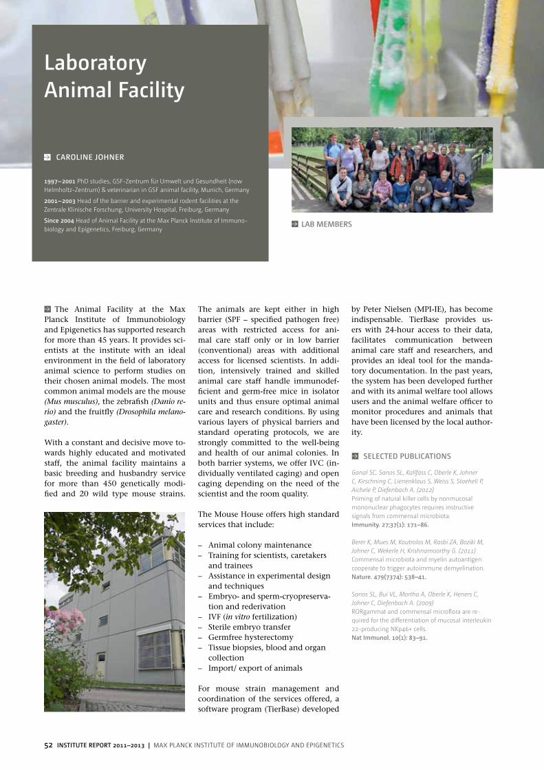
52 INSTITUTE REPORT 2011–2013 | Max Planck InstItute of IMMunobIology and ePIgenetIcs
The Animal Facility at the Max Planck Institute of Immunobiology and Epigenetics has supported research for more than 45 years. It provides sci-entists at the institute with an ideal environment in the field of laboratory animal science to perform studies on their chosen animal models. The most common animal models are the mouse (Mus musculus), the zebrafish (Danio re-rio) and the fruitfly (Drosophila melano-gaster).
With a constant and decisive move to-wards highly educated and motivated staff, the animal facility maintains a basic breeding and husbandry service for more than 450 genetically modi-fied and 20 wild type mouse strains.
The animals are kept either in high barrier (SPF – specified pathogen free) areas with restricted access for ani-mal care staff only or in low barrier (conventional) areas with additional access for licensed scientists. In addi-tion, intensively trained and skilled animal care staff handle immunodef-ficient and germ-free mice in isolator units and thus ensure optimal animal care and research conditions. By using various layers of physical barriers and standard operating protocols, we are strongly committed to the well-being and health of our animal colonies. In both barrier systems, we offer IVC (in-dividually ventilated caging) and open caging depending on the need of the scientist and the room quality.
The Mouse House offers high standard services that include:
– Animal colony maintenance – Training for scientists, caretakers
and trainees – Assistance in experimental design
and techniques – Embryo- and sperm-cryopreserva-
tion and rederivation – IVF (in vitro fertilization) – Sterile embryo transfer – Germfree hysterectomy – Tissue biopsies, blood and organ
collection – Import/ export of animals
For mouse strain management and coordination of the services offered, a software program (TierBase) developed
SElECTED PUblICATIOnS
Ganal SC, Sanos SL, Kallfass C, Oberle K, Johner C, Kirschning C, Lienenklaus S, Weiss S, Staeheli P, Aichele P, Diefenbach A. (2012)Priming of natural killer cells by nonmucosal mononuclear phagocytes requires instructive signals from commensal microbiota.Immunity. 27;37(1): 171–86.
Berer K, Mues M, Koutrolos M, Rasbi ZA, Boziki M, Johner C, Wekerle H, Krishnamoorthy G. (2011)Commensal microbiota and myelin autoantigen cooperate to trigger autoimmune demyelination.nature. 479(7374): 538–41.
Sanos SL, Bui VL, Mortha A, Oberle K, Heners C, Johner C, Diefenbach A. (2009)RoRgammat and commensal microflora are re-quired for the differentiation of mucosal interleukin 22-producing nKp46+ cells.nat Immunol. 10(1): 83–91.
laboratory Animal Facility
CAROlInE JOhnER
by Peter Nielsen (MPI-IE), has become indispensable. TierBase provides us-ers with 24-hour access to their data, facilitates communication between animal care staff and researchers, and provides an ideal tool for the manda-tory documentation. In the past years, the system has been developed further and with its animal welfare tool allows users and the animal welfare officer to monitor procedures and animals that have been licensed by the local author-ity.
1997–2001 PhD studies, GSF-Zentrum für umwelt und Gesundheit (now Helmholtz-Zentrum) & veterinarian in GSF animal facility, Munich, Germany
2001–2003 Head of the barrier and experimental rodent facilities at the Zentrale Klinische Forschung, university Hospital, Freiburg, Germany
Since 2004 Head of Animal Facility at the Max Planck Institute of Immuno-biology and Epigenetics, Freiburg, Germany
lAb MEMbERS

53
SElECTED PUblICATIOnS
Balbach ST, Esteves TC, Houghton FD, Siatkowski M, Pfeiffer MJ, Tsurumi C, Kanzler B, Fuellen G, Boiani M. (2012)nuclear reprogramming: kinetics of cell cycle and metabolic progression as determinants of success. PloS One. 7(4): e35322.
Bedzhov I, Liszewska E, Kanzler B, Stemmler MP. (2012)Igf1r signaling is indispensable for preimplantation development and is activated via a novel function of E-cadherin. PloS Genet. 8(3): e1002609.
Corbeaux T, Hess I, Swann JB, Kanzler B, Haas-Assenbaum A, Boehm T. (2010)Thymopoiesis in mice depends on a Foxn1-positive thymic epithelial cell lineage. Proc natl Acad Sci USA. 107(38): 16613–8.
The Transgenic Mouse Facility of the Max Planck Institute of Immunobi-ology and Epigenetics has been estab-lished in 1995 to enable the successful and efficient generation of genetically modified mice, thus allowing the sci-entific investigators to focus their re-search efforts on biological rather than technological aspects. We strive to pro-vide cutting-edge transgenic and gene-targeting technology, in both a time ef-fective and cost efficient manner. New mouse lines are generated either by transgenesis (via pronuclear microin-jection of specific DNA constructs into fertilized oocytes) or targeted mutagen-esis (via microinjection of embryonic stem (ES) cells into blastocysts or 8-cell stage embryos, as well as via diploid or tetraploid ES/morulas aggregations), e.g. “knockouts” or conditional mu-tants.Our barrier facility maintains a specific pathogen free (SPF) health status, hous-ing gene modified mice in ventilated cage systems (IVCs) that are serviced by intensively trained and skilled ani-mal care staff. Microinjected embryos are reimplanted under strict sterile conditions into pseudopregnant foster mothers inside this barrier. Serology testing of selected animals is systemati-cally completed to confirm their health status, thus allowing the transfer of the transgenic founders or positive gene targeted offspring to other areas of the animal house for further breeding and analysis. After germline transmitters have been identified the investigators become responsible for breeding and
analysis of mouse lines in compliance with the German law.
In the last three years, the Transgenic Mouse Facility has generated:
– Through ES cell microinjection: 238 injection days: 900 ES-cell derived mouse lines or chimeric embryos, corresponding to 48 distinct target-ing constructs
– Through DNA pronuclear injec-tion: 159 injection days: 516 new transgenic mouse lines or embryos, corresponding to 58 distinct trans-genes
In parallel, we strive to implement and develop new technologies, e.g. establishment of new methods for ES cell derivation or targeted mutagen-esis using Zinc-finger nucleases (ZFNs) or Transcription activator-like effector nucleases (TALENs).In order to keep track of all experimen-tal parameters, conduct data analysis and edit statistics and reports, a data-base has been developed. For each new mouse line generated, researchers are responsible for keeping accurate and regularly updated records (e.g. mat-ing details, breeding performances, genotyping and phenotyping results) in our internal mouse colony manage-ment database, TierBase. This software enables efficient communication with the scientists and the German authori-ties (Regierungspräsidium Freiburg), complying with the current legislation on animal welfare.
Transgenic Mouse Facility
lAb MEMbERS
From left: hiltl, Clarissa (Research Assistant), lopez, Elsa (Technical Assistant), Kemmer, Svenja (Research Assistant), Kanzler, benoit (Head of Facility)
1996 PhD thesis in Cellular and Molecular Biology with Prof. Danielle Dhouailly, Albert Bonniot Institute, university Joseph Fourier of Grenoble, France
1996–1999 Postdoctoral Fellow, Max Planck Institute, Freiburg, Germany, Labo-ratory of Dr. Moisés Mallo (Department of Developmental Biology)
Since 1999 Head of the Transgenic Mouse Facility at the Max Planck Institute of Immunobiology and Epigenetics, Freiburg, Germany
bEnOIT KAnzlER

54 INSTITUTE REPORT 2011–2013 | Max Planck InstItute of IMMunobIology and ePIgenetIcs
In general, higher vertebrates such as mouse and rat are used as model organisms in immunological research. Nevertheless, the zebrafish and Medaka model systems offer some advantages over higher vertebrates which can also be exploited to answer questions in immunological science. Zebrafish and Medaka are extraordinarily fecund. This opens up the possibility to carry out genetic studies such as mutagen-esis screens. Such screens are also per-formed to identify genes and pathways which are important for the develop-ment and function of the immune sys-tem. Sequencing of fish genomes and analysis of gene functions have shown that there are less differences between fish and humans than expected. This implies that results from mutagenesis screens in fish can be transferred to and used for studies on hereditary diseases affecting the human immune system.The rapid extracorporal development and the transparency of zebrafish and Medaka embryos is another advantage of these model organisms, making them superior to higher vertebrates for certain experimental approaches. For example, transgenic lines make it possible to monitor the development of the embryo and its organs in vivo on a cellular level. Furthermore, the fish embryo is easily accessible allowing manipulation of and interference with developmental processes.
Service
The fish facility at the Max Planck Institute of Immunobiology and
SElECTED PUblICATIOnS
Hess I, Boehm T. (2012)Intra-vital imaging of thymopoiesis reveals dy-namic lympho-epithelial interactions.Immunity. 36(2): 298–309.
Iwanami N, Mateos F, Hess I, Riff el N, Soza-Ried C, Schorpp M and Boehm T. (2011)Genetic evidence for an evolutionary conserved role of IL-7 signaling in T cell development of zebrafish. J Immunol. 186(12): 7060–7066.
Soza-Ried C, Hess I, Netuschil N, Schorpp M and Boehm T. (2010)Essential role of c-myb in definitive hematopoiesis is evolutionarily conserved. Proc natl Acad Sci USA. 107(40): 17304–17308.
Epigenetics was opened in 2007. It houses zebrafish (Danio rerio) and Me-daka (Oryzias latipes) fish. Each room is equipped with an independent water treatment unit. Access to the facility is restricted and automatically recorded; technical parameters of the entire facil-ity are remotely controlled. A separate quarantine room provides opportunity for short-term experiments without compromising the high hygienic stan-dards of the main facility. Currently, about 50 different lines carrying ENU-induced mutations affecting different developmental processes and several transgenic lines are kept in the sys-tem. Wild-type strains are maintained for breeding experiments as well as for general egg and embryo supply. The facility is run by a group of four staff members.
The following procedures are routinely used:
– Automated whole mount in situ hybridisation
– BAC transgenesis – Cell transplantations in embryos
and adult fish – Homozygosity mapping/positional
cloning – Pressure-driven microinjection of
mRNA, DNA, or antisense morpho-lino oligonucleotides into fertilized eggs
– Sperm cryoconservation and in vitro fertilisation
Fish Facility
MIChAEl SChORPP
1993–1996 PhD at university of Freiburg and German Cancer Research Center, Heidelberg, Germany
1996–2000 Postdoctoral Research Fellow at the German Cancer Research Center, Heidelberg and at the Max Planck Institute of Immunobiology, Freiburg, Germany
Since 2000 Project Leader and Head of Fish Facility (since 2009) at the Max Planck Institute of Immunobiology and Epigenetics, Freiburg, Germany
lAb MEMbERS

55
SElECTED PUblICATIOnS
Ilik IA, Quinn JJ, Georgiev P, Tavares-Cadete F, Mat-iczka D, Toscano S, Wan Y, Spitale RC, Luscombe N, Backofen R, Chang HY, Akhtar A. (2013)Tandem stem-loops in rox RnAs act together to mediate x chromosome dosage compensation in Drosophila.Mol Cell. 51(2): 156–73.
Hallacli E, Lipp M, Georgiev P, Spielman C, Cusack S, Akhtar A*, Kadlec J*. (2012) Msl1-mediated dimerization of the dosage com-pensation complex is essential for male x-Chromo-some regulation in Drosophila.Mol Cell. 48(4): 587–600. *co-corresponding authors
Georgiev P, Chlamydas S, Akhtar A. (2011)Drosophila dosage compensation: males are from Mars, females are from venus.Fly (Austin). 5(2): 147–54.
The fruit fly Drosophila is one of the most extensively characterized meta-zoan organisms. Over more than 100 years since it was introduced as an ex-perimental animal model for biological research, it has allowed for key biologi-cal concepts and phenomena to be elu-cidated, leading to some of biology’s fundamental findings, including the chromosomal theory of heredity and basic genetic mechanisms underlying animal development and evolution.Importantly, many aspects of devel-opment and behaviour in Drosophila parallel those in humans. The comple-tion of both the human and Drosophila genome sequencing projects revealed that more than 75% of human genetic disease genes have clear homologues in the fruit fly. Taking advantage of the significantly shorter life cycle, large number of offspring and powerful array of genetic and molecular tools avail-able in Drosophila, it is now feasible to perform large-scale genetic screens in Drosophila to identify novel drugs and therapeutic targets.The fly facility was set up in 2010 upon recruitment of Dr. Asifa Akhtar. The facility contains a fly room with 7 dis-section microscopes and CO2 supply
for day-to-day fly pushing and a stereo microscope for fluorescent imaging. Attached to the fly room are 25°C and 18°C light-, temperature- and humid-ity-controlled rooms for fly stocks. In addition, there are separate rooms al-located for large population cages al-lowing embryo collections for chro-matin isolation and an 18°C room for a collection of mutant and transgenic lines maintained to facilitate rapid am-plification upon request. In addition, a fly food preparation kitchen with a large production capacity has been es-tablished for the maintenance of these flies as well as for the daily needs of the fly groups. A state-of-the-art microin-jection unit equipped with an inverted microscope, Eppendorf Femtojet, mi-cromanipulator and embryo aligning stage, allows Drosophila embryo trans-formation and generation of transgenic animals using transposase-mediated random insertion as well integrase-me-diated site-specific integration of trans-genic constructs.
At present there are four research groups at the MPI-IE using Drosophila as a model organism and the fly facility aims to offer an organized infrastruc-
ture, consult and support in applying advanced genetic techniques (such as gene targeting by homologous recom-bination or TALENS, ZFNs, CRISPR/Cas-based methods for genome engi-neering), designing large-scale forward genetic screens and developing new ge-netic techniques for the specific needs of the fly-related research of these groups.
Fly Facility
From left: Georgiev, Plamen (Head of Facility), lenz, Ute (Technician). . Not present: Gutierrez, Noel (Technician)
2004–2007 PhD studies in Molecular biology/Genetics at Cambridge uni-versity, Cambridge, uK
2007–2010 Postdoctoral fellow at the Babraham Institute, Cambridge, uK
Since 2010 Head of Fly Facility at the Max Planck Institute of Immunobiol-ogy and Epigenetics, Freiburg, Germany
PlAMEn GEORGIEv
lAb MEMbERS

56 INSTITUTE REPORT 2011–2013 | Max Planck InstItute of IMMunobIology and ePIgenetIcs
The Flow Cytometry Unit, utilizing high quality flow cytometry instru-mentation, provides comprehensive and technically sophisticated cell anal-ysis and sorting services as well as flow cytometry training and education for new users.Flow cytometry is a method that allows measurements of various characteris-tics of individual cells by using fluo-rescent probes. Our laboratory has the capability to run a broad diversity of advanced flow cytometry applications.
The DNA Sequencing Unit provides access to automated DNA sequencing. The sequencing service processes sam-ples on a 48-capillary ABI 3730 DNA Analyser. In 2012, we had a total of 50.000 sequences.
Currently available applications include:
– cloning and cell sorting – multi-color immunofluorescence – DNA cell cycle analysis – measurements of apoptosis – calcium flux – fluorescence resonance energy
transfer (FRET)
The facility is equipped with the most advanced, state-of-the-art flow cyto-meters. For sorting, we have a Beckman Coulter MoFlo XDP sorter, two Becton Dickinson (BD) FACSAria sorters, a (BD) Influx sorter and a Miltenyi au-toMACS magnetic sorter. For analysis of
cell samples our facility provides three LSR II, one LSRFortessa and two FACS-Caliburs (all from BD). The MoFlo, the Influx and the FACSArias are versatile high speed sorters equipped with up to five lasers, enabling the measurement of up to nine fluorescent parameters (MoFlo and FACSAria II) and up to twelve fluorescent parameters (Influx and Aria III), respectively. Addition-ally the Influx is the worldwide unique steam-in-air sorter equipped with the octagon and trigon optical arrays. The light from the five spatially separated laser beam spots is delivered by fiber optics to the octagon and trigon detec-tor arrays increasing the sensitivity and flexibility of the BD Influx, yielding more information from each sample.
All flow cytometers can simultaneously sort four populations at up to 30.000 cells per second with greater than 99,8% purity and high recovery. Select-ed populations can be sorted into test tubes or deposited directly into a 96-well plate. Sterile cell sorting is avail-able upon request allowing subsequent culturing of sorted cells. The magnetic cell separation system (autoMACS) can isolate large numbers of cells for a sin-gle surface marker with high purity in a short period of time. The cell sorters are available on weekdays upon special arrangement with the operators. These instruments are often booked weeks in advance. In 2011 we sorted more than 6.000 samples.
Flow Cytometry and DnA Sequencing Facility
From left: Knoblauch, Maike (Technician), würch, Andreas (Head of Facil-ity), wild, Doris (Technician). Not present: Hobitz, Sebastian (Technician), Burger, Claudia (Technician)
1986 undergraduate studies in Biology, university of Freiburg , Freiburg, Germany
1993–1999 Research Assistant, Department of Cellular and Molecular Im-munolgy, Prof. Dr. Klaus Eichmann, Max Planck Institute of Immunobiology, Freiburg, Germany
Since 2000 Head of FACS Facility at the Max Planck Institute of Immunobiol-ogy and Epigenetics, Freiburg, Germany
AnDREAS wüRCh
lAb MEMbERS
The Influx sorter is a worldwide unique steam-in-air sorter equipped with the octagon and trigon optical arrays.

57
From left: Cohen, Meital (Staff Scientist), nerz, Gabriele (Technician), Mittler, Gerhard (Head of Facility), Richter, Florian (Postdoc)
The proteomics facility is offering a state-of-the-art Mass Spectrometry (MS) analysis service and is collaborating with research groups at the institute in order to develop custom-tailored strat-egies to address important biological questions. We are specialized in func-tional proteomics, which is concerned with correlating physiological changes at the cellular level with alterations in protein composition and protein mod-ifications as a major discovery and hy-pothesis generating tool.
To pursue this we employ metabolic labeling using stable isotopes (SILAC) in order to not only achieve a com-prehensive qualitative but also a quan-titative description of proteomes using nanoLC-MS technology. This enables us to study both complex proteomes (organelles) and interactomes like protein-protein or protein-DNA com-plexes.
The facility is running three hybrid electrospray ionization (ESI) FT-MS instruments that are coupled online to nanobore liquid-chromatography systems. One system consists of a LTQ-FT Ultra mass spectrometer that possesses unrivaled mass resolution (greater one million) and mass accu-racy (sub-ppm). The second system is a LTQ Orbitrap XL+ETD mass spectrom-eter, which offers excellent resolution (>100.000), mass accuracy (sub-ppm) and high sensitivity. This instrument is equipped with ETD (electron transfer dissociation) and HCD (high collision energy dissociation) capabilities that
can give additional structural informa-tion useful for de-novo sequencing and post-translational modification (PTM) analysis. A Q Exactive high-performance benchtop quadrupole Orbitrap MS represents the most recent addition to our facility, offering very high sen-sitivity, resolution (140.000) mass ac-curacy (low ppm) and HCD MS/MS acquisition speed (>10 Hz) making it the instrument of choice for shotgun proteomics as well as targeted ID and quantification experiments (“quanfir-mation”).For offline protein and peptide chro-matography, SMART and ETTAN-LC microbore HPLC systems are used. Isoelectric focusing (IEF) of peptides is performed on an Agilent 3000 off-gel fractionator. Sample preparation com-monly involves in gel, in solution, FASP or on bead trypsin digestion fol-lowed by clean up using C18 and SAX STAGE tip solid phase extraction pro-tocols.
For peptide and protein identifica-tion we use an in-house Mascot data-base server along with tools from the MSQuant open-source environment. Quantitative proteomics experiments (SILAC and label free) are analysed by MaxQuant and Perseus software that are both developed in the department of Prof. Matthias Mann at the MPI for Biochemistry.
Standard service includes protein iden-tification and peptide mapping (using multiple proteases) from both colloi-dal coomassie and silver-stained gel
bands and low-complexity proteomes (e.g. protein complexes, affinity pull-downs etc.). PTM characterization and quantitative proteomic analysis by ei-ther SILAC or label free approaches are much more time consuming and are therefore considered as special analy-sis. Global proteomic projects are eval-uated and ranked by an in-house com-mittee whose current members are A. Pichler, R. Grosschedl and G. Mittler.
Proteomics Facility
SElECTED PUblICATIOnS
Tropberger P, Pott S, Keller C, Kamieniarz-Gdula K, Caron M, Richter F, Li G, Mittler G, Liu ET, Buhler M, Margueron R, Schneider R. (2013)Regulation of transcription through acetylation of H3K122 on the lateral surface of the histone octamer.Cell. 152(4): 859–872.
Flach H, Rosenbaum M, Duchniewicz M, Kim S, Zhang SL, Cahalan MD, Mittler G, Grosschedl R. (2010)Mzb1 (pERp1) regulates calcium homeostasis, anti-body secretion and integrin activation in innate-like B cells.Immunity. 33(5): 723–725.
Mittler G, Butter F and Mann M. (2009)A SILAC-based DnA-protein interaction screen that identifies candidate binding proteins to functional DnA elements.Genome Res. 19(2): 284–293.
1998–2002 PhD studies (Protein Biochemistry and Molecular Cell Biology), Gene Center LMu and Helmholtz Center Munich
2002–2005 Postdoctoral Fellow, laboratory Prof. Dr. Matthias Mann, Center of Experimental Bioinformatics, university of Southern Denmark, odense, Denmark
Since 2005 Group Leader and Head of Proteomics Facility (since 2010), Max Planck Institute of Immunobiology and Epigenetics, Freiburg, Germany
GERhARD MITTlER
lAb MEMbERS

58 INSTITUTE REPORT 2011–2013 | Max Planck InstItute of IMMunobIology and ePIgenetIcs
n.n. (Head of Facility), Kindle, Petra (Interim Head of Facility)
Understanding the roles gene pro-ducts play in biological systems is a key challenge in the post-genomic era. To accomplish this task, visualization techniques based on fluorescence mi-croscopy have become indispensable for gathering information on localiza-tion, morphology and dynamics of biological structures. Today, a broad range of reagents, including organelle-selective dyes and antibody conjugates, permit specific fluorescence labeling of virtually every cellular component. Furthermore, genetically encoded probes, such as fluorescent proteins greatly facilitate labeling and monitor-ing of dynamic events in vivo. Due to improvements in fluorescence label-ing, electronic light sensors and the introduction of digital imaging tech-niques, fluorescence microscopy is a key technology in modern bio-medical research. The imaging facility at the
Max Planck Institute of Immunobiol-ogy and Epigenetics provides a collec-tion of state-of-the-art light microsco-py instruments and image processing tools, including assistance through the microscopic imaging process.
Overview of the Equipment
Confocal microscopes and live cell imaging
– Zeiss LSM 510 Meta NLO with 4 la-sers, Argon (458, 477, 488, 514 nm), DPSS 561 nm, HeNe 633 nm, Cha-meleon (tunable 705-980 nm), heat-ing table for temperature control, HBO 100W for Vis mode, filter sets for GFP, RFP, DAPI, CFP, YFP and NDD (non descanned detection for 2P)
– LSM 780 Zeiss (new GaAsP detec-tors), with integrated Fluorescence Correlation Spectroscopy (FCS), Flu-
orescence Cross-Correlation Spec-troscopy (FCCS), and Raster Image Correlation Spectroscopy (RICS), 4 lasers (Argon (458, 488, 514nm)), DPSS 561nm, HeNe 633nm, Diode 405nm, HXP 120 W for Vis mode, Filtersets for GFP, RFP and DAPI
– Spinning Disc Zeiss: special type of confocal microscope for very fast live cell imaging with 2 cameras Mrm (CCD) and Evolve ( EMCCD), 3 lasers, OPSL 488 nm, Diode 561 nm, Diode 405 nm, small incuba-tion chamber for CO2/Temperature control, HXP for Vis mode, filter sets for GFP, RFP, DAPI
– Axiovert 200M Zeiss with Eppendorf Femto Jet Colibri with LED 385, LED 470, LED 555 and LED 625, DICII and DIC III, filter sets for GFP, RFP, DAPI/ GFP/ Red/ FarRed and Mrm camera
– Tokai Hit Incubation unit for CO2 and temperature control, versatile for all inverted Zeiss microscopes
Structured Illumination
– Zeiss Imager Z1 (upright) with Apo-Tome slider with XBO 75 W, 6 filter cubes for GFP, RFP, CFP, YFP, FarRed and DAPI, DICII and DIC III, Mrm and Mrc camera, Axiovision 4.82 software
– Zeiss Imager Z1 (upright) with Ap-oTome slider with HXP 120 W, 6 filtercubes for GFP, RFP, CFP, YFP, FarRed and DAPI, DICII and DIC III, Mrm camera, Zen 2012 software.
Imaging Facility
1983–1985 Training to medical technical assistant, MTA-Schule Freiburg
1985–2005 Employee at the university Hospital of Freiburg
Since 2005 Research Assistant, Imaging Facility, Max Planck Institute of Immunobiology and Epigenetics, Freiburg, Germany
PETRA KInDlE
lAb MEMbERS
Postdoc Carmen Galán at the spinning disc microscope.

59
From left: bönisch, Ulrike (Scientist), betancourt, Emily (Technician), Arrigoni, laura (Postdoc)
The Deep Sequencing Facility at the Max Planck Institute of Immunobiol-ogy and Epigenetics (MPI-IE) offers high-throughput sequencing services mainly for in-house research groups. The facility is equipped with modern state-of-the-art technology, highly qualified staff and interacts closely with the bioinformatics facility at the MPI-IE. Next to standard sample pro-cessing huge effort is put into the opti-mization, automation and standardiza-tion of existing workflows according to internationally emerging standards.Apart from serving all groups at the in-stitute, the unit is also part of a local (MEDEP) and a large German-wide re-search consortium (DEEP) for which it produces ChIP-seq data and reference epigenomes from various tissue types. As one of the six epigenome production centers in Germany, the facility has set
up a complete semi-automated ChIP-Seq pipeline that enables chromatin extraction, chromatin immunoprecipi-tation, library preparation and deep se-quencing according to highest interna-tional standards and quality controls.
Technology
The unit employs the Illumina tech-nology that uses massively parallel se-quencing to generate several billion bas-es of high quality nucleotide sequences per run. To this end DNA molecules are attached to a surface, amplified and the complementary DNA strand re-synthe-sized by consecutive incorporation of fluorescent reversible terminator de-oxyribonucleotides (a technology re-ferred to as sequencing-by-synthesis). Surface-images are analysed to gener-ate high-quality sequences. Using the Illumina sequencing technology maxi-
mum read length of 100 bp (HiSeq2500®, high-output mode), 150 bp (HiSeq2500®, rapid-run mode) as well as 300 bp (MiSeq®) is feasible. Furthermore paired-end sequencing can be applied as well as barcoding to in-
crease the number of parallel analysed samples.
A robust instrumentation infrastruc-ture is in place for standardized and quality controlled sample preparation. This includes a Covaris S220, an Agi-lent Bioanalyser, a Qubit Fluorometer as well as liquid handling stations for automated chromatin immunopre-cipitation and library preparation. To manage the huge data flow generated by the facility, we utilize a large data center and computer cluster for stor-age, analysis and visualization.
Our service covers – Preparation of multiplexed libraries
for cluster generation and sequenc-ing on the HiSeq2500 and MiSeq instrument
– Tutorials and training of research-ers in sample and library prepara-tion techniques
Main applications supported by the Deep sequencing facility are – ChIP-Seq – RNA-Seq (Poly-A, Ribo-Minus, – directional/non-directional), – gDNA-Seq – Amplicon-Seq
Deep Sequencing Facility
UlRIKE bÖnISCh
2002–2007 undergraduate studies in nutritional Sciences, university of Hohenheim, Stuttgart, Germany
2007–2012 PhD Student, Department of Microsystems Engineering, univer-sity of Freiburg, Freiburg, Germany
Since 2012 Scientist, Deep Sequencing Facility, Max Planck Institute of Im-munobiology and Epigenetics, Freiburg, Germany
ulrike Bönisch loads the MiSeq system. In the foreground: HiSeq2500® that conducts up to 3 billion single reads.
lAb MEMbERS

60 INSTITUTE REPORT 2011–2013 | Max Planck InstItute of IMMunobIology and ePIgenetIcs
From left: Ramirez, Fidel (Postdoc), Richter, Andreas (Postdoc), Diehl, Sarah (Research Assistant), Manke, Thomas (Head of Facility), Dündar, Friederike (PhD Student)
The regulation of gene expression is controlled by multiple mechanisms, such as the sequence-specific binding of transcription factors to DNA, epigen-etic signals and a dynamic chromatin state. An unbiased understanding of these processes requires access to large-scale experiments and the capacity to analyse genome-wide data. Apart from supporting many collaborative projects at the MPI-IE, the group interacts very closely with the Deep Sequencing Fa-cility that generates data at an unprec-edented scope, resolution and rate. The bioinformatics facility is operating a powerful Data Center to process, anal-yse and visualize this information. We also provide regular training and help our colleagues to interpret genome-wide data.
Data Center
We operate a state-of-the-art data cen-ter with a dedicated cooling system, more than 500 high-speed cores, 100TB storage and backup. This infrastructure is constantly upgraded and provides a powerful backbone for primary analy-sis of sequencing data, various web ser-vices, and for the extensive secondary data analysis by the bioinformatics fa-cility and other internal users. We also implemented hundreds of standardized tools, protocols and Linux software for data management and analysis.
Epicenter web Service
Apart from offering direct access to our Linux servers, we also host a number of different web-services (Galaxy, Rstudio, GenomeBrowser) that help to access primary deep-sequencing data, per-form standardized analyses and visual-izations. We provide an interactive web interface for data storage, management and sharing. At its core the Galaxy web-service includes many customiz-able tools and extensive workflows for deep-sequencing analysis, visualization and data integration.
Training
We offer regular bioinformatics train-ing courses and interactive tutorials on genome-wide data analysis, visual-ization, and statistical interpretation. These courses take place twice per year and are open to all members of the MPI-IE and PhD students.
bioinformatics Facility
ThOMAS MAnKE
Raw sequences are obtained as large images with different
colour codes for different nucelotides. Based on the measured intensities, it is
possible to accurately assign a nucleotide to each spot of the image. The enormous degree
of parallelization allows for the simultaneous determina-
tion of billions of basepairs.
SElECTED PUblICATIOnS
Bulut-Karslioglu A, Perrera V, Scaranaro M, de la Rosa-Velazquez IA, van de Nobelen S, Shukeir N, Popow J, Gerle B, Opravil S, Pagani M, Meidhof S, Brabletz T, Manke T, Lachner M, Jenuwein T. (2012)A transcription factor-based mechanism for mouse heterochromatin formation. nat Struct Mol biol. 19(10): 1023–30.
Lam KC, Muhlpfordt F, Vaquerizas JM, Raja SJ, Holz H, Luscombe NM, Manke T, Akhtar A. (2012)The nSL Complex Regulates Housekeeping Genes in Drosophila. PloS Genet. 8(6): e1002736.
Thomas-Chollier M, Hufton A, Heinig M, O‘Keeffe S, Masri NE, Roider HG, Manke T, Vingron M. (2011)Transcription factor binding predictions using TRAP for the analysis of ChIP-seq data and regulatory SnPs. nat Protoc. 6(12): 1860–9.
1994–1998 PhD studies (Theoretical Physics) at Cambridge university, uK
1998–2010 Postdoc at Tsukuba university, Japan, at Columbia university, new york, uSA, and at Max Planck Institute for Molecular Genetics, Berlin, Germany
Since 2010 Group Leader and Head of Bioinformatics/nGS unit, Max Planck Institute of Immunobiology and Epigenetics, Freiburg, Germany lAb MEMbERS

61
04 Around the Institute

62 INSTITUTE REPORT 2011–2013 | Max Planck InstItute of IMMunobIology and ePIgenetIcs
little Scientists
The institute offers child care premises located right next to the institute. The facility is run by Concept Maternel, a well-respected non-profit organization which runs several child care facili-ties and a primary school in Freiburg. Its pedagogical concept is based on the principles of Montessori, Freinet and Pikler and promotes the indivi-dual development of each child. Since June 2012, Concept Maternel provides education for children aged between
Annual Institute Retreat
Each year in October, all scientific staff at the institute, including students and research assistants, take part in a 2-day retreat that aims at an intensified sci-entific exchange between members of the MPI-IE. In 2011 and 2012, the event took place in Saint Hippolyte in Alsace, France. In 2013, Phd students, postdocs and group leaders attended individual retreats that were tailored to focus on specific questions in these peer groups.
Marathon Men and women
It is a great festival every year in April: after winter the runner's scene meets in Southern Baden to start the new season with the Freiburg Marathon. For this event, members of the MPI-IE build a runners team, consisting of both Mara-thon newcomers and experts. Every-body is welcome to take part in the MPI-IE running team.
Max Planck health Day
How to avoid back pain? How to recog-nize burnout? And how to eat healthy even when in a hurry? During June 2013, all employees of the MPI of Im-munobiology and Epigenetics (MPI-IE) and the MPI of Foreign and interna-tional Law (MPICC) were invited to the 1. Max Planck Health Day. With this initiative, the two Max Planck Insti-tutes set a sign for a healthier working environment. The employees received advice for ergonomic work places, par-ticipated in back coachings and learned about strategies of stress prevention.
life at the Institute
AROUnD ThE InSTITUTE
6 months to 3 years. In autumn 2012, building operations started for the erection of a large new building which will also be in immediate vicinity of the MPI-IE. The extension will enable us from December 2013 to add a “Kin-dergarten” group for children aged 3 to 6 years to the facility. This way, MPI-IE employees will have the option of leav-ing their child in the same facility until primary school.
The lounge with its relaxing atmosphere is the central meeting point for members of the institute.

63
AROUnD ThE InSTITUTE
life in and around Freiburg
characteristic cuckoo clocks and tra-ditional costumes. It is one of Ger-many's highest mountain ranges, with the “Feldberg” only 45 minutes from Freiburg. It is an ideal terrain not only for nature lovers for hillwalking ski-ing, mountain biking, and paraglid-ing. The nearby vineyards of Kaiser-stuhl and Markgräfler Land provide Freiburg and beyond with excellent wines such as “Weissburgunder” and “Spätburgunder”. In the French re-gion of Alsace, Colmar and the village Riquewihr are attractive locations, not only due to their historical city center. From Freiburg, the door to Switzer-land is Basel. Many museums reside in or next to the Swiss city. Basel is also called the “pharmaceutical city” since the headquaters of various pharmaceu-tical companies are located here. From there, some of the most prominent Swiss mountains like the Eiger can be reached easily.
Freiburg is most commonly known as the capital of The Black Forest and is located in one of the most scenic corners of Southern Germany, situated between the borders to Switzerland and France. Freiburg is a very young and lively city due to a high percent-age of students. The town with its ap-proximately 200.000 inhabitants is
characterized by 25,000 students at the University of Freiburg. 15% among them are from abroad, half European and non-European, which adds to the
international flair of the town. The calm climate and the local cuisine of Baden, crowned with numerous first class restaurants in and around Freiburg, make Freiburg a town highly attractive to visitors from all over the world.The cultural life of Freiburg is also very diverse. A big concert hall, several the-aters, and many clubs offer events on a regular basis. During the summer time, various open air concerts and festivals take place, the Zeltmusikfestival ZMF being one of the biggest.
life around Freiburg
Within the last 50 years, the upper Rhine valley has undergone a tremen-dous change from a nearly rural area, narrowed by borders, to the heart of Eu-rope, with Milan and Paris being closer than the German capital Berlin. Since August 2013, a TGV train directly con-nects Freiburg and Paris, in 3.5 hours. The Black Forest, flanking Freiburg in the east, has lots to offer, besides the
The district Stühlinger: close to the city center and popular with students.
Daily event: Market in Freiburg.
view from the Feldberg to the Swiss Alps. Mountainbiking and Paragliding in the Black Forest.
© M
anfr
ed G
erbe
r, pi
xelio
.de
© A
xel D
rain
ville

64 INSTITUTE REPORT 2011–2013 | Max Planck InstItute of IMMunobIology and ePIgenetIcs
Special Guest Seminar Series
13. January 2011 Daniela RhodesMRC Laboratory of Molecular Biology, Cambridge, UKChromatin structure and regula-tion of its compaction
03. February 2011 Günter HämmerlingGerman Cancer Research Cen-ter, Heidelberg, GermanyConsequences of weak and strong interactions between T cells and antigen-presenting cells
10. February 2011Tom Owen-HughesUniversity of DundeeDundee, UKChromatin remodelling and genome organization
10. March 2011 Howard CedarHebrew University Medical School, Jerusalem, IsraelProgramming DNA methylation during development
28. March 2011 Mark ShlomchikYale University School of Medi-cine, New Haven, USAGerminal center selection and the development of memory B and plasma cells
07. April 2011 Alfred SingerNational Institute of Cancer, NIH, Bethesda, USAHow MHC specificity is imposed in the thymus Kevin StruhlHarvard Medical SchoolBoston, USAAn epigenetic switch linking in-flammation to cancer and the role of polycomb complexes in cancer stem cells
28. April 2011 Shiv PillaiHarvard Medical SchoolBoston, USAClonal ignorance and genetic susceptibility to human autoim-mune disorders
12. May 2011 Daniela FinkeUniversity of BaselBasel, SwitzerlandIntrinsic and extrinsic signals regulating the development and function of lymphoid tissues
19. May 2011 Ichiro TaniuchiRIKEN Research Center for Al-lergy and ImmunologyYokohama, JapanTranscriptional regulation of thymocyte development
26. May 2011 Evan RosenHarvard Medical SchoolBoston, USAEpigenomic approaches to path-way discovery in adipose biology
07. July 2011 Sidonia FagarasanRIKEN Research Center for Al-lergy and ImmunologyYokohama, JapanDynamic interactions between bacteria and immune cells in the gut
29. September 2011 Gerhard ChristoforiUniversity of BaselBasel, SwitzerlandMolecular dissection of epithelial-mesenchymal transition (EMT)
30. September 2011 Ben BlackUniversity of PennsylvaniaPhiladelphia, USAThe molecular underpinnings of centromere identity and mainte-nance
11. October 2011 Bing RenUCSD School of MedicineSan Diego, USADecoding the epigenome
18. October 2011 Nikolaus RajewskyMax-Delbrück Center for Mo-lecular Medicine Berlin-BuchBerlin, GermanyGene regulation by small RNAs and RNA binding proteins
17. novemeber 2011 Matthias SchäferGerman Cancer Research Cen-ter, Heidelberg, GermanyDnmt2-mediated RNA methyla-tion and endogenous RNA stress
28. november 2011 Mitchell LazarUniversity of PennsylvaniaPhiladelphia, USAEpigenomic regulation of metabo-lism
The Special Guest Seminar Series has long been established as a regular event featur-ing talks by highly recognized scientists from across the globe. The topics range from sci-ence directly relevant to the research of the MPI-IE to novel and innovative research from other areas of biomedical science.
2011
Discussion in the lounge after the seminar.
AROUnD ThE InSTITUTE

65
02. February 2012 Lars JansenInstituto Gulbenkian de CiênciaOeiras, PortugalThe centromere: A showcase for epigenetic inheritance
10. January 2013 Johan AuwerxEcole Polytechnique FédéraleLausanne, SwitzerlandMitochondrial proteostasis and the regulation of metabolism on longevity
09. February 2012 Ronald HayUniversity of DundeeDundee, UKHow SUMO talks to ubiquitin
23. February 2012
Eric VivierCentre of ImmunologyMarseille-Luminy, FranceNatural Killer cells
24. January 2013 Bruce EdgarRuprecht-Karls-University of Heidelberg, Heidelberg, Germany Stem cell-mediated homeostatic and dysplastic growth in the Drosophila intestine
1. March 2012 Neil BrockdorffUniversity of Oxford Oxford, UKPolycomb – a complex problem
7. February 2013 Edvard MoserNorwegian University of Sci-ence and TechnologyTrondheim, NorwayBrain maps for space
15. March 2012Nicholas ProudfootUniversity of OxfordOxford, UKEnding the message: Roles of R-loops, gene loops and non coding RNA in terminating transcription in eukaryotes
7. March 2013Catherine DargemontInstitute Jacques MonodParis, FranceUbiquitin conjugation: a timing mechanism for nuclear functions
14. March 2013 Giacomo CavalliInstitute of Human GeneticsMonpellier, FranceGenome organization and epigen-etic control of development
21. March 2013 Angelika AmonMassachusetts Institute of Tech-nology, Cambridge, USAConsequences of aneuploidy
4. April 2013 Eran SegalWeizmann Institute of ScienceRehovot, IsraelUnraveling the effect of DNA sequence on chromatin and transcriptional regulation using thousands of designed promoters
3. May 2012 Peter LaneUniversity of BirminghamBirmingham, UKEvolution and involution of CD4 memory and effector function
10. May 2012 Peter VogtThe Scripps Research InstituteLa Jolla, USAPI3K signaling in cancer
11. May 2012 Tasuku HonjoKyoto University Graduate School of MedicineKyoto, JapanAn evolutionary view of the mechanism for immune and genome diversity
14. May 2012 Bruce BeutlerUniversity of Texas, Dallas, USAGenetic analysis of immune sens-ing and signaling in mammals
5. June 2012 Howard ChangStanford University School of Medicine, Stanford, USAGenome regulation by long non-coding RNAs
24. April 2013 Steffen JungWeizmann Institute of ScienceRehovot, IsraelIntestinal macrophages in gut homeostasis and inflammation
7. May 2013 Oliver RandoUniversity of MassachusettsMassachusetts, USAGenomic approaches to epigenetic inheritance in yeast and mouse
23. May 2013 Robin AllshireUniversity of EdinburghEdinburgh, UKEstablishing and propagating specialised chromatin states
13. June 2013 Jesper SvejstrupCancer Research UK London Research InstituteLondon, UKThe interface between transcrip-tion and genome instability
5. July 2012 John SchwabeUniversity of LeicesterLeicester, UKInsights into the assembly, regula-tion and specificity of HDAC: corepressor complexes
23. August 2012 Matthias MannMax Planck Institute of Biochemistry, Martinsried, GermanyThe modern, mass spectromet-ric toolbox for proteomics and its application in biology and biomedicine
11. September 2012 Ellen RothenbergCalifornia Institute of Technol-ogy, Pasadena, USAForging T-lymphocyte identity: transcriptional and epigenetic control
12. September 2012 Sadaf FarooqiCambridge Institute for Medical Research, Cambridge, UKGenetic and molecular mecha-nisms involved in human obesity
27. September 2012 Gioacchino NatoliEuropean Institute of OncologyMilan, ItalyThe macrophage epigenome and the control of inflammatory gene expression
27. June 2013 Sten Eirik JacobsenUniversity of OxfordOxford, UKCellular pathways and molecular determinants of early lympho-myeloid fate decisions
7. October 2013 Job DekkerUniversity of MassachusettsWorchester, USA3D folding of genomes
14. October 2013 Thomas EickbushUniversity of RochesterNew York, USADynamic features of the rDNA locus allow the long term survival of a mobile element
7. november 2013 Patrick MatthiasFriedrich Miescher InstituteBasel, SwitzerlandAnalysis of HDAC function in lymphoid cells and beyond
12. October 2012 Patrick TamUniversity of SydneySydney, AustraliaIntersection of transcriptional and signalling activity for head mor-phogenesis in mouse embryos
18. October 2012 Richard TreismanCancer Research UK London Research InstituteLondon, UKG-actin as a regulator of protein function
14. november 2012 Steven ShoelsonHarvard University Medical School, Boston, USATargeting inflammation in diabe-tes and cardiovascular disease
20. november 2012 Karen AdelmanNational Institute of Environ-mental Health Science, NIHNorth Carolina, USAPromoter-proximal pausing of RNA polymerase II: a novel means to regulate immune responses
22. november 2012 Kai SimonsMax Planck Institute of Molecu-lar Cell Biology and GeneticsDresden, GermanyLipids organizing cell membranes
2012
2013

66 INSTITUTE REPORT 2011–2013 | Max Planck InstItute of IMMunobIology and ePIgenetIcs
Exit Freiburg nord
Isfa
han
alle
eZä
hrin
ger
Str.
Stübeweg
Center of Freiburg / Main train stationBase
l
Karl
sru
he
A5
B294
Hans-Bunte-Str.
Tullastr.
Zink
mat
tens
tr.
B3
MPI-IE
Bus 7200/7206
Tram 2
h
h
Directions to the Institute
Max Planck Institute of Immunobiology and EpigeneticsStübeweg 51D-79108 FreiburgGermany
Phone: +49 761 5108 -0 Fax: +49 761 5108 -221Mail: [email protected]: www.ie-freiburg.mpg.de
by public transport (from main train station)
Tramway Station at the south end of the main station. Take either tram no. 1 (dir. Littenweiler), 3 (dir. Vauban) or 5 (dir. Rieselfeld) until “Bertoldsbrun-nen” (2 stops). Then change to tram no. 2 (dir. Zähringen) until “Tullastra-ße” (6 stops from “Bertoldsbrunnen”). Walk along the “Tullastraße” until “Zinkmattenstraße” (roundabout) and follow this street until “Stübeweg” on your left (a 15–20 min walk).Alternatively, you can use busses 7200/7206 from the ZOB (central bus station), next to the main train stati-on. Take the bus until “Stübeweg” (ap-prox. 14 min ride). After you leave the bus, Stübeweg is on the right. The MPI-IE is on the right hand side.
by airplane
Via Frankfurt International Airport After arrival at the Frankfurt Interna-tional Airport go to the long-distance train station (Fernbahnhof). From the-re, it is a 2h train ride to Freiburg main train station.
Via Basel/Mulhouse/Freiburg AirportAfter arrival take the Airport bus to Freiburg (app. 1h bus ride). It will stop at Freiburg main train station.
by car
From the A5 Autobahn, Karlsruhe to Basel, take the Exit “Freiburg Nord”; keep in the left lane and take Highway B294 in the direction of Freiburg. After B 294 runs into Highway B 3 towards Freiburg, take the Exit “Industriegebiet Nord”. At the traffic light, go through the large intersection and make a half-left turn into “Hans-Bunte-Straße”. Turn left at Stübeweg. The MPI-IE is on your right.
AROUnD ThE InSTITUTE
67
Published by Max Planck Institute of Immunobiology and Epigenetics
november 2013
Content responsibilityThomas Jenuwein, Managing Director
TextsJohannes Faber, Team Leaders
Images (unless otherwise stated): Scientific images/illustrations provided by team leaders.Major photo contributions by Carmen Galán, Johannes Faber, May-Brit Hansen and from the MPI-IE archive.
layoutmarkenfaktur, Heidelberg
Printed byLitho Druck GmbH – Schwarz auf Weiss, Freiburg
Printed with environmentally-friendly inks on FSC-certified paper.
The MPI-IE is continually evolving. For up-to-date information please see our website: www.ie-freiburg.mpg.de
Copryright 2013 MPI-IE.

www.ie-freiburg.mpg.de
Max Planck Institute of
Immunobiology and Epigenetics
Stübeweg 51
D-79108 Freiburg
Germany


