"Essential" Hypernatremia Due to Ineffective Osmotic and Intact Volume Regulation of Vasopressin Secretion FREDEmIcK R. DERUBERTIS, MiCHAEL F. MICHELIS, NAMA BECK, JAMES B. FIELD, and BERNARD B. DAVIS From the Department of Medicine, University of Pittsburgh, Pittsburgh, Pennsylvania 15213 A B S T R A C T A physiological explanation for sustained hyperosmolality was sought in a patient with histio- cytosis. During 23 days of observation with only sodium intake regulated at 100 mEq daily, elevation (mean 310 mOsm/kg of water) and fluctuation (range 298-323) of the fasting plasma osmolality were recorded. The presence of endogenous vasopressin was indicated by the patient's ability to concentrate the urine to as high as 710 mOsm/kg of water with a creatinine clearance of 84 cc/min, and by dilution of the urine in response to alcohol. The failure of increasing fluid intake to as high as 6.2 liters daily to lower the plasma osmolality indicated that deficient fluid intake was not solely responsible for the elevated plasma osmolality. Hyper- tonic saline infusion during water diuresis resulted in the excretion of an increased volume of dilute urine. The water diuresis continued despite a rise in plasma osmolality from 287 to 339. An isotonic saline infusion initiated during hydropenia resulted in a water diuresis which continued despite a rise in the plasma osmolality from 303 to 320. Stable water diuresis induced during recumbency by either oral ingestion of water or intra- venous infusion of normal saline was terminated by orthostasis and resumed with the return to the recum- bent position. Antecedent alcohol ingestion blocked the antidiuresis of orthostasis. The data are interpreted as indicating impairment of the osmoreceptor mechanism as the primary cause of the hyperosmolar syndrome. They also indicate that vasopressin secretion was regu- lated primarily by changes in effective blood volume. Chlorpropamide was found to be an effective treatment for the syndrome. This appeared as a preliminary report in 1970 Clin. Res. 18: 554. Received for publication 17 June 1970. INTRODUCTION The association of hypernatremia and hyperosmolality with neurologic lesions is not unusual. Zierler, in re- viewing this subject in 1958, concluded that this in- creased total solute concentration of the body fluids was usually the result of relatively deficient fluid intake in obtunded or comatose patients rather than a specific distortion of water regulating mechanisms (1). While this is true in most instances, a group of patients with neurologic lesions and sustained hyperosmolality has been described with more complex and specific disturb- ances in water metabolism (2-18). In such patients lesions involving the hypothalamic-neurohypophyseal area have frequently been documented (2-7, 10-12, 14, 17, 18) or strongly suspected from associated clinical findings (8, 9, 13, 16). Abnormalities in water metabo- lism associated with such lesions appear to result from (a) impaired thirst (2-7, 19), (b) impaired anti- diuretic hormone production (diabetes insipidus) (20), or (c) altered regulation of antidiuretic hormone (ADH) secretion (21). Since the neuronal "centers" for thirst and ADH production and secretion lie in close proximity to each other in the anterior hypothala- mus (22), these disturbances may occur either singly or in combination. Conscious patients with hypothalamic lesions and sustained hyperosmolality have been de- scribed in whom adipsia or hypodipsia appeared to be the sole or predominant abnormality (2-7). In such patients, like those reviewed by Zierler (1), hyper- osmolality was primarily the result of inadequate fluid intake and was corrected by fluid administration (2-6). However, patients with hypothalamic lesions have been described in whom mechanisms in addition to defective thirst appeared to be operative in maintaining sustained hyperosmolality (8-18). These complex dis- orders have been referred to as "essential" hyper- The Journal of Clinical Investigation Volume 50 1971 97
Transcript
"Essential" Hypernatremia Due to Ineffective Osmotic and
Intact Volume Regulation of Vasopressin Secretion
FREDEmIcKR. DERUBERTIS, MiCHAEL F. MICHELIS, NAMABECK,JAMESB. FIELD, and BERNARDB. DAVIS
From the Department of Medicine, University of Pittsburgh,Pittsburgh, Pennsylvania 15213
A B S T R A C T A physiological explanation for sustainedhyperosmolality was sought in a patient with histio-cytosis. During 23 days of observation with only sodiumintake regulated at 100 mEq daily, elevation (mean 310mOsm/kg of water) and fluctuation (range 298-323)of the fasting plasma osmolality were recorded. Thepresence of endogenous vasopressin was indicated bythe patient's ability to concentrate the urine to as highas 710 mOsm/kg of water with a creatinine clearanceof 84 cc/min, and by dilution of the urine in responseto alcohol. The failure of increasing fluid intake to ashigh as 6.2 liters daily to lower the plasma osmolalityindicated that deficient fluid intake was not solelyresponsible for the elevated plasma osmolality. Hyper-tonic saline infusion during water diuresis resulted inthe excretion of an increased volume of dilute urine.The water diuresis continued despite a rise in plasmaosmolality from 287 to 339. An isotonic saline infusioninitiated during hydropenia resulted in a water diuresiswhich continued despite a rise in the plasma osmolalityfrom 303 to 320. Stable water diuresis induced duringrecumbency by either oral ingestion of water or intra-venous infusion of normal saline was terminated byorthostasis and resumed with the return to the recum-bent position. Antecedent alcohol ingestion blocked theantidiuresis of orthostasis. The data are interpreted asindicating impairment of the osmoreceptor mechanismas the primary cause of the hyperosmolar syndrome.They also indicate that vasopressin secretion was regu-lated primarily by changes in effective blood volume.Chlorpropamide was found to be an effective treatmentfor the syndrome.
This appeared as a preliminary report in 1970 Clin. Res.18: 554.
Received for publication 17 June 1970.
INTRODUCTION
The association of hypernatremia and hyperosmolalitywith neurologic lesions is not unusual. Zierler, in re-viewing this subject in 1958, concluded that this in-creased total solute concentration of the body fluids wasusually the result of relatively deficient fluid intake inobtunded or comatose patients rather than a specificdistortion of water regulating mechanisms (1). Whilethis is true in most instances, a group of patients withneurologic lesions and sustained hyperosmolality hasbeen described with more complex and specific disturb-ances in water metabolism (2-18). In such patientslesions involving the hypothalamic-neurohypophysealarea have frequently been documented (2-7, 10-12, 14,17, 18) or strongly suspected from associated clinicalfindings (8, 9, 13, 16). Abnormalities in water metabo-lism associated with such lesions appear to result from(a) impaired thirst (2-7, 19), (b) impaired anti-diuretic hormone production (diabetes insipidus) (20),or (c) altered regulation of antidiuretic hormone(ADH) secretion (21). Since the neuronal "centers"for thirst and ADH production and secretion lie inclose proximity to each other in the anterior hypothala-mus (22), these disturbances may occur either singly orin combination. Conscious patients with hypothalamiclesions and sustained hyperosmolality have been de-scribed in whom adipsia or hypodipsia appeared to bethe sole or predominant abnormality (2-7). In suchpatients, like those reviewed by Zierler (1), hyper-osmolality was primarily the result of inadequate fluidintake and was corrected by fluid administration (2-6).
However, patients with hypothalamic lesions havebeen described in whom mechanisms in addition todefective thirst appeared to be operative in maintainingsustained hyperosmolality (8-18). These complex dis-orders have been referred to as "essential" hyper-
The Journal of Clinical Investigation Volume 50 1971 97
TABLE I
24 hr Urinary Steroid Excretion in Control State andafter Metapyrone and ACTH
6/22-23 ACTH, 40 U i.m. twice 8.8 6.56/23-24 daily for 3 days 19.0 10.06/24-25 25.0 11.0
natremia. Certain common features characterize suchpatients. The sustained hypernatremia is usually un-associated with a significant deficit of extracellularfluid volume as reflected by an absence of oliguria,azotemia, or decreased urinary sodium content (23).The spontaneous fluid intake is generally low relativeto the elevated plasma osmotic pressure, indicatingdefective thirst. In addition, the release of antidiuretichormone in response to osmotic stimuli appears im-paired. However, endogenous antidiuretic hormone pro-duction is at least partially intact as implied by concen-tration of urine under certain circumstances. In anumber of these patients (9-14) hyperosmolality wasnot completely corrected by acute or chronic fluid load-ing excluding inadequate fluid intake as the predominantfactor in the disruption of osmotic homeostasis. It hasbeen suggested (10-14, 24) that the sustained hyper-osmolality in this group (8-18) is the result of anelevated osmotic threshold for release of antidiuretichormone. With such a disturbance in the osmotic regu-lation of antidiuretic hormone secretion, urine wouldbe concentrated and water conserved only at very highlevels of plasma osmolality (Poam). Thus, a new steadystate at a high plasma osmolality would be maintained bythis proposed upward "resetting of the hypothalamicosmostat."
This paper reports a patient with sustained hyper-osmolality and hypopituitarism whose disorder can beclassified as "essential" hypernatremia. Biopsy of thelung established the diagnosis of histiocytosis. The effectof both osmotic and volume stimuli on dilution and con-centration of the urine were evaluated. The studies indi-cate that antidiuretic hormone was not released bychanges in osmolality but appeared to be controlled byalterations in effective blood volume. The relationship
between this abnormality and the maintenance of herhyperosmolality is discussed.
CASE REPORT
D.F., a 23 yr old white housewife, had been well until18 months before admission when she noted amenorrheaand gradual loss of axillary hair over a 12 month period.The patient had delivered a normal full-term baby girl20 months before admission. Pregnancy, delivery, andimmediate postpartum period were uncomplicated. Shenursed the infant successfully for the next 5 months.However, menses failed to recur after the delivery andpubic hair did not completely regrow after being shavedin preparation for delivery. In the 5 months before herhospitalization the patient experienced weakness, ano-rexia, lethargy, and intermittent low grade fever. Therewas no history of cough or dyspnea. She was admittedto Presbyterian-University Hospital on 6/19/69. Onexamination her vital signs were normal. Height was5 ft 5 inches and weight was 58 kg. The patient waslethargic but oriented. She did not appear dehydrated.Pubic and axillary hair were diminished. Visual fieldand funduscopic examinations were normal. The chestwas clear; the remainder of the examination, includingthe neurological, was normal. The patient was ambula-tory and capable of self care but was noted to sleepthrough much of the day. Laboratory data included ahemoglobin of 12.6 g/100 ml; hematocrit of 38%, whiteblood cells (WBC) of 5700/mm3 with 51% polymorpho-nuclear neutrophils (PMN), 39% lymphocytes, 1%mononuclears, and 9% eosinophils. Urinalysis, electro-cardiogram, fasting blood sugar, uric acid, serum trans-aminase, alkaline phosphatase, Bromsulphalein retention,serum protein electrophoresis, calcium, and phosphoruswere normal. Blood urea nitrogen was 12 mg/100 mlwith serum creatinine of 0.9 mg/100 ml. Serum sodiumwas 157 mEq/liter (range of 11 determinations, 148-160), chloride 116 mEq/liter, potassium 4.3 mEq/liter,and bicarbonate 27 mmoles/liter. Serum osmolality mea-sured on two occasions during this admission were 322
TABLE I IGrowth Hormone Levels by Immunoassay
after Insulin Hypoglycemia
Insulin tolerance test (0.1 U/kg)
Time after insulin Blood sugar Growth hormone
min mg/100 ml Ming/ml0 92 0.8
30 50 2.260 60 1.290 78 1.2
120 88 1.0
98 DeRubertis, Michelis, Beck, Field, and Davis
and 331 mOsm/kg of water. Highest urine osmolalityobserved was 539 mOsm/kg of water. Mean 24 hr fluidintake was 2.4 liters/day with urine output of 2.1liters/day during this admission. Urinary steroid valuesare shown in Table I and growth hormone response toinsulin hypoglycemia in Table II. Maximum plasmafollicle stimulating hormone level observed after 5 daysof chlomiphene stimulation, 200 mg/day, was 1.3 IU/ml(normal 2-10). Murphy-Pattee was 0.7 /Lg/l00 ml andprotein-bound iodine (PBI) 2.9 tLg/100 ml. All ofthese results were compatible with a diagnosis of pitui-tary insufficiency. Lumbar puncture revealed normalpressure with 8 lymphocytes/mm3; protein was 137 mg/100 ml. Cultures were sterile.
Bilateral diffuse interstitial pulmonary infiltrates withpatchy honeycomb changes were seen on chest film.Skull films showed no enlargement of the sella turcica.Skeletal survey, intravenous pyelogram, and bilateralcarotid angiograms were normal. Pneumoencephalogramdemonstrated a mass in the region of the hypothalamus.Bone marrow aspirate and biopsy revealed increasedeosinophils. Needle biopsy of the liver and scalene nodebiopsy were normal. A presumptive diagnosis of histio-cytosis was made and therapy with 60 mg of prednisonedaily was begun. After 1 wk on this dosage, the patientwas discharged on the 27th hospital day. Desiccatedthyroid extract 180 mg/day and prednisone 30 mg/dayin divided doses were taken at home for the next 85days.
She was readmitted on 9/30/69 with acute thrombo-phlebitis of the left lower extremity. In the interim shehad become increasingly lethargic, inactive, and hadgained 22 kg in weight. Appetite had increased markedlyand this persisted 4 months after pharmacologic dosesof prednisone were stopped, suggesting it might berelated to the hypothalamic lesion. On examination bloodpressure was 110/70 mmHg with a pulse of 92 beats/min. Again she did not appear dehydrated. The patienthad Cushingoid facies and was markedly obese. Recentmemory was impaired and there was intermittent dis-orientation to time and place. Examination was other-vise unchanged. Blood urea nitrogen was 17 mg/100 ml
with serum creatinine of 1.2 mg/100 ml. Serum sodiumwas 160 mEq/liter, potassium was 3.5 mEq/liter, plasmaosmolality was 337 mOsm/kg of water with a simul-taneous urine osmolality of 542. Repeat lumbar puncturerevealed normal pressure but cell count had increasedto 95/mm3 with 86% lymphocytes and the remainderPMN's; protein content had increased to 295 mg/100ml. The patient was transferred to the Clinical ResearchUnit of the hospital on 10/6/69 for studies of her saltand water metabolism. On 1/24/70 she underwent openbiopsy of the lingula of the left lung. Microscopic ex-
amination of the biopsy was compatible with histio-cN tosis.
METHODSDuring the period of study on the Clinical Research Unitshe was maintained on 180 mg of desiccated thyroid extractand 5 mg of prednisone daily in divided doses. Activity wasunrestricted. She was offered a diet containing 100 mEq ofsodium daily and fluids were allowed ad lib. Sodium intakewas measured by estimating the content of the diet fromtables and correcting for portions returned to the kitchen.Fluid intake and urine output were quantitated. The patientwas weighed and a venous blood sample obtained eachmorning in the fasting state. During this period of spon-taneous fluid intake, blood volume was estimated by the sumof plasma volume (albumin-'I space) and red cell volume(RBC--1Cr space). Inulin space was determined by calcu-lating the volume distribution of 3.2 g of inulin givenintravenously, with correction for urinary excretion. Thepatient's thirst perception was assessed daily by directquestioning.
The response to the ingestion of either 20 ml/kg of tapwater or 60 ml of 50% alcohol was determined as previouslydescribed (25). The effect of chronic oral water loadingwas assessed by increasing fluid intake to over 3 liters daily.The response to chronic intravenous fluid therapy wasevaluated by supplementing the patient's spontaneous oralfluid intake with 2-4 liters of 5% glucose in water for 4consecutive days.
Water deprivation. The patient was allowed her usualevening meal at 6:00 p.m. At 9:00 p.m. she was asked toempty her bladder. She was then weighed and begun on totalfluid deprivation. The test was continued until 11:00 a.m. thefollowing day. Urine and plasma samples, and body weightin kg, were obtained at the times recorded in Table IV.
Hypertonic saline infusion. After oral water loading,5% saline was infused at a rate of 0.1 ml/kg of bodyweight per min.
Isotonic saline. After overnight dehydration, isotonicsaline was infused at a rate of 5.0 ml/min for 5 hr.
Orthostasis. After overnight dehydration, a water diure-sis was established by the ingestion of 1200 ml of tap waterand maintained by giving orally at the time of each voidinga volume of tap water equal to the amount of urineexcreted. When the water diuresis was stabilized, she wastilted to 750 for 90 min and then returned to the recumbentposition. On another day the study was repeated except 60ml of 50%o alcohol was given orally 15 min before the tilt.
Normal saline, orthostasis, and alcohol. Normal salineinfusion (5 ml/min) was repeated. After stable waterdiuresis was established during recumbency, the patient wastilted to 75° for 90 min. When stable water diuresis resumedafter return to recumbency, the patient was given 60 ml of50% alcohol orally. 15 min after alcohol ingestion tilt wasrepeated. During this study urine was collected from in-dwelling bladder catheter at 20-min intervals.
The patient's ability to alter urinary sodium concentrationand conserve sodium was evaluated by restricting her dietarysodium to 25 mEq/day. Since chlorpropamide therapy hadbeen used successfully in a patient similar to the presentpatient (12), a trial of chlorpropamide using 250 mg orallyper day was undertaken.
During all acute studies blood samples were collected atappropriate intervals and urine samples were quantitatedand accurately timed. The patient remained recumbent ex-
Volume Regulation of Vasopressin Secretion 99
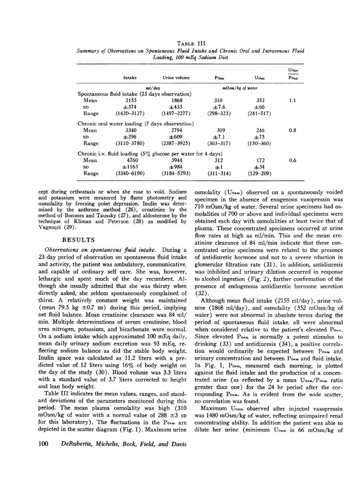
TABLE IIISummary of Observations on Spontaneous Fluid Intake and Chronic Oral and Intravenous Fluid
Loading, 100 mEq Sodium Diet
U0am
Intake Urine volume Po=, U0am Pow
mil/day mOsm/kg of waterSpontaneous fluid intake (23 days observation)
Chronic oral water loading (7 days observation)Mean 3340 2794 309 246 0.8SD ±296 ±609 ±7.1 ±75Range (3110-3780) (2387-3925) (303-317) (150-360)
Chronic iv. fluid loading (5% glucose per water for 4 days)Mean 4760 3944 312 172 0.6SD 41163 ±984 ±1 434Range (3340-6190) (3184-5293) (311-314) (129-209)
cept during orthostasis or when she rose to void. Sodiumand potassium were measured by flame photometry andosmolality by freezing point depression. Inulin was deter-mined by the anthrone method (26), creatinine by themethod of Bonsnes and Taussky (27), and aldosterone by thetechnique of Kliman and Peterson (28) as modified byVagnurci (29).
RESULTSObservations on spontaneous fluid intake. During a
23 day period of observation on spontaneous fluid intakeand activity, the patient was ambulatory, communicative,and capable of ordinary self care. She was, however,lethargic and spent much of the day recumbent. Al-though she usually admitted that she was thirsty whendirectly asked, she seldom spontaneously complained ofthirst. A relatively constant weight was maintained(mean 79.5 kg +0.7 SD) during this period, implyingnet fluid balance. Mean creatinine clearance was 84 ml/min. Multiple determinations of serum creatinine, bloodurea nitrogen, potassium, and bicarbonate were normal.On a sodium intake which approximated 100 mEq daily,mean daily urinary sodium excretion was 93 mEq, re-flecting sodium balance as did the stable body weight.Inulin space was calculated as 11.2 liters with a pre-dicted value of 12 liters using 16% of body weight onthe day of the study (30). Blood volume was 3.3 literswith a standard value of 3.7 liters corrected to heightand lean body weight.
Table III indicates the mean values, ranges, and stand-ard deviations of the parameters monitored during thisperiod. The mean plasma osmolality was high (310mOsm/kg of water with a normal value of 288 +3 SDfor this laboratory). The fluctuations in the Posm aredepicted in the scatter diagram (Fig. 1). Maximum urine
osmolality (Uo0m) observed on a spontaneously voidedspecimen in the absence of exogenous vasopressin was710 mOsm/kg of water. Several urine specimens had os-molalities of 700 or above and individual specimens wereobtained each day with osmolalities at least twice that ofplasma. These concentrated specimens occurred at urineflow rates at high as ml/min. This and the mean cre-atinine clearance of 84 ml/min indicate that these con-centrated urine specimens were related to the presenceof antidiuretic hormone and not to a severe rduction inglomerular filtration rate (31). In addition, antidiuresiswas inhibited and urinary dilution occurred in responseto alcohol ingestion (Fig. 2), further confirmation of thepresence of endogenous antidiuretic hormone secretion(32).
Although mean fluid intake (2155 ml/day), urine vol-ume (1868 ml/day), and osmolality (352 mOsm/kg ofwater) were not abnormal in absolute terms during theperiod of spontaneous fluid intake, all were abnormalwhen considered relative to the patient's elevated Po..,.Since elevated Posm is normally a potent stimulus todrinking (33) and antidiuresis (34), a positive correla-tion would ordinarily be expected between Po0m andurinary concentration and between Posm and fluid intake.In Fig. 1, Po.m, measured each morning, is plottedagainst the fluid intake and the production of a concen-trated urine (as reflected by a mean Uosm/Poam ratiogreater than one) for the 24 hr period after the cor-responding Po... As is evident from the wide scatter,no correlation was found.
Maximum Uoum observed after injected vasopressinwas 1480 mOsm/kg of water, reflecting unimpaired renalconcentrating ability. In addition the patient was able todilute her urine (minimum Uo.m is 66 mOsm/kg of
100 DeRubertis, Michelis, Beck, Field, and Davis
water) and widely vary urinary sodium concentration(1-206 mEq/liter) in response to varying sodium in-takes. On a 25 mEqsodium diet 24 hr urinary aldosteroneexcretion was 15.3 ltg (normal 3-19 /ig). Sodium bal-ance was maintained on this restricted intake over a pe-riod of several months.
Response to acute and chronic water loading. As can
be seen from the data in Table III chronic water loadingeither orally or intravenously up to 6.2 liters/day failedto significantly lower mean Posm but resulted in in-creased volumes of dilute urine. Po.m was 311 mOsm/kg of water at the initiation and 312 at the termina-tion of 5% dextrose and water administration (Fig. 3).The infusion increased mean daily fluid intake by anaverage of 2.6 liters over spontaneous intake. Bloodsugars remained in the euglycemic range and urine re-
ductions done four times daily were negative duringthe infusion. Mean daily Uosr fell from 210 to 129 mOsm/kg water and urine volume rose progressively duringthe period of the infusion.
As can be seen in Fig. 3 urine remained dilute andPosm high in the 24 hr after the cessation of intra-venous infusion. During this period negative fluid bal-ance developed with weight loss. Subsequently urinaryconcentration increased, Posm transiently fell, and weightincreased. The response to chronic oral water loadingwas comparable. Posm was 308 mOsm/kg of water at theinitiation of the study and 314 after 7 days of water load-ing during which an average of 1.1 liters daily abovespontaneous intake was ingested. Mean urine osmolalityagain fell progressively from 360 to 150 mOsm/kg ofwater and urine volumes increased.
E
<
St
40i0 2o4D2 i200 OD 3e0 d? 09 1.0 1.1 h Is 1'4 Ii 51 IarAD LII FLUID INTAKE in ml/day UOs. /POw
FIGURE 1 Correlation of plasma osmolality (Po.m) withad lib. fluid intake and with concentration of the urine.Scattergram depicts the wide range of values of the fastingPosm during a 23 day period of spontaneous fluid intake.No correlation was found between the morning fasting Po.mand the volume of fluid ingested in the succeeding 24 hrperiod. Similarly there was no correlation between Po.m andconcentration of the urine excreted in that period. Concen-tration of the urine is expressed as the mean urine osmo-lality (Uo.m/Po.m) with a value greater than one repre-
senting urinary concentration.
TIME IN HOURS
FIGURE 2 Response to alcohol. After overnight dehydration,administration of ethyl alcohol resulted in prompt urinarydilution and water diuresis.
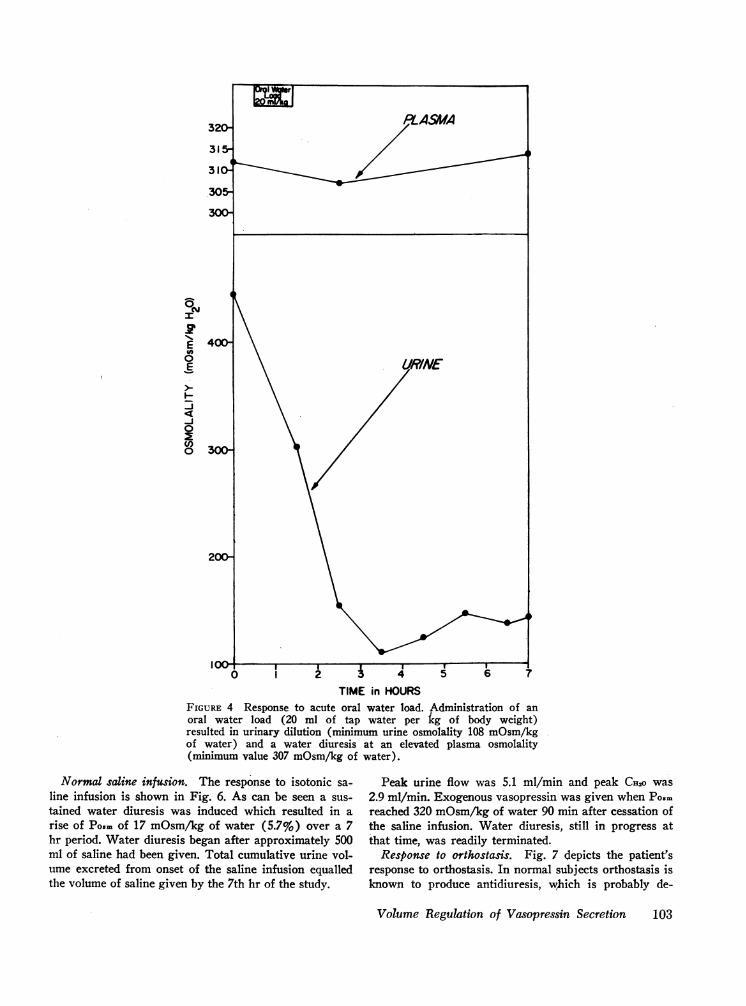
The response to an acute oral water load is shown inFig. 4. Water diuresis occurred even though Po.m re-mained high, the minimum Po.m observed being 307mOsm/kg of water 1 hr after water ingestion. Thisstudy was repeated and quite similar results were
obtained.Response to water deprivation. At the initiation of
total fluid deprivation, the patient's plasma osmolalitywas 303 and she weighed 87.1 kg (Table IV). After10 hr the plasma osmolality had risen to 319 and theweight had decreased to 86 kg. The urine osmolalitywas 411. Over the next 4 hr she lost another 0.3 kg bodyweight, the plasma osmolality rose to 326, and the urineto 604. The rate of urine flow over the last 4 hr was
stable at 0.2-0.3 ml/min.
Volume Regulation of Vasopressin Secretion 101
0
ICYl
.Z
#A0
E
I-
-J
-J
0
0)0o
30 0
* 0
~~0030- *go* 0 0
0 00 0 0 0 0 @0 0 @00
* 0* 0
0 00 0
Response to hypertonic saline. Hypertonic saline wasgiven to evaluate the patient's ability to concentrate theurine in response to an acute rise in plasma osmolality.As shown in Fig. 5, Po.m in response to hypertonic sa-line rose markedly (52 mOsm/kg of water, 18%) in 6hr without a resultant antidiuresis. Urine flow, ratherthan decreasing, rose to a peak of 18.3 ml/min 90 minafter cessation of infusion. This was accompanied by apeak osmolar clearance of 8.3 ml/min and a peak freewater clearance (CHzo) of 10 ml/min. As shown inFig. 5, water diuresis was in progress 5 hr after cessa-tion of the infusion and Pos. rose an additional 25mOsm/kg of water. Total cumulative urine volume ex-creted equalled volume of fluid administered by both
AD LIB. ORALFLUJDI ADI7- ONLY I+1
,,, 6-
5-
4-
3! 3-
2-
315^
310\
305.
ONM 295-
E6>i450
-J 400-~350 A
water ingestion and saline infusion by the 7th hr of thestudy. Approximately 25% of the administered sodiumload was excreted within 4 hr of completion of the infu-sion with a peak urinary sodium excretion of 950 uEq/min. Within 24 hr sodium balance was restored. Thesevalues for sodium excretion are comparable to thosefound in normal subjects given 5% saline at similarrates (35, 36), making it unlikely that an abnormallydelayed excretion of sodium contributed significantly tothe marked rise in Po.m. At a Po.m of 339 mOsm/kg ofwater, the patient's sensorium was seriously clouded.Water diuresis was still in progress but at a decreasedrate. Vasopressin infusion readily terminated the waterdiuresis and markedly improved her sensorium.
6 7 8 9 10 11 12 13TIME in DAYS
FIGURE 3 Response to intravenous fluid loading. Intravenous glucose/water(i.v. 5% D/W) up to 4 liters daily (total fluid intake up to 6.2 liters daily)failed to lower plasma osmolality. Progressive urinary dilution occurredwith plasma osmolality (Po..) still high during the period of infusion(mean Po.m 312 mOsm/kg of water).
102 DeRubertis, Michelis, Beck, Field, and Davis
320O
315-
310-
305-
300
0
1'%EEO0
-j
0(I)0
400-
300-
200-
I i0
PLASMA
URINE
I I I . . I1 2 A 4 5 6
TIME in HOURSFIGURE 4 Response to acute oral water load. Administration of anoral water load (20 ml of tap water per kg of body weight)resulted in urinary dilution (minimum urine osmolality 108 mOsm/kgof water) and a water diuresis at an elevated plasma osmolality(minimum value 307 mOsm/kg of water).
Normal saline infusion. The response to isotonic sa-line infusion is shown in Fig. 6. As can be seen a sus-tained water diuresis was induced which resulted in arise of Po.. of 17 mOsm/kg of water (5.7%) over a 7hr period. Water diuresis began after approximately 500ml of saline had been given. Total cumulative urine vol-ume excreted from onset of the saline infusion equalledthe volume of saline given by the 7th hr of the study.
Peak urine flow was 5.1 ml/min and peak CH2o was2.9 ml/min. Exogenous vasopressin was given when PoSMreached 320 mOsm/kg of water 90 min after cessation ofthe saline infusion. Water diuresis, still in progress atthat time, was readily terminated.
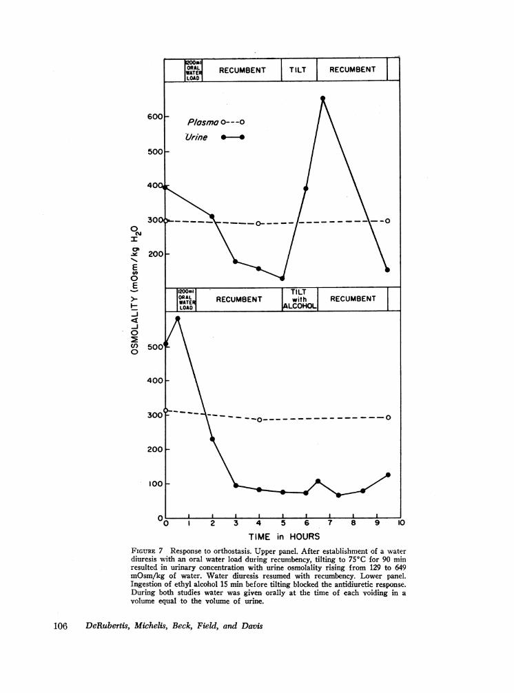
Response to orthostasis. Fig. 7 depicts the patient'sresponse to orthostasis. In normal subjects orthostasis isknown to produce antidiuresis, which is probably de-
l C- ~0.4_>1 . I * I 2 *1.4 11.7 1a|_ 2. 1 _4 32F2.1 t24 _I.3
FIGURE 5 Response to hypertonic saline infusion. After establishment of a waterdiuresis with an oral water load (H20 load), 20 ml/kg body weight, infusion ofintravenous hypertonic saline (5% NaCl i.v.) 0.1 ml/kg of body weight per min,did not result in urinary concentration despite a marked acute rise in plasmaosmolality (52 mOsm/kg of water, 18% in 6 hr). Rather, urine flow (maximum18.3 ml/min) increased with saline infusion. The maximum values were noted90 min after cessation of the infusion. Water diuresis, still in progress at hr 10,was terminated with exogenous vasopressin (Pitressin, 1 mU/mm i.v.). Table, atbottom, depicts V, Cosm, and CH2O in milliliters per minute for individual deter-minations corresponding in sequence to points plotted for urine osmolality.
pendent on a reduction of effective circulating blood vol-ume by pooling of blood in the legs rather than a reduc-tion of total plasma volume, since iso-oncotic albumin in-fusion does not prevent the antidiuresis (37). Alcohol,which blocks the secretion of antidiuretic hormone, canprevent orthostatic antidiuresis (32, 38, 39). As shownin Fig. 7, tilt to 750 interrupted a stable water diuresiswith the Uo.m rising to 649 mOsm/kg of water. Waterdiuresis resumed when the patient was returned to therecumbent position. Posm remained stable during the
study after a small initial fall which resulted from waterloading. When the study was repeated (Fig. 7) giving60 ml of 50% alcohol orally before tilting, antidiuresiswas not observed. Further evaluation of dilution andconcentration of the urine in response to effective vol-ume changes are summarized in Fig. 8. Volume expan-sion caused by infusion of approximately 400 ml of sa-line initiated a water diuresis which was terminated bytilting her despite the continued infusion of saline.Recumbency was associated with restoration of the wa-
Posm = plasma osmolality (mOsm/kg of H20), UOsrm = urineosmolality (mOsm/kg of H20), and V = urine flow rate.
ter diuresis. Antecedent alcohol administration again in-hibited the antidiuresis of tilting.
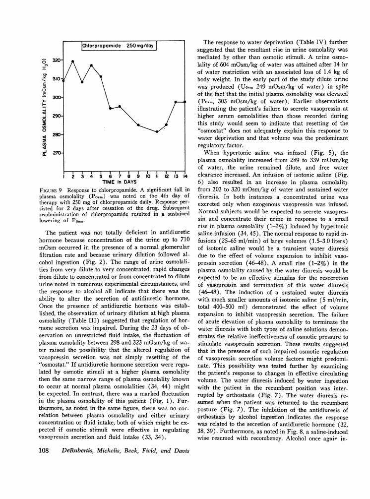
Response to chlorpropamide. The response to chlor-propamide therapy, 250 mg orally per day, is shown inFig. 9. The effectiveness of this agent in diabetes in-
32G r ~~Normal Saline Inusio3i 5-i
310-
ON
-J0
0 300\
sipidus has recently been reported (40-43). Mahoneyand Goodman (12) employed chlorpropamide success-fully in a similar patient with chronic hypernatremia. Inthe present patient, significant reduction of Po.. was notobserved until the 4th day of therapy with chlorpropa-mide and was sustained for 2 days after the drug was
temporarily discontinued. At the time of this report thepatient had received chlorpropamide therapy for a totalof 6 wk. Po.. has recently ranged from 282 to 296mOsm/kg of water, the most sustained reduction ob-served. Clinically, corection of the hyperosmolality wasaccompanied by a marked improvement in the patient'smental status.
DISCUSSION
This patient demonstrates the features of the syndromewhich has been termed "essential hypernatremia" (8-18).Previous examples of essential hypernatremia have beenpostulated to be on the basis of an elevated osmoticthreshold for vasopressin release (10-15, 24). The ob-
FIGuRE 6 Response to normal saline infusion. After overnight dehydration,infusion of approximately 500 ml of normal saline resulted in urinary dilution(Uosm is 102 mOsm/kg of water). With continued saline infusion, a sustainedwater diuresis was observed despite a rising plasma osmolality (301-320mOsm/kg of water). Water diuresis was terminated with exogenous vaso-pressin (Pitressin 1 mU/min i.v.). Table, at bottom, depicts V, Com, andCH20 in milliliters per minute for individual determinations corresponding insequence to points plotted for urine osmolality.
Volume Regulation of Vasopressin Secretion 105
0
Je 200 -
E0E_.., 1200mi TILT
>_ ORAL RECUMBENT with I RECUMBENTWATE LOOLOADOLOH~
-J
0
500
400 \
300 i 0--0
200 -
100 -
C I1 I1 1 1 10 I 2 3 4 5 6 7 8 9 1OTIME in HOURS
FIGURE 7 Response to orthostasis. Upper panel. After establishment of a waterdiuresis with an oral water load during recumbency, tilting to 75'C for 90 minresulted in urinary concentration with urine osmolality rising from 129 to 649mOsm/kg of water. Water diuresis resumed with recumbency. Lower panel.Ingestion of ethyl alcohol 15 min before tilting blocked the antidiuretic response.During both studies water was given orally at the time of each voiding in avolume equal to the volume of urine.
106 DeRubertis, Michelis, Beck, Field, and Davis
Nz
E
I4-J
TIME (20 minute intervals)
ULuuuu UjU U U UM U U0*1. 3.013351 3.01.5 1.50.5 0.1 0.O 1.9 384.1 3. 1414414. 2951203 03iiIA 0.7 1.2 1.0 QS .41ol7 .4aS 021 0.s l 1.321.3 1 1.3 1.2 LI 1.031. O 0.6CHAD°1 @ 22.2 1.1 Q120.92.31*3.232 L 0.1
FIGURE 8 Interruption with orthostasis of a water diuresis induced by normal salineinfusion. After overnight dehydration, a water diuresis was established during recum-bency with normal saline infusion (5 ml/min). Tilting to 750C for 90 min resulted in uri-nary concentration with urine osmolality rising from 87 to 543 mOsm/kg of water. Waterdiuresis resumed with recumbency. Tilting was repeated 15 min after administration ofethyl alcohol orally. Urinary concentration was not observed. During this study urinewas collected at 20-min intervals by an indwelling bladder catheter. Table, at bottom,depicts V, Com, and CH2o in milliliters per minute for individual determinations corre-sponding in sequence to points for urine osmolality.
servations of excretion of a dilute urine despite an ele-vated plasma osmolality (9-14), the failure of fluidloading to return the plasma osmolality to normal (9-14), and concentration of the urine when plasma osmo-lality was raised to high levels by fluid deprivation (10-15) have been used to support this postulated resetting ofthe hypothalamic "osmostat." This and other hypothesesincluding a primary disturbance in sodium metabolism(14) were considered in the investigation of this patientwho presented with a sustained elevation of her plasmaosmolality.
The normal creatinine clearance, the absence of azo-temia and oliguria, and the normal aldosterone excretionrate are all evidence against severe extracellular fluidvolume depletion. The failure of forced fluid administra-tion to lower the plasma osmolality eliminates inadequate
fluid intake as the sole cause of the hypernatremia. Like-wise, the urine osmolality of 1410 mOsm/kg of waterafter exogenous vasopressin, the ability to dilute theurine to an osmolality of 60 mOsm/kg of water, and therange of urinary sodium concentration (1-206 mEq/liter) indicate intact renal tubule function. Overproductionof aldosterone did not appear to be involved in sustain-ing the hypernatremia since aldosterone excretion, se-rum potassium, and bicarbonate were normal. All ofthese observations in addition to the demonstrated abilityto maintain sodium balance under all of the conditions ofthe study indicate that an abnormality in sodium metabo-lism was not the cause of the sustained and fluctuatinghyperosmolality. The evidence, therefore, suggests thatthe hyperosmolar syndrome was related to an abnormalityin water metabolism.
Volume Regulation of Vasopressin Secretion 107
320-
I0'
I 310-E00
E
~300
J 290-0
0
280-
4
-j270-
1 2 3 4 5 6 7 8 9 10 11 12 13 14TIME in DAYS
FIGURE 9 Response to chlorpropamide. A significant fall inplasma osmolality (Posm) was noted on the 4th day oftherapy with 250 mg of chlorpropamide daily. Response per-sisted for 2 days after cessation of the drug. Subsequentreadministration of chlorpropamide resulted in a sustainedlowering of Posm.
The patient was not totally deficient in antidiuretichormone because concentration of the urine up to 710mOsmoccurred in the presence of a normal glomerularfiltration rate and because urinary dilution followed al-cohol ingestion (Fig. 2). The range of urine osmolali-ties from very dilute to very concentrated, rapid changesfrom dilute to concentrated or from concentrated to diluteurine noted in numerous experimental circumstances, andthe response to alcohol all indicate that there was theability to alter the secretion of antidiuretic hormone.Once the presence of antidiuretic hormone was estab-lished, the observation of urinary dilution at high plasmaosmolality (Table III) suggested that regulation of hor-mone secretion was impaired. During the 23 days of ob-servation on unrestricted fluid intake, the fluctuation ofplasma osmolality between 298 and 323 mOsm/kg of wa-
ter raised the possibility that the altered regulation ofvasopressin secretion was not simply resetting of the"osmostat." If antidiuretic hormone secretion were regu-lated by osmotic stimuli at a higher plasma osmolalitythen the same narrow range of plasma osmolality knownto occur at normal plasma osmolalities (34, 44) mightbe expected. In contrast, there was a marked fluctuationin the plasma osmolality of this patient (Fig. 1). Fur-thermore, as noted in the same figure, there was no cor-
relation between plasma osmolality and either urinaryconcentration or fluid intake, both of which might be ex-
pected if osmotic stimuli were effective in regulatingvasopressin secretion and fluid intake (33, 34).
The response to water deprivation (Table IV) furthersuggested that the resultant rise in urine osmolality wasmediated by other than osmotic stimuli. A urine osmo-
lality of 604 mOsm/kg of water was attained after 14 hrof water restriction with an associated loss of 1.4 kg ofbody weight. In the early part of the study dilute urinewas produced (U08m 249 mOsm/kg of water) in spiteof the fact that the initial plasma osmolality was elevated(Po.m, 303 mOsm/kg of water). Earlier observationsillustrating the patient's failure to secrete vasopressin athigher serum osmolalities than those recorded duringthis study would seem to indicate that resetting of the"osmostat" does not adequately explain this response towater deprivation and that volume was the predominantregulatory factor.
When hypertonic saline was infused (Fig. 5), theplasma osmolality increased from 289 to 339 mOsm/kgof water, the urine remained dilute, and free waterclearance increased. An infusion of isotonic saline (Fig.6) also resulted in an increase in plasma osmolalityfrom 303 to 320 mOsm/kg of water and sustained waterdiuresis. In both instances a concentrated urine was
excreted only when exogenous vasopressin was infused.Normal subjects would be expected to secrete vasopres-
sin and concentrate their urine in response to a smallrise in plasma osmolality (1-2%) induced by hypertonicsaline infusion (34, 45). The normal response to rapid in-fusions (25-65 ml/min) of large volumes (1.5-3.0 liters)of isotonic saline would be a transient water diuresisdue to the effect of volume expansion to inhibit vaso-
pressin secretion (46-48). A small rise (1-2%) in theplasma osmolality caused by the water diuresis would beexpected to be an effective stimulus for the resecretionof vasopressin and termination of this water diuresis(46-48). The induction of a sustained water diuresiswith much smaller amounts of isotonic saline (5 ml/min,total 400-500 ml) demonstrated the effect of volumeexpansion to inhibit vasopressin secretion. The failureof acute elevation of plasma osmolality to terminate thewater diuresis with both types of saline solutions demon-strates the relative ineffectiveness of osmotic pressure tostimulate vasopressin secretion. These results suggestedthat in the presence of such impaired osmotic regulationof vasopressin secretion volume factors might predomi-nate. This possibility was tested further by examiningthe patient's response to changes in effective circulatingvolume. The water diuresis induced by water ingestionwith the patient in the recumbent position was inter-rupted by orthostasis (Fig. 7). The water diuresis re-
sumed when the patient was returned to the recumbentposture (Fig. 7). The inhibition of the antidiuresis oforthostasis by alcohol ingestion indicates the response
was related to the secretion of antidiuretic hormone (32,38, 39). Furthermore, as noted in Fig. 8, a saline-inducedwise resumed with recombency. Alcohol once agair in-
108 DeRubertis, Michelis, Beck, Field, and Davis
IChlorpropomide 250mg/doy |
hibited the antidiuretic response to orthostasis. Theplasma osmolality continued to rise (300-316 mOsm/kgof water) and exogenous vasopressin was needed to ter-minate the water diuresis. The induction of a water diu-resis with effective volume expansion, saline or re-cumbent position, and its termination with effectivevolume contraction (orthostasis) indicate that the vaso-pressin secretory mechanism could be regulated by vol-ume stimuli. Once again osmotic stimuli were seen to beineffective in producing an antidiuresis.
The hypothalamic location of this patient's neurologiclesion, almost certainly histiocytic infiltration as wasfound on the lung biopsy, would be quite consistent withdisruption of osmoreception. Histiocytosis has previ-ously been reported as a cause of essential hypernatremia(11, 16, 17) but a variety of other hypothalamic lesionshave also been associated with this syndrome. Theseinclude pineal germinoma (10), surgery for cranio-pharyngioma (12) and cerebral artery aneurysm (14),microcephaly (13), and glioma (17). Verney (34) ini-tially introduced the concept of neural receptive elementsmonitoring plasma osmolality and effecting an appropri-ate release of ADH when osmolality rises. More re-cent studies have similarly indicated that a fall in plasmaosmolality produced by intracarotid water infusion re-sults in a water diuresis by central inhibition of vaso-pressin secretion (49). It has been suggested that theresponsive cells might be activated by shrinking andswelling (50, 51). Jewell and Verney (21) presented evi-dence eliminating many regions of the brain as sites ofosmoreception and implicating the anterior hypothalamicarea. They favored the region of the supraoptic nucleusas the primary site of osmoreception. Much additionalevidence has since been accumulated to support theirview (52), but other nearby regions may also be in-volved (53-55). There is also evidence to sugggest thatthere are blood volume receptors in areas such as the leftatrium, carotid artery, and aortic arch which mediateADH secretion by reflex mechanisms (56-61). Bloodvolume contraction stimulates (57, 62-64) and expan-sion inhibits (61, 64, 65) ADH secretion. However,the stimulation or inhibition of ADH secretion appearsto be governed by the net effect of osmotic and volumefactors operative (64, 66-68). Under ordinary circum-stances, such as hydropenia and overhydration, volumeand osmotic factors are acting in a parallel fashion toeither stimulate or inhibit vasopressin secretion. Whenthese two stimuli act in opposite directions, such as inexpansion with hypertonic saline (45) or in hypotoniccontraction (66, 69-71), vasopressin seems to be se-creted in response to a rising plasma osmolality andfalling volume (45, 66, 69-71). The former observationwater diuresis was also inhibited by orthostasis and like-is the basis of the usefulness of hypertonic saline in the
differential diagnosis of polyuric states. Similarly, it isthe resultant small rise in plasma osmolality which isthought to limit the water diuresis which normally oc-curs in response to isotonic expansion in man (46-48).From these normal physiologic responses and the con-cept that ordinarily both osmotic and volume factors in-fluence the secertion of vasopressin, it might be pre-dicted that in the absence of effective osmotic regulationisotonic or hypertonic expansion, by inhibiting ADHse-cretion, would result in a sustained water diuresis. Suchresponses were indeed observed in the present patient(Figs. 5 and 6). With impaired osmotic mediation ofADH secretion, other stimuli which act to inhibit itssecretion, such as alcohol ingestion or cold exposure,might also result in a water diuresis in spite of a risingPo0m. This was documented in response to alcohol ad-ministration (Fig. 2). In such circumstances, volumecontraction would eventually serve as an effective stimu-lus for resecretion of ADH. However, considerable fluc-tuations of Po.m would result. It is of course possiblethat the patient's failure to terminate water diuresisduring saline infusion despite rising plasma osmolalityreflected an inability to release acutely sufficient vaso-pressin because of partial deficiency of this hormone.However, termination of a saline-induced water diuresismediated by vasopressin secretion in response to ortho-stasis makes this possibility unlikely.
Elevated and fluctuating osmotic pressure would thenbe a consequence of (a) loss of the sensitive osmoticregulation of ADHsecretion which normally maintainsplasma osmolality at a relatively constant value (34)and (b) intact volume modulation of ADH secretionwhich would result in overall water balance but a lessstable plasma osmolality.
Little consideration has been given to the role ofvolume regulation of vasopressin secretion in the previ-ous reports of essential hypernatremia. In the presentpatient and in others reported (9-14), it was not pos-sible to completely correct hyperosmolality with increasedfluid administration. Rather, water diuresis was ob-served with plasma osmolality still elevated. Inhibition ofvasopressin secretion by the volume expansion which ac-companies such fluid loading could explain this observa-tion. Further, several patients with essential hyperna-tremia were noted to concentrate their urine whenplasma osmolality was raised to higher levels by fluiddeprivations (11-15). Such urinary concentration is notnecessarily related to an elevated osmotic threshold forvasopressin release. Volume contraction, a concomitantof fluid deprivation, may have been the important factorin vasopressin release in these instances. In only a fewpatients has hypertonic saline been employed as a meansof evaluating the effectiveness of osmotic stimuli regu-lating vasopressin secretion. A decrease in urinary flow
Volume Regulation of Vasopressin Secretion 109
and a small rise in urine osmolality was reported in each(10, 14, 15), but in only one (10) did the urine osmo-lality rise above plasma in response to hypertonic saline.Such differences might be explained by the degree towhich osmotic regulation of vasopressin secretion is im-paired. However, a more systemic evaluation of the roleof both osmotic and volume factors in the regulation ofvasopressin secretion may serve to clarify further thepathophysiology of this complex disorder.
Finally, based on the formulations proposed, patientssuch as the present one may offer the rare opportunityto examine in man volume regulation of antidiuretichormone secretion relatively independent of the usual in-fluence of concomitant osmotic factors. Indeed, the pres-ent data would seem to lend further support to the con-cept of volume regulation of vasopressin secretion inman.
ACKNOWLEDGMENTSWeare indebted to Rebecca J. Clare for her expert techni-cal assistance and to Sally A. Sawyer and Stephanie Koenigfor their secretarial help. Weare especially grateful to MissRegina Onda and her dietary staff, Mrs. Bonita Levine,Miss Linda Pape, and the excellent nursing staff of theClinical Research Unit. Wewould like to give special thanksto Dr. William M. Cooper for referring this most interestingpatient to us for study.
This work was supported in part by Grants AM11911 02,AM10949, FR 56, and AM05047 from the National Insti-tutes of Health, U. S. Public Health Service.
REFERENCES1. Zierler, K. L. 1958. Hyperosmolarity in adults: a critical
review. J. Chronic Dis. 7: 1.2. Christie, S. B. M., and E. J. Ross. 1968. Ectopic pinea-
loma with adipsia and hypernatremia. Brit. Med. J. 2:669.
3. Skultety, F. M., and R. J. Joynt. 1963. Clinical impli-cations of adipsia. J. Neurosurg. 20: 793.
4. Hays, R. M., P. R. McHugh, and H. E. Williams. 1963.Absence of thirst associated with hydrocephalus. N. Engl.J. Med. 269: 227.
5. Travis, L. B., W. F. Dodge, J. D. Waggener, and C.Kashemsant. 1967. Defective thirst mechanism secondaryto hypothalamic lesion: studies in a child with adipsia,polyphagia, obesity, and persistent hyperosmolarity.J. Pediat. 70: 915.
6. Crigler, J. R., and S. Suh. 1961. Hyperosmolarity fol-lowing radical surgical treatment of craniopharyngioma.Amer. J. Dis. Child. 102: 81.
7. Truniger, B., and D. Kuenzler. 1962. Chronic hyperos-molarity and hypothalamic lesions. On the pathogenesisof occult hypersalemic diabetes insipidus. Klin. I'Vochen-schr. 40: 872.
8. Engstrom, W. W., and A. Liebman. 1953. Chronic hy-perosmolarity of the body fluids with a cerebral lesioninsipidus and anterior pituitary insufficiency. Anmer. J.Med. 15: 180.
9. Goldberg, M., G. Weinstein, J. Adesman, and S. J.Bleicher. 1967. Asymptomatic hypovolemic hypernatre-mia, a variant of essential hypernatremia. Amer. J. Mcd.43: 804.
10. Kastin, A. J., M. B. Lipsett, A. K. Ommaya, and J. M.Moser. 1965. Asymptomatic hypernatremia, physiologicaland clinical study. Amer. J. Med. 38: 306.
11. Avioli, L. V., L. E. Earley, and H. K. Kashima. 1962.Chronic and sustained hypernatremia, absence of thirst,diabetes insipidus, and adrenocorticotrophin insufficiencyresulting from widespread destruction of the hypothala-mus. Ann. Intern. Med. 56: 131.
12. Mahoney, J. H., and A. D. Goodman. 1968. Hyperna-tremia due to hypodypsia and elevated threshold for vaso-pressin release. N. Engl. J. Med. 279: 1191.
13. Segar, W. E. 1966. Chronic hyperosmolality. Amer. J.Dis. Child. 112: 318.
14. Pleasure, D., and M. Goldberg. 1966. Neurogenic hy-pernatremia. Arch. Neurol. 15: 78.
15. Golonka, J. E., and J. A. Richardson. 1970. Postcon-cussive hyperosmolality and deficient thirst. Amer. J.Med. 48: 261.
16. Leaf, A., A. R. Mamby, H. Rasmussen, and J. P.Marasco. 1952. Some hormonal aspects of water excre-tion in man. J. Clin. Invest. 31: 914.
17. Wise, B. L. 1962. Neurogenic hyperosmolarity (hyper-natremia). Neurology. 12: 453.
18. Weitzman, E. D., and M. H. Triedman. 1960. Hyper-osmolarity associated with hypothalamic lesion. Neu-rology. 10: 584.
19. Andersson, B., and S. M. McCann. 1955. The effect ofhypothalamic lesions on the water intake of the dog.Acta Physiol. Scand. 35: 312.
20. Lederis, K. 1962. The distribution of vasopressin andoxytocin in hypothalamic nuclei. In Neurosecretion. H.Heller and R. B. Clark, editors. Academic Press Inc.,New York. 227.
21. Jewell, P. A., and E. B. Verney. 1957. An experimentalattempt to determine the site of neurohypophysial osmo-receptors in the dog. Phil. Trans. Roy. Soc. LondonSer. B Biol. Sci. 240: 197.
22. Andersson, B., and S. M. McCann. 1955. Drinking, anti-diuresis and milk ejection from electrical stimulationwithin the hypothalamus of the goat. Acta Physiol. Scand.35: 191.
23. Peters, J. P. 1950. Sodium, water and edema. J. Mt.SinaiHosp. 17: 159.
24. Welt, L. G. 1962. Hypo- and hypernatremia. Ann. In-tern. Med. 56: 161.
25. Davis, B. B., M. E. Bloom, J. B. Field, and D. H. Mintz.1969. Hyponatremia in pituitary insufficiency. Metab.(Clin. Exp.). 18: 821.
26. Fiuhr, J., J. Kaczmarczyk, and C.-D. Kruittgen. 1955.Eine einfache colorimetrische methode zur insulinbestim-mung fur nieren-clearance-untersuchungen bei stoff-wechselgesunden und diabetikern. Klin. Wochenschr. 33:729.
27. Bonsnes, F. W., and H. H. Taussky. 1945. On the colori-metric determination of creatinine by the Jaffe Reaction.J. Biol. Chem. 158: 581.
28. Kliman, B., and R. E. Peterson. 1960. Double isotopederivative assay of aldosterone in biological extracts.J. Biol. Chem. 235: 1639.
29. Vagnucci, A. H. 1969. Selective aldosterone deficiency.J. Clin. Endocrinol. Metab. 29: 279.
30. Pitts, R. F. 1963. Physiology of the kidney and bodyfluids. Year Book Medical Publishers, Inc., Chicago. 1stedition. 24.
31. Berliner, R. W., and D. G. Davidson. 1956. Production
110 DeRubertis, Michelis, Beck, Field, and Davis
of hypertonic urine in the absence of pituitary antidiu-retic hormone. J. Clin. Invest. 35: 690.
32. Strauss, M. B., J. D. Rosenbaum, and W. P. Nelson III.1950. The effect of alcohol on the renal excretion ofwater and electrolyte. J. Clin. Invest. 29: 1053.
33. Wolf, A. V. 1950. Osmometric analysis of thirst in manand dog. Amer. J. Physiol. 161: 75.
34. Verney, E. B. 1947. The antidiuretic hormone and thefactors which determine its release. Proc. Roy. Soc. Ser.B Biol. Sci. 135: 25.
35. Crawford, B., and H. Ludemann. 1951. The renal re-sponse to intravenous injection of sodium chloride solu-tions in man. J. Clin. Invest. 30: 1456.
36. Papper, S., L. Saxon, J. D. Rosenbaum, and H. W.Cohen. 1956. The effects of isotonic and hypertonic saltsolutions on the renal excretion of sodium. J. Lab. Clint.Med. 47: 776.
37. Epstein, F. H., A. V. N. Goodyer, F. D. Lawrason, andA. S. Relman. 1951. Studies of the antidiuresis of quitestanding: the importance of changes in plasma volume andglomerular filtration rate. J. Clin. Invest. 30: 63.
38. Kleeman, C. R., M. E. Rubini, E. Lamdin, and F. H.Epstein. 1955. Studies on alcohol diuresis. II. The evalu-ation of ethyl alcohol as an inhibitor of the neurohypo-physis. J. Clin. Invest. 34: 448.
39. Pearce, M. L., and E. V. Newman. 1954. Some posturaladjustments of salt and water excretion. J. Clin. In-vest. 33: 1089.
40. Arduino, F., F. P. J. Ferraz, and J. Rodrigues. 1966.Antidiuretic action of chlorpropamide in idiopathic dia-betes insipidus. J. Clin. Endocrinol. Metab. 26: 1325.
41. Meinders, A. E., J. L. Touber, and L. A. DeVries. 1967.Chlorpropamide treatment in diabetes insipidus. Lancet.2: 544.
42. Webster, B., and J. Bain. 1970. Antidiuretic effects andcomplications of chlorpropamide therapy in diabetesinsipidus. J. Clin. Endocrinol. Mletab. 30: 215.
43. Andreani, D., G. A. Cinotti, and G. Stirati. 1969.Chlorpropamide in idiopathic diabetes insipidus. Metab.(Clin. Exp.). 18: 874.
44. Czaczkes, J. W., C. R. Kleeman, and M. Koenig. 1964.Physiologic studies of antidiuretic hormone by its directmeasurement in human plasma. J. Clin. Inivest. 43: 1625.
45. Moses, A. M., and D. H. P. Streeten. 1967. Differentia-tion of polyuric states by measurement of responses tochanges in plasma osmolality induced by hypertonicsaline infusions. Amer. J. Med. 42: 368.
46. Welt, L. G., and J. Orloff. 1951. The effects of an in-crease in plasma volume on the metabolism and excre-tion of water and electrolytes by normal subjects. J.Clin. Invest. 30: 751.
47. Strauss, M. B., R. K. Davis, J. D. Rosenbaum, and E. C.Rossmeisl. 1951. "Water diuresis" produced during re-cumbency by the intravenous infusion of isotonic salinesolution. J. Clin. Invest. 30: 862.
48. Ladd, M. 1950. Effect of prehydration on the responseto saline infusion in man. J. Appl. Physiol. 3: 379.
49. Arndt, J. 0. 1965. Diuresis induced by water infusioninto the carotid loop of unanesthetized dogs. PfleugersArch. Gesamte Physiol. Menschen Tiere. 282: 301.
50. Jewell, P. A. 1953. Occurrence of vesiculated neuronsin the hypothalamus of the dog. J. Physiol. (London).121: 167.
51. Alanis, J., and B. H. C. Matthews. 1952. The mechano-receptor properties of central neurones. J. Physiol. (Lonz-don). 117: 59.
52. Joynt, R. J. 1966. Verney's concept of the osmoreceptor.A review and further experimental observations. Arch.Neurol. 14: 331.
53. Dierickx, K. 1962. The dendrites of the preoptic neuro-secretory nucleous of Rana Temporaria and the osmo-receptors. Arch. Int. Pharmacodyn. Ther. 140: 708.
54. Dingman, J. F., K. Benirschke, and G. W. Thorn. 1957.Studies of neurohypophyseal function in man. Amer. J.Med. 23: 226.
55. Holland, R. C., B. A. Cross, and C. H. Sawyer. 1959.EEG correlates of osmotic activation of the neurohy-pophyseal milk-ejection mechanism. Amner. J. Physiol.196: 796.
56. Henry, J. P., 0. H. Gauer, and J. L. Reeves. 1956.Evidence of the atrial location of receptors influencingurine flow. Circ. Res. 4: 85.
57. Share, L., and M. N. Levy. 1962. Cardiovascular re-ceptors and blood titer of antidiuretic hormone. Amer.J. Physiol. 203: 425.
58. Share, L. 1965. Effects of carotid occlusion and left atrialdistention on plasma vasopressin titer. Amer. J. Physiol.208: 219.
59. Shu'ayb, W. A., W. H. Moran, Jr., and B. Zimmerman.1965. Studies of the mechanism of antidiuretic hormonesecretion and the post-commissurotomy dilutional syn-drome. Ann. Surg. 162: 690.
60. Gupta, P. D., J. P. Henry, R. Sinclair, and R. VonBaumgarten. 1966. Responses to atrial and aortic baro-receptors to nonhypotensive hemorrhage and to trans-fusion. Amer. J. Physiol. 211: 1429.
61. Johnson, J. A., W. W. Moore, and W. E. Segar. 1969.Small changes in left atrial pressure and plasma antidiu-retic hormone titers in dogs. Amer. J. Physiol. 217: 210.
62. Weinstein, H., R. M. Berne, and H. Sachs. 1960. Vaso-pressin in blood; effect of hemorrhage. Endocrinology.66: 712.
63. Henry, J. P., P. D. Gupta, J. P. Meehan, R. Sinclair,and L. Share. 1968. The role of afferents from the lowpressure system in the release of antidiuretic hormoneduring non-hypotensive hemorrhage. Can. J. Physiol.Pharnzacol. 46: 287.
64. Smith, H. W. 1957. Salt and water volume receptors.Amer. J. Med. 23: 623.
65. Baratz, R. A., and R. C. Ingraham. 1960. Renal hemo-dynamics and antidiuretic hormone release associatedwith volume regulation. Amer. J. Physiol. 198: 565.
66. Arndt, J. 0. 1965. Diuresis induced by water infusioninto the carotid loop and its inhibition by small hemor-rhage. The competition of volume- and osmocontrol.Pfleugers Arch. Gesamtf Physiol. Menschen Tiere. 282:313.
67. Johnson, J. A., J. E. Zehr, and W. W. Moore. 1970.Effect of separate and concurrent osmotic and volumestimuli on plasma ADHin sheep. Amer. J. Physiol. 218:1273.
68. Gauer, H. 0. 1968. Osmocontrol versus volume control.Fed. Proc. 27: 1132.
69. McCance, R. A. 1936. Experimental sodium chloride de-ficiency in man. Proc. Roy. Soc. Ser. B Biol. Sci. 119:245.
70. Leaf, A., and A. R. Mamby. 1952. An antidiureticmechanism not regulated by extracellular fluid tonicity.J. Clin. Invest. 31: 60.
71. Young, T. K., and R. A. Phillips. 1967. Antidiuretic hor-mone release in the monkey during hypo- and hyper-os-motic dehydration. Proc. Soc. Exp. Biol. Med. 125: 1174.