Page 1
HAL Id: hal-01726553https://hal.univ-lorraine.fr/hal-01726553
Submitted on 8 Mar 2018
HAL is a multi-disciplinary open accessarchive for the deposit and dissemination of sci-entific research documents, whether they are pub-lished or not. The documents may come fromteaching and research institutions in France orabroad, or from public or private research centers.
L’archive ouverte pluridisciplinaire HAL, estdestinée au dépôt et à la diffusion de documentsscientifiques de niveau recherche, publiés ou non,émanant des établissements d’enseignement et derecherche français ou étrangers, des laboratoirespublics ou privés.
Integrated assessment of ceria nanoparticle impacts onthe freshwater bivalve Dreissena polymorpha
Maël Garaud, Melanie Auffan, Simon Devin, Vincent Felten, ChristophePagnout, Sandrine Pain-Devin, Olivier Proux, François Rodius, Benedicte
Sohm, Laure Giambérini
To cite this version:Maël Garaud, Melanie Auffan, Simon Devin, Vincent Felten, Christophe Pagnout, et al.. Integratedassessment of ceria nanoparticle impacts on the freshwater bivalve Dreissena polymorpha. Nanotoxi-cology, Taylor & Francis, 2016, 10 (7), pp.935 - 944. �10.3109/17435390.2016.1146363�. �hal-01726553�
Page 2
Integrated assessment of ceria nanoparticle impacts onthe freshwater bivalve Dreissena polymorphaMaël Garaud, Mélanie Auffan, Simon Devin, Vincent Felten, Christophe Pagnout,Sandrine Pain-Devin, Olivier Proux, François Rodius, Bénédicte Sohm, Laure Giam-berini
Doi: 10.3109/17435390.2016.1146363
Abstract
Exposures in realistic environmental conditions are essential to properly assess theeffects of emerging pollutants on ecosystems. While ceria nanoparticles (nCeO2)production and use are expanding quickly, ecotoxicity studies remain very scarce. Inthis work, we set up experimental systems reproducing a simplified ecosystem toassess the effects of a chronic exposure to citrate-coated nCeO2 (ci-CeO2) and barenCeO2 (ba-CeO2) on the freshwater mussel Dreissena polymorpha using an inte‐grated multibiomarker approach. The fate of nanoparticles was tightly monitored to
properly characterize the exposure. Organisms were exposed for three weeks and sampled weekly for biomarker analysis.Mussel filter-feeding activity resulted in significant removal of nCeO2 from the water column. At the same time, bioaccumu‐lation was low, reaching its maximum in the first week. Mussels bioaccumulated ci-CeO2 three times more than ba-CeO2,probably due to coating-related differences in their behavior in the water column and in organisms. Meanwhile, biomarkerresults were integrated and synthesized using linear discriminant analysis, highlighting that pi-glutathione-S-transferase(piGST) mRNA, catalase (CAT) activity, and lysosomal system were the most impacted of the seven biomarkers singled outby the discriminant analysis. These biomarker responses indicated that mussels exposed to both forms of nCeO2 werestressed and differentiate from the controls. Moreover,theyresponded differently to ba-CeO2 and ci-CeO2 exposure. How‐ever, biomarkers used in the experimental conditions of the present study did not indicate severe nCeO2 toxicity on mussels,as cellular damage biomarkers and mussel filtering activity were left unimpaired. However, further studies are needed toinvestigate if the slight perturbations observed could lead to populational impacts in the long-term.
Just Accepted by Nanotoxicology
© 2016 Taylor & Francis. This provisional PDF corresponds to the article as it appeared upon acceptance. Fully formattedPDF and full text (HTML) versions will be made available soon.DISCLAIMER: The ideas and opinions expressed in the journal's Just Accepted articles do not necessarily reflect those of Taylor & Francis (the Publisher),the Editors or the journal. The Publisher does not assume any responsibility for any injury and/or damage to persons or property arising from or related toany use of the material contained in these articles. The reader is advised to check the appropriate medical literature and the product information currentlyprovided by the manufacturer of each drug to be administered to verify the dosages, the method and duration of administration, and contraindications. It isthe responsibility of the treating physician or other health care professional, relying on his or her independent experience and knowledge of the patient, todetermine drug dosages and the best treatment for the patient. Just Accepted articles have undergone full scientific review but none of the additional editorialpreparation, such as copyediting, typesetting, and proofreading, as have articles published in the traditional manner. There may, therefore, be errors inJust Accepted articles that will be corrected in the final print and final online version of the article. Any use of the Just Accepted articles is subject to theexpress understanding that the papers have not yet gone through the full quality control process prior to publication.
Page 3
Integrated assessment of ceria nanoparticle impacts on the
freshwater bivalve Dreissena polymorpha
Maël Garaud12
, Mélanie Auffan23
, Simon Devin1, Vincent Felten
1, Christophe
Pagnout12
, Sandrine Pain-Devin1, Olivier Proux
24, François Rodius
1, Bénédicte Sohm
1 &
Laure Giamberini12*
1Université de Lorraine, CNRS UMR 7360, Laboratoire Interdisciplinaire des
Environnements Continentaux (LIEC), Campus Bridoux, Rue du Général Delestraint, 57070
Metz, France.
2International Consortium for the Environmental Implications of Nanotechnology (iCEINT),
Aix en Provence, France.
3CNRS, Aix-Marseille Université, CEREGE UM34, UMR 7330, 13545 Aix en Provence,
France.
4Observatoire des Sciences de l’Univers de Grenoble, UMS 832, CNRS, Universite Joseph
Fourier, 38041 Grenoble cedex 9, France
* Corresponding author e-mail: [email protected] and phone number:
+ 33 (0) 3 87 37 84 15
Keywords: ceria nanoparticle – Dreissena polymorpha - multibiomarker approach–––
discriminant analysisJUST A
CCEPTED
Page 4
Abstract
Exposures in realistic environmental conditions are essential to properly assess the effects of
emerging pollutants on ecosystems. While ceria nanoparticles (nCeO2) production and use
are expanding quickly, ecotoxicity studies remain very scarce. In this work, we set up
experimental systems reproducing a simplified ecosystem to assess the effects of a chronic
exposure to citrate-coated nCeO2 (ci-CeO2) and bare nCeO2 (ba-CeO2) on the freshwater
mussel Dreissena polymorpha using an integrated multibiomarker approach. The fate of
nanoparticles was tightly monitored to properly characterize the exposure. Organisms were
exposed for three weeks and sampled weekly for biomarker analysis. Mussel filter-feeding
activity resulted in significant removal of nCeO2 from the water column. At the same time,
bioaccumulation was low, reaching its maximum in the first week. Mussels bioaccumulated
ci-CeO2 three times more than ba-CeO2, probably due to coating-related differences in their
behavior in the water column and in organisms. Meanwhile, biomarker results were
integrated and synthesized using linear discriminant analysis, highlighting that pi-glutathione-
S-transferase (piGST) mRNA, catalase (CAT) activity, and lysosomal system were the most
impacted of the seven biomarkers singled out by the discriminant analysis. These biomarker
responses indicated that mussels exposed to both forms of nCeO2 were stressed and
differentiate from the controls. Moreover,theyresponded differently to ba-CeO2 and ci-CeO2
exposure. However, biomarkers used in the experimental conditions of the present study did
not indicate severe nCeO2 toxicity on mussels, as cellular damage biomarkers and mussel
filtering activity were left unimpaired.. However, further studies are needed to investigate if
the slight perturbations observed could lead to populational impacts in the long-term.JU
ST ACCEPTED
Page 5
1. Introduction
Production and use of nanomaterials have been expanding quickly in the recent decades,
raising concern about their potential impacts on the environment (Moore, 2006). Due to their
use as catalysts in fuel additives or as UV absorber in paints (Auffan et al., 2014a), CeO2
nanoparticle (nCeO2) production has risen sharply, potentially increasing environmental
exposure (Sun et al., 2014). This led to the recent calculation of predicted environmental
concentrations as high as 1 µg/L in surface waters (O’Brien and Cummins, 2011). To date,
studies on nCeO2 ecotoxicity have focused on acute toxicity assessment in standardized
conditions, generally reporting low toxicity in bacteria, algae, invertebrates and fishes (Collin
et al., 2014a). However, those standardized studies failed to incorporate environmental
parameters likely to impact exposure and effects of nCeO2 in the environment (Auffan et al.,
2014b).
Recent studies have stressed the importance of conducting investigations under more
environmentally relevant conditions, i.e. working in complex media, at realistic
concentrations, in long-term exposure, to correctly assess the potential impacts of
nanoparticles on organisms (Bour et al., 2015). It has been shown that interactions between
nCeO2 and natural inorganic or organic colloids could modify their chemical and colloidal
stability, therefore modulating their bioavailability and toxicity (Conway et al., 2014; Van
Hoecke et al., 2011). Consequently, there is a need to better characterize the fate of
nanoparticles in the exposure media and to relate that to their bioaccumulation,
biotransformation and subsequent biological effects. To meet these goals, the French ANR
MESONET program was launched with the aim of setting up adaptable freshwater
mesocosms (Auffan et al., 2014b) in three French laboratories to study the fate and effects of
manufactured nanoparticles in simplified ecosystems, using species belonging to different
JUST A
CCEPTED
Page 6
functional groups in each laboratory allowing relevant inter-species comparisons of
nanoparticle effects.
In the present study, biological effects were assessed using a multibiomarker approach,
including responses related to various organism functions, and ranging from the molecular to
the individual scale, allowing a better understanding of stressor mechanisms of action
(Garaud et al., 2015). Such multibiomarker studies on the effects of nanoparticles on aquatic
organisms are scarce, probably due to the difficulty to synthesize and analyze properly the
complex data set (Guerlet et al. 2010). Linear discriminant analysis could simplify
multibiomarker data analysis by integrating them into a model allowing (i) the discrimination
of experimental groups as a function of the global biological response pattern and (ii) the
isolation of a minimal biomarker battery allowing group discrimination.
In the light of the shortcomings on current nano-ecotoxicity research, the aims of this study
were to assess nCeO2 effects on the freshwater mussel Dreissena polymorpha by (i) working
in complex experimental systems at low concentrations to enhance environmental relevance,
(ii) linking the fate of nCeO2 to their bioaccumulation, biotransformation and biological
effects, and (iii) using a wide multibiomarker battery integrated using linear discriminant
analysis to obtain an overview of nCeO2 impacts under more environmentally realistic
conditions. We hypothesized that linear discriminant analysis could reveal global biological
response to nCeO2 and isolate the main physiological functions impacted. We investigated
during four weeks the fate and effects of citrate-coated (ci-CeO2) and bare (ba-CeO2) nCeO2
in freshwater experimental systems containing bacteria, algae, and the mussel Dreissena
polymorpha, which as a filter-feeding bivalve could be a primary target for nanoparticle
accumulation and toxicity (Canesi et al., 2012). The multibiomarker approach used to assess
nCeO2 effects on mussels measured responses at different levels of the biological
organization and covered the main physiological functions (figure 1), and the results were
JUST A
CCEPTED
Page 7
integrated using a linear discriminant analysis. The stability of nCeO2 was followed in the
water column, and the organism exposure was characterized measuring bioaccumulation and
biotransformation of nCeO2 in mussel tissues.
2. Materials and methods
2.1 Test material
nCeO2 suspensions were supplied and characterized by the CEREGE (UMR CNRS 7600 Aix
en Provence, France). Detailed characterization is available in Tella et al. (2014). Briefly, ba-
CeO2, commercially available, and ci-CeO2, purchased from Byk as a 2.2 × 10⁵ mg Ce/L
stock suspension in 5.1 ± 0.3 × 10³ mg/L of citrate, are both crystallites of cerianite (3−4 nm
TEM diameter) with an average hydrodynamic diameter centered on 8 nm in stock
suspensions. In milli-Q water; the point of zero charge (PZC) of ba-CeO2 was measured
between 7 and 7.5, while PZC could not be measured for ci-CeO2 as it exhibits a negative
zeta potential from pH 3 to 10 (≈-20 mV between pH 7 and 8; (Auffan et al., 2014a)).
2.2. Collection and acclimatization of mussels
D. polymorpha (2-2.5 cm length) were hand-collected (Vadonville, Meuse, France),
transferred to the laboratory, cleaned up and placed on ceramic tiles (10x15 cm) allowing
byssus attachment and acclimatized during 10 days by increasing gradually the spring water
(Volvic®) percentage to 100% and the temperature to 17+2°C.
2.3. Micro-organism inoculum preparation and analyses
The bacterial inoculum, sampled from the freshwater-aquarium filters of the Museum-
Aquarium of Nancy (France), was prepared and characterized by pryrosequencing as
described in the Supplementary Material (SM). The algal inoculum was composed of
JUST A
CCEPTED
Page 8
Chlorella vulgaris, Raphidocelis (Pseudokirchneriella) subcapitata, and Scenedesmus
capricornus, which were cultured independently and added to the experimental systems to
reach an initial concentration of 1x10⁵ cells/mL (3.3x10
4 cells/mL per species). Micro-
organisms were used as food sources for mussels. Water was sampled weekly for algal and
bacterial biomass determination (Figure 2, details in SM).
2.4. Experimental design
The experimental design is summarized in Figure 2. The experiment was conducted in nine
aquariums (75x20x60cm) containing 7 kg synthetic OECD sediments (89% sand - 10%
kaolin - 1% CaCO3) and filled with 56L of Volvic® (composition in SM) supplemented with
70 mg/L CaCl2.2H2O (final conductivity = 350 µs/cm²). Details on experimental systems are
given in Auffan et al. (2014b). Temperature was kept at 17.5+0.2°C, 18:6 photoperiod was
applied and water was continuously circulated by pumps (70 L/h flow rate). Temperature, O2,
conductivity and pH were continuously recorded at few centimeters from below the surface
(Kit Ponsel Odéon open X with PHEHT, C4E and ODOT probes) and were not influenced by
nCeO2 (Table S3).
Aquatic systems were run in triplicate for each experimental condition (control, ba-CeO2 and
ci-CeO2). Organisms were introduced sequentially and nCeO2 was added regularly in 12
injections (84 µg/L per injection) to reach a final nominal concentration of 1 mg/L. Water
and organisms were sampled for nCeO2 content, microorganism counting and biomarker
analysis as indicated in Figure 2.
2.5. Water and biota Ce concentrations
Total water [Ce] (in µg/L) was measured weekly by ICP-MS (PerkinElmer Elan DRCe) from
samples taken before nCeO2 injection (Figure 2) and microwave mineralized (40 bars and
JUST A
CCEPTED
Page 9
220°C max; Anton Paar Multiwave PRO) with HNO3/H2O2 mixture (1.75/0.75 mL + 3 mL of
sample). Bioaccumulation was measured in the lyophilized digestive glands of three mussels
per aquarium following the same analytical procedure. [Ce] were expressed in µg Ce/g dry
weight. Ce mass balance was defined as % of Ce measured in a given compartment as a
function of the total mass of Ce introduced. Based on the assumption that the % of Ce in the
water column at day 0 would be similar at days 7, 14 and 21 without the mussel filtering
activity, which is a rather conservative hypothesis given the fact that as Ce concentrations
were measured three days after injections during which mussels could be exposed to and
filtrate higher nCeO2 concentrations, we calculated the removal % of Ce from water column
due to mussel filtration activity as the difference between expected and real % of Ce in the
water column.
2.6. Tissue nCeO2 speciation
At the end of the exposure, one pool of three digestive glands per exposure condition was
freeze-dried. Samples were ground, pressed in thin pellets and Ce L3 (5.723 keV) X-ray
Absorption Near Edge Structure (XANES) measurements were performed on the FAME
beamline at the ESRF (Grenoble, France) as described in Tella et al. (2014).
2.7. Biomarker measurements – Detailed protocols in SM
Gill mRNA expressions of metallothionein (RNA MT), pi-glutathione-S-transferase (RNA
piGST) and selenium-dependent glutathione peroxidase (RNA SeGPx) were measured by
RT-qPCR (Pain-Devin et al., 2014). Multi-xenobiotic resistance efflux activity (MXR) was
assessed in gill tissues (Kurelec et al., 2000).
JUST A
CCEPTED
Page 10
Hemocyte viability, phagocytosis, ROS production, lysosomal system size and CSP-3
induction were measured by flow-cytometry (FACScaliburTM, BD Biosciences) using
protocols adapted from Minguez et al. (2012).
Prior to biomarker analysis on the automated chemistry analyzer Konelab 20 Xti (Thermo
Scientific), digestive glands were treated as described in Sroda and Cossu-Leguille (2011).
Antioxidant and antitoxic defenses, i.e. total antioxidant capacity (TAC; Erel (2004)), total
glutathione peroxidase (GPx), glutathione-S-transferase (GST) and the acid phosphatase
activities (ACP) were measured using colorimetric methods adapted and developed on the
Konelab. Catalase activity (CAT) was measured spectrophotometrically (Beer and Sizer,
1952). Lipid hydroperoxide concentration ([LOOH]) was measured following the automated
method developed by Arab and Steghens (2004) and Caspase-3 activity (CSP-3) activity was
assessed following manufacturer instructions (Euromedex). Protein, triglyceride, cholesterol
concentrations ([prot], [trigly.] and [chol.]) and Lactate Dehydrogenase (LDH) activity were
measured using Thermo-Scientifc Konelab ready-to-use reagents. Electron Transport System
(ETS) mitochondrial activity was measured following the method from Coen and Janssen
(1997). Finally, mussel filtration rate was measured according to Palais (2011).
2.8. Statistical analysis
Statistical analyses were conducted using R (R Core Team, 2014). Homoscedasticity and
normality were verified respectively by Levene and Shapiro-Wilk tests, then two-ways
ANOVA were performed to evaluate combined effects of nCeO2 exposure and exposure
duration with a threshold of p 0.05 considered as significant. Post hoc Tukey HSD tests
were done to verify differences between pairs of values. Non-parametric data were analyzed
using Kruskal-Wallis test followed by post-hoc tests.
JUST A
CCEPTED
Page 11
2.9. Discriminant analysis
In order to (i) define whether the exposure conditions led to different patterns of biological
responses and (ii) select a minimal battery of biomarkers that allow identification of the
exposure condition and effects, we performed a linear discriminant analysis (functions lda
and discDA of the MASS and DiscriMiner packages in R). A preliminary analysis of
variance partitioning (functions varpart and rda of the vegan library) revealed that the time
factor did not contribute significantly in explaining the variance (explained variance of 1.5%,
p=0.32). The time factor was thus neglected in our discriminant analysis.
To simplify effect assessment, we had to look for the smallest battery while preserving its
discriminant ability. To achieve this, we dropped one explanatory variable at each step based
on its individual significance in the discriminant analysis. We controlled at each step the
overall signification of the analysis and the classification error rate among our dataset and
stopped when the classification errors started to increase. At this step, we controlled our
model for (i) multicollinearity, (ii) multivariate normality and (iii) variance-covariance
homogeneity.
JUST A
CCEPTED
Page 12
3. Results
3.1. [Ce] in water column
The concentrations of Ce in the water column were below limit of detection in all of the
experimental systems before the first nCeO2 injection (day –7) and in the control tanks
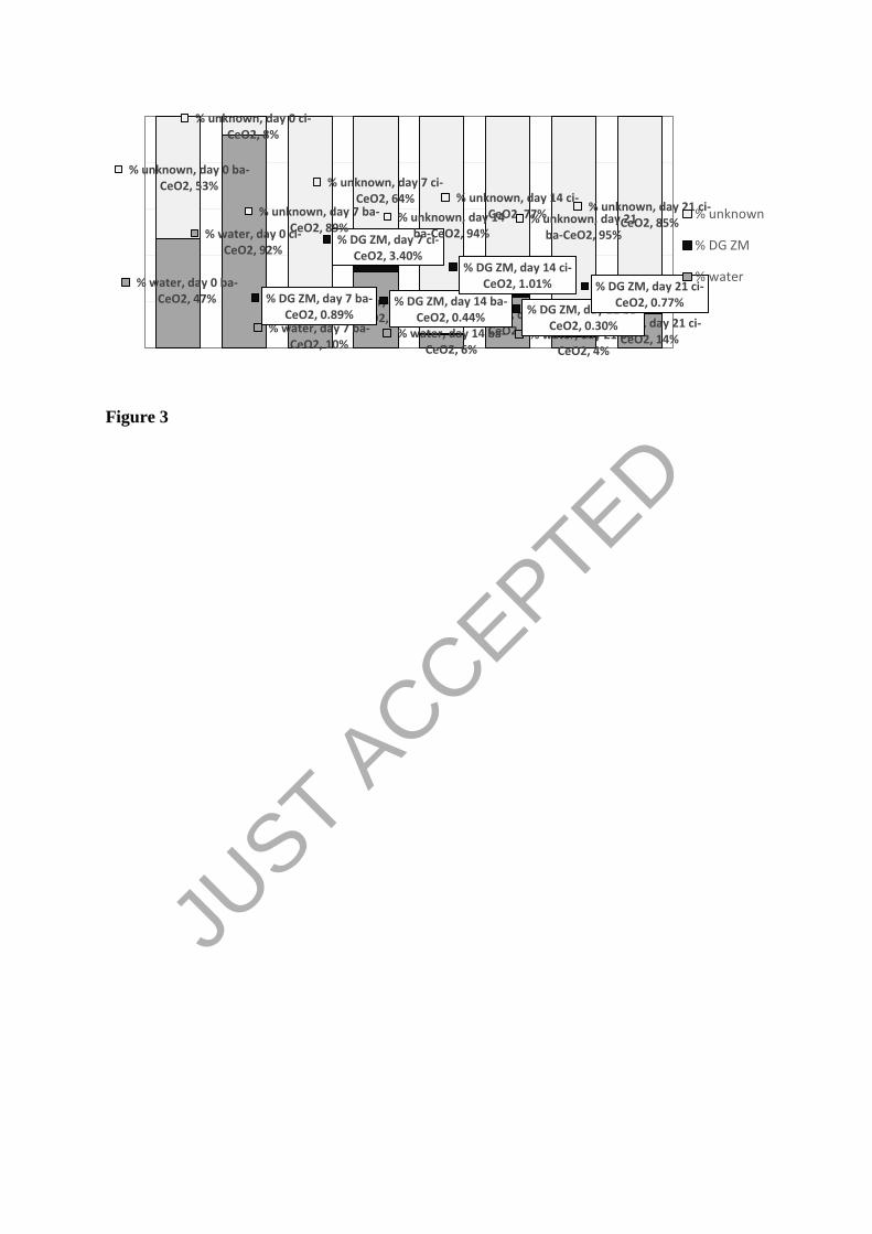
during the whole exposure period. Just before mussel introduction (day 0), 47+6 % (ba-CeO2;
96+12 µg/L) and 92+5 % (ci-CeO2; 187+11 µg/L) of total introduced Ce remained in the
water column (Figures 3 and 4A). [Ce] dropped significantly after mussel insertion : from
day 7 to day 21, [Ce] averaged 39% (ba-CeO2; 37+4 µg/L) and 69% (ci-CeO2; 128+9 µg/L) of
initial [Ce], even though the total amount of Ce introduced was gradually increased by nCeO2
injections. At the end of the experiment Ce left in the water column represented only 4+1%
(ba-CeO2) and 14+3% (ci-CeO2) of the total Ce introduced (Figure 3). Water [Ce] was 1.9-
fold and 3.2-fold higher in aquariums contaminated with ci-CeO2 at days 0 and 7 respectively
(Figure 4A).
Estimation of Ce removal by mussel filtration showed that after 7, 14 and 21 days of
exposure respectively, at least 79+13%, 88+12% and 91+14% of the suspended ba-CeO2,
and 64+15%, 76+17% and 84+13% of the suspended ci-CeO2 could have been removed due
to mussel filtration activity.
3.2. Ce bioaccumulation and speciation in mussel tissues
Results showed a significant coating-dependent Ce bioaccumulation in digestive glands
following nCeO2-exposure (Figure 4B and Table 1), with significantly higher accumulation
for ci-CeO2 (2.7-folds). After 21 days of exposure, 0.3% (ba-CeO2; 10.2+1.3 µg/gdw) and
0.8% (ci-CeO2; 29.1+10.3 µg/gdw) of the total Ce introduced were found in digestive glands
(Figure 3). ba-CeCe bioaccumulation was unaffected by exposure duration.
JUST A
CCEPTED
Page 13
After 21 days of exposure, almost total reduction of the CeIV
originally constituting nCeO2
into CeIII
was reported in the digestive gland (Figure 5). Percentages of CeIII
reached 82+8 %
(ba-CeO2; R factor = 0.0029) and 78+8 % (ci-CeO2; R factor = 0.0029).
3.3. Biomarker responses
Only significantly impacted biomarkers are described in this section (see detailed values in
Tables S5 to S7).
Significant interactive effects of nCeO2 exposure and exposure duration were observed on
SeGPx, piGST and MT mRNA expressions and immuno-efficiency (Table 1). Compared to
control, SeGPx mRNA was transiently decreased (-75% at day 7, Table S5) in mussel
exposed to ba-CeO2 (p=0.01). A 90% reduction of piGST mRNA expression was observed in
mussels 14 and 21 days-exposed to ci-CeO2 (p<0.001), reaching levels significantly lower
than in ba-CeO2-exposed mussels (p=0.035 and 0.002), which exhibited a 75% decrease at
day 14 (p=0.09). MT mRNA expression was transiently induced by ba-CeO2 (14-folds,
p<0.001) and ci-CeO2 (3.4-folds, p<0.001) at day 7, and ba-CeO2 induced MT mRNA 4-folds
more than ci-CeO2 (p<0.001). Higher immuno-efficiency was reported in ba-CeO2-exposed
mussels between days 7 and 21 (+55%, p=0.021, Table S6).
Healthy cells % was impacted by exposure duration (Table 1), with an increase at day 21
compared to day 14 (p=0.003). Increase in hemocyte lysosomal system size was reported for
ci-Ceci-CeO2-exposed mussels compared to ba-CeO2-exposed (+18%, p=0.035) and control
(+16%, p=0.052) mussels.
CAT activity was strongly affected by nCeO2 exposure and exposure duration (Table 1).
Lower CAT activities in ci-Ceci-CeO2-exposed mussels compared to control (p=0.04) and
ba-CeO2-exposed mussels (p=0.004, Table S6) were measured. All conditions combined,
CAT activities decrease at day 21 compared to days 7 (p=0.006) and 14 (p=0.034). Exposure
JUST A
CCEPTED
Page 14
duration also impacted TAC (decrease at day 14 compared to day 7, p=0.026), GST activity
(reduction between days 14 and 21, p=0.014) and [LOOH] (reduction between days 7 and 14;
p=0.04).
3.4. Discriminant analysis
We first get a model with no classification errors with only 9 biomarkers among the 24
initially considered. However, multicollinearity was observed, leading to drop two additional
variables (MT mRNA and ACP). The final classification error rate was 3.7% (i.e., a
classification error among 27 individuals). The final set of variables achieved multivariate
normality (all p-values above 0.05, E-statistic test (Székely and Rizzo, 2005)). The Box M
test revealed slight differences of variance-covariance between groups (p=0.008), but that did
not induce major bias in the LDA, considering the method robustness.
The discriminant power of our analysis was assessed basing on the Wilk's Lambda (0.063, p-
value <0.001). It evidenced that the deployed biomarker battery was able to describe the
effect of exposure to nCeO2, and to depict the two nCeO2 types using two discriminant
functions (DF) DF1 and DF2, with respective discriminant powers of 0.75 and 0.25. We
managed to significantly reduce the biomarker dataset from 24 to 7, which were sufficient to
depict nCeO2 exposure.
The reduced data set of seven biomarkers is shown in Table 2. To consider the relative
importance of the different variables, we used the standardized coefficients and associated p-
values. The three most discriminating biomarkers (p < 0.05) on the two axes were the piGST
mRNA expression, the catalase activity and the hemocyte lysosomal system size, which were
also among the biomarkers the most impacted individually. The four other biomarkers were
less significant but nevertheless critical to avoid classification errors.
JUST A
CCEPTED
Page 15
When plotting the 27 replicates (9 per experimental condition with the exposure duration
factor neglected), the three experimental conditions were clearly separated from each other
(Figure 6). ci-CeO2 and control only differ according to the first DF, while ba-CeO2 differs
from the two other groups on the two axes. Therefore, the reduced set of seven biomarkers
was sufficient to discriminate the three experimental conditions.
4. Discussion
4.1. [Ce] in the water column
ba-CeO2 and ci-CeO2 presented distinct behaviors in the water column. At day 0, ci-CeO2
displayed high stability, with more than 90% of the introduced ci-CeO2 still in suspension
compared to 50% for ba-CeO2. Tella et al. (2014) also showed that ci-CeO2 was relatively
stable in the short term in freshwater mesocosms but aggregated after few days due to coating
degradation. Enhanced ci-CeO2 stability could result from their citrate coating which endows
them with negative zeta potential at pH of the experiment (Auffan et al., 2014a). Therefore,
homo and hetero-aggregation processes, with negatively charged clay particles or
microorganisms, could be hindered by electrostatic repulsions, reducing ci-CeO2 aggregation
and sedimentation (Tella et al., 2014). On the contrary, ba-CeO2 were uncharged at the pH of
the experiment, and therefore aggregated faster (Tella et al., 2014). ba-CeO2 could also
interact strongly with suspended matter (Conway et al., 2014) and the larger hetero-
aggregates produced could sediment faster.
As filter feeding organisms, mussels could greatly impact nanoparticle concentrations in
water column (Conway et al., 2014; Montes et al., 2012). In fact, based on the review of
zebra mussel filtration rate made by Elliott et al. (2008), a median value of ~83 mL/mussel/h
was calculated, meaning that 120 L/day and 60 L/day (compared to a total volume of water of
56L in aquariums) were filtered by mussels in our experimental systems at the beginning and
JUST A
CCEPTED
Page 16
at the end of the experiment respectively. During this filtration process, mussels could trap
nanoparticles, ultimately resulting in their immobilization in the sediment as pseudofaeces or
faeces, which could explain why after mussel introduction at day 7, only 10% (ba-CeO2) and
33% (ci-CeO2) of the introduced Ce were still in suspension compared to 47% and 92% at
day 0. Estimation of nCeO2 removal due to mussel filtering activity showed that more than
70% of the Ce was removed from the water column, similar to other studies (Conway et al.,
2014; Montes et al., 2012). Slightly higher removal of ba-CeO2 could arise from its behavior
in water (see below). In fact, while zebra mussel can filter out sub-micron particles (down to
0.4 µm) from water, particle retention efficiency decrease with size, with a maximum
efficiency between 5 and 35 µm (Sprung and Rose, 1988). Consequently, ba-CeO2 homo-
aggregated or hetero-aggregated with algae and suspended matters could have been more
efficiently trapped by mussel gills (Baker et al. 2014; Ward and Kach, 2009).
As a result of nCeO2 immobilization in faeces and pseudofaeces, natural aggregation and
sedimentation processes, the percentage of Ce still suspended in water or accumulated by
mussels at the end of the experiment was low for both nanoparticles. Previous mesocosm
studies confirmed that sediments would be the primary sink of nCeO2 (Auffan et al., 2014b;
Tella et al., 2014; Zhang et al., 2012a) and while it was not measured in our experiment, the
sediment probably made up most of the balance of Ce indicated as unknown in Figure 3.
4.2. Ce bioaccumulation
Mussels accumulated both types of nCeO2 in their digestive gland, the preferential site of
nanoparticle accumulation in filter-feeding organisms (Baker et al. 2014; Browne et al.,
2008; Canesi et al., 2010).. Ce bioaccumulation presented two distinctive features. Firstly, it
seemed that nCeO2 surface properties influenced bioaccumulation, ci-CeO2 accumulating
three times more than ba-CeO2. Differences in ba-CeO2 and ci-CeO2 surface properties could
JUST A
CCEPTED
Page 17
modulate bioaccumulation at several levels. As evidenced in the previous paragraph, ci-CeO2
could be more stable in the water column, making it more bioavailable to mussels than ba-
CeO2. Once filtrated and trapped by the gills, nCeO2 could undergo chemical sorting (Baker
et al., 1998), and the organic citrate-coating of ci-CeO2 could be recognized as a potential
food source and promote its ingestion compared to ba-CeO2. Furthermore, according to Ward
and Shumway (2004), suspended sediments with organic coating had longer gut retention
time than uncoated ones in M. edulis. Retention time could also be increased for smaller
particles (100 nm) compared to larger (10 µm) ones (Ward and Kach, 2009). Such an
increase in ci-CeO2 retention time could explain part of the observed higher bioaccumulation,
and could facilitate cellular internalization. Once inside digestive glands, nCeO2 coating and
surface charge could modulate their internalization inside cells (Patil et al., 2007). In vitro
data comparing uptake of negatively charged and of neutral nanoparticles are contradictory,
some works showing increased uptake of negative nanoparticles (Patil et al., 2007), which
could support the observed higher ci-CeO2 bioaccumulation, but other works show the
opposite trend (Asati et al., 2010).
The second distinctive feature of nCeO2 bioaccumulation is that for both nCeO2 types,
internal [Ce] remained stable between day 7 and 21, even though total [nCeO2] in
experimental systems increased by repeated injections. A similar pattern was observed for
bivalves exposed to nCeO2 or to citrate-Au (Conway et al., 2014; García-Negrete et al.,
2013). nCeO2 hetero-aggregation with algae and suspended matters (Tella et al., 2014) could
have increased nCeO2 retention during the first week of exposure, when algae and suspended
matter contents were higher (Ward and Kach, 2009). For example, Conway et al. (2014)
reported a transient increase in nCeO2 bioaccumulation in the presence of algae in five days-
exposed mussels. Authors also reported concentration-dependent increases in mussel
clearance rate and pseudofaeces production, probably as a response to low quality food,
JUST A
CCEPTED
Page 18
therefore limiting nCeO2 ingestion and bioaccumulation. These findings could explain the
low recovery (<1%) of total introduced Ce in digestive gland at the end of exposure, coherent
with other works (Conway et al., 2014; Montes et al., 2012).
Finally, even in the case of nCeO2 internalization, bivalves are able to regulate internal
concentrations of some metals (Marigómez et al., 2002; Viarengo and Nott, 1993) and the
strong increase in MT mRNA levels after one week suggests that some regulation
mechanisms were activated and could have also contributed to the stabilization of internal
[Ce].
4.3. Internal nCeO2 speciation
We observed an almost complete reduction of the original CeIV
into CeIII
, which was also
observed in the freshwater snail P. corneus (Tella et al., 2014) and in the nematode C.
elegans (Collin et al. 2014b). In both experiments, no reduction occurred in the water
column, meaning that Ce reduction occurred after particle trapping by the mussels, in the
lumen and/or in contact with the cell lining the gut, the stomach and the digestive tubules,
which are loaded with free amino acids, proteins and digestive enzymes (Zhong and Wang,
2006). The cystein and thiol groups contained in the digestive fluid are known to be strong
metal complexation agents (Zhong and Wang, 2006). However, Collin et al. (2014b) and Liu
et al. (2013) showed that no reduction occurred when nCeO2 was incubated at neutral pH
with a wide range of biological components, suggesting that other mechanisms and molecules
played a role in CeIV
reduction. To unveil reduction mechanisms, the effects of mussel
digestive enzyme mixtures at relevant pH should be investigated thoroughly.
As hypothesized by Zhang et al. (2012b), the other possible mechanism for CeIII
presence in
digestive glands could be the dissolution of nCeO2 following contact with biological
molecules. Solubility of nCeO2 in environmentally relevant aqueous media has been shown
JUST A
CCEPTED
Page 19
to be negligible (Collin et al., 2014a), , while in the range of pH observed in zebra mussel
stomach (6.6-8.8 (Morton, 1969)), Ce predominant form is supposed to be solid CeIV
O2, or
solid CeIII
(OH)3 in the case of highly reducing conditions (Tamilmani et al., 2003).
Furthermore, nCeO2 have been retrieved in faeces following experiments on marine mussels,
even after transit in digestive apparatus, supporting the hypothesis that no dissolution
occurred (Montes et al., 2012). Formation of CeIII
oxides, but also possibly as CePO4, a stable
Ce form observed in phosphate-rich medium (Singh et al., 2011; Zhang et al., 2012b) as is the
intracellular environment, could then arise from surface reduction of nCeO2. Alternatively,
nCeO2 internalized in lysosomes could experiment dissolution due to acidic conditions
(Cornelis et al., 2011) and the soluble CeIII
could then reprecipitate into oxides or phosphates
or be trapped in metal-rich insoluble granules (Marigómez et al., 2002; Viarengo and Nott,
1993).
4.4. Biological effects of nCeO2
We recently stressed the benefit of using integrated biomarker responses to study nCeO2
effects and to identify biomarkers of interest (Garaud et al., 2015). In the present work, a very
synthetic overview of the effects of nCeO2 on mussel biology was provided using
discriminant analysis and we were able i) to clearly differentiate control groups from nCeO2-
exposed groups, ii) to show differential responses depending on nCeO2 type and iii) to
identify a reduced set of biomarkers sufficient to discriminate groups and their respective
significance. However, due to a reduced dataset, this model has no predictive value, and
should only be considered for its ability to synthetize and describe links between nanoparticle
exposition and biomarker responses. Of the seven biomarkers identified, three were of
particular significance (p<0.05; piGST mRNA levels, catalase activity, and hemocyte
lysosomal system size) confirming our previous results (Garaud et al., 2015).
JUST A
CCEPTED
Page 20
Of these most discriminant biomarkers, the gill piGST mRNA expression was the most
impacted biomarker and decreased following nCeO2 exposure, while greater impact was
reported for ci-CeO2. GST are involved in endogenous molecule and xenobiotic detoxication,
and piGST, the principal cytosolic GST, could deactivate lipid hydroperoxides and their by-
products, preventing lipoperoxidation, and directly detoxify ROS through their cysteine
groups (Doyen et al., 2008). GST gene transcription can be induced directly by several
xenobiotics or indirectly following oxidative stress (Blanchette et al. 2007). Park et al. (2008)
observed GST mRNA up-regulation following in vitro nCeO2-exposure (40 mg/L, human
bronchial cells) associated with increased ROS production, GSH decrease and cytotoxicity.
Conversely, in an nCeO2 in vitro exposure on rat neuronal cells, Ciofani et al. (2014) reported
the absence of effects on piGST mRNA expression, but highlighted the down-regulation of
several genes related to ROS presence and inflammatory processes, such as GPx genes
expression, also transitory decreased at day 7 during our experiment. Down-regulation of
piGST and SeGPx genes could result from a trapping of endogenous ROS by nCeO2, whose
antioxidant properties have been observed in many studies (Das et al., 2007; Garaud et al.,
2015). That could be linked to the observed reduction of CeIV
into CeIII
, which implied redox
reactions impacting redox balance of the cell.
The second most discriminative biomarker was the catalase activity, which was reduced
throughout the exposure for ci-CeO2, correlating with the stable internal [nCeO2]. As an
important player in the cellular antioxidant defenses, catalase is classically induced following
pollutant exposure, while inhibition is generally observed in cases of severe toxicity (Osman
et al. 2007). However, no impacts on lipoperoxidation, caspase-3 induction or energy reserve
depletion were observed in ci-CeO2-exposed mussels, refuting the existence of severe ci-
CeO2 toxicity. A similar decrease in catalase activity, associated with reduced cellular
damages, was observed in Garaud et al. (2015) and could arise from nCeO2 ROS scavenging
JUST A
CCEPTED
Page 21
through catalase mimetic activity (Das et al., 2007), or gene down-regulation resulting from
decreased intracellular H2O2 levels (Ciofani et al., 2014). The observed reduction of CeIV
to
CeIII
could also explain the observed decrease in catalase activity, as CeIII
phosphate has been
shown to exert catalase mimetic activity (Singh et al., 2011).
Finally, the third most discriminative biomarker was the lysosomal system in hemocytes,
more developed following ci-CeO2 exposure. In invertebrates, hemocytes are circulating cells
implied in important physiological processes, namely xenobiotic sequestration and
detoxication, immunity, intracellular digestion of nutrients and transport, and endogenous and
exogenous waste disposal (Giamberini and Pihan, 1997). Various nanoparticles were
internalized by mussel hemocytes following short-term in vitro and in vivo exposures (Ciacci
et al., 2012; Couleau et al., 2012). Direct nCeO2 uptake could occur in the haemolymph, in
which nanoparticles were shown to penetrate quickly in marine mussel (Browne et al., 2008),
or in the digestive tubule lumen, in which hemocytes can penetrate to absorb nutrients before
re-infiltrating in the tissues to distribute them into the organism (Cheng, 1981). Alternatively,
a classic detoxication mechanism is the disintegration of mussel digestive cells to eliminate
the indigestible material phagocyted, the distal parts of the cell forming spherules eliminated
through the faeces, and the basal parts forming circulating cells to
transport wastes to excretory organs (Morton, 1969; Viarengo and Nott, 1993). Both direct
uptake and digestive cell fragmentation could explain the presence of nCeO2 inside
hemocytes. However, lysosomal system enlargement is only observed in hemocytes of ci-
CeO2-exposed mussels, which could arise from a weaker ba-CeO2 bioaccumulation. This
could also support the hypothesis that negatively charged nanoparticles accumulate
preferentially inside lysosomes (Asati et al., 2010; García-Negrete et al., 2013; Singh et al.,
2010). Despite potential exposure of hemocytes to nCeO2, no adverse effects on the other
hemocyte parameters were observed, whereas several nanoecotoxicological studies using
JUST A
CCEPTED
Page 22
other metallic nanoparticles showed impacts on phagocytosis, lysosomal membrane stability,
and ROS production following in vitro and in vivo exposures of hemocytes at high
concentrations (Canesi et al., 2010; Ciacci et al., 2012; Couleau et al., 2012).
The four other biomarkers of the reduced data set (i.e. LDH, ETS and GPx activities, and
cholesterol content) were less significant but nonetheless essential to obtain a correct
classification. Taken individually, their trends were not clear, but the discriminant analysis
suggests that they could be impacted by nCeO2 exposure. For example, ETS activity helped
discriminate mussels exposed to ba-CeO2 on the DF2 axis (decreasing trend), suggesting an
impact on mussel metabolic activity. Cholesterol content and LDH activity also played a role
in the discriminant analysis, supporting the hypothesis of an impact on metabolism.
Similarly, GPx activity, linked to nonspecific antioxidant defense systems, could be impacted
by nCeO2 exposure.
MT mRNA where not maintained in the discriminant analysis due to collinearity problems,
but considered individually, this variable exhibited significant variations and is of high
biological significance when studying the effects of metallic contamination on organisms.
MT mRNA levels were strongly induced at day 7 for both nanoparticle types, then dropped
back to basal values. This feature could be linked with the levelling off of the internal [Ce]
indicating that some regulation mechanisms were activated and equilibrated. MT gene
transcription responds to a wide spectrum of stresses, permitting heavy metal chelation and
reducing their cytotoxicity (Viarengo and Nott, 1993). ba-CeO2 induced MTs four-times more
than ci-CeO2, although ci-CeO2 was three times more accumulated by mussels. Two
hypotheses could explain this paradox. Firstly, the citrate coating of ci-CeO2 could hide the
metallic core and therefore reduced MT binding to CeO2 and subsequent mRNA induction.
Secondly, MTs are mostly localized in the cytosol and nucleus (Cherian, 1994), and neutral
nCeO2 could be mostly localized in the cytosol while negatively charged nanoparticles
JUST A
CCEPTED
Page 23
accumulated in lysosomes (Asati et al., 2010; Singh et al., 2010), then ba-CeO2 would be
more likely to bind to and induce MT.
As a whole, although the discriminant analysis showed a clear separation between control and
exposed mussels, we were unable to evidence severe toxicity of nCeO2 since the number of
impacted biomarkers and the variations observed were quite low. Furthermore, on the
impacted biomarkers, MT mRNA levels and hemocyte lysosomal system are considered as
exposure biomarkers, but provide no clues on the toxicity resulting from this exposure. In the
same way, the results on the other two most impacted biomarkers, piGST mRNA levels and
catalase activity, and on the four less significant biomarkers of the reduced data set, are
insufficient to conclude on hypothetic toxic impacts of nCeO2 on mussel biology. No
biomarkers of toxicity (lipoperoxidation, caspase-3) were activated by nCeO2 exposure, nor
was the functional filtering activity of mussels affected, even after a three week exposure. It
suggests that, in our experiment, nCeO2 did not exert severe deleterious effects on mussels.
JUST A
CCEPTED
Page 24
5. Conclusions
This work aimed at assessing the fate and effects of ba-CeO2 and ci-CeO2 nanoparticles on D.
polymorpha, in simplified ecosystems and at low concentrations, using an integrated
multibiomarker approach. ba-CeO2 and ci-CeO2 presented distinct behavior in the water
column, the citrate-coating enhancing ci-CeO2 colloidal stability. This led to different
bioaccumulation patterns, mussels accumulating three times more ci-CeO2 than baCeO2.
However, the same bioreduction of CeIV
into CeIII
was observed inside mussel digestive
glands, but mechanisms pertaining to this biotransformation remain to be uncovered. An
integrated discriminant analysis on the measured biomarkers was performed to assess the
effects of nCeO2 bioaccumulation and biotransformation on mussel biology. This powerful
tool allowed the separation of exposed and control mussels, and also showed that mussels
responded differently to ba-CeO2 and ci-CeO2 exposure. The discriminant analysis also
allowed us to single out a set of seven biomarkers sufficient to discriminate experimental
groups. We showed that two of the three most significant biomarkers, lysosomal system size
and catalase activity, were related to those isolated as the most responsive in a previous short-
term exposure to nCeO2 (Garaud et al., 2015), confirming the usefulness of these biomarkers
in nCeO2 ecotoxicity assessment. MT mRNA induction and lysosomal enlargement were
more indicative of the exposure and stress responses by the organisms, while the lack of
impact on cellular damages, energetic reserves and filtration activity suggested the absence of
serious adverse effects of nCeO2 following more environmentally realistic exposure.
However, further studies are needed to investigate if the slight perturbations observed could
lead to populational impacts in the long-term. JU
ST ACCEPTED
Page 25
Acknowledgements. Financial supports were provided by the French National Agency
(ANR-10-NANO-0006/MESONNET project) for M. Garaud PhD salary and running costs
and CPER Lorraine-ZAM (Contrat Projet Etat Région Lorraine, Zone Atelier Moselle). This
work is a contribution to the Labex Ressources 21, ANR-10-LABX-21-01 (Strategic metal
resources of the 21st century). The authors gratefully acknowledge CNRS for funding the
iCEINT International Consortium for the Environmental Implications of NanoTechnology.
Sharon Kruger is gratefully acknowledged for her English corrections.
Declaration of interest
As indicated in the acknowledgements, financial supports come from different public
institutions. They were provided by the French National Agency (ANR-10-NANO-
0006/MESONNET project) for M. Garaud PhD salary and running costs and CPER Lorraine-
ZAM (Contrat Projet Etat Région Lorraine, Zone Atelier Moselle). It is not necessary for
University, CNRS or ANR to review, comment or approve their manuscript before
publication. We did not receive writing assistance; Sharon Kruger just reviewed the English
version of the manuscript.
JUST A
CCEPTED
Page 26
LITERATURE CITED
Arab, K., Steghens, J.-P., 2004. Plasma lipid hydroperoxides measurement by an automated
xylenol orange method. Anal. Biochem. 325, 158–163. doi:10.1016/j.ab.2003.10.022
Asati, A., Santra, S., Kaittanis, C., Perez, J.M., 2010. Surface-Charge-Dependent Cell
Localization and Cytotoxicity of Cerium Oxide Nanoparticles. ACS Nano 4, 5321–
5331. doi:10.1021/nn100816s
Auffan, M., Masion, A., Labille, J., Diot, M.-A., Liu, W., Olivi, L., Proux, O., Ziarelli, F.,
Chaurand, P., Geantet, C., Bottero, J.-Y., Rose, J., 2014a. Long-term aging of a CeO2
based nanocomposite used for wood protection. Environ. Pollut. 188, 1–7.
doi:10.1016/j.envpol.2014.01.016
Auffan, M., Tella, M., Santaella, C., Brousset, L., Paillès, C., Barakat, M., Espinasse, B.,
Artells, E., Issartel, J., Masion, A., Rose, J., Wiesner, M.R., Achouak, W., Thiéry, A.,
Bottero, J.-Y., 2014b. An adaptable mesocosm platform for performing integrated
assessments of nanomaterial risk in complex environmental systems. Sci. Rep. 4.
doi:10.1038/srep05608
Baker, Levinton, J.S., Kurdziel, J.P., Shumway, S.E., 1998. Selective feeding and
biodeposition by zebra mussels and their relation to changes in phytoplankton
composition and seston load. J. Shellfish Res. 17, 1207–1213.
Baker, T.J., Tyler, C.R., Galloway, T.S., 2014. Impacts of metal and metal oxide
nanoparticles on marine organisms. Environ. Pollut. 186, 257–271.
doi:10.1016/j.envpol.2013.11.014
Beer, R.F., Sizer, I.W., 1952. A spectrophotometric method for measuring the breakdown of
hydrogen peroxide by catalase. J. Biol. Chem. 195, 133–140.
Blanchette, B., Feng, X., Singh, B.R., 2007. Marine Glutathione S-Transferases. Mar.
Biotechnol. 9, 513–542. doi:10.1007/s10126-007-9034-0
Bour, A., Mouchet, F., Silvestre, J., Gauthier, L., Pinelli, E., 2015. Environmentally relevant
approaches to assess nanoparticles ecotoxicity: A review. J. Hazard. Mater. 283, 764–
777. doi:10.1016/j.jhazmat.2014.10.021
Browne, M.A., Dissanayake, A., Galloway, T.S., Lowe, D.M., Thompson, R.C., 2008.
Ingested Microscopic Plastic Translocates to the Circulatory System of the Mussel,
Mytilus edulis (L.). Environ. Sci. Technol. 42, 5026–5031. doi:10.1021/es800249a
Canesi, L., Ciacci, C., Fabbri, R., Marcomini, A., Pojana, G., Gallo, G., 2012. Bivalve
molluscs as a unique target group for nanoparticle toxicity. Mar. Environ. Res.,
Emerging and persistent impacts on Marine Organisms: Detection methods and action
mechanisms 76, 16–21. doi:10.1016/j.marenvres.2011.06.005
Canesi, L., Fabbri, R., Gallo, G., Vallotto, D., Marcomini, A., Pojana, G., 2010. Biomarkers
in Mytilus galloprovincialis exposed to suspensions of selected nanoparticles (Nano
carbon black, C60 fullerene, Nano-TiO2, Nano-SiO2). Aquat. Toxicol. 100, 168–177.
doi:10.1016/j.aquatox.2010.04.009
Cheng, T.C., 1981. 8. Bivalves, in: Invertebrate Blood Cells. N.A. Ratcliffe and A.F. Rowley,
Academic Press, London, p. 314.
Cherian, M.G., 1994. The significance of the nuclear and cytoplasmic localization of
metallothionein in human liver and tumor cells. Environ. Health Perspect. 102, 131–
135. doi:10.1289/ehp.94102s3131
Ciacci, C., Canonico, B., Bilaniĉovă, D., Fabbri, R., Cortese, K., Gallo, G., Marcomini, A.,
Pojana, G., Canesi, L., 2012. Immunomodulation by Different Types of N-Oxides in
the Hemocytes of the Marine Bivalve Mytilus galloprovincialis. PLoS ONE 7,
e36937. doi:10.1371/journal.pone.0036937
JUST A
CCEPTED
Page 27
Ciofani, G., Genchi, G.G., Mazzolai, B., Mattoli, V., 2014. Transcriptional profile of genes
involved in oxidative stress and antioxidant defense in PC12 cells following treatment
with cerium oxide nanoparticles. Biochim. Biophys. Acta BBA - Gen. Subj. 1840,
495–506. doi:10.1016/j.bbagen.2013.10.009
Coen, W.M.D., Janssen, C.R., 1997. The use of biomarkers in Daphnia magna toxicity
testing. IV. Cellular Energy Allocation: a new methodology to assess the energy
budget of toxicant-stressed Daphnia populations. J. Aquat. Ecosyst. Stress Recovery
6, 43–55. doi:10.1023/A:1008228517955
Collin, B., Auffan, M., Johnson, A.C., Kaur, I., Keller, A.A., Lazareva, A., Lead, J.R., Ma,
X., Merrifield, R.C., Svendsen, C., White, J.C., Unrine, J.M., 2014a. Environmental
release, fate and ecotoxicological effects of manufactured ceria nanomaterials. Env.
Sci Nano -. doi:10.1039/C4EN00149D
Collin, B., Oostveen, E., Tsyusko, O.V., Unrine, J.M., 2014b. Influence of Natural Organic
Matter and Surface Charge on the Toxicity and Bioaccumulation of Functionalized
Ceria Nanoparticles in Caenorhabditis elegans. Environ. Sci. Technol. 48, 1280–1289.
doi:10.1021/es404503c
Conway, J.R., Hanna, S.K., Lenihan, H.S., Keller, A.A., 2014. Effects and Implications of
Trophic Transfer and Accumulation of CeO2 Nanoparticles in a Marine Mussel.
Environ. Sci. Technol. 48, 1517–1524. doi:10.1021/es404549u
Cornelis, G., Ryan, B., McLaughlin, M.J., Kirby, J.K., Beak, D., Chittleborough, D., 2011.
Solubility and Batch Retention of CeO2 Nanoparticles in Soils. Environ. Sci.
Technol. 45, 2777–2782. doi:10.1021/es103769k
Couleau, N., Techer, D., Pagnout, C., Jomini, S., Foucaud, L., Laval-Gilly, P., Falla, J.,
Bennasroune, A., 2012. Hemocyte responses of Dreissena polymorpha following a
short-term in vivo exposure to titanium dioxide nanoparticles: Preliminary
investigations. Sci. Total Environ. 438, 490–497. doi:10.1016/j.scitotenv.2012.08.095
Das, M., Patil, S., Bhargava, N., Kang, J.-F., Riedel, L.M., Seal, S., Hickman, J.J., 2007.
Auto-catalytic ceria nanoparticles offer neuroprotection to adult rat spinal cord
neurons. Biomaterials 28, 1918–1925. doi:10.1016/j.biomaterials.2006.11.036
Doyen, P., Bigot, A., Vasseur, P., Rodius, F., 2008. Molecular cloning and expression study
of pi-class glutathione S-transferase (pi-GST) and selenium-dependent glutathione
peroxidase (Se-GPx) transcripts in the freshwater bivalve Dreissena polymorpha.
Comp. Biochem. Physiol. Part C Toxicol. Pharmacol. 147, 69–77.
doi:10.1016/j.cbpc.2007.08.002
Elliott, P., Aldridge, D.C., Moggridge, G.D., 2008. Zebra mussel filtration and its potential
uses in industrial water treatment. Water Res. 42, 1664–1674.
doi:10.1016/j.watres.2007.10.020
Erel, O., 2004. A novel automated direct measurement method for total antioxidant capacity
using a new generation, more stable ABTS radical cation. Clin. Biochem. 37, 277–
285. doi:10.1016/j.clinbiochem.2003.11.015
Garaud, M., Trapp, J., Devin, S., Cossu-Leguille, C., Pain-Devin, S., Felten, V., Giamberini,
L., 2015. Multibiomarker assessment of cerium dioxide nanoparticle (nCeO2)
sublethal effects on two freshwater invertebrates, Dreissena polymorpha and
Gammarus roeseli. Aquat. Toxicol. doi:10.1016/j.aquatox.2014.11.004
García-Negrete, C.A., Blasco, J., Volland, M., Rojas, T.C., Hampel, M., Lapresta-Fernández,
A., Jiménez de Haro, M.C., Soto, M., Fernández, A., 2013. Behaviour of Au-citrate
nanoparticles in seawater and accumulation in bivalves at environmentally relevant
concentrations. Environ. Pollut. 174, 134–141. doi:10.1016/j.envpol.2012.11.014
JUST A
CCEPTED
Page 28
Giamberini, L., Pihan, J.C., 1997. Lysosomal changes in the hemocytes of the freshwater
mussel Dreissena polymorpha experimentally exposed to lead and zinc. Dis. Aquat.
Organ. 28, 221–227.
Guerlet, E., Vasseur, P., Giambérini, L., 2010. Spatial and temporal variations of biological
responses to environmental pollution in the freshwater zebra mussel. Ecotoxicol.
Environ. Saf. 73, 1170–1181. doi:10.1016/j.ecoenv.2010.05.009
Kurelec, B., Smital, T., Pivèeviæ, B., Eufemia, N., Epel, D., 2000. Multixenobiotic
Resistance, P-Glycoprotein, and Chemosensitizers. Ecotoxicology 9, 307–327.
doi:10.1023/A:1026560922731
Liu, W., Rose, J., Plantevin, S., Auffan, M., Bottero, J.-Y., Vidaud, C., 2013. Protein corona
formation for nanomaterials and proteins of a similar size: hard or soft corona?
Nanoscale 5, 1658–1668. doi:10.1039/C2NR33611A
Marigómez, I., Soto, M., Cajaraville, M.P., Angulo, E., Giamberini, L., 2002. Cellular and
subcellular distribution of metals in molluscs. Microsc. Res. Tech. 56, 358–392.
doi:10.1002/jemt.10040
Minguez, L., Boiché, A., Sroda, S., Mastitsky, S., Brulé, N., Bouquerel, J., Giambérini, L.,
2012. Cross-effects of nickel contamination and parasitism on zebra mussel
physiology. Ecotoxicology 21, 538–547. doi:10.1007/s10646-011-0814-y
Montes, M.O., Hanna, S.K., Lenihan, H.S., Keller, A.A., 2012. Uptake, accumulation, and
biotransformation of metal oxide nanoparticles by a marine suspension-feeder. J.
Hazard. Mater. 225-226, 139–145. doi:10.1016/j.jhazmat.2012.05.009
Moore, M.N., 2006. Do nanoparticles present ecotoxicological risks for the health of the
aquatic environment? Environ. Int. 32, 967–976. doi:10.1016/j.envint.2006.06.014
Morton, B., 1969. Studies on the Biology of Dreissena Polymorpha Pall II. Correlation of the
Rhythms of Adductor Activity, Feeding, Digestion and Excretion. J. Molluscan Stud.
38, 401–414.
O’Brien, N.J., Cummins, E.J., 2011. A Risk Assessment Framework for Assessing Metallic
Nanomaterials of Environmental Concern: Aquatic Exposure and Behavior. Risk
Anal. 31, 706–726. doi:10.1111/j.1539-6924.2010.01540.x
Osman, A.M., Heuvel, H. van den, Noort, P.C.M. van, 2007. Differential responses of
biomarkers in tissues of a freshwater mussel, Dreissena polymorpha, to the exposure
of sediment extracts with different levels of contamination. J. Appl. Toxicol. 27, 51–
59. doi:10.1002/jat.1183
Pain-Devin, S., Cossu-Leguille, C., Geffard, A., Giambérini, L., Jouenne, T., Minguez, L.,
Naudin, B., Parant, M., Rodius, F., Rousselle, P., Tarnowska, K., Daguin-Thiébaut,
C., Viard, F., Devin, S., 2014. Towards a better understanding of biomarker response
in field survey: A case study in eight populations of zebra mussels. Aquat. Toxicol.
155, 52–61. doi:10.1016/j.aquatox.2014.06.008
Palais, F., 2011. Potentiel informatif des reponses enzymatiques digestives dans l’evaluation
des effets d’un stress toxique sur l’etat physiologique des invertebres aquatiques :
etude de cas chez un bivalve d’eau douce : Dreissena polymorpha. PhD Reims.
Park, E.-J., Choi, J., Park, Y.-K., Park, K., 2008. Oxidative stress induced by cerium oxide
nanoparticles in cultured BEAS-2B cells. Toxicology 245, 90–100.
doi:10.1016/j.tox.2007.12.022
Patil, S., Sandberg, A., Heckert, E., Self, W., Seal, S., 2007. Protein adsorption and cellular
uptake of cerium oxide nanoparticles as a function of zeta potential. Biomaterials 28,
4600–4607. doi:10.1016/j.biomaterials.2007.07.029
R Core Team, 2014. R: A language and environment for statistical computing. R Foundation
for Statistical Computing, Vienna, Austria. URL http://www.R-project.org/.
JUST A
CCEPTED
Page 29
Singh, S., Dosani, T., Karakoti, A.S., Kumar, A., Seal, S., Self, W.T., 2011. A phosphate-
dependent shift in redox state of cerium oxide nanoparticles and its effects on
catalytic properties. Biomaterials 32, 6745–6753.
doi:10.1016/j.biomaterials.2011.05.073
Singh, S., Kumar, A., Karakoti, A., Seal, S., Self, W.T., 2010. Unveiling the mechanism of
uptake and sub-cellular distribution of cerium oxide nanoparticles. Mol. Biosyst. 6,
1813–1820. doi:10.1039/C0MB00014K
Sprung, M., Rose, U., 1988. Influence of food size and food quantity on the feeding of the
mussel Dreissena polymorpha. Oecologia 77, 526–532. doi:10.1007/BF00377269
Sroda, S., Cossu-Leguille, C., 2011. Seasonal variability of antioxidant biomarkers and
energy reserves in the freshwater gammarid Gammarus roeseli. Chemosphere 83,
538–544. doi:10.1016/j.chemosphere.2010.12.023
Sun, T.Y., Gottschalk, F., Hungerbühler, K., Nowack, B., 2014. Comprehensive probabilistic
modelling of environmental emissions of engineered nanomaterials. Environ. Pollut.
185, 69–76. doi:10.1016/j.envpol.2013.10.004
Székely, G.J., Rizzo, M.L., 2005. A new test for multivariate normality. J. Multivar. Anal. 93,
58–80. doi:10.1016/j.jmva.2003.12.002
Tamilmani, S., Shan, J., Huang, W., Raghavan, S., Small, R., Shang, C., Scott, B., 2003.
Interaction Between Ceria and Hydroxylamine, in: Symposium F – Chemical-
Mechanical Planarization, MRS Online Proceedings Library. doi:10.1557/PROC-767-
F3.3
Tella, M., Auffan, M., Brousset, L., Issartel, J., Kieffer, I., Pailles, C., Morel, E., Santaella,
C., Angeletti, B., Artells, E., Rose, J., Thiéry, A., Bottero, J.-Y., 2014. Transfer,
Transformation, and Impacts of Ceria Nanomaterials in Aquatic Mesocosms
Simulating a Pond Ecosystem. Environ. Sci. Technol. 48, 9004–9013.
doi:10.1021/es501641b
Van Hoecke, K., De Schamphelaere, K.A.C., Van der Meeren, P., Smagghe, G., Janssen,
C.R., 2011. Aggregation and ecotoxicity of CeO2 nanoparticles in synthetic and
natural waters with variable pH, organic matter concentration and ionic strength.
Environ. Pollut. 159, 970–976. doi:10.1016/j.envpol.2010.12.010
Viarengo, A., Nott, J.A., 1993. Mechanisms of heavy metal cation homeostasis in marine
invertebrates. Comp. Biochem. Physiol. Part C Comp. Pharmacol. 104, 355–372.
doi:10.1016/0742-8413(93)90001-2
Ward, J.E., Kach, D.J., 2009. Marine aggregates facilitate ingestion of nanoparticles by
suspension-feeding bivalves. Mar. Environ. Res. 68, 137–142.
doi:10.1016/j.marenvres.2009.05.002
Ward, J.E., Shumway, S.E., 2004. Separating the grain from the chaff: particle selection in
suspension- and deposit-feeding bivalves. J. Exp. Mar. Biol. Ecol. 300, 83–130.
doi:10.1016/j.jembe.2004.03.002
Zhang, P., He, X., Ma, Y., Lu, K., Zhao, Y., Zhang, Z., 2012a. Distribution and
bioavailability of ceria nanoparticles in an aquatic ecosystem model. Chemosphere
89, 530–535. doi:10.1016/j.chemosphere.2012.05.044
Zhang, P., Ma, Y., Zhang, Z., He, X., Zhang, J., Guo, Z., Tai, R., Zhao, Y., Chai, Z., 2012b.
Biotransformation of Ceria Nanoparticles in Cucumber Plants. ACS Nano 6, 9943–
9950. doi:10.1021/nn303543n
Zhong, H., Wang, W.-X., 2006. Sediment-Bound Inorganic Hg Extraction Mechanisms in the
Gut Fluids of Marine Deposit Feeders. Environ. Sci. Technol. 40, 6181–6186.
doi:10.1021/es061195z
JUST A
CCEPTED
Page 30
Factor
exposure duration (df=2)
nCeO2 exposure (df=2) interaction (df=4) d
f
F Pr(F)
F Pr(F)
F Pr(F)
Ce bioacc. 0.03 0.9661 401.34 0.0000 *** 2.72 0.0621 18
RNA MT 162.80 0.0000 *** 173.23 0.0000 *** 93.62 0.0000 *** 18
RNA piGST 7.45 0.0044 ** 53.79 0.0000 *** 4.48 0.0109 * 18
RNA SeGPx 15.56 0.0001 *** 1.10 0.3535 7.27 0.0011 ** 18
Cv 7.41 0.0045 ** 0.18 0.8339 1.94 0.1478 18
Ce 3.00 0.0749
0.57 0.5781
1.50 0.2453
18
Cm 1.93 0.1745
1.01 0.3827
0.54 0.7095
18
ROS 0.82 0.4562
0.74 0.4930
0.96 0.4515
18
Lyso 0.07 0.9350
3.73 0.0442 * 0.52 0.7225
18
phag. 1b 0.53 0.4801
0.62 0.5538
0.15 0.8602
12
phag. 3b 8.94 0.0113 * 2.16 0.1585
3.93 0.0486 * 12
H. CSP-3 Kruskal-Wallis chi-squared = 16.7407, df = 8, p-value = 0.0329*
GPx 1.80 0.1936 1.86 0.1836 0.87 0.4996 18
CAT 7.00 0.0056 ** 7.34 0.0047 ** 1.12 0.3784
18
TAC 4.42 0.0274 * 0.10 0.9027
2.79 0.0577
18
GST 5.10 0.0176 * 0.42 0.6645
0.97 0.4500
18
ACP 1.71 0.2089
1.07 0.3634
1.01 0.4271
18
[LOOH] 3.93 0.0384 * 0.96 0.4021 1.41 0.2715 18
CSP-3 0.59 0.5639 0.72 0.4996 0.57 0.6850 18
[trigly.] 3.38 0.0568 0.35 0.7115 0.48 0.7519 18
[chol.] 0.03 0.9750
1.36 0.2809
0.43 0.7828
18
[prot.] 1.29 0.2986
0.04 0.9646
0.52 0.7213
18
ETS 2.89 0.0818
1.89 0.1796
0.25 0.9081
18
LDH 1.26 0.3085 2.68 0.0956 0.68 0.6133 18
MXR 0.20 0.8222 0.45 0.6432 0.92 0.4760 18
Table 1 : Results of the statistical analysis (2 ways-ANOVA or Kruskal Wallis when non-parametric data)
to assess respective effects of nCeO2 exposure, exposure duration and their interaction on Ce internal
concentration and measured biomarkers after 7, 14 and 21 days of exposure to ba-CeO2 and ci-CeO2 - *
indicates significant effect of the studied factor (*p<0.05; **p<0.01; ***p<0.001)
JUST A
CCEPTED
Page 31
Biomarker Raw coefficients Standardized coefficients
F p value DF1 DF2 DF1 DF2
RNA piGST 0.027 0.029 1.224 1.299 27.49 <0.001 CAT 0.040 -0.036 0.948 -0.837 4.82 0.017 Lyso -0.057 0.040 -0.901 0.627 4.42 0.023 LDH 0.009 0.010 0.213 0.235 2.77 0.083 ETS -0.053 0.098 -0.506 0.931 1.84 0.181 GPx 0.992 2.677 0.110 0.296 1.54 0.190
[chol.] -0.097 0.052 -0.443 0.236 1.78 0.234 Table 2 : Results of the discriminant analysis – final set of biomarkers and their raw coefficients on
discriminant functions 1 and 2 (DF1 and DF2), useful for predicting the group for a new replicate, their
standardized coefficients, useful to hierarchize relative influence of the variables, and their significance.
JUST A
CCEPTED
Page 32
Figure 1
Antioxidant defenses (CAT, GPx activities and total antioxidant capacity)
Antitoxic defenses (GST, MXR and ACP activities)
Cellular damages (lipoperoxidation and apoptosis)
Energetic reserves (cholesterol, glycogen and triglycerides) and energetic metabolism (ETS, LDH)
Gill mRNA expression of metallothionein (MT), SeGPx and piGST
Filtration
Hemocyte parameters (phagocytosis, ROS, viability, lysosome, CSP-3)
-
+
Molecular
Sub-cellular
Cellular scale
Individual
Ecological relevance
JUST A
CCEPTED
Page 33
Figure 2
JUST A
CCEPTED
Page 34
Figure 3
% water, day 0 ba-CeO2, 47%
% water, day 0 ci-CeO2, 92%
% water, day 7 ba-CeO2, 10%
% water, day 7 ci-CeO2, 33%
% water, day 14 ba-CeO2, 6%
% water, day 14 ci-CeO2, 22% % water, day 21 ba-
CeO2, 4%
% water, day 21 ci-CeO2, 14%
% DG ZM, day 7 ba-CeO2, 0.89%
% DG ZM, day 7 ci-CeO2, 3.40%
% DG ZM, day 14 ba-CeO2, 0.44%
% DG ZM, day 14 ci-CeO2, 1.01%
% DG ZM, day 21 ba-CeO2, 0.30%
% DG ZM, day 21 ci-CeO2, 0.77%
% unknown, day 0 ba-CeO2, 53%
% unknown, day 0 ci-CeO2, 8%
% unknown, day 7 ba-CeO2, 89%
% unknown, day 7 ci-CeO2, 64%
% unknown, day 14 ba-CeO2, 94%
% unknown, day 14 ci-CeO2, 77% % unknown, day 21
ba-CeO2, 95%
% unknown, day 21 ci-CeO2, 85%
% unknown
% DG ZM
% water
JUST A
CCEPTED
Page 35
Figure 4
[Ce
] (µ
g/L)
Ba-CeO2Ci-CeO2
ac
b b ab
d cd
c cd
A.
[Ce
] (µ
g/gd
.w.)
Ba-CeO2Ci-CeO2
B.
JUST A
CCEPTED
Page 36
Figure 5
-1
0
1
2
3
4
5
5690 5720 5750 5780
No
rmal
ize
d a
bso
rban
ce
Energy (eV)
CeIII cysteine
Ba-CeO2
DG Ba-CeO2
-1
0
1
2
3
4
5
5690 5720 5750 5780
No
rmal
ize
d a
bso
rban
ce
Energy (eV)
CeIII cysteine
Ci-CeO2
DG Ci-CeO2
A. B.
JUST A
CCEPTED
Page 37
Figure 6
JUST A
CCEPTED
Page 38
Figure captions
Figure 1: Multibiomarker approach used and physiological functions involved, ecological
relevance (left arrow) and level on the biological scale (right arrow).
Figure 2 : Schematic representation of the experimental design.
Figure 3 : Mass balance of Ce in experimental systems at days 0, 7, 14 and 21 (d0, d7, d14
and d21) expressed as % of the total mass of introduced Ce retrieved in water column and
mussel digestive glands (% of Ce in mussel DG are indicated in the with boxes)
Figure 4: Measured [Ce] in experimental systems water column at day 0 (just before mussel
introduction), and after 7, 14 and 21 days of contamination (A) and total Ce bioaccumulation
in mussel digestive glands after 7, 14 and 21 days of exposure to ba-CeO2 and ci-CeO2 (B) –
data are represented as mean + SD (n=3) – groups that do not share a common letter are
statistically different (p<0.05)
Figure 5 : Ce L3-egde XANES in mussel digestive glands (DG) after 7, 14 and 21 days of
exposure to ba-CeO2 (A) and ci-CeO2 (B) - spectra of the original nCeO2 and of CeIII
cysteine
standard are represented on each graph.
Figure 6 : Discriminant analysis plot of the 27 replicates (9 per exposure condition) against
the two discriminant functions DF1 and DF2 – larger symbols represent group barycenters.
The individual in a square corresponds to the unique classification error (a control individual
classified in the Ba-CeO2 group).
JUST A
CCEPTED