21 Mechanical Properties of Tooth Structures Roberto De Santis, Luigi Ambrosio, and Luigi Nicolais 21.1. Introduction Enamel, dentine, cementum, and pulp are the dental tissues. These four materials are joined as shown in Figure 21.1 and characterize the external and internal junctions, CEJ and DEJ, respectively (cementum–enamel and dentine–enamel junctions). The enamel, dentine, and cementum mineralized structures with the bone structure are the connective hard tissues. Dentine is the bulk of a tooth and, as compact bone material, is formed by an inorganic mineral part (hydroxyapatite) and an organic matrix (mainly formed by collagen). Type I fibrillar collagen is the main constituent of bone and the dentine extracellular matrix. However, chroma- tography tests suggest a different cross-link distribution between bone and dentine collagen (Kuboky and Mechanic, 1982). Dentinal tubules across dentine (Veis, 1996) and the intertubular and intratubular dentine is distinguished (ITD and PTD, respectively). Dentinal tubules course through dentine following an S-shaped curva- ture and change their diameter. Lateral branches of tubules with smaller diameter complete the tubules’ network beside the DEJ (Cagidiaco and Ferrari, 1995). There is a different distribution of dentinal tubules and diameters in the P-DEJ direction (Garberoglio and Brannstrom, 1976). Tubules are wider and more numerous near the pulp. Enamel covers the crown of the tooth and its structure consists of a tightly packed mass of hydroxyapatite crystals which are organized in highly Roberto De Santis, Luigi Ambrosio, and Luigi Nicolais Materials Technology C.N.R., and C.R.I.B., University of Naples, “Federico II”, Piazzale Tecchio 80, 80125 Naples, Italy. Integrated Biomaterials Science, edited by R. Barbucci. Kluwer Academic/Plenum Publishers, New York, 2002. 589 Institute of Composite
Transcript
21Mechanical Properties ofTooth Structures
Roberto De Santis, Luigi Ambrosio, and Luigi Nicolais
21.1. Introduction
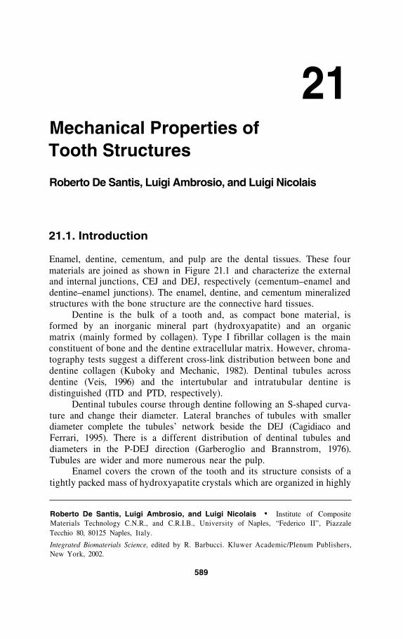
Enamel, dentine, cementum, and pulp are the dental tissues. These fourmaterials are joined as shown in Figure 21.1 and characterize the externaland internal junctions, CEJ and DEJ, respectively (cementum–enamel anddentine–enamel junctions). The enamel, dentine, and cementum mineralizedstructures with the bone structure are the connective hard tissues.
Dentine is the bulk of a tooth and, as compact bone material, isformed by an inorganic mineral part (hydroxyapatite) and an organicmatrix (mainly formed by collagen). Type I fibrillar collagen is the mainconstituent of bone and the dentine extracellular matrix. However, chroma-tography tests suggest a different cross-link distribution between bone anddentine collagen (Kuboky and Mechanic, 1982). Dentinal tubules acrossdentine (Veis, 1996) and the intertubular and intratubular dentine isdistinguished (ITD and PTD, respectively).
Dentinal tubules course through dentine following an S-shaped curva-ture and change their diameter. Lateral branches of tubules with smallerdiameter complete the tubules’ network beside the DEJ (Cagidiaco andFerrari, 1995). There is a different distribution of dentinal tubules anddiameters in the P-DEJ direction (Garberoglio and Brannstrom, 1976).Tubules are wider and more numerous near the pulp.
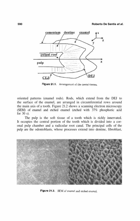
Enamel covers the crown of the tooth and its structure consists of atightly packed mass of hydroxyapatite crystals which are organized in highly
Roberto De Santis, Luigi Ambrosio, and Luigi NicolaisMaterials Technology C.N.R., and C.R.I.B., University of Naples, “Federico II”, PiazzaleTecchio 80, 80125 Naples, Italy.
Integrated Biomaterials Science, edited by R. Barbucci. Kluwer Academic/Plenum Publishers,New York, 2002.
589
Institute of Composite
590 Roberto De Santis et al.
oriented patterns (enamel rods). Rods, which extend from the DEJ tothe surface of the enamel, are arranged in circumferential rows aroundthe main axis of a tooth. Figure 21.2 shows a scanning electron microscopy(SEM) of enamel and etched enamel (etched with 37% phosphoric acidfor 30 s).
The pulp is the soft tissue of a tooth which is richly innervated.It occupies the central portion of the tooth which is divided into a cor-onal pulp chamber and a radicular root canal. The principal cells of thepulp are the odontoblasts, whose processes extend into dentine, fibroblast,
Mechanical Properties of Tooth Structures 591
mesenchymal cells and cells of connective tissue related to the neural,vascular, and immune systems. Vessels enter into the pulp through theapical foramina.
Cementum is the mineralized tissue which covers the root of a tooth;it is a bone-like structure. This tissue is deposited as a thin layer from theCEJ to the apex of the tooth; the thickness of this layer is higher at the apex.Cementoblasts and cementocytes are the cells of cementum which aresimilar to the osteoblasts and osteocytes of bone; however, unlike bone,cementum is avascular and incapable of remodeling. Cementum anchors thetooth to the surrounding alveolar bone through the periodontal ligament.
The periodontal ligament system is the soft connective tissue madeof fibers spanning from the cement of the root to the alveolar bone fol-lowing an obliquely cervical direction. The periodontal ligament charac-terizes the articulation between the tooth and bone; during the masticatoryfunctions the periodontal ligaments act as a natural shock absorbingsystem.
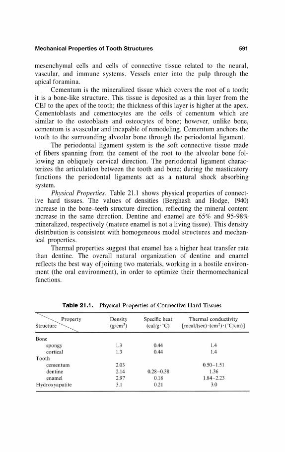
Physical Properties. Table 21.1 shows physical properties of connect-ive hard tissues. The values of densities (Berghash and Hodge, 1940)increase in the bone–teeth structure direction, reflecting the mineral contentincrease in the same direction. Dentine and enamel are 65% and 95-98%mineralized, respectively (mature enamel is not a living tissue). This densitydistribution is consistent with homogeneous model structures and mechan-ical properties.
Thermal properties suggest that enamel has a higher heat transfer ratethan dentine. The overall natural organization of dentine and enamelreflects the best way of joining two materials, working in a hostile environ-ment (the oral environment), in order to optimize their thermomechanicalfunctions.
592 Roberto De Santis et al.
Heterogeneity of natural tissue and the anisotropy in their mechanicalbehaviors are a fact regarded as a source for high-performance engineeringdesigns. However, modes of failure and bond strengths depend on the modeof testing, and this is a problem that needs to be resolved if strength andbond strength data are to be believed (Stanley, 1990; Drummond et al.,1996; van Noort, 1998).
21.2. Mechanical Properties
Dentine, as compact bone, is a heterogeneous multiphase materialwhich exhibits a multiscale composite structure; thus mechanical propertiesdepend on the testing conditions. Classical mechanical tests (macroscopictests in tension, compression, torsion, flexion, etc.) on connective tissuespecimens suggest a minimum cross-sectional area of Such speci-mens contain various Haversian systems (Reilly et al., 1974) and thusprovide significant data. Unfortunately this cross-sectional value is high ifcompared to the average of overall dentine dimensions, preventing thedistinguishing of material properties.
Human third molars and the diaphyses of femoral compact bone havebeen the main source for material research because of their geometricalconstraints.
Reductions in cross-sectional values lead to micromechanical testswhose results suggested that there is no relation between the applied loadand the cross-sectional area (Sano et al., 1994).
Indentation tests, by means of atomic force microscopes (AFM), arean alternative to classical micromechanical testing (Xu et al., 1998). UsingAFM it has been possible to apply loads in the range of to 100 Nthrough the tip of the indenter (Balooch et al., 1998).
21.2.1. Static Mechanical Properties
Figure 21.3 presents typical stress–strain curves of dentine and com-pact bone. Young’s moduli of hydroxyapatite and collagen are 114 GPa and1.2 GPa, respectively, so upper and lower bounds of the elastic modulus ofhard tissues may be derived from composite models where the mineralcrystal is the reinforcement while the surrounding collagen is the matrixphase.
The averaged values of the elastic modulus of dentine (Craig et al.,1961) and enamel (van Meerbeek et al., 1993; Willems et al., 1993) are10–20 GPa and 75–90 GPa, respectively.
Mechanical Properties of Tooth Structures 593
The anisotropy in the mechanical properties of bone derives from theultrastructural organization of collagen fibrils and mineral crystals withinthe osteons and the lamellar microstructure. The partial alignment of theosteons in the longitudinal direction of long bones makes this the stiffer andstronger axis of the material. This microstructure organization controls therelationship between loading conditions and fracture patterns in bone. Theelastic modulus of femoral compact bone (Bonfield and Grypass, 1977)varies from 14 GPa to 17 GPa (measured in the longitudinal direction of thefemur). The average mechanical properties of compact femoral bone anddentine (Table 21.2) show that the elastic modulus of dentine is similar tothat of cortical bone. However, the different hydroxyapatite–collagen net-work grants higher compression performances to dentine and higher tensileperformances to compact bone (Figure 21.3).
The collagen contribution to the elastic modulus of enamel is negli-gible; even viscoelastic properties do not change in the pulp DEJ direction.Instead, dentine strength, toughness, and bonding performances are depend-ent on the collagenic network properties.
Based on measured Young’s moduli of 30 GPa for PTD and 15 GPafor ITD and the tubule density in dentine, a slight variation was found inthe axial and transverse shear moduli with position in the tooth and themean values were 16 GPa and 6.2 GPa for the Young’s modulus and shearmodulus, respectively (Kinney et al., 1999).
Figure 21.4 presents the effect of tubule organization (Garberoglio andBrannstrom, 1976) on shear strength (measured with dentine specimens of
594 Roberto De Santis et al.
5 x 1 x 1 mm) (Watanabe et al., 1996), where the x, y, and z directions areshown in Figure 21.1. It can be seen that the shear strength increases in thepulp–enamel direction while tubule density and diameter decrease in thesame direction.
Microtensile tests assess local variation in dentine bonding strength(van Noort, 1998). These tests showed that bond strength to root dentine islower than that to coronal dentine (Yoshiyama et al., 1996). Micromechani-cal tests are important, especially when the specimen sizes are constrained.
Mechanical Properties of Tooth Structures 595
21.2.2. Hardness
Investigations using a modified atomic-force microscope suggest thatthe hardness of hydrated PTD and ITD is 2.3 ± 0.3 GPa and 0.5 ± 0.1 GPa,respectively (Kinney et al., 1996a). The hardness of ITD increased in thepulp–DEJ direction (0.15 ± 0.03 to 0.50 ± 0.02 GPa, respectively) while thatof PTD had a homogeneous distribution (Kinney et al., 1996b).
The impression size in dentine using loads of the order of 10 mN issimilar to heterogeneities such us enamel rods or dentinal tubules, preven-ting one from distinguishing material properties (Xu et al., 1998). This load,applied through a nano-indentator, suggested an elastic modulus of19.3±2.3 GPa and 90.6 + 16.1 GPa for dentine (van Meerbeek et al., 1993)and enamel (Willems et al., 1993), respectively. By using higher loads (2 to50 N) the resulting elastic modulus of dentine was 20±2 GPa and theanisotropy in the mechanical properties of enamel were related to micro-structural organization: the crack propagating toward the DEJ arrests atDEJ (Xu et al., 1998).
The averaged values of Vickers’ hardness of dentine and enamel areand respectively (Willems et al., 1992;
Forss et al., 1991).
21.2.3. Fracture Toughness
Linear elastic fracture mechanics (LEFM) have been applied to boneand dentine in order to characterize its resistance to fracture. The stressintensity factor K characterizes the stress amplification at the crack tip of aloaded material. According to LEFM each material has a critical stressintensity factor also known as fracture toughness. Fracture occurs whenK reaches the value
The fracture toughness reflects the ability of a material to resist crackinitiation and propagation. The critical value is a material characteristic,which provides a basis for predicting the onset of unstable crack propaga-tion as a function of stress and crack dimensions (Suresh, 1991; Rooke,1993).
In the last decades LEFM and mechanical tests have been used toevaluate the fracture toughness of cortical bone and dentine according tomode I fracture (crack extension in which the tensile stresses, acting normalto crack faces, are responsible for crack propagation). Toughness wasevaluated in the direction parallel to the long axis of a long bone (longitudi-nal direction) and in the direction parallel to dentinal tubules according toa variety of precracked specimens: the compact tension specimen (CT)
596 Roberto De Santis et al.
(Bonfield and Datta, 1976; Behiri and Bonfield, 1984; Vashishth et al., 1997;Norman et al., 1995, 1996), the single edge notched specimen (SEN) (Melvinand Evans, 1973; Moyle and Gavens, 1986), the center notched cylindricalspecimen (CNC) (Bonfield, 1987), the compact sandwich specimens (Wangand Agrawal, 1996), and the 3-point bending specimen (Robertson et al.,1978). The aim of these investigations is propagation rather than initiationof the crack.
The CT geometry has proven to be the most useful for studying bonefracture mechanics in the longitudinal direction, even if the dimensionrequirement (ASTM E 399-72, 1983) was difficult to match with connectivetissues specimens. Plane strain fracture toughness using CT specimensof coronal dentine, with DT parallel to the notch plane, suggested a valueof (standard deviation 0.33) (El Mowafy and Watts, 1986).
The main features of the CNT specimen are its geometry and theV-shaped notch. The latter constrains the crack to a steady-state propaga-tion in the chevron-notch ligament (ASTM B 771-87, 1987) while the formerallows a diameter 40% smaller than the thickness of a standard CT (Barker,1997). These features are essential to distinguish the anisotropy in thefracture properties of a material like bone and dentine. Moreover, by usingthe CNT geometry, fracture toughness is computed as a function of themaximum load and the specimen’s geometry:
where A is a CNT constant (A = 20.8 for ASTM specimens), B is the
Mechanical Properties of Tooth Structures 597
diameter, and v is Poisson’s ratio. Figure 21.5 shows the mechanicalbehavior of CNT short-rod specimens of dentine and bone.
A CNT short-bar bovine specimen (with the notch tip in enamel)suggested that the lower boundary value of and (critical strain energyrelease), in the direction perpendicular to the DEJ, areand respectively (Lin and Douglas, 1994a); of thebonding system–dentine interface is one order of magnitude lower (Lin andDouglas, 1994b).
The value of measured by using CNT short-rod specimens of adentine–bonding system, with the interface positioned in the notch plane,suggests a range of 0.20 to (a significant lower limit offracture toughness is reached in a deep dentine–bonding system interface)(Tam and Yim, 1997).
The fracture toughness of dentine is midway in the rangeobserved for cortical bone and is at least one order of magnitude
greater than the value for dentine-restorative materials.
References
ASTM Standards E 399-72. 1983. Standard Method of Test for Plane-Strain Fracture Toughnessof Metallic Materials.
ASTM Standards B 771-87. 1987. Standard Test for Short Rod Fracture Toughness of CementedCarbides.
Balooch, M., Wu-Magidi, I.C., Balazs, A., Lundkvist, A. S., Marshall, S.J., Marshall, G.W.,Siekhaus, W.J., Kinney, J.H. 1998. Viscoelastic properties of demineralised human dentinmeasured in water with atomic force microscope (AFM)-based indentation, J. Biomed.Mater. Res. 40, 539–544.
Barker, L.M. 1997. http ://www.terratek.com7fracto_2.htmBehiri, J.C., Bonfield, W. 1984. Fracture of bone: the effect of density, specimen thickness and
crack velocity on longitudinal fracture, J. Biomech. 17, 25–34.Berghash, S.R., Hodge, H.C. 1940. http://www.lib.umich.edu/libhome/Dentistry.lib/Dental_
tables/intro.html.Bonfield, W. 1987. Advances in the fracture mechanics of cortical bone, J. Biomech. 20, 1071 –1081.Bonfield, W., Datta, P. K. 1976. Fracture toughness of compact bone, J. Biomech. 9, 131–134.Bonfield, W., Grynpass, M.D. 1977. Anisotropy of the Young’s modulus of bone, Nature 270,
453–454.Cagidiaco, M.C., Ferrari, M. 1995. Dentinal tubules, in: Bonding to Dentin, O. Debatte & F.
cements and gold, J. Dent. Res. 40, 936–945.Drummond, J.L., Sakaguchi, R.L., Racean, D.C., Wozny, J., Steinberg, A.D. 1996. Testing mode
and surface treatment effects on dentin bonding, J. Biomed. Mater. Res. 32, 533–541.El Mowafy, O.M., Watts, D.C. 1986. Fracture of human dentin, J. Dent. Res. 35, 677–681.Forss, H., Seppa, L., Lappalainen, R. 1991. In vitro abrasion resistance and hardness of
glass-ionomer cements, Dent. Mater. 7, 36–39.
598 Roberto De Santis et al.
Garberoglio, R., Brannstrom, M. 1976. Scanning electron microscopic investigation of humandentinal tubules, Arch. Oral Biol. 21, 355–362.
Kinney, J. H., Balooch, M., Marshall, S.J., Marshall, G.W., Weihs, T.P. 1996a. Atomic forcemicroscope measurements of the hardness and elasticity of peritubular and intertubularhuman dentin, J. Biomech. Eng. 118, 133–135.
Kinney, J.H., Balooch, M., Marshall, S.J., Marshall, G.W., Weihs, T.P. 1996b. Hardness andYoung’s modulus of human peritubular and intertubular dentine, Arch. Oral Biol. 41,9–13.
Kinney, J. H., Balooch, M., Marshall, G. W., Marshall, S. J. 1999. A micromechanics model ofthe elastic properties of human dentine, Arch. Oral Biol. 44, 813–822.
Kuboky, Y., Mechanic, G.L. 1982. Comparative molecular distribution of cross-link in boneand dentine collagen: structure-function relationship, Calcif. Tissue Int. 34, 306–308.
Lin, C.P., Douglas, W.H. 1994a. Structure–property relations and crack resistance at thebovine dentin–enamel junction, J. Dent. Res. 73, 1072–1078.
Lin, C.P., Douglas, W.H. 1994b. Failure mechanism at the human dentin–resin interface: afracture mechanism approach, J. Biomech. 27, 1037–1047.
Melvin, J.W., Evans, F.G. 1973. Crack propagation in bone, pp. 87–88, BiomechanicsSymposium, ASME New York.
Moyle, D.D., Gavens, A.J. 1986. Fracture propeties of bovine tibial bone, J. Biomech. 19,919–927.
Norman, T.L., Vashishth, D., Burt, D. 1995. Fracture toughness of human bone under tension,J. Biomech. 28, 309–320.
Norman, T.L., Vashishth, D., Burt, D. 1996. Resistance to crack growth in human cortical boneis greater in shear than in tension, J. Biomech. 29, 1023–1031.
Reilly, D.T., Burstein, A.H., Frankel, V.H. 1974. The elastic modulus for bone, J. Biomech. 7,271–275.
Robertson, D.M., Robertson, D., Barret, C.G. 1978. Fracture toughness, critical crack lengthand plastic zone size in bone, J. Biomech. 11, 359–364.
Rooke, D.P. 1993. Development of fracture mechanics, in: Static and Dynamic FractureMechanisms (M.H. Aliabad, C.A. Brebbia, V.Z. Parton, eds.), pp. 3–35, ComputationalMechanics Publications, Portland.
Sano, H., Shono, T., Sonoda, H., Takatsu, T., Ciucchi, B., Carvalho, R., Pashley, D.H. 1994.Relationship between surface area for adhesion and tensile bond strength — evaluation ofa micro-tensile bond test. Dent Mater, 10, 236–40.
Stanley, H.R. 1990. Pulpal responses to ionomer cements — biological characteristics, J. Am.Dem. Assoc. 120, 25–29.
Suresh, S. 1991. Principles of fracture mechanics and their implication for fatigue, in: Fatigueof Materials (D.R. Clarke, ed.), Cambridge University Press.
Tam, L.E., Yim, D. 1997. Effect of dentine depth on the fracture toughness of dentine–composite adhesive interfaces, J. Dent. 25, 339–346.
van Meerbeek, B., Willems, G., Celis, J.P., Roos, J.R., Braem, M. 1993. Lambrechts. Assessmentby nano-indentation technique of the hardness and elasticity of the resin dentin bondingarea, J. Dent. Res. 72, 1434–1442.
van Noort, R. 1998. Dental Materials: 1996. Dentine bonding, J. Dent. 26, 191–207.Vashishth, D., Behiri, J.C., Bonfield, W. 1997. Crack growth resistance in cortical bone: Concept
of microcrack toughness, J. Biomech. 30, 763–769.Veis, A. 1996. in: Dentin. Extracellular Matrix. Tissue Function (Wayne D. Comper, ed.), Vol.
1, Amsterdam.Wang, X., Agrawal, C.M. 1996. Fracture toughness of bone using a compact sandwich
specimen: effect of sampling sites and crack orientations, J. Biomed. Mater. Res. 33, 13–21.
Mechanical Properties of Tooth Structures 599
Watanabe, L.G., Marshall, G.W., Marshall, S.J. 1996. Dentin shear strength: effects of tubuleorientation and intratooth location, Dent. Mater. 12, 109–115.
Willems, G., Lambrechts, P., Braem, M., Celis, J.P., Vanherle, G.A. 1992. Classification ofdental composites according to their morphological and mechanical characteristics, Dent.Mater. 8, 310–331.
Willems, G. Celis, J.P., Lambrechts, P., Braem, M. 1993. Hardness and Young’s modulusdetermined by nano-indentation technique of filler particles of dental restorative materialscompared with human enamel, J. Biomed. Mater. Res. 27, 747–755.
Xu, H.H.K., Smith, D.T., Jahanmir, S., Romberg, E., Kelly, J.R., Thompson, V.P., Rekow, E.D.1998. Indentation damage and mechanical properties of human enamel and dentin, J.Dent. Res. 77, 472–480.
Yoshiyama, M., Carvalho, R.M., Sano, H., Horner, J.A., Brewer, P.D., Pashley, D.H. 1996.Regional bond strengths of resins to human root dentine, J. Dent, 24, 435–442.