Integration of faces and vocalizations in ventral prefrontal cortex: Implications for the evolution of audiovisual speech Lizabeth M. Romanski 1 Department of Neurobiology & Anatomy, University of Rochester School of Medicine, Rochester, NY 14642 Edited by Francisco J. Ayala, University of California, Irvine, CA, and approved May 14, 2012 (received for review March 13, 2012) The integration of facial gestures and vocal signals is an essential process in human communication and relies on an interconnected circuit of brain regions, including language regions in the inferior frontal gyrus (IFG). Studies have determined that ventral prefrontal cortical regions in macaques [e.g., the ventrolateral prefrontal cortex (VLPFC)] share similar cytoarchitectonic features as cortical areas in the human IFG, suggesting structural homology. Anterog- rade and retrograde tracing studies show that macaque VLPFC receives afferents from the superior and inferior temporal gyrus, which provide complex auditory and visual information, respec- tively. Moreover, physiological studies have shown that single neurons in VLPFC integrate species-specific face and vocal stimuli. Although bimodal responses may be found across a wide region of prefrontal cortex, vocalization responsive cells, which also respond to faces, are mainly found in anterior VLPFC. This suggests that VLPFC may be specialized to process and integrate social commu- nication information, just as the IFG is specialized to process and integrate speech and gestures in the human brain. frontal lobe | sensory integration | primate | neurophysiology T he area dedicated to language processing in the frontal lobe is located within the inferior frontal gyrus (IFG), which can be further subdivided into the pars opercularis (most posterior portion of the IFG), the pars triangularis, and the pars orbitalis (cortex inferior and anterior to the horizontal ramus of the lat- eral fissure). These subdivisions include Brodmann areas 44, 45, and 47. Our understanding of the functions within this special- ized area of cortex is hampered by the fact that no other mammal has a frontal lobe of similar organization or complexity, leaving few animal models to investigate. Within nonhuman primates, only catarrhines have a well-developed frontal lobe with cytoar- chitectonic evidence of Brodmann areas 44, 45, and 47 (1). In contrast, New World monkeys including marmosets and squirrel monkeys have a lissencephalic frontal lobe with previously identified motor and premotor cortices but less clearly defined prefrontal regions (2). Recent work has identified an area in the ventrolateral prefrontal cortex (VLPFC) of rhesus macaques (Macaca mulatta) that is involved in the processing and in- tegration of vocalizations and faces. We have hypothesized that the ventral prefrontal cortex (VLPFC) became specialized for the processing and integration of auditory and visual communi- cation signals, in at least early anthropoid primates, and ulti- mately this region was modified and lateralized to the left cerebral hemisphere to subserve language in modern humans. Ventral PFC: Anatomical Considerations Organization of VLPFC. The frontal lobe of the macaque monkey has been studied extensively with anatomical, electrophysiologi- cal, and functional methodologies compared with other primate species. The area of the VLPFC, also referred to as the inferior convexity of the PFC, in the macaque monkey, includes the cortical region ventral to the principal sulcus and anterior to the inferior limb of the arcuate sulcus (Fig. 1). The cytoarchitectonic areas of VLPFC in the macaque are arranged in a similar fashion to that of the human frontal lobe and include regions on the lateral frontal surface: area 45, which lies just anterior to the inferior arcuate sulcus; area 12 (or 12/47), which lies anterior to area 45 and ventral to area 46; and the most ventrolateral extent of the inferior convexity, which wraps around the inferior gyral surface and extends to the lateral orbital sulcus: area 12 orbital (3). Additional architectonic studies have described areas within the arcuate sulcus and premotor cortex as well as the sub- divisions of the orbital cortex (1, 4–6), but we will confine our discussion to VLPFC, areas 45 and 12/47. We will refer to area 12 in general as area 12/47 to convey the homology of area 12 in the macaque with human area 47 as introduced by Petrides and Pandya (1). However, to differentiate between the ventrolateral region below the principal sulcus and the lateral orbital cortex we will use 12 ventrolateral (12vl) and 12 orbital (12o), respectively, as defined by Preuss and Goldman-Rakic (3). The VLPFC also commonly includes a small sulcus—termed the inferior frontal sulcus by Winters et al. (7) and the infrap- rincipal dimple or the inferior prefrontal dimple (IPD) by others (1, 8, 9)—which varies in its position and depth in M. mulatta. Some schematics of VLPFC depict the IPD as running in a ros- tral-to-caudal direction and separating area 45A from area 46 (1). However, in our neurophysiological recordings (10, 11) and in other studies (8), it is depicted as running dorsal to ventral and separating area 45 from area 12/47. It is not always described or visible in studies of other subspecies of macaque monkeys. Thus, there is variability in the position of area 12/47 and area 45 not only between the subspecies of macaques but also within M. mulatta individuals. As explained later, the IPD is the primary location in which auditory responsive neurons and audiovisual responsive cells have been reliably located in several studies, and may be a critical landmark for delineating the functional auditory responsive prefrontal region in macaques. Whether it defines the border of areas 12/47 and 45 is unclear. In the human brain, areas 44 and 45 have been associated with language processing confirmed by electrical stimulation, PET and functional MRI (fMRI). However, the areas that control vocaliza- tion production in Old World monkeys are not as well understood and could include VLPFC, whereas other studies have implicated ventral premotor and the cingulate vocalization area (8, 12, 13). Cytoarchitectonic Organization of VLPFC. The cytoarchitectonic descriptions here are taken from Preuss and Goldman-Rakic (3), who described the frontal lobe of the rhesus macaque, M. This paper results from the Arthur M. Sackler Colloquium of the National Academy of Sciences, “In the Light of Evolution VI: Brain and Behavior,” held January 19–21, 2012, at the Arnold and Mabel Beckman Center of the National Academies of Sciences and Engi- neering in Irvine, CA. The complete program and audio files of most presentations are available on the NAS Web site at www.nasonline.org/evolution_vi. Author contributions: L.M.R. designed research and wrote the paper. The author declares no conflict of interest. This article is a PNAS Direct Submission. 1 E-mail: [email protected]. www.pnas.org/cgi/doi/10.1073/pnas.1204335109 PNAS Early Edition | 1 of 8
Transcript
Integration of faces and vocalizations in ventralprefrontal cortex: Implications for the evolutionof audiovisual speechLizabeth M. Romanski1
Department of Neurobiology & Anatomy, University of Rochester School of Medicine, Rochester, NY 14642
Edited by Francisco J. Ayala, University of California, Irvine, CA, and approved May 14, 2012 (received for review March 13, 2012)
The integration of facial gestures and vocal signals is an essentialprocess in human communication and relies on an interconnectedcircuit of brain regions, including language regions in the inferiorfrontal gyrus (IFG). Studies have determined that ventral prefrontalcortical regions in macaques [e.g., the ventrolateral prefrontalcortex (VLPFC)] share similar cytoarchitectonic features as corticalareas in the human IFG, suggesting structural homology. Anterog-rade and retrograde tracing studies show that macaque VLPFCreceives afferents from the superior and inferior temporal gyrus,which provide complex auditory and visual information, respec-tively. Moreover, physiological studies have shown that singleneurons in VLPFC integrate species-specific face and vocal stimuli.Although bimodal responses may be found across a wide region ofprefrontal cortex, vocalization responsive cells, which also respondto faces, are mainly found in anterior VLPFC. This suggests thatVLPFC may be specialized to process and integrate social commu-nication information, just as the IFG is specialized to process andintegrate speech and gestures in the human brain.
The area dedicated to language processing in the frontal lobeis located within the inferior frontal gyrus (IFG), which can
be further subdivided into the pars opercularis (most posteriorportion of the IFG), the pars triangularis, and the pars orbitalis(cortex inferior and anterior to the horizontal ramus of the lat-eral fissure). These subdivisions include Brodmann areas 44, 45,and 47. Our understanding of the functions within this special-ized area of cortex is hampered by the fact that no other mammalhas a frontal lobe of similar organization or complexity, leavingfew animal models to investigate. Within nonhuman primates,only catarrhines have a well-developed frontal lobe with cytoar-chitectonic evidence of Brodmann areas 44, 45, and 47 (1). Incontrast, New World monkeys including marmosets and squirrelmonkeys have a lissencephalic frontal lobe with previouslyidentified motor and premotor cortices but less clearly definedprefrontal regions (2). Recent work has identified an area in theventrolateral prefrontal cortex (VLPFC) of rhesus macaques(Macaca mulatta) that is involved in the processing and in-tegration of vocalizations and faces. We have hypothesized thatthe ventral prefrontal cortex (VLPFC) became specialized forthe processing and integration of auditory and visual communi-cation signals, in at least early anthropoid primates, and ulti-mately this region was modified and lateralized to the leftcerebral hemisphere to subserve language in modern humans.
Ventral PFC: Anatomical ConsiderationsOrganization of VLPFC. The frontal lobe of the macaque monkeyhas been studied extensively with anatomical, electrophysiologi-cal, and functional methodologies compared with other primatespecies. The area of the VLPFC, also referred to as the inferiorconvexity of the PFC, in the macaque monkey, includes thecortical region ventral to the principal sulcus and anterior to theinferior limb of the arcuate sulcus (Fig. 1). The cytoarchitectonicareas of VLPFC in the macaque are arranged in a similar fashion
to that of the human frontal lobe and include regions on thelateral frontal surface: area 45, which lies just anterior to theinferior arcuate sulcus; area 12 (or 12/47), which lies anterior toarea 45 and ventral to area 46; and the most ventrolateral extentof the inferior convexity, which wraps around the inferior gyralsurface and extends to the lateral orbital sulcus: area 12 orbital(3). Additional architectonic studies have described areas withinthe arcuate sulcus and premotor cortex as well as the sub-divisions of the orbital cortex (1, 4–6), but we will confine ourdiscussion to VLPFC, areas 45 and 12/47. We will refer to area12 in general as area 12/47 to convey the homology of area 12 inthe macaque with human area 47 as introduced by Petrides andPandya (1). However, to differentiate between the ventrolateralregion below the principal sulcus and the lateral orbital cortex wewill use 12 ventrolateral (12vl) and 12 orbital (12o), respectively,as defined by Preuss and Goldman-Rakic (3).The VLPFC also commonly includes a small sulcus—termed
the inferior frontal sulcus by Winters et al. (7) and the infrap-rincipal dimple or the inferior prefrontal dimple (IPD) by others(1, 8, 9)—which varies in its position and depth in M. mulatta.Some schematics of VLPFC depict the IPD as running in a ros-tral-to-caudal direction and separating area 45A from area 46(1). However, in our neurophysiological recordings (10, 11) andin other studies (8), it is depicted as running dorsal to ventral andseparating area 45 from area 12/47. It is not always described orvisible in studies of other subspecies of macaque monkeys. Thus,there is variability in the position of area 12/47 and area 45 notonly between the subspecies of macaques but also within M.mulatta individuals. As explained later, the IPD is the primarylocation in which auditory responsive neurons and audiovisualresponsive cells have been reliably located in several studies, andmay be a critical landmark for delineating the functional auditoryresponsive prefrontal region in macaques. Whether it defines theborder of areas 12/47 and 45 is unclear.In the human brain, areas 44 and 45 have been associated with
language processing confirmed by electrical stimulation, PET andfunctional MRI (fMRI). However, the areas that control vocaliza-tion production in Old World monkeys are not as well understoodand could include VLPFC, whereas other studies have implicatedventral premotor and the cingulate vocalization area (8, 12, 13).
Cytoarchitectonic Organization of VLPFC. The cytoarchitectonicdescriptions here are taken from Preuss and Goldman-Rakic (3),who described the frontal lobe of the rhesus macaque, M.
This paper results from the Arthur M. Sackler Colloquium of the National Academy ofSciences, “In the Light of Evolution VI: Brain and Behavior,” held January 19–21, 2012, atthe Arnold and Mabel Beckman Center of the National Academies of Sciences and Engi-neering in Irvine, CA. The complete program and audio files of most presentations areavailable on the NAS Web site at www.nasonline.org/evolution_vi.
Author contributions: L.M.R. designed research and wrote the paper.
The author declares no conflict of interest.
This article is a PNAS Direct Submission.1E-mail: [email protected].
www.pnas.org/cgi/doi/10.1073/pnas.1204335109 PNAS Early Edition | 1 of 8
mulatta, which is the same species that has been examined inmost neurophysiology studies of VLPFC. These descriptions arein general agreement with Petrides and Pandya (1). Area 45 islocated ventral to the caudal principal sulcus within the ventrallimb of the arcuate and extends onto the cortical surface (Fig. 1C–E). It is composed of large pyramidal cells in layer V and deeplayer III. Layer IV is thick with densely packed small cells, withsome of the larger pyramidal cells from deep layer III and su-perficial layer V intruding on layer IV. It is densely myelinated.Area 45 is bordered dorsally by area 8a (3), which can be dis-tinguished from area 45 by the presence of extremely large py-ramidal cells in layer Va of area 8. Area 12 (areas 12vl and 12o),which covers the surface of the ventrolateral convexity andextends onto the lateral orbital surface as far as the lateral orbitalsulcus, can be distinguished from area 46 by its more heavilymyelinated appearance. The disappearance of the large layer IIIpyramidal cells marks the transition from area 45 to area 12vl.Area 12vl on the ventrolateral surface has been distinguishedfrom area 12o by the more diffuse myelinated appearance of 12oand the more granular layer IV of 12vl. A series of comparativecytoarchitectonic studies have examined the similarities of area12 in macaque and area 47 in the human brain (1). As a result,area 12 has been referred to as area 12/47 even though the as-signation of 12 was renewed in a recent analysis of VLPFCconnections (5). In addition, studies by Petrides and Pandya (1)and Gerbella et al. (5) have suggested that area 45 be divided
into subdivisions 45B, closest to and within the anterior limb ofthe anterior bank of the arcuate sulcus, and area 45A, locatedrostral to 45B and extending across the surface of the inferiorconvexity to the IPD, with area 12vl (i.e., 47) bordering 45Arostrally and ventrally (Fig. 1). The precise location of area 12vlrelative to area 45A may vary somewhat in individuals and maybe better determined from a combination of connectivity studiesand physiological recordings.
Cortical Connectivity of VLPFC. Connectivity of VLPFC with cortical visualprocessing regions. Much of what we know about the cellularfunctions of the primate PFC is based on the processing of visualinformation. Thus, it is not surprising that many studies haveexamined projections from visual association cortex to the pri-mate PFC. Results indicate that VLPFC receives afferents fromextrastriate visual cortical areas in the inferotemporal cortex,including area TE. Early anatomical studies by Barbas, Pandya,and others examined the innervation of the entire prefrontalmantle by visual association areas (14–16). Barbas was amongthe first to note that basoventral prefrontal cortices were morestrongly connected with extrastriate, ventral visual areas, whichhave been implicated in object recognition and feature discrim-ination. In contrast, medial and dorsal prefrontal cortices aremore densely connected with medial and dorsolateral occipitaland parietal areas, which are associated with visuospatial func-tions (14). This dissociation was confirmed by Bullier et al. (17),
A B
E
DC
4612
45a
8a
45b
12l
r
IPD
IPD
Fig. 1. Organization of ventral PFC. Maps of the cytoarchi-tectonic organization of ventral PFC are shown for the human(A and B) and macaque brain (C–E). (A) Brodmann map (1909)of the human brain with areas 44, 45, and 47 marked on theIFG. Reproduced with permission from Brodmann (1909). (B)Map of the human brain color-coded to match the corre-sponding homologous regions in the macaque brain shown inD. Reproduced with permission from ref. 1. (C) Map of M.mulatta. Reproduced with permission from ref. 3. (D) Map ofmacaque brain. Reproduced with permission from ref. 1. (E)Photomicrograph of VLPFC in macaque monkey withcytoarchitectonic areas labeled. The IPD is marked in D and inE. Image courtesy of G. Luppino (ref. 5).
2 of 8 | www.pnas.org/cgi/doi/10.1073/pnas.1204335109 Romanski
who found a segregation of inputs to caudal PFC when pairedinjections of tracers were placed into temporal and parietal vi-sual processing regions. In their study, visual temporal cortexprojected mainly to ventrolateral PFC, area 45, whereas parietalcortex sent projections to ventrolateral PFC and dorsolateralPFC (areas 8a and 46) (17). Tracing and lesion studies byUngerleider et al. (18) showed that area TE projected specifi-cally to three ventral prefrontal targets, including the ventrallimb of the arcuate sulcus (area 45), the inferior convexity justventral to the principal sulcus (area 12vl), and within the lateralorbital cortex (areas 11 and 12o). These projections are via theuncinate fasciculus (18). Furthermore, ventrolateral PFC areas12vl and 45, which contain object- and face-selective neurons(19–21), were shown to be connected with inferotemporal areasTE and TEO (22), with the strongest innervation of ventrolateralPFC and orbitofrontal areas 11 and 12o originating in TE.Auditory projections to PFC. In contrast to the visual pathways, theprefrontal targets of central auditory pathways have not beenstudied as extensively despite the accepted role of the frontal lobein language. In early anatomical studies, lesion/degenerationtechniques were used to reveal projections from the caudal su-perior temporal gyrus (STG) to the principal sulcus region, thearcuate cortex, and the inferior convexity of the frontal lobe, andfrom the middle and rostral STG to the rostral principal sulcusand orbital regions (16, 23, 24). Additional studies revealedconnections between the lateral PFC and cortical areas withinthe STG (14, 15, 25, 26). There was a suggestion of rostrocaudaltopography in these studies whereby anterior and middle aspectsof the principal sulcus, including areas 9, 10, and rostral 46, wereconnected with the middle STG, whereas area 8 received pro-jections from mostly caudal STG (14, 26, 27). It became clearthat VLPFC received afferents from the STG, inferotemporalcortex, and multisensory regions within the superior temporalsulcus (STS).Importantly, detailed anatomical studies by Morel et al. (28),
Jones et al. (29), and Hackett et al. (30), together with parallelneurophysiological studies by Rauschecker et al. (31), providedevidence that primate auditory cortices were organized as a core-belt system with a third zone, the parabelt just lateral to the belt(28–30). A series of landmark neurophysiology studies providedthe first electrophysiological evidence for three separate tono-topic regions (AL, ML, and CL) in the belt cortex that could bedistinguished from the core A1 (31). Additional studies havedescribed functional dissociations of anterior and posterior beltand parabelt regions (32). Rauschecker et al. (31), Morel et al.(28), and Hackett et al. (30) used a common terminology todelineate auditory cortex in anatomical and physiological studies,which enabled cross-talk and comparisons that fostered progressin the study of auditory cortical processing and organization.Combining physiological recording with anatomical tract
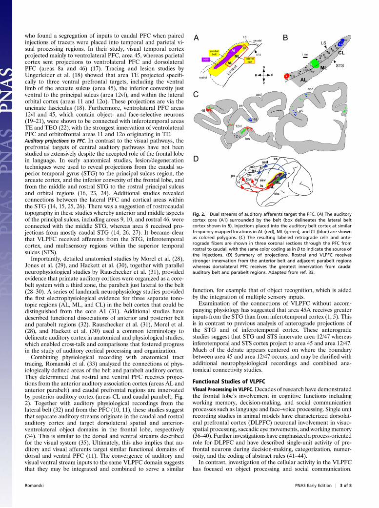
tracing, Romanski et al. (33) analyzed the connections of phys-iologically defined areas of the belt and parabelt auditory cortex.They determined that rostral and ventral PFC receives projec-tions from the anterior auditory association cortex (areas AL andanterior parabelt) and caudal prefrontal regions are innervatedby posterior auditory cortex (areas CL and caudal parabelt; Fig.2). Together with auditory physiological recordings from thelateral belt (32) and from the PFC (10, 11), these studies suggestthat separate auditory streams originate in the caudal and rostralauditory cortex and target dorsolateral spatial and anterior-ventrolateral object domains in the frontal lobe, respectively(34). This is similar to the dorsal and ventral streams describedfor the visual system (35). Ultimately, this also implies that au-ditory and visual afferents target similar functional domains ofdorsal and ventral PFC (11). The convergence of auditory andvisual ventral stream inputs to the same VLPFC domain suggeststhat they may be integrated and combined to serve a similar
function, for example that of object recognition, which is aidedby the integration of multiple sensory inputs.Examination of the connections of VLPFC without accom-
panying physiology has suggested that area 45A receives greaterinputs from the STG than from inferotemporal cortex (1, 5). Thisis in contrast to previous analysis of anterograde projections ofthe STG and of inferotemporal cortex. These anterogradestudies suggest that STG and STS innervate area 12/47 whereasinferotemporal and STS cortex project to area 45 and area 12/47.Much of the debate appears centered on where the boundarybetween area 45 and area 12/47 occurs, and may be clarified withadditional neurophysiological recordings and combined ana-tomical connectivity studies.
Functional Studies of VLPFCVisual Processing in VLPFC.Decades of research have demonstratedthe frontal lobe’s involvement in cognitive functions includingworking memory, decision-making, and social communicationprocesses such as language and face–voice processing. Single unitrecording studies in animal models have characterized dorsolat-eral prefrontal cortex (DLPFC) neuronal involvement in visuo-spatial processing, saccadic eye movements, and working memory(36–40). Further investigations have emphasized a process-orientedrole for DLPFC and have described single-unit activity of pre-frontal neurons during decision-making, categorization, numer-osity, and the coding of abstract rules (41–44).In contrast, investigation of the cellular activity in the VLPFC
has focused on object processing and social communication.
B
46
A
C asd
12vl
12 olos
46
ps
asd
45
cs
sts
asd
asv
ps
8a8b
9
10
12vl 45
46d
46v
ls
D
D
V
R C
RP
caudal
AI
rostral
CL
AL
MLSTS
medialbelt
core
LS
lateralbelt
2.51k
0.63
6.3
10 168k
6k5k
3.2ncr
ncr
STS
LS
AL
ML
CL1 mm
Fig. 2. Dual streams of auditory afferents target the PFC. (A) The auditorycortex core (A1) surrounded by the belt (box delineates the lateral beltcortex shown in B). Injections placed into the auditory belt cortex at similarfrequency mapped locations in AL (red), ML (green), and CL (blue) are shownas colored polygons. (C) The resulting labeled retrograde cells and ante-rograde fibers are shown in three coronal sections through the PFC fromrostral to caudal, with the same color coding as in B to indicate the source ofthe injections. (D) Summary of projections. Rostral and VLPFC receivesstronger innervation from the anterior belt and adjacent parabelt regionswhereas dorsolateral PFC receives the greatest innervation from caudalauditory belt and parabelt regions. Adapted from ref. 33.
Romanski PNAS Early Edition | 3 of 8
Early studies of VLPFC showed that neurons in this region wereresponsive to simple and complex visual stimuli presented at thefovea (45, 46). Face-responsive neurons were documented byThorpe et al. and Rolls et al. (47, 48) and later described in detailby Goldman-Rakic and coworkers (19–21). In these studies,Wilson et al. (21) showed that DLPFC and VLPFC neuronsresponded differentially to spatial and object features of visualstimuli. These studies were the first to demonstrate a functionaldissociation between DLPFC and VLPFC by using single-unitelectrophysiology. Wilson et al. (21) showed that DLPFC neu-rons were selectively engaged by visuospatial memory tasks andVLPFC neurons were selective for color, shape, or type of visualobjects. An earlier study by Mishkin and Manning (49) showedthat lesions of VLPFC in nonhuman primates interfere with theprocessing of nonspatial information, including color and form.Electrophysiological recordings demonstrated that VLPFC facecells had a twofold increase in firing rate to face stimuli com-pared with nonface stimuli during passive presentations or dur-ing working memory tasks (19, 20). Face cells were found only inthe VLPFC and not in DLPFC, and were localized to three smallparts of VLPFC, including a patch on the lateral convexity closeto the lower limb of the arcuate sulcus (area 45), within andaround the IPD (area 12vl), and a small number of cells in thelateral orbital cortex (19). VLPFC face cells were sensitive tochanges in facial features, expressions, or the angle of gaze,much like the inferotemporal cortical regions, which project tothese VLPFC cells. These studies have suggested that VLPFCcells may encode identity, expression, and face view (19, 20, 48,50). Data from the single-unit recordings have been confirmedwith fMRI studies in macaque monkeys (51), which have dem-onstrated activation of face-responsive “patches” in the samearcuate, ventrolateral, and orbitofrontal locations shown byO’Scalaidhe et al. (19, 20). Demonstration by both methods ofvisual responsiveness and face selectivity substantiates the notionthat VLPFC in the macaque monkey is involved in object andface processing (Fig. 3).
Auditory Responses and Function in Ventral PFC. The ventral frontallobe has long been linked with complex auditory function throughits association with language functions in the IFG. The results ofsome studies have suggested parcellation of function in the humanIFG. The anterior region, the pars triangularis (area 45), alongwith the pars orbitalis (area 47), has been suggested to be moreinvolved in semantic processing, comprehension, and auditoryworking memory (52–59). In contrast, the pars opercularis (area44) and ventral premotor cortex are more active during phono-logical processing and speech production. The precise neuronalmechanisms that occur in the frontal lobe during the processing ofcomplex auditory information are unknown but might be indirectlyassessed with neurophysiological recordings in animals with simi-lar ventral frontal lobe regions, such as macaque monkeys.Neuronal responses to acoustic stimuli have been sporadically
noted in the frontal lobes of Old and New World monkeys (60–62). However, when recordings targeted cortical areas that hadbeen shown to receive projections from acoustically character-ized regions of the auditory belt and parabelt cortex (33), a dis-crete auditory responsive region was localized in VLPFC (10).This VLPFC cortical region is thought to be the termination ofa ventral auditory processing stream, specialized for the pro-cessing of nonspatial (i.e., object) auditory information (33, 34,63, 64). The auditory responsive region of VLPFC is locatedrostral to the ventral limb of the arcuate sulcus below the prin-cipal sulcus, in the area of the IPD. This region receives projec-tions from ventral stream auditory cortical regions and polymodalcortex of the STS, as discussed earlier (1, 4, 33, 65, 66). VLPFCauditory neurons are responsive to complex auditory stimuli, in-cluding vocalization and complex nonvocal stimuli (10). Thissmall ventrolateral prefrontal auditory region has also beenshown to be active in neuroimaging studies in rhesus monkeysduring presentation of complex acoustic stimuli (67).The VLPFC auditory area was analyzed with a large library of
rhesus macaque vocalizations to test selectivity to specific callcategories, as previous analysis had implied some selectivity for
BA
C
70
-500 0 1000 1500 20000
spik
e/se
c
D
Fig. 3. Face-responsive neuronsin the VLPFC. (A and B) Face-re-sponsive neurons recorded by O’Sca-laidhe et al. (19) are depicted.Adapted from ref. 19. (A) Regionrecorded in the PFC is indicated witha circle on the lateral brain schematicof the rhesus macaque brain. (B) Flatmap of the recorded region in thePFC is shown with face cells outlinedin black and gray. (C) Face responsivecells that were selective for forwardand 30° rotated face view (50).Stimulus images courtesy of MichaelJ. Tarr, Center for the Neural Basis ofCognition and Department of Psy-chology, Carnegie Mellon University,http://www.tarrlab.org/. (D) Locationof face selective patches in the VLPFCand in the orbitofrontal cortex isshown in yellow (51). A red arrowindicates a similar portion of theVLPFC in the single unit data por-trayed in B and in the fMRI data in D.Reprinted by permission from Mac-millan Publishers Ltd: Nature Neuro-science (ref. 51), copyright (2008).
4 of 8 | www.pnas.org/cgi/doi/10.1073/pnas.1204335109 Romanski
calls with common functions (68). Analysis of these vocalizationresponses with exemplars from 10 different types of calls dem-onstrated that neurons tended to respond to two or three vo-calization types that had similar acoustic morphology rather thansimilar behavioral referents (Fig. 4; 11). Additional electro-physiological recording studies by Gifford et al. (68) and Russet al. (69) have suggested that VLPFC neuronal activity ismodulated during categorization of acoustic stimuli and in au-ditory decision-making (70). These combined data are consistentwith a role for VLPFC in a ventral auditory processing stream forauditory objects, including vocalizations. The localization of thisauditory processing area to the ventral prefrontal region of OldWorld monkeys suggests a functional similarity between it andhuman language-processing regions in the ventral or inferiorfrontal lobe of the human brain (10, 71, 72).
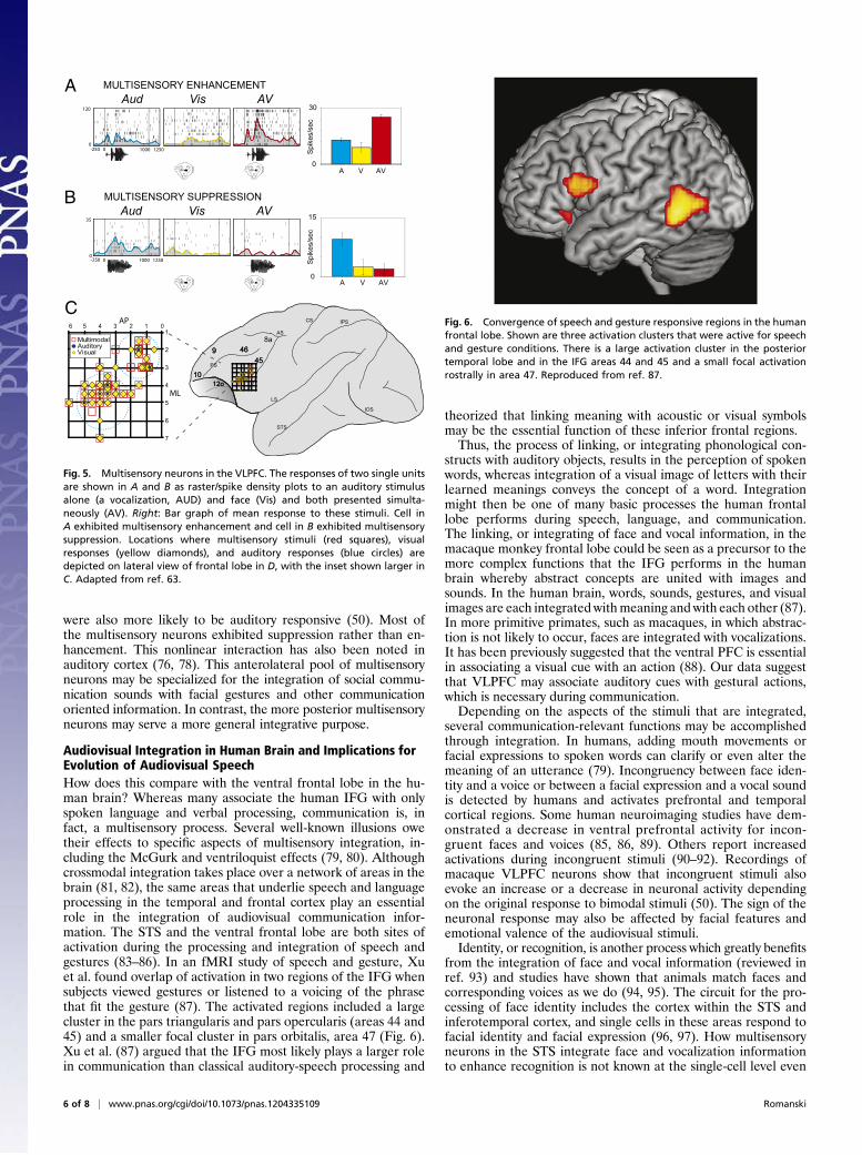
Multisensory Responses in VLPFC. The initial physiological studiesof VLPFC suggested that auditory and visual object processingregions were located adjacent to one another in VLFPC. Anyoverlap in these auditory and visual responsive zones couldthus be sites for multisensory integration of complex auditoryand visual information. As neurons in this region are face- andvocalization-responsive, multisensory neurons in the macaqueVLPFC might integrate face and vocal information. Given thatthe percentage of neurons responsive to visual stimuli was muchgreater than the number of auditory responsive cells (55% vs.18%), we reasoned that multisensory cells are more likely to belocated in regions where auditory cells had been recorded andpredicted that multisensory neurons might be found only in thisregion. In our neurophysiological investigation, we presentedmovies of familiar monkeys vocalizing to macaque monkeyswhile single neurons were recorded from the VLPFC (73). Thesemovies were separated into audio and video streams, and neuralresponses to the unimodal stimuli were compared with the re-sponses to the combined audiovisual stimuli. Interestingly, ap-proximately half the neurons recorded in the VLPFC were mul-tisensory in that they responded to unimodal auditory and visualstimuli, i.e., bimodal responses; or were multisensory because ofan enhanced or decreased response to the combined audiovisualstimulus (face and vocalization) compared with the response tothe unimodal stimuli (73). This is likely to be an underestimate ofthe percentage of multisensory responses because we useda limited set of audiovisual stimuli and neurons were found to beselective for particular face–vocalization pairs.
VLPFC neurons exhibited multisensory enhancement or sup-pression (Fig. 5) just as neurons do in the superior colliculus,the STS, and auditory cortex during multisensory integration(74–77). It was also interesting that face/voice stimuli evokedmultisensory responses more frequently than nonface/nonvoiceaudiovisual stimuli. This adds support to the notion that VLPFCis part of a circuit that is specialized for the integration of socialcommunication information rather than sensory stimuli ina general sense. In localizing these multisensory responses to thePFC, there appeared to be two somewhat separate VLPFCregions for multisensory processing. Interestingly, these twoseparate clusters of multisensory neurons overlap with two pre-frontal face patches described by O’Scalaidhe et al. (19) andTsao et al. (51) in the arcuate and ventrolateral PFC areas. Inour study, there was a large pool of unimodal visual neurons witha small number of multisensory cells located in posterior VLPFC(area 45). Unimodal neurons in this area are mostly visual andrespond to faces and nonface stimuli such as objects, shapes, andpatterns. The multisensory neurons in this arcuate region (Fig. 5)have strong visual responses modulated by the simultaneouspresentation of auditory stimuli. There are strong projections tothis area from the inferotemporal cortex and the polymodal STS,which have been associated with the processing of facial identityand facial expression. Previous studies in nonhuman primates ofvisual working memory, decision-making, and visual search (21,41, 42) have noted responsive neurons within this arcuate region aswell as in the more commonly recorded principal sulcus region.A smaller, potentially more specialized pool of multisensory
neurons is located in VLPFC, anterior and lateral to the firstpool (Fig. 5). These neurons are found near the IPD, and withinits banks in area 12vl of Preuss and Goldman-Rakic (3). This isthe region where unimodal auditory responsive neurons werepredominantly localized in previous studies (10, 11). Theseanterolateral VLPFC neurons respond to vocalizations and tofaces, but only weakly to other visual stimuli (10, 73). This areareceives afferents from mainly polymodal STS cortical regionsand also from auditory association cortex, including a smallamount of afferents from the belt, more from the parabelt, andthe largest contribution from the rostral temporal lobe (33, 65,66). Multisensory responses here favor vocalizations and theircorresponding faces, suggesting a more specialized role in theintegration of social communication information. Face respon-sive cells recorded in this area, which were selective for forwardgaze, such as that which occurs in face-to-face communication,
GRUNT COO HARMONIC ARCHAGGRESSIVE100
00010-2500
9 468a
1012o
LS
STS
45PS
AS
xx x
Fig. 4. Auditory responsive neuronsin VLPFC. A single-cell example ofresponses to four different vocaliza-tion stimuli is shown in the top partof the figure. The response is shownas raster and shaded spike densityfunction in response to a “grunt” vo-calization (low-value food call), anaggressive “pant threat” vocalization,a “coo” vocalization (low-value foodand affiliative) and a harmonic arch(high-value food). The waveforms ofthe calls are shown below the rasters.A schematic of the PFC is shown indi-cating the locations of auditory respon-sive neurons. Adapted from ref. 11.
Romanski PNAS Early Edition | 5 of 8
were also more likely to be auditory responsive (50). Most ofthe multisensory neurons exhibited suppression rather than en-hancement. This nonlinear interaction has also been noted inauditory cortex (76, 78). This anterolateral pool of multisensoryneurons may be specialized for the integration of social commu-nication sounds with facial gestures and other communicationoriented information. In contrast, the more posterior multisensoryneurons may serve a more general integrative purpose.
Audiovisual Integration in Human Brain and Implications forEvolution of Audiovisual SpeechHow does this compare with the ventral frontal lobe in the hu-man brain? Whereas many associate the human IFG with onlyspoken language and verbal processing, communication is, infact, a multisensory process. Several well-known illusions owetheir effects to specific aspects of multisensory integration, in-cluding the McGurk and ventriloquist effects (79, 80). Althoughcrossmodal integration takes place over a network of areas in thebrain (81, 82), the same areas that underlie speech and languageprocessing in the temporal and frontal cortex play an essentialrole in the integration of audiovisual communication infor-mation. The STS and the ventral frontal lobe are both sites ofactivation during the processing and integration of speech andgestures (83–86). In an fMRI study of speech and gesture, Xuet al. found overlap of activation in two regions of the IFG whensubjects viewed gestures or listened to a voicing of the phrasethat fit the gesture (87). The activated regions included a largecluster in the pars triangularis and pars opercularis (areas 44 and45) and a smaller focal cluster in pars orbitalis, area 47 (Fig. 6).Xu et al. (87) argued that the IFG most likely plays a larger rolein communication than classical auditory-speech processing and
theorized that linking meaning with acoustic or visual symbolsmay be the essential function of these inferior frontal regions.Thus, the process of linking, or integrating phonological con-
structs with auditory objects, results in the perception of spokenwords, whereas integration of a visual image of letters with theirlearned meanings conveys the concept of a word. Integrationmight then be one of many basic processes the human frontallobe performs during speech, language, and communication.The linking, or integrating of face and vocal information, in themacaque monkey frontal lobe could be seen as a precursor to themore complex functions that the IFG performs in the humanbrain whereby abstract concepts are united with images andsounds. In the human brain, words, sounds, gestures, and visualimages are each integrated withmeaning and with each other (87).In more primitive primates, such as macaques, in which abstrac-tion is not likely to occur, faces are integrated with vocalizations.It has been previously suggested that the ventral PFC is essentialin associating a visual cue with an action (88). Our data suggestthat VLPFC may associate auditory cues with gestural actions,which is necessary during communication.Depending on the aspects of the stimuli that are integrated,
several communication-relevant functions may be accomplishedthrough integration. In humans, adding mouth movements orfacial expressions to spoken words can clarify or even alter themeaning of an utterance (79). Incongruency between face iden-tity and a voice or between a facial expression and a vocal soundis detected by humans and activates prefrontal and temporalcortical regions. Some human neuroimaging studies have dem-onstrated a decrease in ventral prefrontal activity for incon-gruent faces and voices (85, 86, 89). Others report increasedactivations during incongruent stimuli (90–92). Recordings ofmacaque VLPFC neurons show that incongruent stimuli alsoevoke an increase or a decrease in neuronal activity dependingon the original response to bimodal stimuli (50). The sign of theneuronal response may also be affected by facial features andemotional valence of the audiovisual stimuli.Identity, or recognition, is another process which greatly benefits
from the integration of face and vocal information (reviewed inref. 93) and studies have shown that animals match faces andcorresponding voices as we do (94, 95). The circuit for the pro-cessing of face identity includes the cortex within the STS andinferotemporal cortex, and single cells in these areas respond tofacial identity and facial expression (96, 97). How multisensoryneurons in the STS integrate face and vocalization informationto enhance recognition is not known at the single-cell level even
0
0
Aud Vis AV
15
0
Spi
kes/
sec
A AVV
Spi
kes/
sec
30
0A AVV
0
35
0 1000
120
-250
-250 1000
1250
1250
A
BAud Vis AV
MULTISENSORY ENHANCEMENT
MULTISENSORY SUPPRESSION
C
MultimodalAuditoryVisual
01234561
2
3
4
5
6
7
AP
ML
99 46468a
101012o12o
LS
STS
IOS
4545PS
AS
CS IPS
Fig. 5. Multisensory neurons in the VLPFC. The responses of two single unitsare shown in A and B as raster/spike density plots to an auditory stimulusalone (a vocalization, AUD) and face (Vis) and both presented simulta-neously (AV). Right: Bar graph of mean response to these stimuli. Cell inA exhibited multisensory enhancement and cell in B exhibited multisensorysuppression. Locations where multisensory stimuli (red squares), visualresponses (yellow diamonds), and auditory responses (blue circles) aredepicted on lateral view of frontal lobe in D, with the inset shown larger inC. Adapted from ref. 63.
Fig. 6. Convergence of speech and gesture responsive regions in the humanfrontal lobe. Shown are three activation clusters that were active for speechand gesture conditions. There is a large activation cluster in the posteriortemporal lobe and in the IFG areas 44 and 45 and a small focal activationrostrally in area 47. Reproduced from ref. 87.
6 of 8 | www.pnas.org/cgi/doi/10.1073/pnas.1204335109 Romanski
though pairing of incongruent faces and vocalizations alters ac-tivity in this region. The STS has a robust connection withVLPFC and is likely to send unimodal and multisensory identityinformation to VLPFC neurons. Selectivity of face-responsivecells in VLPFC has been shown for particular individuals, ex-pressions, or categories of face stimuli (19, 20, 48, 50).The accumulation of evidence to date shows that cells in the
ventral PFC of the macaque monkey respond to and integrateaudiovisual information. VLPFC cells respond optimally to face
and vocalization stimuli and exhibit multisensory enhancement orsuppression when face-vocalization stimuli are combined. Thus,the ventral frontal lobe of nonhuman primates may have somebasic functional homologies to the human frontal lobe, althoughmore evidence from additional primate species is needed. Thebasic process of associating a face, or facial gesture, with a vocalstimulus, which occurs in the macaque PFC, may be a precursor tothe more complex functions of the human frontal lobe, wheresemantic meaning is linked with acoustic or visual symbols.
1. Petrides M, Pandya DN (2002) Comparative cytoarchitectonic analysis of the humanand the macaque ventrolateral prefrontal cortex and corticocortical connectionpatterns in the monkey. Eur J Neurosci 16:291–310.
2. Preuss T (2007) Primate Origins: Adaptations and Evolution, eds Ravosa M, Dagosto M(Springer, New York).
3. Preuss TM, Goldman-Rakic PS (1991) Myelo- and cytoarchitecture of the granularfrontal cortex and surrounding regions in the strepsirhine primate Galago and theanthropoid primate Macaca. J Comp Neurol 310:429–474.
4. Carmichael ST, Price JL (1995) Sensory and premotor connections of the orbital andmedial prefrontal cortex of macaque monkeys. J Comp Neurol 363:642–664.
5. Gerbella M, Belmalih A, Borra E, Rozzi S, Luppino G (2010) Cortical connections of themacaque caudal ventrolateral prefrontal areas 45A and 45B. Cereb Cortex 20:141–168.
6. Saleem KS, Kondo H, Price JL (2008) Complementary circuits connecting the orbitaland medial prefrontal networks with the temporal, insular, and opercular cortex inthe macaque monkey. J Comp Neurol 506:659–693.
7. Winters W, Kado R, Adey W (1969) A Stereotaxic Brain Atlas for Macaca nemestrina(Univ California Press, Los Angeles).
8. Petrides M, Cadoret G, Mackey S (2005) Orofacial somatomotor responses in themacaque monkey homologue of Broca’s area. Nature 435:1235–1238.
9. Paxinos G, Huang X, Toga AW (2000) The Rhesus Monkey Brain (Academic, SanDiego).
10. Romanski LM, Goldman-Rakic PS (2002) An auditory domain in primate prefrontalcortex. Nat Neurosci 5:15–16.
11. Romanski LM, Averbeck BB, Diltz M (2005) Neural representation of vocalizations inthe primate ventrolateral prefrontal cortex. J Neurophysiol 93:734–747.
12. Jürgens U (2009) The neural control of vocalization in mammals: A review. J Voice 23:1–10.
13. Coudé G, et al. (2011) Neurons controlling voluntary vocalization in the macaqueventral premotor cortex. PLoS ONE 6:e26822.
14. Barbas H (1988) Anatomic organization of basoventral and mediodorsal visual re-cipient prefrontal regions in the rhesus monkey. J Comp Neurol 276:313–342.
15. Barbas H, Pandya DN (1989) Architecture and intrinsic connections of the prefrontalcortex in the rhesus monkey. J Comp Neurol 286:353–375.
16. Chavis DA, Pandya DN (1976) Further observations on corticofrontal connections inthe rhesus monkey. Brain Res 117:369–386.
17. Bullier J, Schall JD, Morel A (1996) Functional streams in occipito-frontal connectionsin the monkey. Behav Brain Res 76:89–97.
18. Ungerleider LG, Gaffan D, Pelak VS (1989) Projections from inferior temporal cortexto prefrontal cortex via the uncinate fascicle in rhesus monkeys. Exp Brain Res 76:473–484.
19. O Scalaidhe SP, Wilson FA, Goldman-Rakic PS (1997) Areal segregation of face-pro-cessing neurons in prefrontal cortex. Science 278:1135–1138.
20. Scalaidhe SP, Wilson FAW, Goldman-Rakic PS (1999) Face-selective neurons duringpassive viewing and working memory performance of rhesus monkeys: Evidence forintrinsic specialization of neuronal coding. Cereb Cortex 9:459–475.
21. Wilson FA, Scalaidhe SP, Goldman-Rakic PS (1993) Dissociation of object and spatialprocessing domains in primate prefrontal cortex. Science 260:1955–1958.
22. Webster MJ, Bachevalier J, Ungerleider LG (1994) Connections of inferior temporalareas TEO and TE with parietal and frontal cortex in macaque monkeys. Cereb Cortex4:470–483.
23. Pandya DN, Hallett M, Kmukherjee SK (1969) Intra- and interhemispheric connectionsof the neocortical auditory system in the rhesus monkey. Brain Res 14:49–65.
24. Jones EG, Powell TP (1970) An anatomical study of converging sensory pathwayswithin the cerebral cortex of the monkey. Brain 93:793–820.
25. Galaburda AM, Pandya DN (1983) The intrinsic architectonic and connectional orga-nization of the superior temporal region of the rhesus monkey. J Comp Neurol 221:169–184.
26. Barbas H, Mesulam MM (1985) Cortical afferent input to the principalis region of therhesus monkey. Neuroscience 15:619–637.
27. Petrides M, Pandya DN (1988) Association fiber pathways to the frontal cortex fromthe superior temporal region in the rhesus monkey. J Comp Neurol 273:52–66.
28. Morel A, Garraghty PE, Kaas JH (1993) Tonotopic organization, architectonic fields,and connections of auditory cortex in macaque monkeys. J Comp Neurol 335:437–459.
29. Jones EG, Dell’Anna ME, Molinari M, Rausell E, Hashikawa T (1995) Subdivisions ofmacaque monkey auditory cortex revealed by calcium-binding protein immunore-activity. J Comp Neurol 362:153–170.
30. Hackett TA, Stepniewska I, Kaas JH (1998) Thalamocortical connections of the para-belt auditory cortex in macaque monkeys. J Comp Neurol 400:271–286.
31. Rauschecker JP, Tian B, Hauser M (1995) Processing of complex sounds in the macaquenonprimary auditory cortex. Science 268:111–114.
32. Tian B, Reser D, Durham A, Kustov A, Rauschecker JP (2001) Functional specializationin rhesus monkey auditory cortex. Science 292:290–293.
33. Romanski LM, et al. (1999) Dual streams of auditory afferents target multiple domainsin the primate prefrontal cortex. Nat Neurosci 2:1131–1136.
34. Romanski LM (2007) Representation and integration of auditory and visual stimuli inthe primate ventral lateral prefrontal cortex. Cereb Cortex 17(suppl 1):i61–i69.
35. Ungerleider LG, Mishkin M (1982) Two cortical visual systems. In Analysis of VisualBehavior, eds Ingle DJ, Goodale MA, Mansfield RJW (MIT Press, Cambridge, MA), pp549–586.
36. Bruce CJ, Goldberg ME (1985) Primate frontal eye fields. I. Single neurons dischargingbefore saccades. J Neurophysiol 53:603–635.
37. Funahashi S, Bruce CJ, Goldman-Rakic PS (1989) Mnemonic coding of visual space inthe monkey’s dorsolateral prefrontal cortex. J Neurophysiol 61:1–19.
39. Quintana J, Fuster JM (1992) Mnemonic and predictive functions of cortical neuronsin a memory task. Neuroreport 3:721–724.
40. Chafee MV, Goldman-Rakic PS (1998) Matching patterns of activity in primate pre-frontal area 8a and parietal area 7ip neurons during a spatial working memory task.J Neurophysiol 79:2919–2940.
41. Kim JN, Shadlen MN (1999) Neural correlates of a decision in the dorsolateral pre-frontal cortex of the macaque. Nat Neurosci 2:176–185.
42. Freedman DJ, Miller EK (2008) Neural mechanisms of visual categorization: Insightsfrom neurophysiology. Neurosci Biobehav Rev 32:311–329.
43. Nieder A, Freedman DJ, Miller EK (2002) Representation of the quantity of visualitems in the primate prefrontal cortex. Science 297:1708–1711.
44. Miller EK, Cohen JD (2001) An integrative theory of prefrontal cortex function. AnnuRev Neurosci 24:167–202.
45. Rosenkilde CE, Bauer RH, Fuster JM (1981) Single cell activity in ventral prefrontalcortex of behaving monkeys. Brain Res 209:375–394.
46. Suzuki H, Azuma M (1983) Topographic studies on visual neurons in the dorsolateralprefrontal cortex of the monkey. Exp Brain Res 53:47–58.
47. Thorpe SJ, Rolls ET, Maddison S (1983) The orbitofrontal cortex: Neuronal activity inthe behaving monkey. Exp Brain Res 49:93–115.
48. Rolls ET, Critchley HD, Browning AS, Inoue K (2006) Face-selective and auditoryneurons in the primate orbitofrontal cortex. Exp Brain Res 170:74–87.
49. Mishkin M, Manning FJ (1978) Non-spatial memory after selective prefrontal lesionsin monkeys. Brain Res 143:313–323.
50. Romanski LM, Diehl MM (2011) Neurons responsive to face-view in the primateventrolateral prefrontal cortex. Neuroscience 189:223–235.
51. Tsao DY, Schweers N, Moeller S, Freiwald WA (2008) Patches of face-selective cortexin the macaque frontal lobe. Nat Neurosci 11:877–879.
52. Démonet JF, et al. (1992) The anatomy of phonological and semantic processing innormal subjects. Brain 115:1753–1768.
53. Paulesu E, Frith CD, Frackowiak RSJ (1993) The neural correlates of the verbal com-ponent of working memory. Nature 362:342–345.
54. Buckner RL, Raichle ME, Petersen SE (1995) Dissociation of human prefrontal corticalareas across different speech production tasks and gender groups. J Neurophysiol 74:2163–2173.
55. Demb JB, et al. (1995) Semantic encoding and retrieval in the left inferior prefrontalcortex: A functional MRI study of task difficulty and process specificity. J Neurosci 15:5870–5878.
56. Stromswold K, Caplan D, Alpert N, Rauch S (1996) Localization of syntactic compre-hension by positron emission tomography. Brain Lang 52:452–473.
57. Price CJ (1998) The functional anatomy of word comprehension and production.Trends Cogn Sci 2:281–288.
58. Gelfand JR, Bookheimer SY (2003) Dissociating neural mechanisms of temporal se-quencing and processing phonemes. Neuron 38:831–842.
59. Poldrack RA, et al. (1999) Functional specialization for semantic and phonologicalprocessing in the left inferior prefrontal cortex. Neuroimage 10:15–35.
60. Newman JD, Lindsley DF (1976) Single unit analysis of auditory processing in squirrelmonkey frontal cortex. Exp Brain Res 25:169–181.
61. Tanila H, Carlson S, Linnankoski I, Kahila H (1993) Regional distribution of functions indorsolateral prefrontal cortex of the monkey. Behav Brain Res 53:63–71.
62. Wollberg Z, Sela J (1980) Frontal cortex of the awake squirrel monkey: responses ofsingle cells to visual and auditory stimuli. Brain Res 198:216–220.
63. Romanski LM, Averbeck BB (2009) The primate cortical auditory system and neuralrepresentation of conspecific vocalizations. Annu Rev Neurosci 32:315–346.
64. Cohen YE, et al. (2009) A functional role for the ventrolateral prefrontal cortex innon-spatial auditory cognition. Proc Natl Acad Sci USA 106:20045–20050.
Romanski PNAS Early Edition | 7 of 8
65. Romanski LM, Bates JF, Goldman-Rakic PS (1999) Auditory belt and parabelt projec-tions to the prefrontal cortex in the rhesus monkey. J Comp Neurol 403:141–157.
66. Hackett TA, Stepniewska I, Kaas JH (1999) Prefrontal connections of the parabeltauditory cortex in macaque monkeys. Brain Res 817:45–58.
67. Poremba A, Mishkin M (2007) Exploring the extent and function of higher-orderauditory cortex in rhesus monkeys. Hear Res 229:14–23.
68. Gifford GW, 3rd, MacLean KA, Hauser MD, Cohen YE (2005) The neurophysiology offunctionally meaningful categories: Macaque ventrolateral prefrontal cortex playsa critical role in spontaneous categorization of species-specific vocalizations. J CognNeurosci 17:1471–1482.
69. Russ BE, Lee YS, Cohen YE (2007) Neural and behavioral correlates of auditory cate-gorization. Hear Res 229:204–212.
70. Lee JH, Russ BE, Orr LE, Cohen YE (2009) Prefrontal activity predicts monkeys’ deci-sions during an auditory category task. Front Integr Neurosci 3:16.
71. Deacon TW (1992) Cortical connections of the inferior arcuate sulcus cortex in themacaque brain. Brain Res 573:8–26.
72. Aboitiz F (2012) Gestures, vocalizations, and memory in language origins. Front EvolNeurosci 4:2.
73. Sugihara T, Diltz MD, Averbeck BB, Romanski LM (2006) Integration of auditory andvisual communication information in the primate ventrolateral prefrontal cortex.J Neurosci 26:11138–11147.
74. Stein BE, Meredith MA (1993) The Merging of the Senses (MIT Press, Cambridge, MA).75. Barraclough NE, Xiao D, Baker CI, Oram MW, Perrett DI (2005) Integration of visual
and auditory information by superior temporal sulcus neurons responsive to the sightof actions. J Cogn Neurosci 17:377–391.
76. Ghazanfar AA, Maier JX, Hoffman KL, Logothetis NK (2005) Multisensory integrationof dynamic faces and voices in rhesus monkey auditory cortex. J Neurosci 25:5004–5012.
77. Lakatos P, et al. (2009) The leading sense: Supramodal control of neurophysiologicalcontext by attention. Neuron 64:419–430.
78. Kayser C, Logothetis NK, Panzeri S (2010) Visual enhancement of the informationrepresentation in auditory cortex. Curr Biol 20:19–24.
on the time dimension. 1. Evidence from auditory-visual temporal order judgment.Int J Psychophysiol 50:147–155.
81. Driver J, Noesselt T (2008) Multisensory interplay reveals crossmodal influences on‘sensory-specific’ brain regions, neural responses, and judgments. Neuron 57:11–23.
82. Stein BE, Stanford TR (2008) Multisensory integration: Current issues from the per-spective of the single neuron. Nat Rev Neurosci 9:255–266.
83. Noppeney U, Ostwald D, Werner S (2010) Perceptual decisions formed by accumula-tion of audiovisual evidence in prefrontal cortex. J Neurosci 30:7434–7446.
84. Beauchamp MS, Nath AR, Pasalar S (2010) fMRI-guided transcranial magnetic stimu-lation reveals that the superior temporal sulcus is a cortical locus of the McGurk ef-fect. J Neurosci 30:2414–2417.
85. Jones JA, Callan DE (2003) Brain activity during audiovisual speech perception: AnfMRI study of the McGurk effect. Neuroreport 14:1129–1133.
86. Homae F, Hashimoto R, Nakajima K, Miyashita Y, Sakai KL (2002) From perception tosentence comprehension: The convergence of auditory and visual information oflanguage in the left inferior frontal cortex. Neuroimage 16:883–900.
87. Xu J, Gannon PJ, Emmorey K, Smith JF, Braun AR (2009) Symbolic gestures and spokenlanguage are processed by a common neural system. Proc Natl Acad Sci USA 106:20664–20669.
88. Passingham RE, Toni I, Rushworth MF (2000) Specialisation within the prefrontalcortex: The ventral prefrontal cortex and associative learning. Exp Brain Res 133:103–113.
89. Calvert GA, Hansen PC, Iversen SD, Brammer MJ (2001) Detection of audio-visual in-tegration sites in humans by application of electrophysiological criteria to the BOLDeffect. Neuroimage 14:427–438.
90. Miller LM, D’Esposito M (2005) Perceptual fusion and stimulus coincidence in thecross-modal integration of speech. J Neurosci 25:5884–5893.
91. Ojanen V, et al. (2005) Processing of audiovisual speech in Broca’s area. Neuroimage25:333–338.
92. Hein G, et al. (2007) Object familiarity and semantic congruency modulate responsesin cortical audiovisual integration areas. J Neurosci 27:7881–7887.
93. Campanella S, Belin P (2007) Integrating face and voice in person perception. TrendsCogn Sci 11:535–543.
94. Jordan KE, Brannon EM, Logothetis NK, Ghazanfar AA (2005) Monkeys match thenumber of voices they hear to the number of faces they see. Curr Biol 15:1034–1038.
95. Sliwa J, Duhamel JR, Pascalis O, Wirth S (2011) Spontaneous voice-face identitymatching by rhesus monkeys for familiar conspecifics and humans. Proc Natl Acad SciUSA 108:1735–1740.
96. Eifuku S, De Souza WC, Tamura R, Nishijo H, Ono T (2004) Neuronal correlates of faceidentification in the monkey anterior temporal cortical areas. J Neurophysiol 91:358–371.
97. Sugase Y, Yamane S, Ueno S, Kawano K (1999) Global and fine information coded bysingle neurons in the temporal visual cortex. Nature 400:869–873.
8 of 8 | www.pnas.org/cgi/doi/10.1073/pnas.1204335109 Romanski