Inter-DNA Attraction Mediated by Divalent Counterions
Xiangyun Qiu, Kurt Andresen, Lisa W. Kwok, Jessica S. Lamb, Hye Yoon Park, and Lois PollackSchool of Applied and Engineering Physics, Cornell University, Ithaca, New York 14853, USA
(Received 2 March 2007; published 20 July 2007)
Can nonspecifically bound divalent counterions induce attraction between DNA strands? Here, wepresent experimental evidence demonstrating attraction between short DNA strands mediated by Mg2�
ions. Solution small angle x-ray scattering data collected as a function of DNA concentration enablemodel independent extraction of the second virial coefficient. As the [Mg2�] increases, this coefficientturns from positive to negative reflecting the transition from repulsive to attractive inter-DNA interaction.This surprising observation is corroborated by independent light scattering experiments. The dependenceof the observed attraction on experimental parameters including DNA length provides valuable clues to itsorigin.
Electrostatic interactions are fundamental to the com-plex structure and dynamics of nucleic acids due to theirhighly charged nature. Positively charged ions counteractand can even reverse the repulsion between negativelycharged nucleic acids [1,2]. Quantitative studies of coun-terion mediated interactions are essential in understandingphenomena such as DNA condensation and packaging[1,2] and RNA folding [3].
Counterion valence is a critical factor in modulatinginter-DNA forces. In the presence of monovalent cations(e.g., Na�), the dominant force between DNA strands islike charge repulsion. Following the seminal Derjaguin,Landau, Verwey, and Overbeek (DLVO) mean field theory[4], this interaction can be described by a Yukawa pairpotential. To correct for the linear Poisson-Boltzmannapproximation in the DLVO theory, the charge renormal-ization prescription [5] assigns an effective charge Zeff toeach DNA. The Yukawa form of the repulsive inter-DNApotential has been validated by our recent measurements,though the measured effective charges are smaller thanpredicted [6]. In the presence of low concentrations ofcounterions of tri- or higher valence, strong attractiveforces between DNA strands (e.g., precipitation) are ob-served [1,2]. The phase diagram of such ‘‘precipitated’’liquid crystalline DNA complexes has been studied in de-tail [7]. However, the exact physical origin of the likecharge attraction remains elusive [8]. Proposed competingmechanisms for attraction include counterion local densityfluctuations [9,10], positional correlation between con-densed counterions [11,12], and tight binding of counter-ions along discrete charged DNA monomers [13–15].Notably, strong electrostatic coupling may lead to over-charging due to counterion ‘‘Wigner-lattice’’ [16] orBjerrum pairing correlations [17].
The ability of nonspecifically bound divalent counter-ions (e.g., Mg2�) to induce attractive interaction betweenDNA strands remains controversial. Both analytical theo-ries and simulations have predicted net short range attrac-tive forces between DNA strands in divalent (2:1) salts
[11,13,18,19]. Experimental support for the predicted at-tractions remains tentative. Our previous measurement ofthe inter-DNA forces in the presence of divalent ionssuggested a weak attractive interaction under �Mg2��>16 mM [6], though this conclusion was based on theassumption of a particular model calculation. It shouldalso be noted that inter-DNA attraction has been proposedeven in monovalent salts based on observations of the‘‘slow mode’’ in dynamic light scattering and low angle‘‘upturns’’ from small angle neutron scattering [20].Notably, condensation of DNA by divalent Mg2� counter-ions has never been observed in bulk solution [21]. Bymonitoring the conformational changes of a tethered DNAsystem [22], Bai et al. rule out possible strong inter-DNAattraction up to 600 mM Mg2�, which is also consistentwith the lack of discontinuity in inter-DNA spacings uponincreasing osmotic stress [23]. Here, we aim to establishbeyond doubt the existence of inter-DNA attraction medi-ated by divalent counterions.
Small angle x-ray scattering (SAXS) measurements ofsemidilute double strand DNA (dsDNA) solutions werecarried out at the C1 and G1 stations of the Cornell HighEnergy Synchrotron Source (CHESS). The x-ray exposuretime was chosen to ensure time-independent scatteringprofiles. Single strand DNA oligomers were purchasedfrom Integrated DNA Technologies and annealed atpH 7.5 to obtain rigid rodlike dsDNA (8, 16, and 25 basepairs). Each sample was dialyzed against monovalent(NaCl) or divalent (MgCl2) salt solutions buffered with1 mM pH � 7 NaMOPS [sodium 3-(N-morpholino)pro-panesulfonic acid]. Neither Na� nor Mg2� displays site-specific binding to DNA [24]. The measured scatteringintensity I�Q� [Q � �4�=�� sin�, � is the x-ray wave-length, and 2� is the scattering angle] has two compo-nents: the form factor P�Q� of a single DNA and thestructure factor S�Q�. The inter-DNA interference func-tion S�Q� arises from long range structural correlations,modulates the SAXS profile, and is most pronounced atlow Q [25].
PRL 99, 038104 (2007) P H Y S I C A L R E V I E W L E T T E R S week ending20 JULY 2007
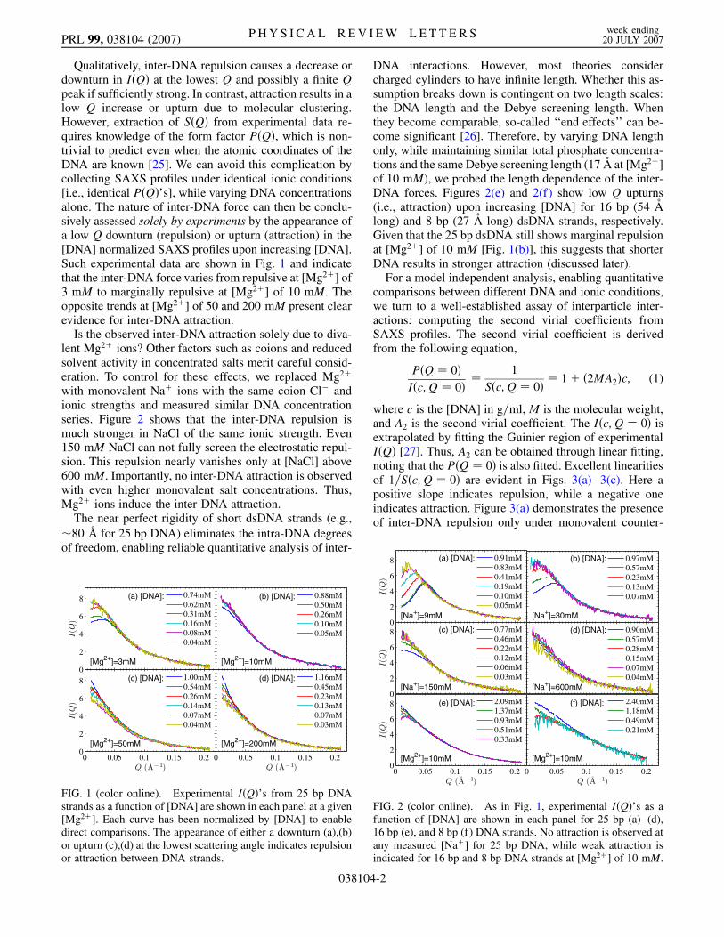
Qualitatively, inter-DNA repulsion causes a decrease ordownturn in I�Q� at the lowest Q and possibly a finite Qpeak if sufficiently strong. In contrast, attraction results in alow Q increase or upturn due to molecular clustering.However, extraction of S�Q� from experimental data re-quires knowledge of the form factor P�Q�, which is non-trivial to predict even when the atomic coordinates of theDNA are known [25]. We can avoid this complication bycollecting SAXS profiles under identical ionic conditions[i.e., identical P�Q�’s], while varying DNA concentrationsalone. The nature of inter-DNA force can then be conclu-sively assessed solely by experiments by the appearance ofa low Q downturn (repulsion) or upturn (attraction) in the[DNA] normalized SAXS profiles upon increasing [DNA].Such experimental data are shown in Fig. 1 and indicatethat the inter-DNA force varies from repulsive at [Mg2�] of3 mM to marginally repulsive at [Mg2�] of 10 mM. Theopposite trends at [Mg2�] of 50 and 200 mM present clearevidence for inter-DNA attraction.
Is the observed inter-DNA attraction solely due to diva-lent Mg2� ions? Other factors such as coions and reducedsolvent activity in concentrated salts merit careful consid-eration. To control for these effects, we replaced Mg2�
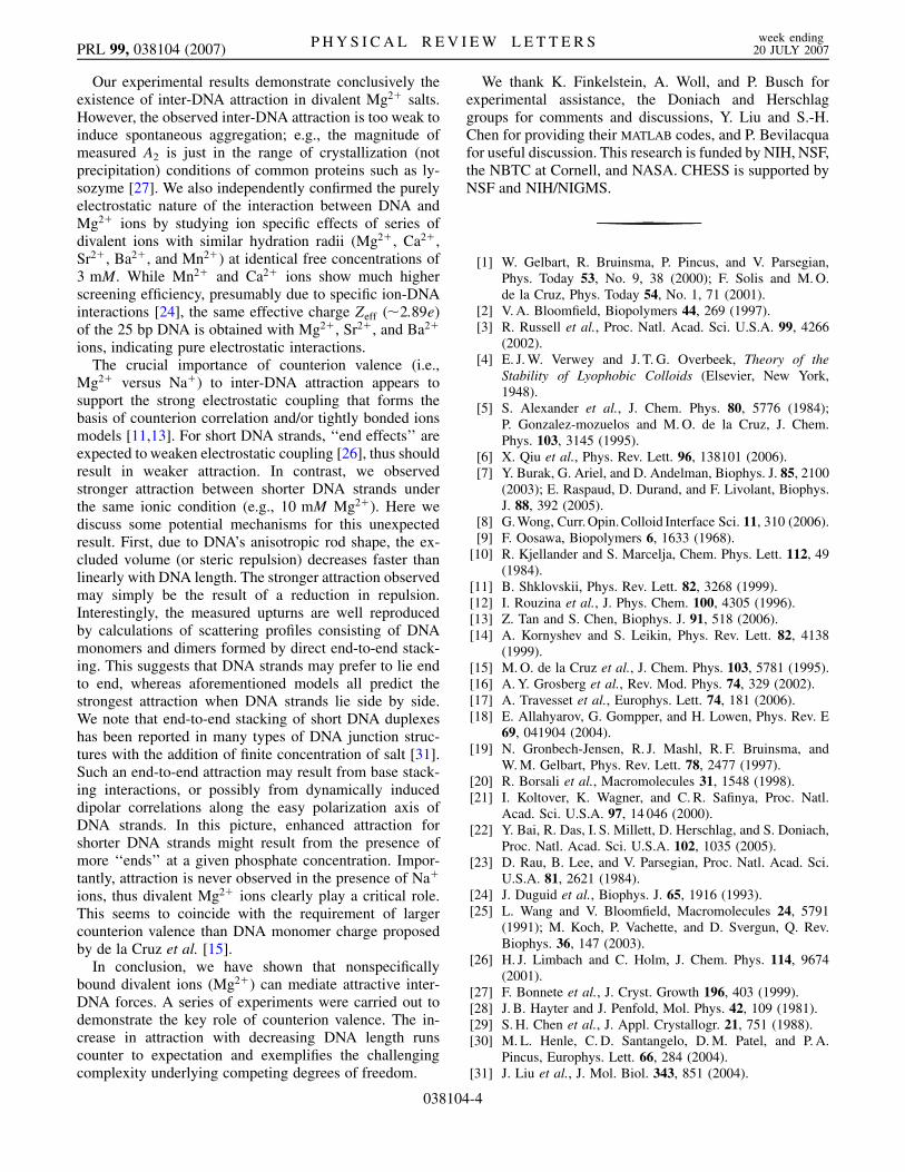
with monovalent Na� ions with the same coion Cl� andionic strengths and measured similar DNA concentrationseries. Figure 2 shows that the inter-DNA repulsion ismuch stronger in NaCl of the same ionic strength. Even150 mM NaCl can not fully screen the electrostatic repul-sion. This repulsion nearly vanishes only at [NaCl] above600 mM. Importantly, no inter-DNA attraction is observedwith even higher monovalent salt concentrations. Thus,Mg2� ions induce the inter-DNA attraction.
The near perfect rigidity of short dsDNA strands (e.g.,�80 �A for 25 bp DNA) eliminates the intra-DNA degreesof freedom, enabling reliable quantitative analysis of inter-
DNA interactions. However, most theories considercharged cylinders to have infinite length. Whether this as-sumption breaks down is contingent on two length scales:the DNA length and the Debye screening length. Whenthey become comparable, so-called ‘‘end effects’’ can be-come significant [26]. Therefore, by varying DNA lengthonly, while maintaining similar total phosphate concentra-tions and the same Debye screening length (17 A at [Mg2�]of 10 mM), we probed the length dependence of the inter-DNA forces. Figures 2(e) and 2(f) show low Q upturns(i.e., attraction) upon increasing [DNA] for 16 bp (54 Along) and 8 bp (27 A long) dsDNA strands, respectively.Given that the 25 bp dsDNA still shows marginal repulsionat [Mg2�] of 10 mM [Fig. 1(b)], this suggests that shorterDNA results in stronger attraction (discussed later).
For a model independent analysis, enabling quantitativecomparisons between different DNA and ionic conditions,we turn to a well-established assay of interparticle inter-actions: computing the second virial coefficients fromSAXS profiles. The second virial coefficient is derivedfrom the following equation,
P�Q � 0�
I�c;Q � 0��
1
S�c;Q � 0�� 1� �2MA2�c; (1)
where c is the [DNA] in g=ml, M is the molecular weight,and A2 is the second virial coefficient. The I�c;Q � 0� isextrapolated by fitting the Guinier region of experimentalI�Q� [27]. Thus, A2 can be obtained through linear fitting,noting that the P�Q � 0� is also fitted. Excellent linearitiesof 1=S�c;Q � 0� are evident in Figs. 3(a)–3(c). Here apositive slope indicates repulsion, while a negative oneindicates attraction. Figure 3(a) demonstrates the presenceof inter-DNA repulsion only under monovalent counter-
FIG. 1 (color online). Experimental I�Q�’s from 25 bp DNAstrands as a function of [DNA] are shown in each panel at a given[Mg2�]. Each curve has been normalized by [DNA] to enabledirect comparisons. The appearance of either a downturn (a),(b)or upturn (c),(d) at the lowest scattering angle indicates repulsionor attraction between DNA strands.
FIG. 2 (color online). As in Fig. 1, experimental I�Q�’s as afunction of [DNA] are shown in each panel for 25 bp (a)–(d),16 bp (e), and 8 bp (f) DNA strands. No attraction is observed atany measured [Na�] for 25 bp DNA, while weak attraction isindicated for 16 bp and 8 bp DNA strands at [Mg2�] of 10 mM.
PRL 99, 038104 (2007) P H Y S I C A L R E V I E W L E T T E R S week ending20 JULY 2007
038104-2
ions, while Fig. 3(b) shows the crossover from repulsion toattraction upon increasing [Mg2�]. The comparison ofDNA strands of different lengths [Fig. 3(c)] confirmsstronger attraction (i.e., larger negative slope) with shorterDNA strands. Figure 3(d) compares the second virial co-efficient A2 as a function of ionic strength for all DNAlengths and counterion types. This chart makes it possibleto find equivalent salt conditions giving the same inter-DNA interaction, e.g., 150 mM Na� 3 mM Mg2�.
To further substantiate the observed inter-DNA at-traction from SAXS experiments, we carried out indepen-dent light scattering measurements of the second virialcoefficients. Selected 25 bp DNA samples were preparedfollowing the same protocol and were measured with thedynamic light scattering instrument Malvin ZetasizerNano Series (Laser 4 mW He-Ne, 633 nm). At each saltcondition, a series of samples with different DNA con-centrations was measured at 25 C. All solutions werefiltered twice to eliminate dust. The measured A2 valuesat [Mg2�] of 3, 5, and 200 mM are 2:2e–3, �6:8e–4, and�1:1e–3 mol ml g�2, respectively. The values not onlyqualitatively confirm the nature of attractive interactionat 50 and 200 mM Mg2�, but also show reasonable quanti-tative agreement with SAXS measurements. We also mea-sured at conditions of 10 mM pH�7 NaMOPS and/or0:1 mM pH � 7 ethylenediamine tetraacetic acid(EDTA) to check for possible pH dependence and highvalence metal ion contamination. No detectable changewas observed with the addition of 0:1 mM EDTA, whileonly a slight decrease of inter-DNA attraction (A2 from
�6:8e–4 to �6:3e–4 mol ml g�2) results from the 1 to10 mM increase in NaMOPS concentration at [Mg2�] of50 mM, presumably due to the effect of counterion com-petition. Possible multivalent ion contamination or pHeffects are thus eliminated.
In addition to second virial coefficients, detailed infor-mation on inter-DNA potentials can be obtained by quan-titatively analyzing the full SAXS profiles. As in our recentstudy [6], the generalized one-component method(GOCM) [28,29] is used to compute the structure factorS�Q� from a model Yukawa pair potential with a hard corerepulsion (as in DLVO theory). Instead of measuring theform factor at each condition, we numerically compute theP�Q�’s from known DNA structure, accounting for solventeffects such as hydration and ‘‘condensed’’ counterions.Comparisons with available measured form factors showreasonable agreement. In the refinement of the full SAXSprofile, the only free parameter is the effective charge Zeff
in the Yukawa potential. A more detailed description ofanalysis methods can be found in Ref. [6]. Although themean spherical approximation (MSA) holds only in thedilute DNA limit, application of MSA allows for conve-nient quantitative analysis, and is supported by the agree-ment between predictions and experimental data in thesemidilute DNA regime.
Figure 4(a) shows the SAXS profiles as a function of[Mg2�] at [DNA] �0:7 mM. The fitted structure factorS�Q�’s shown in Fig. 4(b) demonstrate the transition fromrepulsion (low Q downturn) to attraction (low Q upturn)upon increasing [Mg2�]. The measured effective charges(Zeff) are 13.23, 7.14, 4.50, 2.89, and 0.71 elemental chargewith [Mg2�] of 0, 1, 2, 3, and 4 mM, respectively. This is indrastic contrast with the measured Zeff � 12e with mono-valent Na� ions which only weakly depends on ionicstrength [6]. At [Mg2�] of 10 mM, the interaction betweenDNA strands vanishes as the structure factor approachesunity. While the much higher screening efficiency of Mg2�
versus Na� is not surprising [30], inter-DNA attraction,i.e., the lowQ upturn, is observed when �Mg2��> 10 mM,and increases with [Mg2�]. Addition of a second Yukawapotential of short range in the GOCM is able to model theobserved attraction [6].
FIG. 4 (color online). The structure factor S�Q� can be ex-tracted from the measured I�Q� using models (see text). Panel (a)shows measured I�Q�’s of 25 bp DNA (� 0:7 mM) (symbols)and the fits (lines). Panel (b) shows the corresponding S�Q�’sonly with the same color annotation as in (a).
0 5 10 15 20
2
4
6
DNA Concentration (mg/ml)
1/S(
Q=
0)(a) 9mM [Na+]
30mM150mM600mM
0 5 10 15 20
1
1.5
2
DNA Concentration (mg/ml)
1/S(
Q=
0)
(b) 3mM [Mg2+]10mM50mM200mM
0 5 10 15 20
0.8
1
1.2
DNA Concentration (mg/ml)
1/S(
Q=
0)
(c)
[Mg2+]=10mM
25bp DNA16bp 8bp
0 200 400 600−2
0
2
4
6
8
x 10−3
Ionic Strength (mM)
A2 (
mol
⋅ml⋅g
−2 ) (d) Na+, 25bp DNA
Mg2+, 25bp (x3)
Mg2+, 16bp
Mg2+, 8bp
FIG. 3 (color online). Interactions between DNA strands arereflected by changes in the y intercept of I�Q� [ / S�c;Q � 0�].Here 1=S�c;Q � 0� (symbols) is shown as a function of [DNA](a) at different [Na�], (b) at different [Mg2�], and (c) as afunction of DNA length at 10 mM Mg2�. The second virialcoefficient can be derived from these data according to Eq. (1).The variation of A2 as a function of ionic strength is shown in (d).Both repulsion (A2 > 0) and attraction (A2 < 0) are observed as[Mg2�] increases; however, only repulsion is observed in thepresence of monovalent ions. The data for 25 bp DNA in Mg2�
are multiplied by a factor of 3 for clarity. Most error bars havesizes comparable to symbols.
PRL 99, 038104 (2007) P H Y S I C A L R E V I E W L E T T E R S week ending20 JULY 2007
038104-3
Our experimental results demonstrate conclusively theexistence of inter-DNA attraction in divalent Mg2� salts.However, the observed inter-DNA attraction is too weak toinduce spontaneous aggregation; e.g., the magnitude ofmeasured A2 is just in the range of crystallization (notprecipitation) conditions of common proteins such as ly-sozyme [27]. We also independently confirmed the purelyelectrostatic nature of the interaction between DNA andMg2� ions by studying ion specific effects of series ofdivalent ions with similar hydration radii (Mg2�, Ca2�,Sr2�, Ba2�, and Mn2�) at identical free concentrations of3 mM. While Mn2� and Ca2� ions show much higherscreening efficiency, presumably due to specific ion-DNAinteractions [24], the same effective charge Zeff (�2:89e)of the 25 bp DNA is obtained with Mg2�, Sr2�, and Ba2�
ions, indicating pure electrostatic interactions.The crucial importance of counterion valence (i.e.,
Mg2� versus Na�) to inter-DNA attraction appears tosupport the strong electrostatic coupling that forms thebasis of counterion correlation and/or tightly bonded ionsmodels [11,13]. For short DNA strands, ‘‘end effects’’ areexpected to weaken electrostatic coupling [26], thus shouldresult in weaker attraction. In contrast, we observedstronger attraction between shorter DNA strands underthe same ionic condition (e.g., 10 mM Mg2�). Here wediscuss some potential mechanisms for this unexpectedresult. First, due to DNA’s anisotropic rod shape, the ex-cluded volume (or steric repulsion) decreases faster thanlinearly with DNA length. The stronger attraction observedmay simply be the result of a reduction in repulsion.Interestingly, the measured upturns are well reproducedby calculations of scattering profiles consisting of DNAmonomers and dimers formed by direct end-to-end stack-ing. This suggests that DNA strands may prefer to lie endto end, whereas aforementioned models all predict thestrongest attraction when DNA strands lie side by side.We note that end-to-end stacking of short DNA duplexeshas been reported in many types of DNA junction struc-tures with the addition of finite concentration of salt [31].Such an end-to-end attraction may result from base stack-ing interactions, or possibly from dynamically induceddipolar correlations along the easy polarization axis ofDNA strands. In this picture, enhanced attraction forshorter DNA strands might result from the presence ofmore ‘‘ends’’ at a given phosphate concentration. Impor-tantly, attraction is never observed in the presence of Na�
ions, thus divalent Mg2� ions clearly play a critical role.This seems to coincide with the requirement of largercounterion valence than DNA monomer charge proposedby de la Cruz et al. [15].
In conclusion, we have shown that nonspecificallybound divalent ions (Mg2�) can mediate attractive inter-DNA forces. A series of experiments were carried out todemonstrate the key role of counterion valence. The in-crease in attraction with decreasing DNA length runscounter to expectation and exemplifies the challengingcomplexity underlying competing degrees of freedom.
We thank K. Finkelstein, A. Woll, and P. Busch forexperimental assistance, the Doniach and Herschlaggroups for comments and discussions, Y. Liu and S.-H.Chen for providing their MATLAB codes, and P. Bevilacquafor useful discussion. This research is funded by NIH, NSF,the NBTC at Cornell, and NASA. CHESS is supported byNSF and NIH/NIGMS.
[1] W. Gelbart, R. Bruinsma, P. Pincus, and V. Parsegian,Phys. Today 53, No. 9, 38 (2000); F. Solis and M. O.de la Cruz, Phys. Today 54, No. 1, 71 (2001).
[2] V. A. Bloomfield, Biopolymers 44, 269 (1997).[3] R. Russell et al., Proc. Natl. Acad. Sci. U.S.A. 99, 4266
(2002).[4] E. J. W. Verwey and J. T. G. Overbeek, Theory of the
Stability of Lyophobic Colloids (Elsevier, New York,1948).
[5] S. Alexander et al., J. Chem. Phys. 80, 5776 (1984);P. Gonzalez-mozuelos and M. O. de la Cruz, J. Chem.Phys. 103, 3145 (1995).
[6] X. Qiu et al., Phys. Rev. Lett. 96, 138101 (2006).[7] Y. Burak, G. Ariel, and D. Andelman, Biophys. J. 85, 2100
(2003); E. Raspaud, D. Durand, and F. Livolant, Biophys.J. 88, 392 (2005).
[8] G. Wong, Curr. Opin. Colloid Interface Sci. 11, 310 (2006).[9] F. Oosawa, Biopolymers 6, 1633 (1968).
[10] R. Kjellander and S. Marcelja, Chem. Phys. Lett. 112, 49(1984).
[11] B. Shklovskii, Phys. Rev. Lett. 82, 3268 (1999).[12] I. Rouzina et al., J. Phys. Chem. 100, 4305 (1996).[13] Z. Tan and S. Chen, Biophys. J. 91, 518 (2006).[14] A. Kornyshev and S. Leikin, Phys. Rev. Lett. 82, 4138
(1999).[15] M. O. de la Cruz et al., J. Chem. Phys. 103, 5781 (1995).[16] A. Y. Grosberg et al., Rev. Mod. Phys. 74, 329 (2002).[17] A. Travesset et al., Europhys. Lett. 74, 181 (2006).[18] E. Allahyarov, G. Gompper, and H. Lowen, Phys. Rev. E
69, 041904 (2004).[19] N. Gronbech-Jensen, R. J. Mashl, R. F. Bruinsma, and
W. M. Gelbart, Phys. Rev. Lett. 78, 2477 (1997).[20] R. Borsali et al., Macromolecules 31, 1548 (1998).[21] I. Koltover, K. Wagner, and C. R. Safinya, Proc. Natl.
Acad. Sci. U.S.A. 97, 14 046 (2000).[22] Y. Bai, R. Das, I. S. Millett, D. Herschlag, and S. Doniach,
Proc. Natl. Acad. Sci. U.S.A. 102, 1035 (2005).[23] D. Rau, B. Lee, and V. Parsegian, Proc. Natl. Acad. Sci.
U.S.A. 81, 2621 (1984).[24] J. Duguid et al., Biophys. J. 65, 1916 (1993).[25] L. Wang and V. Bloomfield, Macromolecules 24, 5791
(1991); M. Koch, P. Vachette, and D. Svergun, Q. Rev.Biophys. 36, 147 (2003).
[26] H. J. Limbach and C. Holm, J. Chem. Phys. 114, 9674(2001).
[27] F. Bonnete et al., J. Cryst. Growth 196, 403 (1999).[28] J. B. Hayter and J. Penfold, Mol. Phys. 42, 109 (1981).[29] S. H. Chen et al., J. Appl. Crystallogr. 21, 751 (1988).[30] M. L. Henle, C. D. Santangelo, D. M. Patel, and P. A.
Pincus, Europhys. Lett. 66, 284 (2004).[31] J. Liu et al., J. Mol. Biol. 343, 851 (2004).
PRL 99, 038104 (2007) P H Y S I C A L R E V I E W L E T T E R S week ending20 JULY 2007