106 Biochemistry 1989, 28, 106-1 14 cross-linked with the N-terminal segment of actin. The myosin sequence around these lysine residues is homologous neither to the depactin sequence of residues 1-20 nor to the profilin sequence around Lys- 1 15 (Figure 6). Thus no homology has been detected among sequences (depactin, profilin, and myosin) which participate in interaction with the N- and/or C-terminal segments of actin. These results are contrary to the idea that some actin-binding proteins have a common sequence which recognizes the N- and/or C-terminal segment of actin. It seems that a variety of se- quences can recognize these actin segments. ACKNOWLEDGMENTS of depactin and other actin-binding proteins. REFERENCES Ampe, C., & Vandekerckhove, J. (1987) EMBO J. 6, Ampe, C., Vandekerckhove, J., Brenner, J. L., Tobacman, L., & Korn, E. D. (1985) J. Biol. Chem. 260, 834-840. Gallagher, M., & Elzinga, M. (1980) Fed. Proc., Fed. Am. SOC. Exp. Biol. 39, 296 1. Grabarek, A,, & Gergely, J. (1987) Biophys. J. 51, 331a. Kwiatkowski, D. J., Stossel, T. P., Orkin, S. H., Mole, J. E., Colten, H. R., & Yin, H. L. (1986) Nature (London) 323, We thank Dr. T. Takagi for carrying out homology search 4149-41 57. 455-458. Laemmli, U. K. (1970) Nature (London) 227, 680-685. Mabuchi, I. (1981) J. Biochem. (Tokyo) 89, 1341-1344. Mabuchi, I. (1982) J. Biochem. (Tokyo) 92, 1439-1447. Mabuchi, I. (1983) J. Cell Biol. 97, 1612-1621. Maita, T., Hayashida, M., Tanioka, Y., Komine, Y., & Matsuda, G. (1987) Proc. Natl. Acad. Sci. U.S.A. 84, Mimura, N., & Asano, A. (1987) J. Biol. Chem. 262, Mornet, D., Bertrand, R., Pantel, P., Audemard, E., & Kassab, Spudich, J. A., & Watt, S. (1971) J. Biol. Chem. 246, Sutoh, K. (1982a) Biochemistry 21, 3654-3661. Sutoh, K. (1 982b) Biochemistry 21, 4800-4804. Sutoh, K. (1983) Biochemistry 22, 1579-1585. Sutoh, K., & Mabuchi, I. (1984) Biochemistry 23,6757-6761. Sutoh, K., & Hatano, S. (1986) Biochemistry 25, 435-440. Sutoh, K., & Mabuchi, I. (1 986) Biochemistry 25,6186-61 92. Sutoh, K., Tokunaga, M., & Wakabayashi, T. (1987) J. Mol. Takagi, T., Konishi, K., & Mabuchi, I. (1988) J. Biol. Chem. Towbin, H. M., Staehelin, T. M., & Gordon, J. (1979) Proc. Wilkinson, J. M., & Grand, R. J. A. (1978) Nature (London) 4 16-420. 47 17-4723. R. (1981) Nature (London) 292, 301-306. 4866-487 1. Biol. 195, 953-956. 263, 3097-3102. Natl. Acad. Sci. U.S.A. 76, 4350-4354. 271, 31-35. Interaction of Tetrahydropteroylpolyglutamates with Two Enzymes from Mitochondria? William B. Strong,* Robert Cook,§ and Verne Schirch**t Department of Biochemistry, Virginia Commonwealth University, Richmond, Virginia 23298, and Department of Biochemistry, Vanderbilt University School of Medicine, Nashville, Tennessee 37232 Received May 27, 1988; Revised Manuscript Received August 3, 1988 ABSTRACT: The dissociation constants of tetrahydropteroylpolyglutamates, having from one to six glutamate residues, have been determined for the two mitochondrial enzymes serine hydroxymethyltransferase and dimethylglycine dehydrogenase. The ratios of the dissociation constants for the mono- and hexaglutamate forms of the coenzyme were 200 and less than 10 for serine hydroxymethyltransferase and dimethylglycine dehydrogenase, respectively. K, and k,, values were determined for the reversible interconversion of serine and glycine as a function of the number of glutamyl residues on the coenzyme. The values in the serine to glycine direction did not significantly change with the number of glutamyl residues, but in the glycine to serine direction, there was a 9-fold increase in the k,,JK, when the longer chain polyglutamates were used as the coenzyme substrate. A sensitive and rapid method for determining the dissociation constants of proteins which bind either tetrahydropteroylpolyglutamates or their 5-methyl and 5-formyl conjugates is described. %e intracellular folate pool exists mainly as polyglutamates having from four to eight glutamyl residues linked through the y-carboxyl group (Baugh & Krumdieck, 1971; Brown et al., 1974; Scott & Weir, 1976). The number of glutamyl residues varies with both species and tissue (Cossins, 1984; Krumdieck & Eto, 1986). The active forms of this coenzyme 'This work was supported by Grant GM 28143 from the National *Virginia Commonwealth University. are the reduced compounds, which are referred to collectively as tetrahydropteroylpolyglutamates. Cook and Blair ( 1979) found that in rat liver nearly all the intracellular folate pool is localized in the cytosol and the mitochondria, with the coenzyme being distributed about equally between these two cellular compartments. Exchange between the two folate pools is not known to occur with the monoglutamyl derivative (Cybulski & Fisher, 1976). Many of the enzymes which utilize this coenzyme have been catagorized with regard both to the specificity of these enzymes for a particular polyglutamate chain length and to the effect Institutes of Health. Vanderbilt University School of Medicine. 0006-2960/89/0428-OlO6$01 SO10 0 1989 American Chemical Society
Transcript
106 Biochemistry 1989, 28, 106-1 14
cross-linked with the N-terminal segment of actin. The myosin sequence around these lysine residues is homologous neither to the depactin sequence of residues 1-20 nor to the profilin sequence around Lys- 1 15 (Figure 6).
Thus no homology has been detected among sequences (depactin, profilin, and myosin) which participate in interaction with the N- and/or C-terminal segments of actin. These results are contrary to the idea that some actin-binding proteins have a common sequence which recognizes the N- and/or C-terminal segment of actin. It seems that a variety of se- quences can recognize these actin segments.
Mornet, D., Bertrand, R., Pantel, P., Audemard, E., & Kassab,
Spudich, J. A., & Watt, S . (1971) J . Biol. Chem. 246,
Sutoh, K. (1982a) Biochemistry 21, 3654-3661. Sutoh, K. (1 982b) Biochemistry 21, 4800-4804. Sutoh, K. (1983) Biochemistry 22, 1579-1585. Sutoh, K., & Mabuchi, I. (1984) Biochemistry 23,6757-6761. Sutoh, K., & Hatano, S. (1986) Biochemistry 25, 435-440. Sutoh, K., & Mabuchi, I. (1 986) Biochemistry 25,6186-61 92. Sutoh, K., Tokunaga, M., & Wakabayashi, T. (1987) J. Mol.
Takagi, T., Konishi, K., & Mabuchi, I. (1988) J. Biol. Chem.
Towbin, H. M., Staehelin, T. M., & Gordon, J. (1979) Proc.
Wilkinson, J. M., & Grand, R. J. A. (1978) Nature (London)
4 16-420.
47 17-4723.
R. (1981) Nature (London) 292, 301-306.
4866-487 1.
Biol. 195, 953-956.
263, 3097-3102.
Natl. Acad. Sci. U.S.A. 76, 4350-4354.
271, 31-35.
Interaction of Tetrahydropteroylpolyglutamates with Two Enzymes from Mitochondria?
William B. Strong,* Robert Cook,§ and Verne Schirch**t Department of Biochemistry, Virginia Commonwealth University, Richmond, Virginia 23298, and Department of Biochemistry,
Vanderbilt University School of Medicine, Nashville, Tennessee 37232 Received May 27, 1988; Revised Manuscript Received August 3, 1988
ABSTRACT: The dissociation constants of tetrahydropteroylpolyglutamates, having from one to six glutamate residues, have been determined for the two mitochondrial enzymes serine hydroxymethyltransferase and dimethylglycine dehydrogenase. The ratios of the dissociation constants for the mono- and hexaglutamate forms of the coenzyme were 200 and less than 10 for serine hydroxymethyltransferase and dimethylglycine dehydrogenase, respectively. K , and k,, values were determined for the reversible interconversion of serine and glycine as a function of the number of glutamyl residues on the coenzyme. The values in the serine to glycine direction did not significantly change with the number of glutamyl residues, but in the glycine to serine direction, there was a 9-fold increase in the k,,JK, when the longer chain polyglutamates were used as the coenzyme substrate. A sensitive and rapid method for determining the dissociation constants of proteins which bind either tetrahydropteroylpolyglutamates or their 5-methyl and 5-formyl conjugates is described.
%e intracellular folate pool exists mainly as polyglutamates having from four to eight glutamyl residues linked through the y-carboxyl group (Baugh & Krumdieck, 1971; Brown et al., 1974; Scott & Weir, 1976). The number of glutamyl residues varies with both species and tissue (Cossins, 1984; Krumdieck & Eto, 1986). The active forms of this coenzyme
'This work was supported by Grant GM 28143 from the National
*Virginia Commonwealth University.
are the reduced compounds, which are referred to collectively as tetrahydropteroylpolyglutamates. Cook and Blair ( 1979) found that in rat liver nearly all the intracellular folate pool is localized in the cytosol and the mitochondria, with the coenzyme being distributed about equally between these two cellular compartments. Exchange between the two folate pools is not known to occur with the monoglutamyl derivative (Cybulski & Fisher, 1976).
Many of the enzymes which utilize this coenzyme have been catagorized with regard both to the specificity of these enzymes for a particular polyglutamate chain length and to the effect
Institutes of Health.
Vanderbilt University School of Medicine.
0006-2960/89/0428-OlO6$01 SO10 0 1989 American Chemical Society
Serine Hydroxymethyltransferase
that varying polyglutamate chain length has on their kinetic parameters (McGuire & Coward, 1984; Matthews et al., 1985; MacKenzie & Baugh, 1980). These studies have been limited, however, to enzymes purified from the cell cytosol. The ex- ception to this has been the folate binding proteins di- methylglycine dehydrogenase (EC 1 S.99.2) and sarcosine dehydrogenase (EC 1.5.99.1), which have been purified from rat liver mitochondria (Wittwer & Wagner, 1981). These two enzymes have been isolated with tightly associated H4ReGlu5.' The dissociation constant for this coenzyme derivative was determined to be 0.2 pM for dimethylglycine dehydrogenase (Wittwer & Wagner, 1981). To date, however, no one has yet systematically examined the polyglutamate specificity of a mitochondrial folate-dependent enzyme.
The enzyme serine hydroxymethyltransferase (EC 2.1.2.1) catalyzes the reversible interconversion of L-serine and glycine, with H,PteGlu, being the one-carbon carrier. This enzyme also contains the coenzyme pyridoxal-P covalently bound at the active site via a Schiff base linkage with the eamino group of a lysyl residue. Binding of an amino acid substrate, such as glycine, displaces the lysine at the active site, forming a new Schiff base linkage with the substrate amino group and the pyridoxal-P. A base on the enzyme is then known to abstract the pro-2s proton from the a-carbon of glycine, resulting in a resonance-stabilized species referred to as the quinonoid intermediate (Davis & Metzler, 1972; Schirch et al., 1977). This quinonoid complex absorbs maximally near 500 nm. In the absence of the H4PteGlu, cosubstrate, the internal equi- librium favors the protonated from of the enzyme-glycine complex. The addition of the folate coenzyme increases the equilibrium concentration of the 500-nm-absorbing species nearly 100-fold, indicating that the deprotonated form of glycine is favored in the presence of tetrahydrofolate. Several investigators have previously made use of the absorbance at 500 nm of the quinonoid complex to measure the binding affinities of H,PteGlu, (Schirch & Mason, 1963), 5-CH3- H4PteGlul (Schirch & Ropp, 1967), and H4PteGlu, ( n = 1, 2, 3, 7) (Matthews et al., 1982) for the cytosolic form of this enzyme.
In this paper, using the absorbance of the quinonoid in- termediate, we have examined the specificity of the mito- chondrial isozyme of serine hydroxymethyltransferase for binding H,PteGlu, having from one to six glutamyl residues. We also describe the steady-state kinetic parameters associated with both the formation of glycine and the formation of L- serine (forward and reverse reactions) in the presence of either H4PteGlu, or 5,l 0-CH2-H4PteGlu,, respectively. These types of studies are important in order to ascertain whether fo- late-utilizing enzymes from mitochondria have a different specificity for the polyglutamate forms of the reduced co- enzyme and, hence, perhaps an alternate function in vivo as compared to their cytosolic counterparts.
Further, we present a general method for determining the dissociation constants for H4PteGlu, for other enzymes which bind this coenzyme. This method is based on the ability of the second enzyme to compete with mitochondrial (or Es- cherichia coli) serine hydroxymethyltransferase for H4PteGlu,
Biochemistry, Vole 28, No. I, 1989 107
when bound in ternary complexes with glycine. Using this competitive binding method, we have determined the disso- ciation constants of H,PteGlu, for rat liver dimethylglycine dehydrogenase. The limitations of this method are discussed.
EXPERIMENTAL PROCEDURES Materials. The mitochondrial and cytosolic isozymes of
serine hydroxymethyltransferase and Cl-tetrahydrofolate synthase were purified from fresh frozen rabbit livers as previously described (Schirch & Peterson, 1980; Villar et al., 1985). The Schiff base linkage between pyridoxal-P and the amino group of the active-site lysine of cytosolic serine hy- droxymethyltransferase was reduced by treating a concentrated solution of the enzyme with a 1.2 molar excess of sodium cyanoborohydride (Schirch & Mason, 1963). E. coli serine hydroxymethyltransferase was purified as described by Schirch et al. (1985). Dimethylglycine dehydrogenase was purified from rat liver mitochondria as described by Cook and Wagner (1986). Glycine, L-serine, MgATP, Na2ADP, MgC12, NADP+, NADPH, NaCNBH,, and 2-mercaptoethanol were purchased from Sigma. Stock ammonium formate solution was prepared as previously described (Strong et al., 1987). Pteroylpolyglutamates were purchased from Dr. B. Schircks Labs in Switzerland, and were reduced to the tetrahydro form as described elsewhere (Strong et al., 1987). Cuvettes having a 10-cm path length were purchased from Uvonics. All spectroscopic measurements were made with a Cary 210 spectrophotometer equipped with a circulating water bath to maintain a constant temperature.
Determination of Enzyme and H,PteGlu, Concentrations. The concentrations of all forms of serine hydroxymethyl- transferase were determined from the absorbance at 280 nm (Gavilanes et al., 1982) and from the concentration of bound pyridoxal-P released from the enzyme upon the addition of 0.1 N NaOH. An extinction coefficient of 6550 M-' cm-' at 388 nm was used to quantitate the released pyridoxal-P (Harruff & Jenkins, 1976). Both methods gave nearly iden- tical values (<5% difference) for the enzyme concentrations.
The concentration of dimethylglycine dehydrogenase was determined from the absorbance of the covalently bound flavin coenzyme, assuming an extinction coefficient of 11 300 M-' cm-' at 459 nm (Steenkamp & Husain, 1982).
The concentrations of stock solutions of H,PteGlu, were determined by a coupled enzymatic assay using cSHMT and 5, 10-CH2-H4PteGlu, dehydrogenase and measuring the in- crease in absorbance at 340 nm attributed to the reduction of NADP' (Strong et al., 1987). The combined extinction coefficient for NADPH and 5,10-CH+-H4PteGlu, produced in this assay at 340 nm is 7200 M-' cm-' at pH 7.3 (Schirch, 1978).
Determination of Dissociation Constants for H,PteGlu,. Dissociation constants for H4PteGlu14 for mSHMT were determined by titrating a mixture of enzyme and glycine with increasing amounts of the coenzyme and recording an ab- sorbance spectrum from 540 to 460 nm. The increase in absorbance at 492 nm, associated with the formation of the deprotonated mSHMT-glycine-H,PteGlu, ternary complex (quinonoid species), was used to measure the amount of bound H4PteGlu,.
In order to accurately determine dissociation constants, enzyme concentrations similar to K , values were used (Cleland, 1967). These concentrations (in enzyme subunits) varied from 1 X M for the monoglutamate, to 2 X lo6 M for the diglutamate, to 2 X lo-' M for the tri- through hexa- glutamates. In the case of the mono- or diglutamate form of the coenzyme, the titration was performed in 1 mL of 50 mM
~ ~~~~~
Abbreviations: H,PteGlu,, tetrahydropteroylpolyglutamate with n glutamyl residues; 5,10-CHz-H4PteGlu,, 5,lO-methylenetetrahydro- pteroylpolyglutamate with n glutamyl residues; 5-CH3-H4PteGlu,, 5- methyltetrahydropteroylpolyglutamate with n glutamyl residues; pyri- doxal-P, pyridoxal phosphate; mSHMT, mitochondrial serine hydroxy- methyltransferase; cSHMT, cytosolic serine hydroxymethyltransferase; red-cSHMT, cytosolic serine hydroxymethyltransferase in which the internal Schiff base has been reduced with sodium cyanoborohydride; DMGDH, dimethylglycine dehydrogenase.
108
potassium phosphate buffer containing 50 mM glycine and 5 mM 2-mercaptoethanol. This buffer was filtered to remove any light-scattering particulates and purged with argon. The titrations were performed at 24 OC. Mitochondrial serine hydroxymethyltransferase was added to the cuvette at the concentration indicated above, and a base-line spectrum was recorded from 540 to 460 nm. H4PteGlul (1-200 pM) or H4PteGlu2 (0.8-85 pM) was added to the cuvette using a Hamilton syringe. The absorbance spectrum of the contents of the cuvette was recorded after each addition. The titrations using the tri- through hexaglutamate derivatives were per- formed as described above, except in a cuvette with a IO-cm path length in a total volume of 25 mL. To increase sensitivity, the spectrophotometer was set to a 0 4 . 1 full-scale absorbance range. The concentration of the coenzyme in these titrations ranged from 0.01 to 40 pM. In all cases, the final concen- tration of added H,PteGlu, contributed less than 5% to the total absorbance change.
The concentration of bound H,PteGlu, was determined by using an apparent extinction coefficient of 37 300 M-' cm-' at 492 nm determined for the fully formed ternary complex under the conditions described above. This extinction coef- ficient did not change appreciably with increasing poly- glutamate chain length. The concentration of free H,PteGlu, was determined by subtracting the concentration of bound H,PteGlu, from the total coenzyme concentration at each titration point.
The data obtained for each polyglutamate coenzyme de- rivative were then plotted according to the method of Scatchard (1949). The individual plots were normalized by dividing both the ordinate ( [ H , P ~ ~ G ~ U , ] ~ ~ ~ / [H,PteGlu,lf,) and the abscissa ( [ H , P ~ ~ G ~ U , ] ~ ~ ~ ~ ) by the total enzyme concentration (in moles per liter of mSHMT tetramers) used for that particular coenzyme derivative. Each titration for the various polyglutamyl forms of the coenzyme was performed in at least triplicate. Scatchard plots were least-squares fit to a linear equation, and the Kd value and apparent number of binding sites were determined from the slope and x-axis intercept, respectively.
Determination of Kq, K,, and k,, Values for Mitochondrial Serine Hydroxymethyltransferase. Mitochondrial serine hydroxymethyltransferase activity in the direction of L-serine to glycine was measured by using a coupled cyclic assay with C1-tetrahydrofolate synthase as previously described (Strong et al., 1987). Excess Cl-tetrahydrofolate synthase was used to make the mSHMT reaction rate determining. In this assay, the product 5,10-CH2-H4PteGlu, is oxidized to 5,10-CH+- H,PteGlu, by NADP' and an excess of the 5,lO-methylene- tetrahydrofolate dehydrogenase (EC 1.5.1.5) activity of C1- tetrahydrofolate synthase. The remaining two activities of this trifunctional enzyme, 5,lO-methenyltetrahydrofolate cyclo- hydrolase (EC 3.5.4.9) and 10-formyltetrahydrofolate synthetase (EC 6.3.4.3), in the presence of phosphate and MgADP, effectively regenerate H,PteGlu,, the cosubstrate for mSHMT. The assays were performed at 24 OC, and the progress of the reaction was monitored by measuring the in- crease in absorbance at 340 nm attributed to the formation of NADPH (extinction coefficient = 6220 M-' cm-l). Each assay contained the following in 1 mL: 300 pM NADP+, 1.5 mM MgADP, 1 pM Cl-tetrahydrofolate synthase subunits, 0.2-10 mM L-serine, and 5-100 pM H,PteGlu, in 50 mM potassium phosphate buffer (pH 7.0) containing 5 mM 2- mercaptoethanol and 10 mM ammonium sulfate (purged with argon). The assay was initiated by the addition of 25 nM mSHMT. The assay was linear with time and mSHMT
Biochemistry, Vol. 28, No. 1, 1989 Strong et al.
concentration with each H,PteGlu, derivative used. The activity of mSHMT, in the direction of glycine to L-
serine, was also measured by using the cyclic assay described above, but where the decrease in the absorbance at 340 nm, as NADPH is converted to NADP' by the methylenetetra- hydrofolate dehydrogenase activity of the trifunctional enzyme, was used to monitor the progress of the reaction. Each assay contained the following in 1 mL: 200 pM NADPH, 2 mM MgATP, 50 mM ammonium formate, 1 pM Cl-tetrahydro- folate synthase subunits, 1.0-50 mM glycine, and 20-100 pM H,PteGlu, in 50 mM potassium phosphate buffer (pH 7.0) containing 5 mM 2-mercaptoethanol and 10 mM ammonium sulfate. The reactions were performed at 24 OC. The buffer used in these assays was purged with argon prior to use. After the addition of H,PteGlu,, there is a rapid decrease in the absorbance at 340 nm over a period of 20-30 s, which is attributable to the conversion of added H,PteGlu, to 5,lO- CH2-H,PteGlu, by the combined synthetase/cyclo- hydrolase/dehydrogenase activities of the trifunctional enzyme. The total concentration of NADPH consumed during this time nearly equals the concentration of added H4PteGlu,, indicating that a majority of the H,PteGlu, added to the reaction has been converted to 5,10-CH2-H4PteGlu,. The reaction was initiated by the addition of 56 nM mSHMT. The rate of change in the absorbance at 340 nm is linear with time and mSHMT concentration.
K , values for glycine and serine and the apparent V,,, values for the enzyme in the forward and reverse directions were calculated from the negative abscissa intercepts and ordinate intercepts of double-reciprocal plots of triplicate data points, respectively.
The equilibrium constant for the conversion of L-serine and H,PteGlu,,, to glycine and 5,1 O-CH2-H4PteGlul,, was deter- mined essentially as described by Schirch et al. (1977) for H4PteGlu1. The reactions were allowed to come to equilibrium over 45 min at 24 OC in a 0.1-mL volume containing 20 mM potassium phosphate buffer, pH 7.5 , extensively purged with argon, and containing 5 mM 2-mercaptoethanol, 8 pg of mSHMT, 0.64 mM H4PteGlul,,, 50 mM glycine, and 10 mM L-serine. The total concentration of H4PteGlul,, was deter- mined by adding 0.9 mL of a dilution buffer containing 50 mM sodium borate buffer, pH 8.3, 5 mM 2-mercaptoethanol, and 75 pg of Cl-tetrahydrofolate synthase. After a base-line absorbance value was recorded, NADP+ (300 pM) was added and the increase in the absorbance at 340 nm measured. As described by Schirch et al. (1977), under these conditions, both H4PteGlu1,, and 5,10-CH2-H4PteGlu1,S are converted to 10- CHO-H4PteGlul,,. The equilibrium concentration of 5,lO- CH2-H4PteGlu1,, alone was determined by first inactivating mSHMT by including 10 mM L-cysteine in the dilution buffer, therefore blocking the conversion of H4PteGlul,, to 5,lO- CI-12-H4PteGlu1,5 and then performing the assay described above (Schirch & Mason, 1962). The concentration of H,PteGl~t~,~ at equilibrium was determined from the difference between the total H,PteGlul,, and the measured 5,10-CH2- H4PteGlul,, concentrations. The equilibrium concentrations of glycine and L-serine were assumed to be unchanged from the initial concentrations indicated above, because they were used in large excess over the concentration of H,pteGl~,,~. The final equilibrium constant was determined from the ratio of the concentrations of products (glycine and 5,10-CH2- H4PteGlul,,) to the concentrations of reactants (L-serine and H,PteGlu ,,).
Synergistic Binding of Glycine and H4PteGlu5 with Mito- chondrial Serine Hydroxymethyltransferase. The effect on
the dissociation constant of glycine with increasing concen- trations of H4PteGlu5 was investigated by titrating mSHMT, at different fixed concentrations of glycine, and measuring the increase in the absorbance at 492 nm. The titrations were performed in a 10-cm path-length cell at 24 OC in 50 mM potassium phosphate, pH 7.0, and 5 mM 2-mercaptoethanol buffer which was purged with argon. Concentrated solutions of the reduced coenzyme were added with a Hamilton syringe. The mSHMT concentration was 0.27 pM. Titrations at each glycine concentration were performed in triplicate. The data were analyzed by double-reciprocal plots, where the inverse of the absorbance changes at 492 nm was plotted versus the inverse of the free coenzyme concentrations at each fixed glycine concentration. Dissociation constants for the coenzyme and for glycine were determined from the negative x-axis intercepts of primary and secondary plots of the data as de- scribed by Segal (1975a).
Determination of Dissociation Constants for H,PteGlu, Binding Enzymes. By titrating a solution of the SHMT- glycine binary complex with H,PteGlu, in the absence and presence of a second H,PteGlu, binding enzyme, it is possible to determine the value for the dissociation constant of the H4PteGlu, coenzyme for the competing enzyme. The decrease in the concentration of the quinonoid intermediate, as deter- mined by the decrease in absorbance at 492 nm, in the presence of a second folate binding enzyme is a measure of the amount of the folate coenzyme bound to the second enzyme (E2). In this paper, E2 refers either to red-cSHMT or to rat liver mitochondrial DMGDH.
Scheme I illustrates the equilibrium situation occurring in these competitive binding experiments, and eq 1 and 2 show
the pertinent equilibrium expressions. In order to determine the dissociation constants of the H,PteGlu, derivatives of the coenzyme for the second enzyme, it is necessary to know both the concentration of free H4PteGlu, and the concentration of H,PteGlu, bound to the second enzyme. When SHMT is half-saturated with H,PteGlu,, the concentration of (SHMT.Gly)fr,, is equal to the concentration of SHMT. Gly.H,PteGlu,, and so Kdl equals the concentration of (H4PteGlu,Jfm (eq 1). The values for Kdl for each H4F'teGlu, were determined independently as described under Experi- mental Procedures, and therefore the concentration of free H,PteGlu, is known for each polyglutamyl derivative when SHMT is half-saturated.
The data from a series of titrations, with and without an added second folate binding enzyme (E2), were plotted as the
Scheme I1 KA
absorbance at 492 nm versus the total concentration of H,PteGlu, added at each particular point. The difference between the curve with E2 and the curve without E2, at half-maximal absorbance, is equal to the concentration of H,PteGlu, bound to the second enzyme. The values for [H4PteGlu,lfr, and [E2.H4PteGlu,] were substituted into the equilibrium expression for Kd2 (eq 2). Knowing the total concentration of E2, eq 2 was rearranged to a linear format as described by eq 3. A series of titrations with increasing
[E21total = [E2'H4PteGlu,l(Kd2/Kdl + 1) (3)
concentrations of E, can be used to generate a plot of [E21total vs [E2.H4PteGlu,], the slope of which is equal to Kdz/Kdl + 1.
Titrations were carried out in 0.7 mL of 50 mM potassium phosphate, pH 7.0, 5 mM 2-mercaptoethanol, and 50 mM glycine buffer, which had been purged with argon. Mito- chondrial or E. coli serine hydroxymethyltransferase was added (usually at a concentration of 1-2 pM) and a base-line spectrum recorded. This solution was then titrated by the addition of a concentrated solution of H4PteGlu,, and the spectrum of the sample was recorded after each addition. Similar titrations were repeated, but which included increasing concentrations of the second enzyme.
RESULTS
Dissociation Constants of H,PteGlu, for Mitochondrial Serine Hydroxymethyltransferase. Scheme I1 represents the known enzymesubstrate complexes involved when H4ReGlu, and glycine bind to mSHMT. The scheme and nomenclature system are the same as described for the pig liver cytosolic SHMT reaction (Matthews et al., 1982). In the absence of H4PteGlu,, the ratio of the protonated to unprotonated binary complex (mSHMT.Gly/mSHMT.Gly-) is determined by K,, which greatly favors the protonated form. The addition of H4PteGlu, results in a several order of magnitude increase in the absorbance at 492 nm, suggesting the accumulation of the ternary complex mSHMT.Gly-.H,PteGlu, relative to the protonated ternary complex mSHMT-Gly-H,PteGlu,. Pre- vious studies have shown that binding of H,PteGlu, to the binary enzyme-glycine complexes, leading to an accumulation of the quinonoid complex, is the result of both the parameters a and 0 being less than 1. However, the primary effect of H,PteGlu, binding is reflected in the value of 0, which reflects the shift in equilibrium from the protonated to the un- protonated complex (Schirch & Ropp, 1967; Matthews et al., 1982).
We first determined if increasing the number of glutamyl residues on H,PteGlu, changed the value of 8. This was done by determining the apparent molar absorptivity coefficient at 492 nm for the ternary complex after extrapolating double- reciprocal plots to infinite glycine and H,PteGlu, concentra- tions. We found that all glutamyl chain lengths, except H4PteGlu2, gave a value of about 37 000 f 1 100 M-' cm-' for this constant, using the assumption that tetrameric mSHMT has four binding sites. The diglutamate gave a value of about
KC mSHMT + Gly ' , rnSHMT-Gly (p mSHMT*Gly-
+ + + H4PteG Iu, H 4Pte G Iu, H4PieG I u,
J r K O J r aK0 J /' aBK0
mSHMT*H4PteGlun + Gly e mSHMT*Gly*H4PteGlun mSHMT*Gly-. H4PteGlu, aKA BKC
110 Biochemistry, Vol. 28, No. 1, 1989 Strong et al.
Table I: Parameters Associated with Binding of Tetrahydropteroylpolyglutamates with the Mitochondrial Serine Hydroxymethyltransferase-Glycine Binary Complex'
re1 free energy
no. of sites decrease derivative ICd (bM) per tetramer (kcal/mol) H,PteGlu, 14 f 2 3.7 * 0.2 0
H4PteGlu3 2.3 f 0.5 3.9 f 0.4 -1.1 H4PteGlu2 3.7 f 1 4.3 * 0.2 -0.8
H,PteGlu, 0.53 f 0.1 3.8 * 0.3 -1.9 H4PteGluS 0.29 f 0.1 3.6 * 0.2 -2.3 H4PteGlu6 0.07 * 0.02 3.7 f 0.2 -3.1
'Values were determined from the inverse of the slopes of Scatchard plots. The values reported represent the mean f the standard error from 23 titrations. The number of binding sites per mSHMT tetramer was determined from the x-axis intercepts of Scatchard plots. It was assumed that each subunit of the tetrameric enzyme can bind one co- enzyme molecule. Kd values listed above were used to calculate the relative decrease in free energy associated with the binding of H4PteGlun as compared to H,PteGlu,.
40 000 M-' cm-'. This constant value for the molar absorp- tivity coefficient shows that the effect of folate binding on the value of 6 is attributable to the H,PteGlu, portion of the molecule and not to the polyglutamyl portion.
We have taken advantage of the unique absorbance band at 492 nm upon the formation of the deprotonated ternary mSHMT.Gly-.H4PteGlu, complex to determine dissociation constants for H,PteGlu, with glycine at near-saturating con- centrations. From these titrations, we determined the con- centrations of bound and free coenzyme. The coenzyme having from one to six glutamyl residues have Scatchard plots which were linear, the slopes of which are inversely proportional to the dissociation constant for the coenzyme. The linearity of the data and the nearly identical point of intersection of the lines on the x axis of such plots suggest that the mono- through hexaglutamates bind at independent sites in the tetrameric enzyme.
The binding parameters calculated from these Scatchard plots are listed in Table I. The Kd for the monoglutamate was found to be 14 pM, with nearly a 200-fold decrease in the Kd value observed upon the addition of the sixth glutamyl residue. Values for the decrease in free energy associated with the binding of H,PteClu,, as compared to the value for H4PteGlul, were also determined. The binding of each ad- ditional H,PteGlu, giving an even number of glutamate res- idues was associated with a relative free energy decrease of about 0.8 kcal/mol, where the binding of each additional H,PteGlu, resulting in an odd number of glutamate residues contributed about 0.4 kcal/mol to the total decrease in free energy (Table I).
Synergistic Binding of Glycine and H4PteGlu, with Mito- chondrial Serine Hydroxymethyltransferase. As noted above, the polyglutamate portion of H,PteGlu, does not affect the value of (Scheme 11). We next wanted to determine if the polyglutamate portion of the molecule affects the factor CY,
which reflects the degree of synergistic binding of glycine and H,PteGlu,. Matthews et al. (1982) have pointed out that the value of CY cannot be determined directly from double-recip- rocal plots, since the value for K, is not known. The experi- mentally determined value is a*, which is defined by eq 4 and 5 (Matthews et al., 1982). We have previously shown with
(4)
( 5 )
a*KA = aKA/( 1 + 1 / @ K c )
a*KB = aKB/(1 + I /PKc)
mSHMT that the vallue of a* is 0.2-0.25 for the mono-
Y I , ' \ 1 4 i
1 2 3 4 5 6 hO OF GLUTAIIYL RESIDUES
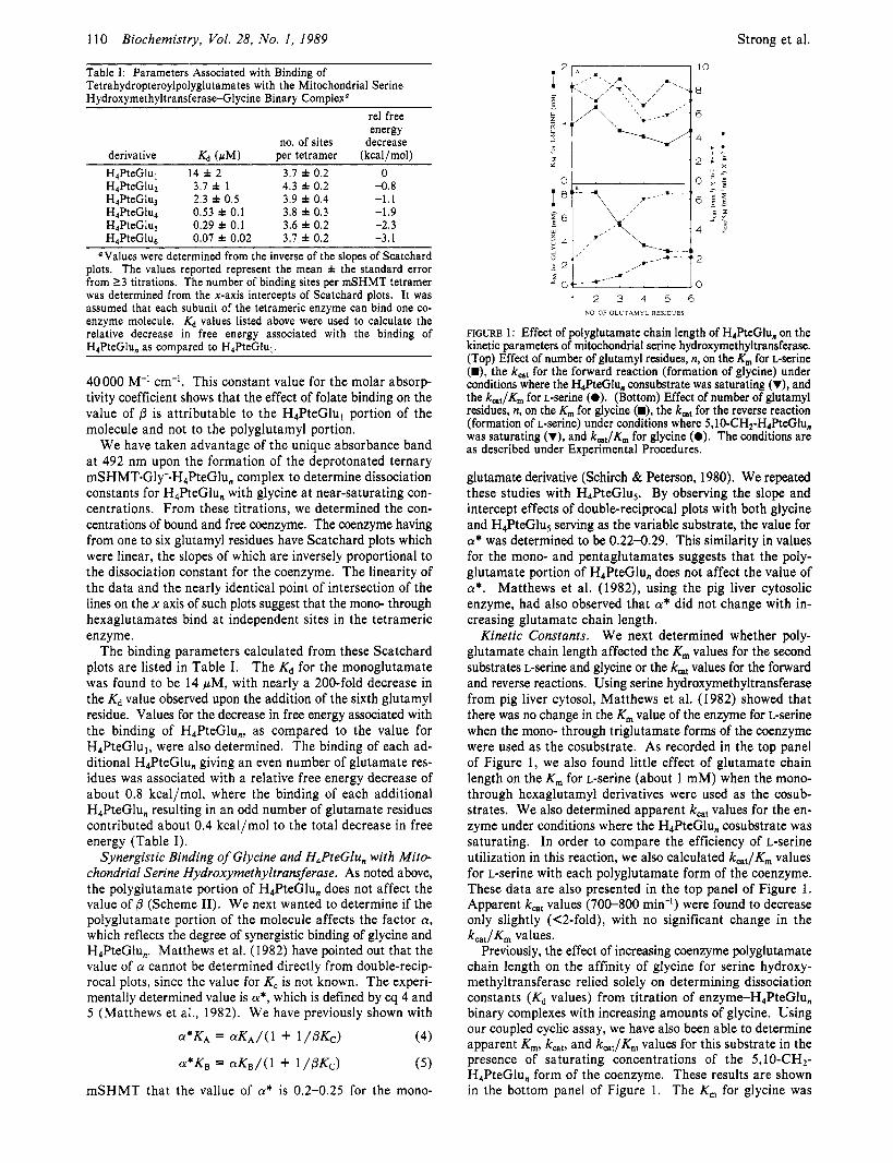
FIGURE 1: Effect of polyglutamate chain length of H,PteGlu, on the kinetic parameters of mitochondrial serine hydroxymethyltransferase. (Top) Effect of number of glutamyl residues, n, on the K , for L-serine (D), the k,, for the forward reaction (formation of glycine) under conditions where the H4ReGlu, consubstrate was saturating (V), and the k,,/Km for L-serine (0). (Bottom) Effect of number of glutamyl residues, n, on the Km for glycine (U), the k,, for the reverse reaction (formation of L-serine) under conditions where 5 , 10-CH2-H4PteGlu, was saturating (V), and k,,/Km for glycine (0). The conditions are as described under Experimental Procedures.
glutamate derivative (Schirch & Peterson, 1980). We repeated these studies with H,PteGluS. By observing the slope and intercept effects of double-reciprocal plots with both glycine and H4PteGlu5 serving as the variable substrate, the value for CY* was determined to be 0.22-0.29. This similarity in values for the mono- and pentaglutamates suggests that the poly- glutamate portion of H,PteGlu, does not affect the value of a*. Matthews et al. (1982), using the pig liver cytosolic enzyme, had also observed that CY* did not change with in- creasing glutamate chain length.
Kinetic Constants. We next determined whether poly- glutamate chain length affected the K , values for the second substrates L-serine and glycine or the k,, values for the forward and reverse reactions. Using serine hydroxymethyltransferase from pig liver cytosol, Matthews et al. (1982) showed that there was no change in the K , value of the enzyme for L-serine when the mono- through triglutamate forms of the coenzyme were used as the cosubstrate. As recorded in the top panel of Figure 1, we also found little effect of glutamate chain length on the K , for L-serine (about 1 mM) when the mono- through hexaglutamyl derivatives were used as the cosub- strates. We also determined apparent k,, values for the en- zyme under conditions where the H,PteGlu, cosubstrate was saturating. In order to compare the efficiency of L-serine utilization in this reaction, we also calculated k,,/K, values for L-serine with each polyglutamate form of the coenzyme. These data are also presented in the top panel of Figure 1. Apparent k,, values (700-800 m i d ) were found to decrease only slightly (<2-fold), with no significant change in the k,,,/K, values.
Previously, the effect of increasing coenzyme polyglutamate chain length on the affinity of glycine for serine hydroxy- methyltransferase relied solely on determining dissociation constants (Kd values) from titration of enzyme-H4PteGlu, binary complexes with increasing amounts of glycine. Using our coupled cyclic assay, we have also been able to determine apparent K,, kcat, and k,,/K, values for this substrate in the presence of saturating concentrations of the 5,10-CH2- H,PteGlu, form of the coenzyme. These results are shown in the bottom panel of Figure 1. The K , for glycine was
FiciuRe 2: Absorbance spectra associated with the titration of 2.5 pM mitochondrial serine hydroxymethyltransferase with H4PteGlu4 in the presence of 0 (right spectra) or 10 pM reduced cytosolic serine hydroxymethyltransferase and 50 mM glycine. The numbers on each curve represent the micromolar concentration of H4PteGlu4 added at each point in the titration. The conditions are as described under Experimental Procedures.
observed to decrease from about 8 mM with the mono- glutamate to about 3 mM with the penta- and hexaglutamates. Unlike what was observed with the k,, value of the L-serine reaction, the k,, of the glycine reaction increased greater than 3-fold, to a value nearly equal to that found for the L-serine reaction when the penta- or hexaglutamates were used as the cosubstrate. The decrease in the K,,, values and the increase in the k,, values are reflected in the nearly 9-fold increase in the enzyme efficiency with H4PteGlus and H4PteGlu6, as compared to the monoglutamate, for this reaction.
One explanation for the increased efficiency of the reverse reaction (glycine to L-serine), as compared to the forward reaction (L-serine to glycine), is that the equilibrium constant changes as the number of glutamyl residues on the coenzyme are increased. This would mean that H4PteGlu, would have to be more stable than 5,lO-CH2-H4PteGlu,. We therefore measured the equilibrium constant for the reaction catalyzed by mSHMT using the monoglutamate and pentaglutamate derivatives of the coenzyme. The Keq values determined from these experiments were 8.0 for the monoglutamate and 7.5 for the pentaglutamate, suggesting that the increased glutamate chain length has not altered the equilibrium constant.
With serine hydroxy- methyltransferase, the ability of the enzyme to form the quinonoid intermediate has repeatedly been used to determine dissociation constants for the folate substrate (Schirch & Mason, 1963; Schirch & Ropp, 1967; Matthews et al., 1982). Relying on this property of serine hydroxymethyltransferase, we have developed a general competitive binding method for the determination of dissociation constants of H4PteGlu, for other enzymes which bind this coenzyme.
In order to check the validity of this method in determining these Kd values, we have measured the dissociation constants for H4PteGlu, and H4PteGlus binding to rabbit liver cytosolic serine hydroxymethyltransferase in which the internal pyri-
Competitive Binding Method.
10
2 8
B - 6
X
% w
g 4 L
E 2
0 0 4 8 12 16
W@teGl%)totai (W
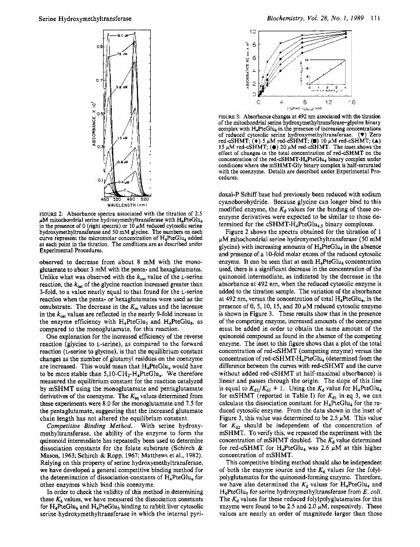
FIGURE 3: Absorbance changes at 492 nm associated with the titration of the mitochondrial serine hydroxymethyltransferase-glycine binary complex with H4PteGlu4 in the presence of increasing concentrations of reduced cytosolic serine hydroxymethyltransferase. (V) Zero red-cSHMT; (+) 5 p M red-cSHMT; (H) 10 pM red-cSHMT; (A) 15 pM red-cSHMT; (0) 20 pM red-cSHMT. The inset shows the effect of changes in the total concentration of red-cSHMT on the concentration of the red-cSHMT.H4PteGlu4 binary complex under conditions where the mSHMT-Gly binary complex is half-saturated with the coenzyme. Details are described under Experimental Pro- cedures.
doxal-P Schiff base had previously been reduced with sodium cyanoborohydride. Because glycine can longer bind to this modified enzyme, the Kd values for the binding of these co- enzyme derivatives were expected to be similar to those de- termined for the cSHMT.H4PteGlu4,, binary complexes.
Figure 2 shows the spectra obtained for the titration of 1 pM mitochondrial serine hydroxymethyltransferase (50 mM glycine) with increasing amounts of H4PteGlu4 in the absence and presence of a 10-fold molar excess of the reduced cytosolic enzyme. It can be seen that at each H4PteGlu4 concentration used, there is a significant decrease in the concentration of the quinonoid intermediate, as indicated by the decrease in the absorbance at 492 nm, when the reduced cytosolic enzyme is added to the titration sample. The variation of the absorbance at 492 nm, versus the concentration of total H4PteGlu4, in the presence of 0, 5, 10, 15, and 20 pM reduced cytosolic enzyme is shown in Figure 3. These results show that in the presence of the competing enzyme, increased amounts of the coenzyme must be added in order to obtain the same amount of the quinonoid compound as found in the absence of the competing enzyme. The inset to this figure shows that a plot of the total concentration of red-cSHMT (competing enzyme) versus the concentration of red-cSHMT.H4PteGlu4 (determined from the difference between the curves with red-cSHMT and the curve without added red-cSHMT at half-maximal absorbance) is linear and passes through the origin. The slope of this line is equal to Kdz/Kdl + 1. Using the Kd value for H4PteGlu4 for mSHMT (reported in Table I) for Kdl in eq 3, we can calculate the dissociation constant for H4PteGlu4 for the re- duced cytosolic enzyme. From the data shown in the inset of Figure 3, this value was determined to be 2.5 pM. This value for Kd2 should be independent of the concentration of mSHMT. To verify this, we repeated the experiment with the concentration of mSHMT doubled. The Kd value determined for red-cSHMT for H4PteGlu4 was 2.6 pM at this higher concentration of mSHMT.
This competitive binding method should also be independent of both the enzyme source and the Kd values for the folyl- polyglutamates for the quinonoid-forming enzyme. Therefore, we have also determined the Kd values for H4PteGlu4 and H4PteGluS for serine hydroxymethyltransferase from E . coli. The Kd values for these reduced folylpolyglutamates for this enzyme were found to be 2.5 and 2.0 pM, respectively. These values are nearly an order of magnitude larger than those
112 Biochemistry, Vol. 28, No. 1, 1989 Strong et al.
olism. This analysis of each enzyme in one-carbon metabolism with the coenzyme having up to seven glutamate residues is a difficult task. Part of the difficulty is that the higher chain polyglutamates bind very tightly to these enzymes, usually having Kd values less than 1 pM. This makes kinetic studies difficult except under saturation conditions. The interactions of the reduced pteroylpolyglutamates with several enzymes have been reported. In general, the polyglutamate forms of the coenzyme both bind more tightly than the monoglutamate form and reduce the value of kcat, but increase significantly the value of k,,,/K, (McGuire & Bertino, 1981). However, more subtle effects have also been observed. In several cases, the K,,, values of the non-folate substrates are also significantly altered in the presence of the polyglutamate forms of the coenzyme. Altered inhibition by other forms of the coenzyme has been observed, and in one case, the polyglutamate portion of the coenzyme induces a conformational change in the en- zyme (Matthews et al., 1982; Strong et al., 1987). In at least one case the addition of the glutamate residues changes the mechanism of the enzyme (Lu et al., 1984). Evidence has also been presented for channeling occurring between two folate- requiring enzymes with the polyglutamate forms of the co- enzyme (MacKenzie & Baugh, 1980).
The cell contains H,PteGlu, in primarily two cell com- partments, the cytosol and the mitochondria. To date, most of the data on the interaction of enzymes in one-carbon me- tabolism with the polyglutamate forms of the coenzyme have been restricted to enzymes found in the cytosol. In this study, we report the first extensive study of the interaction of the tetrahydropteroylpolyglutamates with the mitochondrial en- zymes serine hydroxymethyltransferase and dimethylglycine dehydrogenase. The study of mSHMT also offers the ad- vantage that an extensive study of the interaction of the re- duced pteroylpolyglutamates with the cytosolic form of this enzyme has been done (Matthews et al., 1982). The results in this paper permit us to determine if there are any different interactions of these two isozymes with the folylpolyglutamates. Unfortunately, an analysis of the distribution of the pteroyl- polyglutamate chain length in mammalian liver has only been determined with whole cells. In rabbit liver, the major de- rivatives are the penta- and hexaglutamate forms of the co- enzyme. Whether there are differences in the distribution of polyglutamates between the cytosol and mitochondria remains to be determined.
Our first experiments were to determine how the affinity of H,PteGlu, varied with glutamate chain length with mSHMT. Our results show that this enzyme has a broad specificity for the glutamate chain length, with each additional glutamate residue resulting in increased affinity (Table I). The results are similar to the data obtained with porcine liver cytosolic SHMT (Matthews et al., 1982). In each case, the penta- and hexaglutamates have Kd values in the 0.1 pM range. The major difference between the cytosolic and mitochondrial isoenzymes is in the affinity of the triglutamate. The cytosolic enzyme binds this form of the coenzyme 5 times more tightly than the mitochondrial enzyme. We have determined the Kd values of the polyglutamates for the rabbit cytosolic enzyme and find that they also show this difference in affinity for the triglutamate (unpublished results). Since the cell contains very little of the triglutamate, this observation will be important only if the levels of H4PteGlu3 are very different in the cytosol and mitochondria.
In view of the known mechanism for the formation of the quinonoid complex, Matthews et al. (1982) demonstrated that the increased affinity of the polyglutamates for cytosolic
Table 11: Dissociation Constants for Tetrahydropteroylpolyglutamates for Sodium Cyanoborohydride Reduced Cytosolic Serine Hydroxymethyltransferase and Mitochondrial Dimethylglycine Dehydrogenase As Determined by Competitive Binding with Mitochondrial Serine Hvdroxvmethvltransferase"
values were calculated by using eq 3 as described under Ex- perimental Procedures. Values for Kdl were taken from Table I. For studies with red-cSHMT, the mSHMT concentration was 2.5 r M , and red-cSHMT concentration was 5-20 pM. For studies with dimethyl- glycine dehydrogenase, the mSHMT concentration was 1 pM, and the dimethylglycine dehydrogenase concentration was 1.45 r M . Values from Wittwer and Wagner C. (1981). cValues determined by using 2 pM E . coli serine hydroxymethyltransferase as the quinonoid-forming enzyme and 5-15 pM red-cSHMT as the competing enzyme. K d , values for H , P ~ ~ G ~ U , ~ for the E. coli enzyme, in the absence of E*, were determined as described for mSHMT under Experimental Proce- dures.
obtained for the mitochondrial enzyme. However, it should be pointed out that these Kd values may not reflect the values for the natural coenzymes, since it has been shown that the folylpolyglutamates of E . coli contain both a and y linkages (Ferone et al., 1986). When the above experiment, using reduced cytosolic serine hydroxymethyltransferase as the competing enzyme, was repeated with the enzyme purified from E . coli, a value of 2.6 pM was obtained for the Kd for red-cSHMT for H4PteGlu4 and a value of 1.2 pM for the Kd value for H,PteGlu, (Table 11).
A number of years ago, extracts from rat liver mitochondria were shown to contain two proteins, which when taken through several purification steps still contained bound H,PteGlu, (Zamierowski & Wagner, 1977). More recently, these pro- teins have been identified as sarcosine dehydrogenase and dimethylglycine dehydrogenase (Wittwer & Wagner, 198 1 ), which like serine hydroxymethyltransferase are involved in glycine metabolism. Dimethylglycine dehydrogenase was found by these authors to bind H,PteGlu, and H,PteGlu, with Kd values of 0.4 and 0.2 pM, respectively. It was therefore of interest, using the competitive binding method described above with mitochondrial serine hydroxymethyltransferase, to determine the Kd values for the tetrahydropteroylpoly- glutamates, both as a control for the method and to obtain a complete list of how Kd varies with the number of glutamyl residues for this mitochondrial enzyme. It was only possible to obtain absolute values for H4PteGlu3 through H,PteGlu, (0.76-0.1 1 pM, respectively), but Kd values for H4PteGlul and H4PteGlu2 were estimated from the data to be in the same range as the coenzyme with higher numbers of glutamates (Kd values less than 1 pM). Table I1 lists the calculated and reported dissociation constants both for reduced cytosolic serine hydroxymethyltransferase and for dimethylglycine de- hydrogenase.
DISCUSSION The physiological function of the polyglutamate chain on
H,PteGlu, remains largely unsolved. It appears that to solve this problem will require the determination of the effect of additional glutamate residues, to the monoglutamate form of the coenzyme, on most of the enzymes in one-carbon metab-
Serine Hydroxymethyltransferase
SHMT was seen as an effect only on KB (Scheme 11). We have found the same to be true for mSHMT. Previous studies have shown that the effect of H,PteGlu, binding in shifting the equilibrium (change in K,-) toward the unprotonated ternary complex resides primarily in the reduced pteridine ring portion of the molecule (Jones et al., 1978).
We next addressed the question of how increased glutamate chain length affected the synergistic binding with glycine, which we had previously demonstrated with the mono- glutamate (Schirch & Peterson, 1980). Our results with the pentaglutamate show that the value of a* (eq 4 and 5) is about 0.25, giving a 4-fold synergism in binding between the co- enzyme and glycine. This is essentially the same value we found for the monoglutamate, suggesting that addition of the polyglutamate chain has not altered the synergism of binding between folate and glycine. The same observation was found for the cytosolic enzyme with H4PteGlu,, but the value was changed markedly when the competitive inhibitor 5-CH3- H,PteGlu, was used (Matthews et al., 1982). For this com- petitive feedback inhibitor, the degree of synergism with glycine increased markedly with the addition of the poly- glutamate side chain. Matthews et al. (1982) have proposed that this increased sensitivity of binding of 5-CH3-H,PteGlu, to glycine concentration may be an important regulatory mechanism in the cell.
The effect of polyglutamate chain length on the kinetic parameters for both the forward and reverse reactions cata- lyzed by mSHMT was also determined. We found that in the direction of serine to glycine, increasing the number of glu- tamate residues from 1 to 6 had less than a 2-fold effect on the K,, kat, and k,,/K, values for serine. Studies with the porcine cytosolic enzyme using the mono- through tri- glutamates also showed no effect on the K, for serine, but they did observe an almost 2-fold decrease in k,, (Matthews et al., 1982). However, when we determined the effect of glutamate chain length on the kinetic parameters in the glycine to serine direction, we found that in going from the monoglutamate to the hexaglutamate there was a 3-fold decrease in K , and a 3-fold increase in k,,,. This results in a 9-fold increase in
The 9-fold increase in the k,,/K, for glycine reflects only the effect of the amino acid substrate, since in each experiment the 5,l 0-CH,-H,PteGlu, was present at saturating concen- trations. We have not been able to determine K , values for 5,10-CH2-H,PteGlu,. However, if these K , values vary as much as the Kd values for H4PteGlu, (Table I), then the k,,/K, values with respect to the 5,10-CH2-H,PteGlu, sub- strate would increase 600-fold in going from the mono- glutamate to the hexaglutamate.
We have shown that the Kq for the reaction is not affected by increasing the number of glutamyl residues on the co- enzyme. Therefore, the 3-fold increase in k,, in the glycine to serine reaction and a 2-fold decrease in k,, in the serine to glycine reaction must be accommodated by a change in the values of K , for the substrates. The cytosolic enzyme has been shown to proceed by a sequential random mechanism (Schirch et al., 1977). We know that the mitochondrial enzyme is also sequential, but we have not determined if it is random or ordered. The K , values we determined for glycine and serine do not account for the changes in k,, for the forward and reverse reactions. This suggests that the ratio of K , values for H,PteGlu, and 5,10-CH2-H4PteGlu, must account for these k,,, effects. Using the Haldane relationship for a two- substrate/two-product random equilibrium mechanism permits us to calculate the ratio of K, values for 5,10-CH2-
kcatl Km.
Biochemistry, Vol. 28, No. 1, 1989 113
H4PteGlu,/H4PteGlu,, as the number of glutamates increases from 1 to 6 (Segal, 1975b). When the values for k,, forward and k,,, reverse and the K , values for glycine and serine are used, the ratio of K, values for the folate substrates varies from 0.2 for the monoglutamate to 2 for the hexaglutamate. We currently have no method for verifying this change in ratio of K , values, but it offers yet another possibility of the effect of the polyglutamate portion of the coenzyme on an enzyme reaction. To our knowledge, K,,, values for both the substrate and product forms of a folate coenzyme have not been de- termined as a function of glutamate chain length for any enzyme in one-carbon metabolism.
The intense absorption at 492 nm of the SHMT. H,PteGlu,.Gly complex permits a simple and rapid method for determining the Kd of other proteins which bind H,PteGlu,. In this paper, we have tested this method and shown that we can determine Kd values which are independent of the amount of SHMT and the source of SHMT. The method does have one limitation. You cannot accurately determine the Kd of the competing enzyme if its Kd is more than about 8-fold lower than the & for SHMT. We found that with dimethylglycine dehydrogenase, we could not accurately determine the Kd values for the monoglutamate or the diglutamate by this method, because the dehydrogenase binds these two folates with a Kd less than 1 FM compared to SHMT which has Kd values well above 1 pM. These results are of interest because in the mitochondria these two enzymes must compete for the coenzyme. The data show that when the number of glutamate residues on the coenzyme is greater than 3 the two enzymes have similar affinities for the coenzyme. However, with the mono- and diglutamate forms of the coenzyme, the de- hydrogenase must be almost completely saturated with co- enzyme before mSHMT will bind the coenzyme. There is reason to believe that the concentration of H,PteGlu, in the mitochondria may be similar to the concentrations of active sites on enzymes which utilize this coenzyme. Under these conditions, it is important for all enzymes to be able to compete for the low levels of free H,PteGlu,.
This competitive binding approach to determining disso- ciation constants for folylpolyglutamates can also be used for proteins which bind either 5-methyl- or 5-formyl-H4PteGlu,, since these two compounds also form the quinonoid complex with SHMT (Schirch & Ropp, 1967).
REFERENCES Baugh, C. M., & Krumdieck, C. L. (197 1) Ann. N.Y. Acad.
Brown, J. P., Davidson, G. E., & Scott, J. M. (1 974) Biochim.
Cleland, W. W. (1967) Adv. Enzymol. Relat. Areas Mol. Biol.
Cook, R. J., & Blair, J. A. (1979) Biochem. J . 178, 651-659. Cook, R. J., & Wagner, C. (1986) Methods Enzymol. 122,
Cossins, E. A. (1984) in Folates and Pterins (Blakely, R. L., & Benkovic, S . J., Eds.) Vol. I, pp 1-59, Wiley, New York.
Cybulski, R. L., & Fisher, R. R. (1976) Biochemistry 15,
Davis, L., & Metzler, D. E. (1972) Enzymes (3rd Ed.) 7,
Ferone, R., Hanlon, M. H., Singer, S . C., & Hunt, D. F.
Registry No.
Sci. 186, 7-28.
Biophys. Acta 343, 78-88.
29, 1.
255-260.
3 183-3 187.
33-74.
(1986) J . Biol. Chem. 261, 16356-16362.
114 Biochemistry 1989, 28, 114-123
Gavilanes, F., Peterson, D., & Schirch, L. (1982) J. Biol.
Harruff, R. C., & Jenkins, W. T. (1976) Arch. Biochem.
Jones, C. W., Hynes, J . B., & Priest, D. G. (1978) Biochim. Biophys. Acta 524, 55-59.
Krumdieck, C. L., & Eto, I . (1986) in Chemistry and Biology of Pteridines (Cooper, B. A,, & Whitehead, V. M., Eds.) pp 447-466, de Gruyter, New York and Berlin.
Lu, Y.-Z., Aiello, P. D., & Matthews, R. G. (1984) Bio- chemistry 23, 6870-6876.
MacKenzie, R. E., & Baugh, C. M. (1980) Biochim. Biophys. Acta 611, 187-195.
Matthews, R. G., Lu, Y.-Z., Green, J. M., & MacKenzie, R. E. (1985) in Proceedings of the Second Workshop on Folyl and Antifolyl Polyglutamates (Goldman, D. I., Ed.) pp 65-75, Praeger, New York.
Matthews, R. G., Ross, J., Baugh, C. M., Cook, J. D., & Davis, L. (1982) Biochemistry 21, 1230-1238.
McGuire, J. J., & Bertino, J. R. (1981) Mol. Cell. Biochem.
McGuire, J. J., & Coward, J. K. (1984) in Folates and Pterins (Blakely, R. L., & Benkovic, S. J., Us.) Vol. I, pp 135-190, Wiley, New York.
Priest, D. G., & Doig, M. T. (1986) Methods Enzymol. 122,
Scatchard, G . (1949) Ann. N.Y. Acad. Sci. 51, 660. Schirch, L. (1978) Arch. Biochem. Biophys. 189, 283-290.
Chem. 257, 11431-1 1436.
Biophys. 176, 206-2 13.
38, 19-48.
31 3-319.
Human T-Lymphoblast Deoxycytidine
Schirch, L., & Mason, M. (1962) J. Biol. Chem. 237,
Schirch, L. G., & Mason, M. (1963) J. Biol. Chem. 238,
Schirch, L., & Ropp, M. (1967) Biochemistry 6, 253-257. Schirch, L., & Peterson, D. (1980) J. Biol. Chem. 255,
Schirch, L., Tatum, C. M., & Benkovic,,S. J. (1977) Bio-
Schirch, V., Hopkins, S., Villar, E., & Angelaccio, S. (1985)
Scott, J. M., & Weir, D. G. (1976) Clin. Haematol. 5,
Segal, I. H. (1975a) in Enzyme Kinetics, pp 273-329, Wiley,
Segal, I. H. (1975b) in Enzyme Kinetics, pp 643-646, Wiley,
Steenkamp, D. J., & Husain, M. (1982) Biochem. J. 203,
Strong, W., Joshi, G., Lura, R., Muthukumaraswamy, N., & Schirch, V. (1987) J. Biol. Chem. 262, 12519-12525.
Villar, E., Schuster, B., Peterson, D., & Schirch, V. (1985) J. Biol. Chem. 260, 2245-2252.
Wittwer, A. J., & Wagner, C . (1981) J. Biol. Chem. 256,
Zamierowski, M. M., & Wagner, C. (1977) J. Biol. Chem.
2578-258 1 .
1032-1037.
780 1-7806.
chemistry 16, 410-419.
J. Bacteriol. 163, 1-7.
547-568.
New York.
New York.
707-7 1 5 .
4 102-4 108.
252, 933-938.
Kinase: Purification and Properties? Nabanita S. Datta, Donna S. Shewach, Mary C . Hurley, Beverly S. Mitchell, and Irving H. Fox*
Human Purine Research Center, Departments of Internal Medicine and Biological Chemistry, Clinical Research Center, University Hospital, Ann Arbor, Michigan 48109-01 08
Received June 1 . 1988; Revised Manuscript Received August 12, 1988
ABSTRACT: Previous observations present tremendous variations in the properties of deoxycytidine kinase. To clarify the properties and physiologic role of deoxycytidine kinase, we have undertaken its purification. Deoxycytidine kinase was purified from cultured human T-lymphoblasts (MOLT-4) to 90% purity with an estimated specific activity of 8 pmol min-' (mg of protein)-'. The purification procedure included ammonium sulfate precipitation, Superose- 12 HPLC gel filtration chromatography, DE-52 ion-exchange chromatography, AMP-Sepharose 4B affinity chromatography, and dCTP-Sepharose-4B affinity chro- matography. Deoxyguanosine, deoxyadenosine, and cytidine phosphorylating activities copurified with deoxycytidine kinase to final specific activities of 7.2, 13.5, and 4 pmol m i d (mg of protein)-', respectively. The enzyme is very unstable at low protein concentration and is stabilized by storage at -85 O C with 1 mg/mL bovine serum albumin, 20% glycerol (v/v), 200 m M potassium chloride, and 25 mM dithiothreitol. The molecular weight was 60000, and the Stokes radius was 32 A by gel filtration chromatography. The subunit molecular weight was 30 500. This enzyme had apparent K , values of 1.5, 430, 500, 450, and 40 p M for deoxycytidine, deoxyguanosine, deoxyadenosine, cytidine, and cytosine arabinoside, respectively. The pH optimum ranged from 6.5 to 9.0. Mg2+ and Mn2+ were the preferred divalent cations. ATP, GTP, dGTP, ITP, dITP, TTP, and XTP were substrates for the enzymes. Our study indicates that deoxycytidine kinase is a dimer with two subunits and has phosphorylating activity for deoxyguanosine, deoxyadenosine, cytidine, and cytosine arabinoside. This highly purified enzyme will facilitate the study of its regulation and phosphorylation of anticancer or antiviral nucleoside analogues.
x e phosphorylation of deoxyribonucleosides to deoxyribo- nucleotides is a critical step in the pathogenesis of inherited
immunodeficiency diseases and in the activation of specific anticancer and antiviral drugs (Carson et al., 1977, 1979; Cohen. 1966: Cohen et al.. 1978a.b: Coleman et al.. 1978:
'This work was supported by U S . Public Health Service Grants 1 ROI GM 32837, 5 MOI RR 00042, and 2 ROI CA 34085.
* Correspondence should be addressed to this author at the Clinical Research Center, Room A7 1 19, University of Michigan Hospital, 1500
Gudaset 1978; ~ ~ j ~ ~ , 1982; Mitchell et 1979; ullmai et al., 1978; Wortmann et al., 1979). Our previous studies have defined three distinct deoxyribonucleoside phosphory-
E. Medical Center Dr., Ann Arbor, MI 48109-0168. lating activities in human tissue: adenosine-deoxyadenosine
0006-2960/89/0428-0114$01 S O / O 0 1989 American Chemical Society