International Journal of Dentistry and Oral Health Volume 6 Issue 4, April 2020
Citation: Digka et al. (2020), Regenerative procedures in mature teeth: a new era in endodontics? A systematic review. Int J Dent & Oral Heal. 6:4
International Journal of Dentistry and Oral Health
Review Article ISSN 2471-657X
1
1DDS, PhD, Department of Endodontology, Dental School of Aristotle University of Thessaloniki, Thessaloniki, Greece, ORCHID ID: 0000-0002-3167-75542Postgraduate student, Department of Endodontology, Dental School of Aristotle University of Thessaloniki, Thessaloniki, Greece, ORCHID ID: 0000-0001-8362-69703Professor, Department of Endodontology, Dental School, Aristotle University of Thessaloniki, Thessaloniki, Greece
Corresponding author: Digka AnnaDDS, PhD, Department of Endodontology, Dental School of Aristotle University of Thessaloniki, Thessaloniki, Greece, ORCHID ID: 0000-0002-3167-7554. Email: [email protected]
Citation: Digka et al. (2020), Regenerative procedures in mature teeth: a new era in endodontics? A systematic review. Int J Dent & Oral Heal. 6:4
Received: 20 March, 2020 Accepted: 04 April, 2020 Published: 24 April, 2020
Objective: This review investigated the potential of application of regenerative endodontic procedures (REPs) in mature necrotic permanent teeth with apical pathology, as an alternative to root canal treatment (RCT), in terms of elimination of signs and symptoms and healing of apical lesions.
Material and methods: Electronic searches were performed in the MEDLINE, Web of Science, Scopus, and Cochrane library databases. The retrieved studies were evaluated according to pre-established inclusion and exclusion criteria.
Results: Among 344 retrieved studies, 10 fit the inclusion criteria and were critically appraised.
Conclusions: Although only few clinical reports could be retrieved, their findings were promising and indicated that REP can to some extent replace RCT, since elimination of clinical signs and symptoms and apical lesion resolution could be accomplished with REPs. Re-establishment of tooth sensation may also be possible. Further research is necessary to establish reliable protocols for REPs in mature teeth.
Regenerative procedures in mature teeth: a new era in endodontics? A systematic review Digka Anna1*, Sakka Dimitra2, Lyroudia Kleoniki3
Introduction The main goal of endodontics remains the preservation of natural dentition, ensuring function and aesthetics, by curing or prevent-ing periapical pathology. This is accomplished by conventional root canal treatment (RCT), which involves cleaning and shaping of the root canal system and obturation of these canals with filling mate-rials. Although orthograde RCT is a predictable treatment with very high success rates (1-3), it requires replacement of the permanently lost pulpal tissue with biocompatible, non-vital, foreign materials.
Tissue engineering, a new evolving domain of bioscience, is a multi-dimensional scientific area, which combines biology, phar-macology and engineering, and aims to restore, maintain, and re-generate injured or lost tissues (4). It provides a new perception and perspective in many dental specialties, including endodontics.
Regenerative endodontics procedures (REPs) have been defined as “biologically based procedures designed to replace damaged struc-tures, including dentin and root structures, as well as cells of the pulp–dentin complex” (5). During the last two decades, REPs have been applied successfully as treatment for immature permanent necrotic teeth with arrested root development, replacing the conventional apexification treatment with calcium hydroxide (Ca(OH)2) or mineral
Abstract:
International Journal of Dentistry and Oral Health Volume 6 Issue 4, April 2020
Citation: Digka et al. (2020), Regenerative procedures in mature teeth: a new era in endodontics? A systematic review. Int J Dent & Oral Heal. 6:4
trioxide aggregate (MTA). More than 400 published reports show very promising results, such as attenuation of clinical signs and symptoms, healing of periapical lesions, continuation and completion of root development, and even re-establishment of tooth sensation (6-8). To date, application of REPs has been limited mainly to imma-ture teeth. Nevertheless, there are reports with surprising and outstanding results of utilizing these procedures in mature teeth. Application of REPs in mature teeth holds several appre-ciable advantages, such as re-establishment of the immune sys-tem and proprioception in the tooth, properties that are other-wise irreversibly lost when conventional RCT is implemented.
Accordingly, the aim of this review was to investigate the poten-tial of REP application in mature necrotic permanent teeth with periapical pathology, as an alternative to RCT, in terms elimina-tion of signs and symptoms and healing of periapical lesions.
Materials and Methods: This systematic review was conducted in accordance with the Pre-ferred Reporting Items for Systematic Reviews and Meta-Analyses Statement (PRISMA) (9). In terms of the PICO question, we asked “Do REPs, which are mainly applied in immature teeth (P), have potential
for utilization in mature permanent necrotic teeth with periradicular pathology (I), as an alternative to conventional RCT (C), in terms of elimination of signs and symptoms and resolution of apical lesions (O)?The inclusion criteria for this systematic review were studies published in the English language, that were related to human mature teeth with closed apices, necrotic pulp, and apical periodontitis that underwent REPs according to the guidelines of the American Association of En-dodontists (AAE) (10) and European Society of Endodontics (ESE) (11). Studies in other languages, animal studies, studies of immature teeth, apicoectomized teeth, or teeth without pulp necrosis and periapical lesions, and studies without regenerative protocols were excluded.
Literature search Electronic searches were performed in the MEDLINE (via the PubMed search engine), Web of Science, Scopus, and the Cochrane library databases. The following key words were combined to op-timize the search strategy: “mature permanent tooth” or “mature permanent teeth” or “closed apex” or “closed apices” and “pulp necrosis” or “necrotic teeth” or “apical periodontitis” or “apical lesion” or “periapical” and “regenerat*” or “revital*” or “revas-culari*” (Table 1). Additionally, endodontic textbooks and the refer-ences of the studies that emerged from the electronic search were searched manually. The last search was performed on 8 October 2019.
OR “mature permanent teeth” [all] OR “necrotic teeth” [all] OR “revital*” ” [all]
OR “closed apex” [all] OR “apical periodontitis” [all] OR “revasculari*”[all]
OR “closed apices” [all] OR “apical lesion” [all]
OR “periapical” [all]
*: means any suffix. Table 1: Search Strategy Pubmed
Study selection Two authors (AD and DS) screened the titles and abstracts of all articles that emerged by the electronic research and excluded those that did not meet the inclusion criteria. The remaining articles’ full-text was evaluat-ed and disagreements were resolved by a third author (KL) to arrive at a final decision regarding inclusion or exclusion of an article in the study.
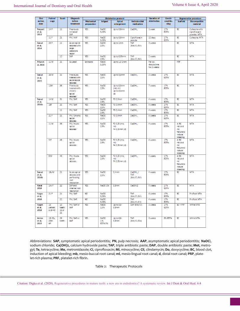
Data extraction All included studies were carefully read and data collection sheets were used to collect the following extracted information: first author, publication date, patient’s age, tooth, diagnosis before treatment, pretreatment radiographic findings, disinfection pro-tocol (mechanical preparation, irrigant, apical enlargement, and antimicrobial medication), duration of disinfection, dentin con-
ditioning, regenerative procedure (scaffold and biocompatible material), clinical and radiographic treatment outcomes (clinical signs and symptoms, apical lesion resolution, response to cold and time of response, response to electric stimulation and time of re-sponse, root canal thinning, and time of last control (Tables 2 and 3).The names of the authors of included studies were kept from the re-viewers (AD and SD) during data extraction to eliminate risk of bias. .
International Journal of Dentistry and Oral Health Volume 6 Issue 4, April 2020
Citation: Digka et al. (2020), Regenerative procedures in mature teeth: a new era in endodontics? A systematic review. Int J Dent & Oral Heal. 6:4
International Journal of Dentistry and Oral Health Volume 6 Issue 4, April 2020
Citation: Digka et al. (2020), Regenerative procedures in mature teeth: a new era in endodontics? A systematic review. Int J Dent & Oral Heal. 6:4
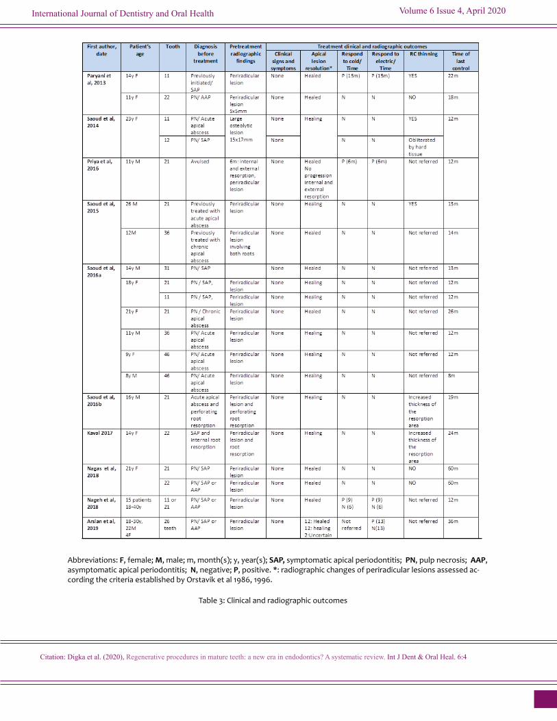
Abbreviations: F, female; M, male; m, month(s); y, year(s); SAP, symptomatic apical periodontitis; PN, pulp necrosis; AAP, asymptomatic apical periodontitis; N, negative; P, positive. *: radiographic changes of periradicular lesions assessed ac-cording the criteria established by Orstavik et al 1986, 1996.
Table 3: Clinical and radiographic outcomes
International Journal of Dentistry and Oral Health Volume 6 Issue 4, April 2020
Citation: Digka et al. (2020), Regenerative procedures in mature teeth: a new era in endodontics? A systematic review. Int J Dent & Oral Heal. 6:4
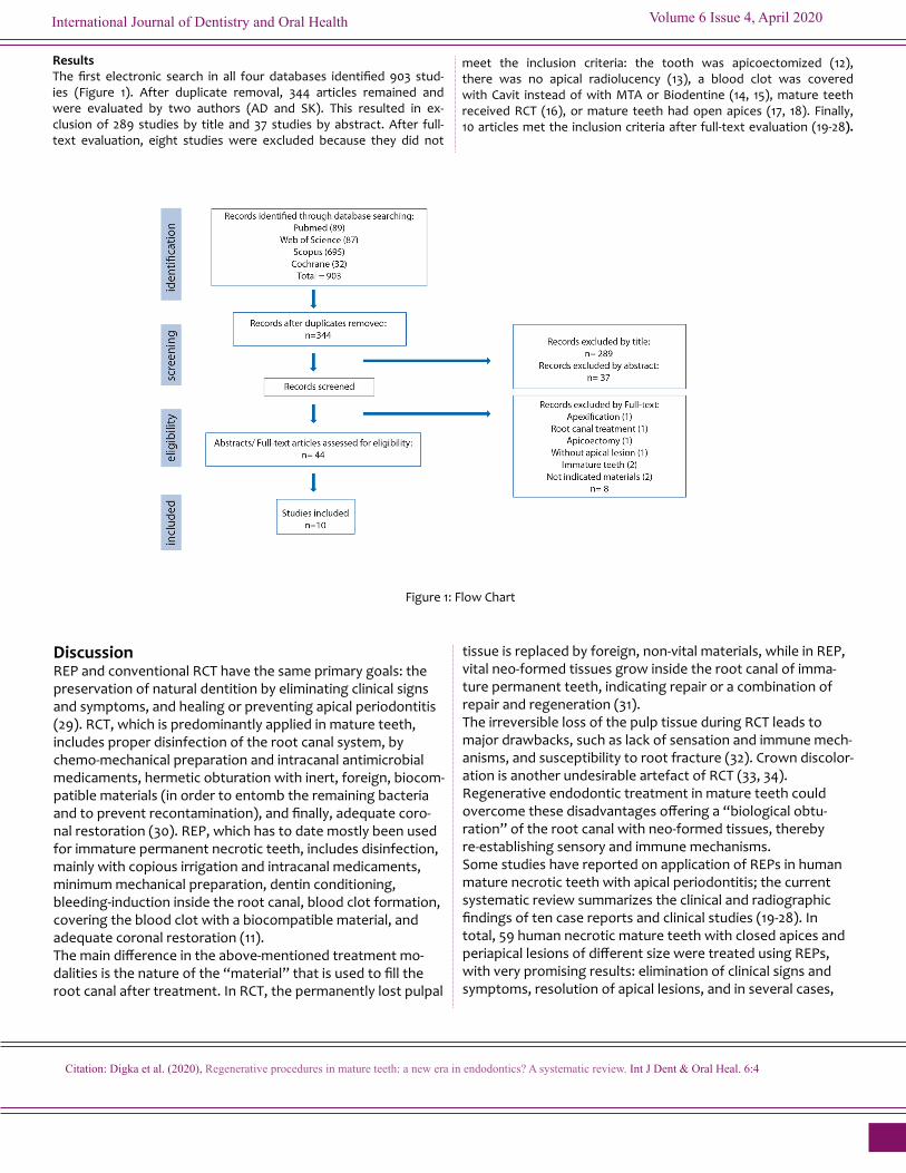
Results The first electronic search in all four databases identified 903 stud-ies (Figure 1). After duplicate removal, 344 articles remained and were evaluated by two authors (AD and SK). This resulted in ex-clusion of 289 studies by title and 37 studies by abstract. After full-text evaluation, eight studies were excluded because they did not
meet the inclusion criteria: the tooth was apicoectomized (12), there was no apical radiolucency (13), a blood clot was covered with Cavit instead of with MTA or Biodentine (14, 15), mature teeth received RCT (16), or mature teeth had open apices (17, 18). Finally, 10 articles met the inclusion criteria after full-text evaluation (19-28).
Figure 1: Flow Chart
Discussion REP and conventional RCT have the same primary goals: the preservation of natural dentition by eliminating clinical signs and symptoms, and healing or preventing apical periodontitis (29). RCT, which is predominantly applied in mature teeth, includes proper disinfection of the root canal system, by chemo-mechanical preparation and intracanal antimicrobial medicaments, hermetic obturation with inert, foreign, biocom-patible materials (in order to entomb the remaining bacteria and to prevent recontamination), and finally, adequate coro-nal restoration (30). REP, which has to date mostly been used for immature permanent necrotic teeth, includes disinfection, mainly with copious irrigation and intracanal medicaments, minimum mechanical preparation, dentin conditioning, bleeding-induction inside the root canal, blood clot formation, covering the blood clot with a biocompatible material, and adequate coronal restoration (11). The main difference in the above-mentioned treatment mo-dalities is the nature of the “material” that is used to fill the root canal after treatment. In RCT, the permanently lost pulpal
tissue is replaced by foreign, non-vital materials, while in REP, vital neo-formed tissues grow inside the root canal of imma-ture permanent teeth, indicating repair or a combination of repair and regeneration (31).The irreversible loss of the pulp tissue during RCT leads to major drawbacks, such as lack of sensation and immune mech-anisms, and susceptibility to root fracture (32). Crown discolor-ation is another undesirable artefact of RCT (33, 34).Regenerative endodontic treatment in mature teeth could overcome these disadvantages offering a “biological obtu-ration” of the root canal with neo-formed tissues, thereby re-establishing sensory and immune mechanisms.Some studies have reported on application of REPs in human mature necrotic teeth with apical periodontitis; the current systematic review summarizes the clinical and radiographic findings of ten case reports and clinical studies (19-28). In total, 59 human necrotic mature teeth with closed apices and periapical lesions of different size were treated using REPs, with very promising results: elimination of clinical signs and symptoms, resolution of apical lesions, and in several cases,
International Journal of Dentistry and Oral Health Volume 6 Issue 4, April 2020
Citation: Digka et al. (2020), Regenerative procedures in mature teeth: a new era in endodontics? A systematic review. Int J Dent & Oral Heal. 6:4
re-establishment of tooth sensation.The applied regenerative protocols that were followed were according to the guidelines of AAE (10) and ESE (11), with slight modifications (19-28) (Table 2). The main modification was the mechanical preparation and debridement of the root canal walls. In immature teeth, because of the very thin and fragile dentin root walls, mechanical preparation is restricted to the minimum if at all and disinfection is accomplished with copious irrigation and intracanal medicaments (10, 11). In most cases included in this review, the teeth were mechanically prepared and the apical foramen was enlarged from 2 to 6 sizes larger than the master apical file (MAF) (see below). As an irrigant, sodium hypochlorite solution in concentrations ranging from 1% to 5.25% was used in all cases (19-28). As intracanal medica-ment, calcium hydroxide paste alone was applied in 12 teeth (19 21, 22) and antibiotic paste alone (triple, double, or modi-fied) in 46 (19, 20, 25-28). In one case, calcium hydroxide was used as the first intracanal medicament, and triple antibiotic paste as a second medicament, with a 2-week interval (23). As a scaffold, amongst the 59 teeth, bleeding-induction alone was evoked in 57 (19-13, 26), bleeding-induction along with PRF (platelet-rich fibrin) in one (27), and only PRP (platelet-rich plasma) in one (25). After bleeding-induction, the blood clot was covered with MTA, as a biocompatible material, in all cases included (19-28). All included teeth were free of clinical signs and symptoms, without pain, not sensitive to palpation or percussion, without palatal, buccal, lingual swelling or a sinus tract, during the fol-low-up visit after the REP (19-28). One of the inclusion criteria was the existence of periapical radiolucency. The size of the apical lesion before treatment ranged from 2 to 5 according to periapical index scoring system (19-28, 35). A very large osteo-lytic lesion, 15 × 17 mm, involving teeth 11 and 12, was present in one case (20), while in another case involving a mandibular first molar, the periapical lesion involved both mesial and distal roots (21). The radiographic findings of the changes in periapi-cal lesions were assessed according to the criteria established by Orstavik (36). Amongst the 59 included teeth, 35 were healed (without clinical signs and symptoms, and with resolu-tion of the periapical lesion, with normal periradicular tissues) (19-22, 25-28), 22 were still in the process of healing (without clinical signs and symptoms, and with a reduced periapical le-sion) (20-24, 28). Two cases were characterized as “uncertain” due to the difficulty of appraising the absence, reduction, or enlargement of the lesion (28). Interestingly, the large osteo-lytic lesion (15×17 mm) was in the process of healing at a 12-m control (20) and the apical lesion of a 36 tooth, which involved both roots, showed healing at the 14-m control (21) (Table 3).Of particular interest was the re-establishment of tooth sensa-tion in 23 of 59 mature necrotic teeth that were treated with REPs (19, 25, 27, 28). Eleven of the teeth responded to both electric and cold stimuli, whereas 23 responded only to electric
stimuli. The time range for restored reaction to stimuli was from 6 to 15 months (Table 3).Application of REP in mature teeth features three main chal-lenges and limitations, as compared to its application in imma-ture teeth: the source/type, amount/number and age of stem cells, the narrower apical pathway for stem cells’ migration, and the complex root canal anatomy that hampers appropri-ate disinfection.Tissue regeneration is based on three key elements: stem cells, growth factors, and scaffolds (4). In immature necrotic teeth, the existence of the apical papilla constitutes the main source of stem cells for “dental pulp” regeneration (37, 38). In mature teeth, there is no evidence of the existence of apical papilla. Stem cells from the periapical tissues that are available for regeneration in mature teeth are periodontal ligament stem cells (PDLSCs) (39), bone marrow stem cells (BMSCs) (40), stem cells from inflamed periapical tissues (iPAPCs) (41), and possibly, a few surviving dental pulp stem cells (DPSCs) (42). BMSCs possess dentinogenic, osteogenic, angiogenic, and neurogenic differentiation potential (43-45). PDLSCs have the ability for fibroblastic, osteoblastic, cementoblastic, adipo-genic, chondrogenic, neurogenic, and angiogenic differentia-tion (39, 46-48). IPAPCs, mesenchymal stem/progenitor cells that are present in periapical inflamed tissues associated with endodontic infections, exhibit osteogenic capabilities (41). DPSCs are cells that possess odontoblastic, osteoblastic, neu-rogenic, angiogenic, chondrogenic, and osteogenic differen-tiation capabilities (49-53). Theoretically, the periapical area’s stem cells have the potential for regenerating dental pulp through neurovascular system reconstruction, mineralized tissue deposition, and by giving rise to new odontoblasts.Another crucial element in tissue regeneration is the migration of stem cells inside the empty root canal, after chemomechan-ical debridement. Cell homing by intracanal bleeding evoked by the instrumentation of periapical tissues is the most simplis-tic endodontic approach in clinical practice. Chrepa et al. (54), in a clinical human study, investigated whether the induction of apical bleeding in mature teeth with closed apices and peri-radicular lesions can elicit an influx of mesenchymal stem cells (MSCs) inside the root canals. They concluded that the cells delivered by bleeding-induction inside the root canals of ma-ture teeth expressed MSC markers and demonstrated intense mineralizing differentiation potential, and thus possess MSC properties. Additionally, the isolated cells expressed several gene transcripts that are essential for the regulation of migra-tion, proliferation, and differentiation of dental stem cells. The above results indicate that bleeding-induction in mature teeth can play the same role to ensure an influx of stem cells as in immature teeth. In the studies that were included in this review, bleeding-in-duction and usage of blood clot as a scaffold was applied in most cases (58/59) (19-24, 26-28). The apical lesions of those
International Journal of Dentistry and Oral Health Volume 6 Issue 4, April 2020
Citation: Digka et al. (2020), Regenerative procedures in mature teeth: a new era in endodontics? A systematic review. Int J Dent & Oral Heal. 6:4
teeth were either healed (35 teeth) or were in the process of healing (22 teeth). Furthermore, 22 of these teeth responded positively to electric or cold stimuli (Table 3). These clinical and radiographic findings indicated that periapical bleeding-prov-ocation in mature necrotic teeth with closed apices and apical lesions led to adequate tissue regeneration inflow of stem cells inside the root canals. The age of the patient, and subsequently, the age of the stem cells, is another parameter that plays a significant role in tissue regeneration. MSCs, as somatic stem cells, are subjected to cellular senescence (55, 56). Aging leads firstly to a decrease in the amount of stem/progenitor cells (57) and secondly to the decrease in their stemness and the loss of their capability to proliferate, differentiate, and support tissue regeneration (58). Cellular senescence should be considered in case selec-tion of the treatment protocol, RCT or REP, in older patients. Nevertheless, Chrepa et al. (54) found no correlation between age and MSC marker expression in cells isolated after bleed-ing-induction inside the root canals of mature teeth. Mature teeth have closed apices, and thus they have narrower apical pathways for stem cell migration than immature teeth. Therefore, in most studies included in this review, the apices were enlarged two to six sizes larger than the MAF. The diam-eter of the maxillary anterior teeth was estimated to range from 0.369 to 0.425 mm (59); the mean narrow and wide diameters in the maxillary first molars were estimated to be 0.24/0.33 mm for the mesio-buccal root canal, 0.22/0.31 mm for the disto-buccal root canal, and 0.33/0.42 mm for the palatal root canal. In maxillary second molars, this was estimated to be 0.24/0.41 mm for the mesio-buccal root canal, 0.22/0.33 mm for the disto-buccal root canal, and 0.33/0.44 mm for the palatal root canal. In the mandibular first molar, the diameter was considered to be 0.24/0.39 mm for the mesial root and 0.30/0.46 mm for the distal root, while in mandibular second molars, the diameter was 0.25/0.47 mm for the mesial root and 0.31/0.47 mm for the distal root (60). The average diameter of human MSCs ranges from 17.9 μm to 30.4 μm (61). Thus, from a strictly mechanical view, the apical diameter is very large as compared to the diameter of MSCs, which can facilitate their passage inside the root canal. Ingrowth of neo-formed tissues inside the root canals after replantation of teeth in dogs, with an apical diameter of 0.32 mm (62), and in implanted cathe-ter tubes, with a diameter < 1 mm (63), has been reported. Additionally, a recent study investigated the influence of apical foramen enlargement of mature teeth in the formation of den-tinal cracks: in cases with instrumentation at the apical level, the incidence of apical cracks was estimated at 40%, while it was considered to be 60% in over-instrumentation cases (64). Although it has been suggested earlier that wide open apices can facilitate faster tissue ingrowth inside root canals (65), an apex opening of much less than 1 mm did not prevent revascularization (62). In a case-series study included in this review, the apices were not enlarged; yet, the apical lesions
were healed or were in the process of healing (22).On the other hand, apical enlargement has been found to favour both bacterial elimination in the apical third of the root canal (66) and more rapid apical lesion repair (based on histological and radiographic assessments in an animal study) (67). Although the apex diameter may play some role in revascularization, the critical apex size has not been determined. Apical enlargement should be carefully considered to avoid undermining the physi-cal durability and resistance of the root to fracture. Pulpal and periradicular diseases are mainly of microbiotic origin (68, 69). Apical periodontitis is considered to be a bio-film-related disease (70). Disinfection of the root canal system plays a crucial role in the success of REP and is much more difficult to achieve in mature than in immature teeth, because mature teeth have more complex root canal system anatomy, with narrow canals, lateral canals, isthmuses, fins, and ramifi-cations, or multiple foramina. In REP in immature teeth, with simplistic root canal anatomy, disinfection is accomplished with minimum mechanical preparation, copious irrigation, and use of intracanal antimicrobial medicaments (11). In mature teeth, mechanical preparation is necessary to disturb the bio-film structure on root canal walls, remove infected dentin, and create adequate space for the action of irrigants (71). Antimi-crobial irrigants and intracanal medicaments are also needed for the elimination of the microbial load in the root canal (72). Chemical disinfection should balance the bacteriostatic and bactericidal action with the preservation and induction of survival, proliferation, and differentiation properties of stem cells (7).Sodium hypochlorite solution (NaOCl) is the most potent irrigant solution used in endodontic procedures (73). For revascularization of immature teeth, NaOCl solution has been used at different concentrations, from 1% to 5.25% (8). The same type of concentrations were used in the cases included in this review (Table 2). Survival and differentiation ability of stem cells have been found to depend reversibly on the con-centration of NaOCl (74). NaOCl 5.25% was reported to have cytotoxic effects on stem cells from periapical tissues and to decrease odontoblastic differentiation (75). Although 1.5% NaOCl is suggested for REP in immature teeth, it is reported to be insufficient for root canal disinfection (76). The concen-trations used (1–5.25%) reduce cultivable microbial remnants by 40–60% (66, 77). Nevertheless, with the currently available knowledge, there is no significance in using concentrations of NaOCl higher than 1.5%. The optimal residual microbial load for endodontic success remains unknown. Although irrigation with NaOCl is required for root canal disinfection, it appears to have some drawbacks that interfere with the regenerative process: dentin alter-ation, which detrimentally influences stem cell attachment to dentin, and growth factor release (78, 79). The use of EDTA 17%, as a final irrigation solution, can moderate or improve the above-mentioned side-effects of NaOCl (70, 80). Additionally,
International Journal of Dentistry and Oral Health Volume 6 Issue 4, April 2020
Citation: Digka et al. (2020), Regenerative procedures in mature teeth: a new era in endodontics? A systematic review. Int J Dent & Oral Heal. 6:4
it can affect the release of growth factors embedded in dentin (80).Intracanal medication is also, suggested for REP. Calcium hydroxide (Ca(OH)2) or antibiotic paste has been used in im-mature teeth (8) as well as in the mature teeth included in this review (19-28). In a recent study, the direct and residual effects of intracanal medicaments in biofilms obtained from necrotic immature and mature teeth were investigated (81). Compar-ison of Ca(OH)2, 5 mg/ml double antibiotic paste (DAP), and 1 mg/ml DAP did not show significant differences in direct antimicrobial efficacy. Use of 1 mg/ml DAP eliminated biofilms from mature teeth, but Ca(OH)2 was not as effective as DAP in terms of residual biofilms in mature teeth. Additionally, there was an increase in the microbial load in infected root canals after Ca(OH)2 removal, compared with the bacterial count im-mediately after preparation (82, 83). Intracanal medication is suggested for at least 1 week after REP for at least one week. Although studies have shown the effectiveness of triple antibiotic paste (TAP) (consisting of equal parts on minocy-cline, metronidazole, and ciprofloxacin), modified TAP, or DAP after REP in immature teeth and in mature teeth, these treatments also exhibit cytotoxic effects on stem cells. Differ-ent concentrations of TAP (100 mg/ml, 10 mg/ml, and 1 mg/ml) reduce stem cell survival by 50% or more (84). Therefore, lower concentrations have been suggested. Additionally, it has been reported that various antimicrobial agents affect the differentiation of MSCs, either by promoting or inhibiting their differentiation (85). The use of scaffolds enriched with lower concentrations of antibiotics could balance the cytotoxic effect with the antimicrobial action and concurrently promote the maintenance of a disinfected environment until new tis-sues are formed (86). There is some controversy about the optimal duration of appli-cation of antibiotic paste. Shorter time-periods (1–2 days) have been suggested by Sato et al. (87), while longer periods (up to 6 weeks) were proposed by Austah et al. (88). In the studies included in this review, the disinfection period with antibiotic paste ranged from 1 to 7 weeks (19, 20, 26-28). In all cases, except for two teeth (28), there was a reversal of clinical signs and symptoms, and the apical lesion was resolved. Neverthe-less, no clear conclusion on this matter could be reached by this review. Although, there are no current techniques or medicaments that can fully eliminate the microbial load of the infected root canals, the possibility that the neo-formed tissues inside the root canals, which can completely fill the canal space, could play a role in microbial “extermination.” It cannot be ruled out that the new tissues may have innate and adaptive immune mechanisms. Furthermore, during bleeding-provocation, along with stem cells, humoral and cellular components of the host’s immune defense-mechanisms are brought inside the root canal. In an immunohistological investigation of two immature
teeth previously treated with REP, immune cell markers (CD45, pan-leukocytic marker, and human leukocyte antigen-antigen D-related protein, a marker of antigen-presenting cells) were expressed robustly (88), supporting the above notion.An undesirable side-effect of TAP, is crown discoloration; this is due to minocycline, a semi-synthetic tetracycline (89). Mino-cycline is considered to cause reduction of teeth’s resistance to fracture (90, 91). Clindamycin, a semisynthetic derivative of lincomycin, is active against anaerobic bacteria, and can replace minocycline in TAP (76, 92). DAP (ciprofloxacin and metronidazole) is less acidic than TAP, exhibits lower deminer-alizing activity on intertubular dentin than TAP (93), and does not discolor dentin, due to the absence of tetracyclines. In all cases included in this review, the scaffold, either blood clot or PRP, was covered by MTA, according the guidelines of the AAE (10) and ESE (11). MTA is considered the medicament of choice for scaffold coverage in REP, due to its biocompati-bility and excellent mechanical and sealing properties, ensur-ing the necessary microbe-free environment for REP (94). Nevertheless, MTA can cause crown discoloration; the usage of white MTA can overcome this side-effect (95). In our liter-ature search, three studies emerged in which Cavit (zinc-sul-phate based temporary filling material) was used instead of MTA (14, 15, 96). They were excluded from this review because they did not fit the inclusion criteria. According to the authors, Cavit was used due to its good sealing properties and its easier removal compared to MTA in case of failure. To date, there is no comparison of the properties of these two materials. The biocompatibility of MTA is well documented, but this has not been reported for Cavit (94). Nevertheless, the 153 teeth with pulpal necrosis and acute or chronic apical periodontitis that were treated in those studies showed reversal of clinical signs and symptoms, as well as healing of the periapical lesions in most of the cases. These findings raise questions about whether simply controlling microbial infection, through thor-ough disinfection of the root canal system and proper crown restoration, is adequate for a successful REP, and whether the use of an expensive biocompatible material, such MTA, is necessary.There is little evidence of the histological nature of the neo-formed tissues inside the root canals of mature necrotic teeth with apical periodontitis after REP. In two animal studies, the neo-formed tissues were characterized as vascular-en-riched connective tissue, cementum-like or bone-like, with few inflammatory elements, in some cases (97, 98). There is, however, a human case report on the matter (99). Two central incisors were previously treated with REP but demonstrated horizontal crown fractures 3 years and 5 months respectively, after treatment. The neo-formed tissues were processed for histological and immunohistochemical examination. Fibrous connective tissue, with vessels and bone-like or cementum-like structures and inflammation were found. These findings indi-
International Journal of Dentistry and Oral Health Volume 6 Issue 4, April 2020
Citation: Digka et al. (2020), Regenerative procedures in mature teeth: a new era in endodontics? A systematic review. Int J Dent & Oral Heal. 6:4
cate repair, but not true dental pulp regeneration. Nevertheless, taken together with clinical (re-establishment of tooth sensation) and radiographic (root canal thickening) findings of the studies included in this review, these findings indicate that tissue regeneration inside the root canals of mature necrotic teeth with or without apical periodontitis is possible after REP. This “biological obturation” offers vitality to previously necrotic teeth, and the capacity for innate and adaptive immune mechanisms and re-sensation, which can protect teeth from future microbial attacks and act as an alarm for harmful stimuli.The teeth in the studies included in this review, were mature necrotic teeth with apical periodontitis that were treated with REP. Amongst these, two had previous root canal treatment with persistent apical periodontitis (21), one was avulsed and transplanted (25), and two demonstrated internal root resorp-tion (23, 24). In all these cases, REP led to the remission of clin-ical and radiographic signs and symptoms. For the cases with root resorption, one had perforated dentinal walls (23); after treatment, the resorbed area inside the root canal decreased significantly through hard tissue deposition. In addition to the above cases, REP had been applied to two mature teeth with irreversible pulpitis; in the first case, the tooth regained pulp sensibility at 6 months after treatment and remained asymptomatic at the 30-month follow-up (100), while in the second tooth, where personalized cell therapy with autolo-gous DPSCs and leukocyte platelet-rich plasma was applied, laser doppler flowmetry showed blood perfusion in the tooth (13). REP was also applied in a mature tooth with a mid-root horizontal fracture (23): progressive healing of the fracture line with deposition of hard tissue at the 19-month follow-up was apparent, while the tooth remained stable and functional. Although, there are few clinical reports in the literature, they cover a large diversity of cases treated with REP. Their very promising findings indicate that REP can to some extent re-place RCT, since elimination of clinical signs and symptoms, as well as apical lesion resolution, can be accomplished by using
REP. Re-establishment of tooth sensation and obliteration of fracture line and resorption areas, as reported for some cases, further support this notion.In conclusion, in future, more sophisticated and organized studies are necessary to investigate the following: • the influence of aging, with clinical studies in older patients,• the critical apex diameter size for REP in mature teeth,• more adequate disinfection protocols and biocompatible materials that will not alter the stemness of stem cells or cause crown discoloration,• more suitable scaffolds, enriched with antimicrobial agents and/or growth and differentiation factors• more accessible diagnostic methods and tools for the assessment of the presence of vital neo-formed tissues inside root canals.Additionally, large-scale clinical studies with patients of differ-ent ages, more complex tooth types and with longer follow-up periods are necessary for the establishment of reliable proto-cols of REP in mature teeth.Endodontic treatment has used the same approach for the last few decades. REPs offer a new alternative to the treatment of necrotic teeth with apical periodontitis, pulpitis, retreatment of persistent apical periodontitis, or even of avulsed teeth, fractured teeth, or teeth with internal resorption. Regenera-tive-based endodontic protocols, by which “biological obtura-tion” of root canals can be achieved, which are replacing the older approaches, are becoming part of mainstream clinical practice in dentistry, introducing a new era in endodontics. Data accessibility statement: The authors are willing to share the supporting data that were used for this systematic review. The supporting data for this systematic review can be found in this link: https://drive.google.com/open?id=1g0Gh80L-P9OV0l7H-cISCAHWXX7ZiaNsgDisclosure of interest: The authors report no conflict of inter-est.
References1. Chugal NM, Clive JM, Spångberg LS. A prognostic model for assessment of the outcome of endodontic treatment: Effect of biologic and diagnostic variables. Oral Surg Oral Med Oral Pathol Oral Radiol Endod 2001;91:342-52.2. Friedman S. Prognosis of initial endodontic therapy. Endod Topics 2002;2:59-88. 3. Ricucci D, Russo J, Rutberg M, et al. A prospective cohort study of endodontic treatments of 1,369 root canals: results after 5 years. Oral Surg Oral Med Oral Pathol Oral Radiol Endod 2011;112:825-42.4. Langer R, Vacanti JP. Tissue engineering. Science 1993; 260:920-926.5. Murray PE, Garcia-Godoy F, Hargreaves KM. Regenerative endodontics: a review of current status and a call for action. J En-dod 2007;33:377–90.6. Bose R, Nummikoski P, Hargreaves K. A retrospective evaluation of radiographic outcomes in immature teeth with necrotic root canal systems treated with regenerative endodontic procedures. J Endod 2009;35:1343–9.7. Diogenes A, Henry MA, Teixeira FB, et al. An update on clinical regenerative endodontics. Endod Top 2013;28:2–23.8. Torabinejad M, Nosrat A, Verma P, et al. Regenerative endodontic treatment or mineral trioxide aggregate apical plug in teeth with necrotic pulps and open apices: a systematic review and meta-analysis. J Endod 2017;43:1806–20.9. Moher D, Liberati A, Tetzlaff J, et al. Preferred reporting items for systematic reviews and meta-analyses: the PRISMA state-ment. PLoS Med 2009;6:e1000097.
International Journal of Dentistry and Oral Health Volume 6 Issue 4, April 2020
Citation: Digka et al. (2020), Regenerative procedures in mature teeth: a new era in endodontics? A systematic review. Int J Dent & Oral Heal. 6:4
10. American Association of Endodontists. AAE clinical considerationsfor a regenaretive procedure. 2016. Available at https://www.aae.org/uploadfiles/publications_and _research/research/currentregenerativeendodonticconsiderations.pdf. Accessed June 8, 2016.11. Galler KM. Clinical procedures for revitalization: current knowledge and considerations. Int Endod J 2016;49:926-36.12. Jakse N, Ruckenstuhl M, Rugani P, et al. Influence of Extraoral Apicoectomy on Revascularization of an Autotransplanted Tooth: A Case Report. J Endod 2018;44:1298-1302.13. Meza G, Urrejola D, Saint Jean N, et al. Personalized Cell Therapy for Pulpitis Using Autologous Dental Pulp Stem Cells and Leukocyte Platelet-rich Fibrin: A Case Report. J Endod 2019;45:144-149.14. Shah N, Logani A. SealBio: A novel, non-obturation endodontic treatment based on concept of regeneration.J Conserv Dent 2012;15:328-32. 15. Shah N. A regeneration-based, nonobturation root-canal treatment for fully-mature teeth: Six years’ experience with “Seal-Bio”. Contemp Clin Dent. 2016;7:296-301.16. Plascencia H, Díaz M, Moldauer BI, et al.. Non-Surgical Endodontic Management of Type II Dens Invaginatus with Closed and Open Apex. Iran Endod J. 2017;12:534-539.17. Shiehzadeh V, Aghmasheh F, Shiehzadeh F, et al. Healing of large periapical lesions following delivery of dental stem cells with an injectable scaffold: new method and three case reports. Indian J Dent Res. 2014 Mar-Apr;25(2):248-53. doi: 10.4103/0970-9290.135937.18. Neelamurthy PS, Kumar RA, Balakrishnan V, et al. Revascularization in Immature and Mature Teeth with Necrotic Pulp: A Clini-cal Study. J Contemp Dent Pract 2018;19:1393-1399.19. Paryani K, Kim SG. Regenerative endodontic treatment of permanent teeth after completion of root development: a report of 2 cases. J Endod 2013;39:929-34.20. Saoud TM, Sigurdsson A, Rosenberg PA, et al. Treatment of a large cystlike inflammatory periapical lesion associated with mature necrotic teeth using regenerative endodontic therapy. J Endod 2014;40:2081-6. 21. Saoud TM, Huang GT, Gibbs JL, et al. Management of Teeth with Persistent Apical Periodontitis after Root Canal Treatment Using Regenerative Endodontic Therapy. J Endod 2015;41:1743-8. 22. Saoud TM, Martin G, Chen YH, et al. Treatment of Mature Permanent Teeth with Necrotic Pulps and Apical Periodontitis Us-ing Regenerative Endodontic Procedures: A Case Series. J Endod 2016a;42:57-65. 23. Saoud TM, Mistry S, Kahler B, et al. Regenerative Endodontic Procedures for Traumatized Teeth after Horizontal Root Frac-ture, Avulsion, and Perforating Root Resorption. J Endod 2016b;42:1476-82.24. Kaval ME, Güneri P, Çalışkan MK. Regenerative endodontic treatment of perforated internal root resorption: a case report. Int Endod J 2018;51:128-137.25. Priya MH, Tambakad PB, Naidu J. Pulp and Periodontal Regeneration of an Avulsed Permanent Mature Incisor Using Plate-let-rich Plasma after Delayed Replantation: A 12-month Clinical Case Study. J Endod 2016;42:66-71. 26. Nagas E, Uyanik MO, Cehreli ZC. Revitalization of necrotic mature permanent incisors with apical periodontitis: a case report. Restor Dent Endod 2018;43:e31. 27. Nageh M, Ahmed GM, El-Baz AA. Assessment of Regaining Pulp Sensibility in Mature Necrotic Teeth Using a Modified Revas-cularization Technique with Platelet-rich Fibrin: A Clinical Study. J Endod 2018;44:1526-1533.28. Arslan H, Ahmed HMA, Şahin Y, et al. Regenerative Endodontic Procedures in Necrotic Mature Teeth with Periapical Radiolu-cencies: A Preliminary Randomized Clinical Study. J Endod 2019;45:863-872.29. Hargreaves KM, Diogenes A, Teixeira FB. Paradigm lost: a perspective on the design and interpretation of regenerative end-odontic research. J Endod 2014;40:S65-9.30. European Society of Endodontology. Quality guidelines for endodontic treatment: consensus report of the European Soci-ety of Endodontology. Inter Endod J 2006;39:921–930. 31. Digka A, Sakka D, Lyroudia K. Histological assessment of human regenerative endodontic procedures (REP) of immature per-manent teeth with necrotic pulp/apical periodontitis: A systematic review. Aust Endod J 2019;20. [Epub ahead of print]32. Schestatsky R, Dartora G, Felberg R, et al. Do endodontic retreatment techniques influence the fracture strength of end-odontically treated teeth? A systematic review and meta-analysis. J Mech Behav Biomed Mater 2019;90:306-312.33. Ahmed HM, Abbott PV. Discolouration potential of endodontic procedures and materials: a review. Int Endod J 2012;45:883-97. 34. Afkhami F, Elahy S, Nahavandi AM, et al. Discoloration of teeth due to different intracanal medicaments. Restor Dent Endod. 2019;12;44:e10.
International Journal of Dentistry and Oral Health Volume 6 Issue 4, April 2020
Citation: Digka et al. (2020), Regenerative procedures in mature teeth: a new era in endodontics? A systematic review. Int J Dent & Oral Heal. 6:4
35. Orstavik D, Kerekes K, Eriksen HM. The periapical index: a scoring system for radiographic assessment of apical periodontitis. Endod Dent Traumatol 1986;2:20-34.36. Orstavik D. Time-course and risk analyses of the development and healing of chronic apical periodontitis in man. Int Endod J 1996;29:150-5.37. Huang GT, Sonoyama W, Liu Y, et al.. The hidden treasure in apical papilla: the potential role in pulp/dentin regeneration and bioroot engineering. J Endod 2008;34:645-51.38. Sonoyama W, Liu Y, Yamaza T, et al. Characterization of the apical papilla and its residing stem cells from human immature permanent teeth: a pilot study. J Endod 2008;34:166-71.39. Seo BM, Miura M, Gronthos S, et al. Investigation of multipotent postnatal stem cells from human periodontal ligament. Lancet 2004;10-16;364(9429):149-55.40. Haynesworth SE, Goshima J, Goldberg VM, et al. Characterization of cells with osteogenic potential from human marrow. Bone 1992;13:81-8.41. Liao J, Al Shahrani M, Al-Habib M, et al. Cells isolated from inflamed periapical tissue express mesenchymal stem cell markers and are highly osteogenic. J Endod 2011;37:1217-24.42. Gronthos S, Brahim J, L W, Fishe, L W, et al. Stem cell properties of human dental pulp stem cells. J Dent Res 2002;81:531–5.43. Zeng R, Wang LW, Hu ZB, et al. Differentiation of human bone marrow mesenchymal stem cells into neuron-like cells in vitro. Spine (Phila Pa 1976). 2011;36:997-1005.44. Yu Y, Wang L, Yu J, et al. Dentin matrix proteins (DMPs) enhance differentiation of BMMSCs via ERK and P38 MAPK path-ways. Cell Tissue Res 2014;356:171-82. 45. Zhang M, Yu W, Niibe K, et al. The Effects of Platelet-Derived Growth Factor-BB on Bone Marrow Stromal Cell-Mediated Vas-cularized Bone Regeneration. Stem Cells Int 2018;31;2018:3272098.46. Okubo N, Ishisaki A, Iizuka T, et al. Vascular cell-like potential of undifferentiated ligament fibroblasts to construct vascular cell-specific marker-positive blood vessel structures in a PI3K activation-dependent manner. J Vasc Res 2010;47:369-83.47. Vera-Sánchez M, Aznar-Cervantes S, Jover E, et al. Silk-Fibroin and Graphene Oxide Composites Promote Human Periodontal Ligament Stem Cell Spontaneous Differentiation into Osteo/Cementoblast-Like Cells. Stem Cells Dev. 2016;15;25:1742-1754.48. Trubiani O, Guarnieri S, Diomede F, et al. Nuclear translocation of PKCα isoenzyme is involved in neurogenic commitment of human neural crest-derived periodontal ligament stem cells. Cell Signal 2016;28:1631-41.49. Ferro F, Spelat R, Baheney CS. Dental pulp stem cell (DPSC) isolation, characterization, and differentiation. Methods Mol Biol 2014;1210:91-115.50. Zhan FL, Liu XY, Wang XB. The Role of MicroRNA-143-5p in the Differentiation of Dental Pulp Stem Cells into Odontoblasts by Targeting Runx2 via the OPG/RANKL Signaling Pathway. J Cell Biochem 2018;119:536-546.51. Hossein-Khannazer N, Hashemi SM, Namaki S, et al. Study of the immunomodulatory effects of osteogenic differentiated human dental pulp stem cells. Life Sci. 2019 1;216:111-118.52. Kim H, Park S, Kim K, et al. Enterococcus faecium L-15 Cell-Free Extract Improves the Chondrogenic Differentiation of Human Dental Pulp Stem Cells. Int J Mol Sci. 2019;31;20(3).53. Rapino M, Di Valerio V, Zara S, et al. Chitlac-coated Thermosets Enhance Osteogenesis and Angiogenesis in a Co-culture of Dental Pulp Stem Cells and Endothelial Cells. Nanomaterials (Basel).2019;27:9(7).54. Chrepa V, Henry MA, Daniel BJ, et al.. Delivery of Apical Mesenchymal Stem Cells into Root Canals of Mature Teeth. J Dent Res 2015;94:1653-9.55. Sharpless NE, DePinho RA. How stem cells age and why this makes us grow old. Nat Rev Mol Cell Biol 2007;8:703-13. 56. Mohrin M, Shin J, Liu Y, et al. Stem cell aging. A mitochondrial UPR-mediated metabolic checkpoint regulates hematopoietic stem cell aging. Science 2015;347:1374-7. 57. Brohlin M, Kingham PJ, Novikova LN, et al.. Aging effect on neurotrophic activity of human mesenchymal stem cells. PLoS One 2012;7:e45052.58. Roobrouck VD, Ulloa-Montoya F, Verfaillie CM. Self-renewal and differentiation capacity of young and aged stem cells. Exp Cell Res 2008;314:1937-44.59. Mizutani T, Ohno N, Nakamura H. Anatomical study of the root apex in the maxillary anterior teeth. J Endod 1992;18:344-7.60. Wolf TG, Paqué F, Sven Patyna M, et al. Three-dimensional analysis of the physiological foramen geometry of maxillary and mandibular molars by means of micro-CT. Int J Oral Sci 2017;9:151-157.61. Ge J, Guo L, Wang S, et al. The size of mesenchymal stem cells is a significant cause of vascular obstructions and stroke. Stem Cell Rev Rep 2014;10:295-303.
International Journal of Dentistry and Oral Health Volume 6 Issue 4, April 2020
Citation: Digka et al. (2020), Regenerative procedures in mature teeth: a new era in endodontics? A systematic review. Int J Dent & Oral Heal. 6:4
62. Laureys WG, Cuvelier CA, Dermaut LR, et al.. The critical apical diameter to obtain regeneration of the pulp tissue after tooth transplantation, replantation, or regenerative endodontic treatment. J Endod 2013;39:759-63.63. Karsten AC, Laureys W, Dermaut L. Abstrract 78: Ingrowth of blood vessels in implanted catheter tubes with varying diam-eter. Abstract of Posters from European Orthodontic Society80th Congress at Aarhus, Denmark 7-11 June 2004. Eue J Orthod 2004;26:e39-40.64. Bucchi C, Gimeno-Sandig A, Manzanares-Céspedes C. Enlargement of the apical foramen of mature teeth by instrumentation and apicoectomy. A study of effectiveness and the formation of dentinal cracks. Acta Odontol Scand 2017;75:488-495.65. Kling M, Cvek M, Mejare I. Rate and predictability of pulp revascularization in therapeutically reimplanted permanent inci-sors. Endod Dent Traumatol 1986;2:83-9.66. Card SJ, Sigurdsson A, Orstavik D, et al. The effectiveness of increased apical enlargement in reducing intracanal bacteria. J Endod.2002;28:779-83.67. Jara CM, Hartmann RC, Böttcher DE, et al. Influence of apical enlargement on the repair of apical periodontitis in rats. Int Endod J 2018;51:1261-1270.68. Kakehashi S, Stanley HR, Fitzgerald RJ. The effects of surgical exposures of dental pulps in germ-free and conventional labo-ratory rats. Oral Surg Oral Med Oral Pathol 1965;20:340-9.69. Möller AJ, Fabricius L, Dahlén G, et al.. Influence on periapical tissues of indigenous oral bacteria and necrotic pulp tissue in monkeys. Scand J Dent Res 1981;89:475-84.70. Ricucci D, Siqueira JF Jr. Biofilms and apical periodontitis: study of prevalence and association with clinical and histopatho-logic findings. J Endod 2010;36:1277-88.71. Peters O, Peters C, Basrani B. Cleaning and shaping the root canal system. In “pathways of the pulp”. Hargreaves K, Berman L eds. 11th edition. ELSEVIER, St. Louis, Missouri, USA, 2016.72. Waltimo T, Zehnder M. Topical antimicrobials in endodontic therapy. In “Endodontic microbiology”. Fouad AF ed. 1rst edition. WILEY-BLACKWELL, Ames, Iowa, USA, 2009.73. Fouad AF, Barry J. The effect of antibiotics and endodontic antimicrobials on the polymerase chain reaction. J Endod 2005;31:510-3.74. Martin DE, De Almeida JF, Henry MA, et al. Concentration-dependent effect of sodium hypochlorite on stem cells of apical papilla survival and differentiation. J Endod.2014;40:51-5.75. Trevino EG, Patwardhan AN, Henry MA, et al. Effect of irrigants on the survival of human stem cells of the apical papilla in a platelet-rich plasma scaffold in human root tips. J Endod 2011;37:1109-15.76. Windley W 3rd, Teixeira F, Levin L, et al. Disinfection of immature teeth with a triple antibiotic paste. J Endod 2005;31:439-43.77. Bystrom A, Sundqvist G. The antibacterial action of sodium hypochlorite and EDTA in 60 cases of endodontic therapy. Int Endod J 1985;18:35-40.78. Ring KC, Murray PE, Namerow KN, et al. The comparison of the effect of endodontic irrigation on cell adherence to root canal dentin. J Endod 2008;34:1474-9.79. Casagrande L, Demarco FF, Zhang Z, et al. Dentin-derived BMP-2 and odontoblast differentiation. J Dent Res 2010;89:603-8.80. Galler KM, D’Souza RN, Federlin M, et al. Dentin conditioning codetermines cell fate in regenerative endodontics. J Endod 2011;37:1536-41.81. Jacobs JC, Troxel A, Ehrlich Y, et al. Antibacterial Effects of Antimicrobials Used in Regenerative Endodontics against Biofilm Bacteria Obtained from Mature and Immature Teeth with Necrotic Pulps. J Endod 2017;43:575-579.82. Peters LB, van Winkelhoff AJ, Buijs JF, et al. Effects of instrumentation, irrigation and dressing with calcium hydroxide on infection in pulpless teeth with periapical bone lesions. Int Endod J 2002;35:13-21.83. Zandi H, Rodrigues RC, Kristoffersen AK, et al. Antibacterial Effectiveness of 2 Root Canal Irrigants in Root-filled Teeth with Infection: A Randomized Clinical Trial. J Endod 2016;42:1307-13.84. Ruparel NB, Teixeira FB, Ferraz CC, et al. Direct effect of intracanal medicaments on survival of stem cells of the apical papilla. J Endod 2012;38:1372-5.85. Li H, Yue B. Effects of various antimicrobial agents on multi-directional differentiation potential of bone marrow-derived mes-enchymal stem cells. World J Stem Cells 2019 26;11:322-336.86. Pankajakshan D, Albuquerque MT, Evans JD, et al. Triple Antibiotic Polymer Nanofibers for Intracanal Drug Delivery: Effects on Dual Species Biofilm and Cell Function. J Endod 2016;42:1490-5.87. Sato I, Ando-Kurihara N, Kota K, et al. Sterilization of infected root-canal dentine by topical application of a mixture of cipro-floxacin, metronidazole and minocycline in situ. Int Endod J 1996;29:118-24.88. Austah O, Joon R, Fath WM, et al. Comprehensive Characterization of 2 Immature Teeth Treated with Regenerative End-odontic Procedures. Endod 2018;44:1802-1811.
International Journal of Dentistry and Oral Health Volume 6 Issue 4, April 2020
Citation: Digka et al. (2020), Regenerative procedures in mature teeth: a new era in endodontics? A systematic review. Int J Dent & Oral Heal. 6:4
89. Akcay M, Arslan H, Yasa B, et al. Spectrophotometric analysis of crown discoloration induced by various antibiotic pastes used in revascularization. J Endod 2014;40:845-8.90. Yassen GH, Chu TM, Eckert G, et al. Effect of medicaments used in endodontic regeneration technique on the chemical struc-ture of human immature radicular dentin: an in vitro study. J Endod 2013;39:269-73.91. Yassen GH, Al-Angari SS, Platt JA. The use of traditional and novel techniques to determine the hardness and indentation properties of immature radicular dentin treated with antibiotic medicaments followed by ethylenediaminetetraacetic acid. Eur J Dent 2014;8:521-527.92. Brook I, Lewis MA, Sándor GK, et al. Clindamycin in dentistry: more than just effective prophylaxis for endocarditis? Oral Surg Oral Med Oral Pathol Oral Radiol Endod 2005;100:550-893. Tanase S, Tsuchiya H, Yao J, et al. Reversed-phase ion-pair chromatographic analysis of tetracycline antibiotics. Application to discolored teeth. J Chromatogr B Biomed Sci Appl 1998;20;706:279-85.94. Rajasekharan S, Martens LC, Cauwels RG, et al. Biodentine™ material characteristics and clinical applications: a review of the literature. Eur Arch Paediatr Dent. 2014;15:147-58.95. Shah N, Jadhav GR, Mittal P, et al. Conservative management of dens evaginatus and attached supernumerary tooth/odon-tome in mandibular premolar with dual radiolucencies. Contemp Clin Dent. 2015 Sep; 6(Suppl 1): S269–S273.96. Roberts HW, Toth JM, Berzins DW, et al. Mineral trioxide aggregate material use in endodontic treatment: a review of the literature. Dent Mater. 2008;24:149-64.97. Gomes-Filho JE, Duarte PC, Ervolino E, Mogami Bomfim SR, et al. Histologic characterization of engineered tissues in the canal space of closed-apex teeth with apical periodontitis. J Endod 2013;39:1549-56.98. Fahmy SH, Hassanien EES, Nagy MM, et al. Investigation of the regenerative potential of necrotic mature teeth following different revascularisation protocols. Aust Endod J 2017;43:73-82.99. Arslan H, Şahin Y, Topçuoğlu HS, et al. Histologic Evaluation of Regenerated Tissues in the Pulp Spaces of Teeth with Mature Roots at the Time of the Regenerative Endodontic Procedures. J Endod 2019 Sep 9. 100. Xu Q, Li Z. Regenerative Endodontic Treatment of a Maxillary Mature Premolar. Case Rep Dent. 2018 Jan 18;2018:5234136.
Acknowledgements:This research is co-financed by Greece and the European Union (European Social Fund- ESF) through the Operational Programme «Human Resources Development, Education and Lifelong Learning» in the context of the project “Reinforcement of Postdoctoral Researchers - 2nd Cycle” (MIS-5033021), implemented by the State Scholarships Foundation (ΙΚΥ).