Consensus document on controversial issues in the treatment of complicated skin and skin-structure infections Angelo Pan a, *, Roberto Cauda b , Ercole Concia c , Silvano Esposito d , Gabriele Sganga e , Stefania Stefani f , Emanuele Nicastri g,j , Francesco N. Lauria g,j , Giampiero Carosi h,j , Mauro Moroni i,j , Giuseppe Ippolito g,j , and the GISIG (Gruppo Italiano di Studio sulle Infezioni Gravi) Working Group on Complicated Skin and Skin-Structure Infections 1 a Divisione di Malattie Infettive e Tropicali, Istituti Ospitalieri di Cremona, Cremona, Italy b Istituto di Malattie Infettive, Universita ` Cattolica del Sacro Cuore, Rome, Italy c Dipartimento di Malattie Infettive, University of Verona, Italy d Dipartimento di Malattie Infettive, Seconda Universita ` degli Studi di Napoli, Naples, Italy e Istituto di Clinica Chirurgica, Universita ` Cattolica del Sacro Cuore, Rome, Italy f Dipartimento di Microbiologia, Universita ` di Catania, Italy g Istituto Nazionale di Malattie Infettive Lazzaro Spallanzani, Rome, Italy h Istituto di Malattie Infettive e Tropicali, Universita ` degli Studi di Brescia, Brescia, Italy i Istituto di Malattie Infettive e Tropicali, Universita ` degli Studi di Milano, Milan, Italy j GISIG (Gruppo Italiano di Studio sulle Infezioni Gravi) Coordinating Committee, Italy 1. Introduction Complicated skin and skin-structure infections (cSSSI), includ- ing surgical site infections, cellulites, and abscesses, are common infections, generally caused by Gram-positive cocci, with Staphy- lococcus aureus and streptococci being the most common etiologic agents. In many countries throughout the world, these infections in the hospital setting are due in a worryingly increasing proportion to antibiotic-resistant strains, such as methicillin-resistant S. aureus (MRSA). 1 Over the last few years, community-acquired MRSA (CA-MRSA) has become a common problem in North America, 2 while CA-MRSA of pig or cattle origin, also known as livestock-associated MRSA (LA-MRSA), has been identified in International Journal of Infectious Diseases 14S4 (2010) S39–S53 ARTICLE INFO Keywords: Staphylococcus aureus MRSA Complicated skin and skin-structure infections Topical negative therapy Antibiotic therapy SUMMARY Background: Complicated skin and skin-structure infections (cSSSI), including surgical site infections (SSI), cellulitis, and abscesses, have been extensively studied, but controversial issues still exist. Controversial issues: The aim of this GISIG (Gruppo Italiano di Studio sulle Infezioni Gravi) working group – a panel of multidisciplinary experts – was to define recommendations for the following controversial issues: (1) What is the efficacy of topical negative pressure wound treatment as compared to standard of care in the treatment of severe surgical site infections, i.e., deep infections, caused by Gram-positive microorganisms? (2) Which are the most effective antibiotic therapies in the treatment of cSSSI, including SSI, due to methicillin-resistant staphylococci? Results are presented and discussed. Methods: A systematic literature search using the MEDLINE, EMBASE, Cochrane Central Register of Controlled Trials, and www.clinicaltrials.gov databases of randomized controlled trials and/or non- randomized studies was performed. A matrix was created to extract evidence from original studies using the CONSORT method to evaluate randomized clinical trials and the Newcastle–Ottawa Quality Assessment Scale for case–control studies, longitudinal cohorts, and retrospective studies. The GRADE method was used for grading quality of evidence. An analysis of the studies published between 1990 and 2008 is presented and discussed in detail. ß 2010 International Society for Infectious Diseases. Published by Elsevier Ltd. All rights reserved. * Corresponding author. Tel.: +39 372 405518; fax: +39 372 405600. E-mail address: [email protected](A. Pan). 1 Members of the working group are: A. Albanese, Policlinico Gaetano Martino, Messina; A. Biglino, Ospedale Cardinal Massaia, Asti; E. Brigati, IRCCS Ospedale Maggiore Policlinico, Milano; P. Chiriaco ` , Ospedale Perrino, Brindisi; F. Ferraro, INMI L. Spallanzani, Roma; E.P. Melada, IRCCS Ospedale Maggiore Policlinico, Milano; G. Pellizzer, Ospedale San Bortolo - USSL 6 Vicenza, Vicenza; L. E. Ruscitti, INMI L. Spallanzani, Roma; R. Russo, Presidio Ospedaliera Garibaldi-Nesima, Catania; L. Soavi, Azienda Ospedaliera Spedali Civili di Brescia, Brescia; R. Urso, INMI L. Spallanzani, Roma; M. Tinelli, Azienda Ospedaliera di Lodi, Lodi; F. Tumietto, Policlinico S. Orsola-Malpighi, Bologna. Contents lists available at ScienceDirect International Journal of Infectious Diseases journal homepage: www.elsevier.com/locate/ijid 1201-9712/$36.00 – see front matter ß 2010 International Society for Infectious Diseases. Published by Elsevier Ltd. All rights reserved. doi:10.1016/j.ijid.2010.05.007
Transcript
International Journal of Infectious Diseases 14S4 (2010) S39–S53
Consensus document on controversial issues in the treatment of complicatedskin and skin-structure infections
Angelo Pan a,*, Roberto Cauda b, Ercole Concia c, Silvano Esposito d, Gabriele Sganga e, Stefania Stefani f,Emanuele Nicastri g,j, Francesco N. Lauria g,j, Giampiero Carosi h,j, Mauro Moroni i,j, Giuseppe Ippolito g,j,
and the GISIG (Gruppo Italiano di Studio sulle Infezioni Gravi) Working Group on Complicated Skin andSkin-Structure Infections1
a Divisione di Malattie Infettive e Tropicali, Istituti Ospitalieri di Cremona, Cremona, Italyb Istituto di Malattie Infettive, Universita Cattolica del Sacro Cuore, Rome, Italyc Dipartimento di Malattie Infettive, University of Verona, Italyd Dipartimento di Malattie Infettive, Seconda Universita degli Studi di Napoli, Naples, Italye Istituto di Clinica Chirurgica, Universita Cattolica del Sacro Cuore, Rome, Italyf Dipartimento di Microbiologia, Universita di Catania, Italyg Istituto Nazionale di Malattie Infettive Lazzaro Spallanzani, Rome, Italyh Istituto di Malattie Infettive e Tropicali, Universita degli Studi di Brescia, Brescia, Italyi Istituto di Malattie Infettive e Tropicali, Universita degli Studi di Milano, Milan, Italyj GISIG (Gruppo Italiano di Studio sulle Infezioni Gravi) Coordinating Committee, Italy
A R T I C L E I N F O
Keywords:
Staphylococcus aureus
MRSA
Complicated skin and skin-structure infections
Topical negative therapy
Antibiotic therapy
S U M M A R Y
Background: Complicated skin and skin-structure infections (cSSSI), including surgical site infections
(SSI), cellulitis, and abscesses, have been extensively studied, but controversial issues still exist.
Controversial issues: The aim of this GISIG (Gruppo Italiano di Studio sulle Infezioni Gravi) working group
– a panel of multidisciplinary experts – was to define recommendations for the following controversial
issues: (1) What is the efficacy of topical negative pressure wound treatment as compared to standard of
care in the treatment of severe surgical site infections, i.e., deep infections, caused by Gram-positive
microorganisms? (2) Which are the most effective antibiotic therapies in the treatment of cSSSI,
including SSI, due to methicillin-resistant staphylococci? Results are presented and discussed.
Methods: A systematic literature search using the MEDLINE, EMBASE, Cochrane Central Register of
Controlled Trials, and www.clinicaltrials.gov databases of randomized controlled trials and/or non-
randomized studies was performed. A matrix was created to extract evidence from original studies using
the CONSORT method to evaluate randomized clinical trials and the Newcastle–Ottawa Quality
Assessment Scale for case–control studies, longitudinal cohorts, and retrospective studies. The GRADE
method was used for grading quality of evidence. An analysis of the studies published between 1990 and
2008 is presented and discussed in detail.
� 2010 International Society for Infectious Diseases. Published by Elsevier Ltd. All rights reserved.
Contents lists available at ScienceDirect
International Journal of Infectious Diseases
journa l homepage: www.e lsev ier .com/ locate / i j id
E-mail address: [email protected] (A. Pan).1 Members of the working group are: A. Albanese, Policlinico Gaetano Martino,
Messina; A. Biglino, Ospedale Cardinal Massaia, Asti; E. Brigati, IRCCS Ospedale
Maggiore Policlinico, Milano; P. Chiriaco, Ospedale Perrino, Brindisi; F. Ferraro,
INMI L. Spallanzani, Roma; E.P. Melada, IRCCS Ospedale Maggiore Policlinico,
Milano; G. Pellizzer, Ospedale San Bortolo - USSL 6 Vicenza, Vicenza; L. E. Ruscitti,
INMI L. Spallanzani, Roma; R. Russo, Presidio Ospedaliera Garibaldi-Nesima,
Catania; L. Soavi, Azienda Ospedaliera Spedali Civili di Brescia, Brescia; R. Urso, INMI
L. Spallanzani, Roma; M. Tinelli, Azienda Ospedaliera di Lodi, Lodi; F. Tumietto,
Policlinico S. Orsola-Malpighi, Bologna.
1201-9712/$36.00 – see front matter � 2010 International Society for Infectious Disea
doi:10.1016/j.ijid.2010.05.007
1. Introduction
Complicated skin and skin-structure infections (cSSSI), includ-ing surgical site infections, cellulites, and abscesses, are commoninfections, generally caused by Gram-positive cocci, with Staphy-
lococcus aureus and streptococci being the most common etiologicagents. In many countries throughout the world, these infections inthe hospital setting are due in a worryingly increasing proportionto antibiotic-resistant strains, such as methicillin-resistant S.
aureus (MRSA).1 Over the last few years, community-acquiredMRSA (CA-MRSA) has become a common problem in NorthAmerica,2 while CA-MRSA of pig or cattle origin, also known aslivestock-associated MRSA (LA-MRSA), has been identified in
ses. Published by Elsevier Ltd. All rights reserved.
A. Pan et al. / International Journal of Infectious Diseases 14S4 (2010) S39–S53S40
different countries, including the Netherlands, Italy, and the USA.3–5
These epidemiological changes are important and should hamper arevision of the literature regarding different aspects of the treatmentof cSSSI, with a special interest in surgical site infection (SSI).Different aspects have emerged as interesting in the field of cSSSI,particularly of those caused by MRSA: prevention and antibiotictherapy, as well as non-antibiotic therapy of SSI.
First, the availability of rapid identification systems for S.
aureus, mostly based upon molecular techniques, now permit theidentification of subjects colonized by these germs in a few hours,either methicillin-resistant (MRSA) or methicillin-sensitive(MSSA). The early identification and treatment of these subjectscan be both clinically and epidemiologically useful, with the aim ofreducing infections in colonized subjects, tailoring antibioticprophylaxis, and limiting the nosocomial spread of the bacterium.
Second, cSSSI have represented a common setting for theregistration of many new antibiotics, including linezolid,6,7
tigecycline,8 ceftobiprole,9 and daptomycin.10 Most recent com-parative studies have evaluated the non-inferiority of a newer drugcompared with the standard of care, i.e., a glycopeptide, with costsof the newer drugs being generally much higher than the olderones. A global revision of the results, taking into account thequality of the different studies, to better define the best clinicalsetting for newer drugs, is needed.
Third, treatment of infected post-surgical wounds may be basedupon different strategies, including surgery, antibiotics, dressings,and topical negative pressure (TNP) therapy, defined also as vacuumassociated closure (VAC).5,11–16 TNP/VAC is becoming a standard ofcare, particularly in the treatment of post-sternotomy infections.17
Although the system may be effective in treating these infections,the high costs of such an approach and the wide diffusion that TNP/VAC has reached over recent years, particularly in the treatment ofpost-sternotomy infections, including mediastinitis, make this areaof research interesting for a systematic review.
2. Objective
The aim of this study was to review the literature on the optimaltreatment of cSSSI, including SSI, caused by resistant Gram-positive strains, with a special focus on studies on newerantibiotics against Gram-positive resistant microorganisms.
3. Methods
3.1. Controversial issues
A group of experts in the field of cSSSI was identified and enrolledin a faculty. The faculty was in charge of defining controversialissues, developing a search strategy, and reviewing the retrievedliterature in order to obtain data on controversial issues and to drawrecommendations based on the best available evidence.
During two workshop meetings held in Milan, Italy, the group ofexperts, after discussion within the group, and with the board ofthe project, identified the following questions to be addressed:
1. ‘‘Do topical nasal mupirocin or other local treatments reduce theincidence of surgical site infections?’’ (decolonization). Regard-ing this question, a meta-analysis was published by theCochrane collaboration18 that covered the same target. Sinceno relevant paper had been published from May 29, 2008through February 28, 2009, this analysis was not performed.
2. ‘‘What is the efficacy of topical negative pressure woundtreatment as compared to the standard of care, in the treatmentof severe surgical site infections, i.e., deep, under the fascialand muscle layers, due to Gram-positive microorganisms?’’(TNP/VAC).
3. ‘‘Which are the most effective therapies in the treatment ofcomplicated skin and skin-structure infections, includingsurgical site infections?’’ (cSSSI).
3.2. Literature search and study selection
To these aims, we systematically reviewed comparative studieson the above-mentioned controversial issues on cSSSI. Fivedifferent databases were thoroughly searched, namely PubMed,EMBASE, the Cochrane Central Register of Controlled Trials, UKClinical Research Network Study Portfolio and www.clinicaltrials.-gov. In each database the following search terms were used for thetwo questions:
1. TNP: (a) ‘vacuum assisted closure’ OR ‘VAC’ OR ‘topical negativepressure’ OR ‘TNP’ OR ‘vacuum’ AND (b) ‘wound’ OR ‘chronicwound’ OR ‘ulcer’ AND (c) ‘infection’.
2. cSSSI: (a) ‘skin infection’ OR ‘soft tissue infection’ OR ‘surgicalwound infection’ OR ‘surgical site infection’ AND (b) ‘Gram-positive bacteria’ OR ‘Staphylococcus’ OR ‘Staphylococcus aureus’
OR MRSA AND (c) ‘infection’ AND (d) ‘randomized controlledtrial’ (RCT).
A study was considered eligible for analysis if the criteria listed belowwere met. If data were missing for the programmed analysis in theselected studies, an e-mail requiring data clarification was sent to thecorresponding author.
3.3. Question 1 – TNP/VAC
1. Population: any person aged �13 years who developed a deepsurgical site infection. A deep surgical site infection was defined asinfection involving the deep soft tissues (e.g., fascial and musclelayers) of the incision, following the Hospital Infection ControlPractices Advisory Committee 1999 guideline definition.19
2. Intervention: use of any kind of TNP/VAC to treat the infectedsurgical wound.
3. Control: any type of dressing, including traditional wet gauzedressing and the newer moist dressings, with or without topicalagents.
4. Outcome: infection cure/wound resolution, time to completehealing, incidence of complications, duration of hospital stay,incremental costs, quality of life, mortality.
5. Study design: any comparative study either RCT or comparativenon-randomized study (CS), either a case–control or a cohortcomparative study.
3.4. Question 2 - cSSSI
1. Population: patients aged �13 years with a diagnosis ofcomplicated skin and skin-structure infection.
2. Intervention: intervention drug, i.e., antibiotic with anti-MRSAactivity.
3. Control: comparator, i.e., a second antibiotic or an association ofantibiotics, with anti-MRSA activity.
4. Outcome: clinical cure at the test of cure (TOC) visit, so that nofurther antibiotic or surgery was necessary, microbiological cureat the TOC visit, incidence of adverse events (AEs), duration ofintravenous therapy, duration of hospital stay, incrementalcosts, mortality.
5. Study design: RCT.
The studies were considered eligible if they assessed clinicaland/or microbiological effectiveness, toxicity, or mortality of boththerapeutic regimens. We included both blinded and unblindedtrials as well as any type of statistical design, such as equivalence,non-inferiority, and superiority studies. Only studies written in
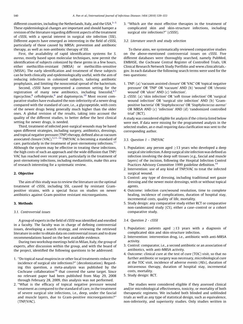
[(Figure_1)TD$FIG]
Figure 1. Flow diagram of trial selection: use of vacuum-assisted closure (VAC) in infected wounds.
A. Pan et al. / International Journal of Infectious Diseases 14S4 (2010) S39–S53 S41
English, French, Italian, or Spanish were included in the analysis. Forquestion 2 (cSSSI), RCTs that did not include any MRSA patient wereexcluded, as well as those in which one of the study regimens did nothave any anti-MRSA activity. Trials focusing on pharmacokinetic orpharmacodynamic variables were also excluded. RCTs that studiedadditional antimicrobial agents, generally with anti-Gram-negativerods and/or anti-anaerobic activity (as is the case in patients withpolymicrobial infections) were included in the analysis.
3.5. Classification and evaluation of the selected evidence
A matrix was made to extract evidence from individual originalstudies using the CONSORT method for the evaluation of random-ized clinical trials and the Newcastle–Ottawa Quality AssessmentScale for the evaluation of case–control trials, longitudinal cohorts,and retrospective studies with comparative groups.20 The originaldata from case studies were considered homogeneous after using apredefined format both for single case reports and series of reportedcases.20 In the discussion section, to assign the strength to the level ofthe recommendations, a methodology adapted from the GRADEWorking Group was applied. The details of the methodology arereported in this supplement.20
3.6. Definition of infection
3.6.1. Deep surgical site infection
A deep surgical site infection was defined as infection involvingthe deep soft tissues (e.g., fascial and muscle layers) of the incision,following the Hospital Infection Control Practices AdvisoryCommittee 1999 guideline definition.19 Complicated skin andskin-structure infections (cSSSI) were defined as infections
involving deeper soft tissue and/or requiring significant surgicalintervention (e.g., surgical or traumatic wound infection, majorabscess, infected ulcer, or deep and extensive cellulitis) or that haddeveloped on a lower extremity in a subject with diabetes mellitusor well-documented peripheral vascular disease. The presence ofat least one local sign of cSSSI (i.e., erythema, fluctuance, purulentor seropurulent drainage/discharge, heat/localized warmth, pain/tenderness to palpation, swelling/induration) or one systemic sign(oral temperature of >38 8C, white blood cell count of >10 � 109/l,>10% immature neutrophils) were necessary to define a cSSSI.
4. Results
4.1. Question 1 – TNP/VAC
‘‘What is the efficacy of the topical negative pressure woundtreatment as compared to the standard of care, in the treatment ofsevere surgical site infections, i.e., deep, under the fascial andmuscle layers, due to Gram-positive micro-organisms?’’
A total of 10 comparative studies were identified (see Figure 1).Of these, six were on post-sternotomy deep surgical site infection,with or without mediastinitis,21–26 three on post-sternotomymediastinitis,27–29 and one on early groin vascular by-pass graftinfection30 (see Tables 1 and 2).
In all studies the main outcome was the cure of the infection orthe failure of the therapy. Although the definition of wound cure wasnot standardized throughout the studies, the definition of woundresolution was based upon the appearance of the wound, thepresence of wound granulation and/or resolution of local signs ofinflammation, and/or negative cultures in six studies (see Table 2).Two studies referred to a definition of failure, including the need for
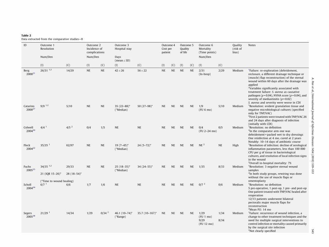
Table 1Data extracted from the comparative studies—I
ID Aim Study design Population Intervention Comparator
Berg 200021 Compare the TNP/VAC and closed
drainage technique
Retrospective comparative
cohort study
Deep surgical site infection of the
sternotomy site with positive cultures
Vacuum suction through 3–6
redon catheters (300–600 mmHg);
no polyester dressing used
2–4 catheters with CDI (2 l of 0.5%
povidone–iodine solution per 24 h
continuously)
Catarino 200027 Compare the TNP/VAC and standard therapy Retrospective comparative
cohort study
Patients with early post-sternotomy
mediastinitis
TNP/VAC 125 mmHg; changed
every 2–3 days
Debridement, CDI with normal saline
(1 l every 6 h until the effluent was
microbiologically clear)
Colwell 200430 Compare debridement/TNP/VAC vs.
incision/drainage + sartorius or rectus
femoris muscle flaps
Retrospective comparative
cohort study
Patients with early groin vascular
by-pass graft infection
TNP/VAC: not specified Incision and drainage
Fleck 200422 Compare preconditioning of the wound
with TNP/VAC with conventional
debridement and immediate primary closure
Retrospective comparative
cohort study
Patients with post-sternotomy
wound infection
TNP/VAC 125 mmHg; changed
every 2–3 days
Rewiring and primary
wound closure with insertion of a
mediastinal drain; daily dressing
changes
Fuchs 200523 Compare TNP/VAC with open pack procedure Retrospective comparative
A. Pan et al. / International Journal of Infectious Diseases 14S4 (2010) S39–S53 S45
re-operation.21,27 In two studies no definition of resolution wasreported.24,30
4.2. Patient populations
The patient populations were similar between the two studygroups throughout most studies, although in one study no dataregarding the demographic and general characteristics of the twogroups were reported22 and in another overall data only wereavailable.24 The mean age was similar between the two treatmentgroups in all the studies, ranging between 61 and 72.6 years. Asignificantly higher proportion of females in the TNP/VAC arm wasobserved in two studies.28,29 Finally, one study reported a longerduration of intervention28 and another a higher EUROscore, anindex of surgical complexity,29 and a lower proportion of S. aureus
infections21 in the TNP/VAC arm.
4.3. Intervention
The modalities of TNP/VAC were relatively similar throughoutthe studies: a negative pressure of 75–125 mmHg was used inseven studies, as was the time interval between dressing changes,i.e. 48–72 h (see Table 1). One study used higher pressures, 300–600 mmHg,21 another lower pressures (25–200 mmHg).24 In onestudy the pressure used was not specified.30
4.4. Control
The comparative conventional therapies were continuousdrainage irrigation in two studies21,27 and closed drainageirrigation in five.22,23,25,28,30
4.5. Study design
Nine studies were retrospective comparative cohort studies,while a single study was prospective (see Tables 1 and 2).25 No RCTwas retrieved.
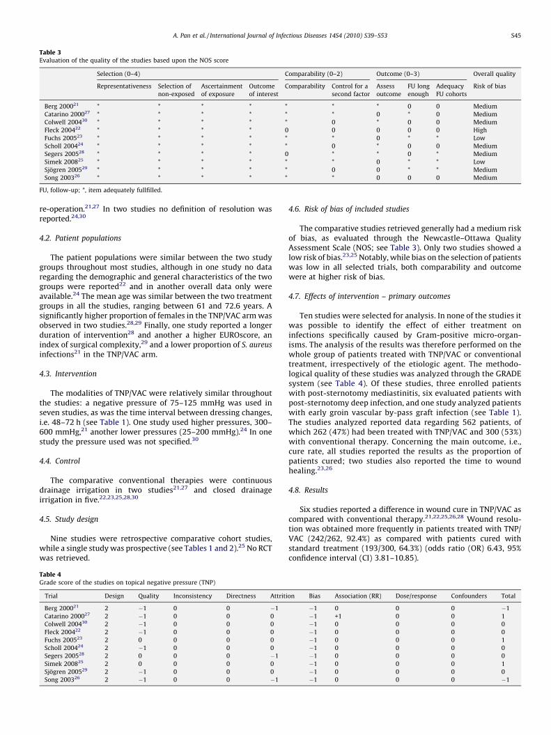
Table 4Grade score of the studies on topical negative pressure (TNP)
The comparative studies retrieved generally had a medium riskof bias, as evaluated through the Newcastle–Ottawa QualityAssessment Scale (NOS; see Table 3). Only two studies showed alow risk of bias.23,25 Notably, while bias on the selection of patientswas low in all selected trials, both comparability and outcomewere at higher risk of bias.
4.7. Effects of intervention – primary outcomes
Ten studies were selected for analysis. In none of the studies itwas possible to identify the effect of either treatment oninfections specifically caused by Gram-positive micro-organ-isms. The analysis of the results was therefore performed on thewhole group of patients treated with TNP/VAC or conventionaltreatment, irrespectively of the etiologic agent. The methodo-logical quality of these studies was analyzed through the GRADEsystem (see Table 4). Of these studies, three enrolled patientswith post-sternotomy mediastinitis, six evaluated patients withpost-sternotomy deep infection, and one study analyzed patientswith early groin vascular by-pass graft infection (see Table 1).The studies analyzed reported data regarding 562 patients, ofwhich 262 (47%) had been treated with TNP/VAC and 300 (53%)with conventional therapy. Concerning the main outcome, i.e.,cure rate, all studies reported the results as the proportion ofpatients cured; two studies also reported the time to woundhealing.23,26
4.8. Results
Six studies reported a difference in wound cure in TNP/VAC ascompared with conventional therapy.21,22,25,26,28 Wound resolu-tion was obtained more frequently in patients treated with TNP/VAC (242/262, 92.4%) as compared with patients cured withstandard treatment (193/300, 64.3%) (odds ratio (OR) 6.43, 95%confidence interval (CI) 3.81–10.85).
on Bias Association (RR) Dose/response Confounders Total
�1 0 0 0 �1
�1 +1 0 0 1
�1 0 0 0 0
�1 0 0 0 0
�1 0 0 0 1
�1 0 0 0 0
�1 0 0 0 0
�1 0 0 0 1
�1 0 0 0 0
�1 0 0 0 �1
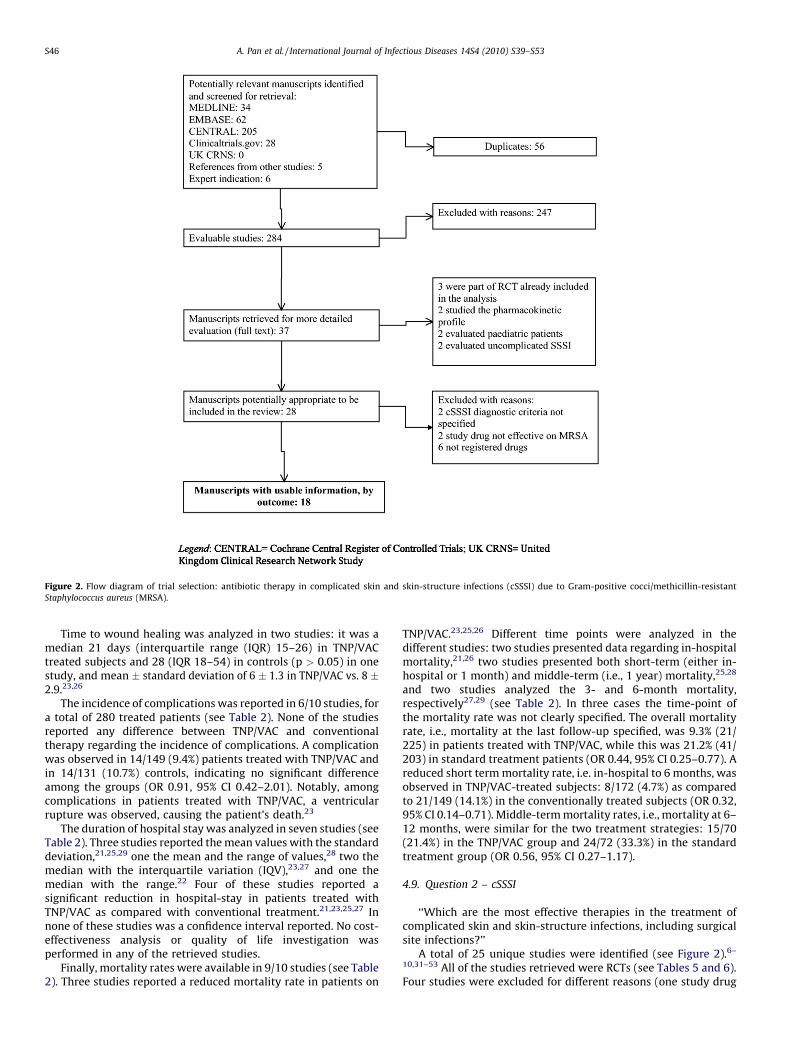
[(Figure_2)TD$FIG]
Figure 2. Flow diagram of trial selection: antibiotic therapy in complicated skin and skin-structure infections (cSSSI) due to Gram-positive cocci/methicillin-resistant
Staphylococcus aureus (MRSA).
A. Pan et al. / International Journal of Infectious Diseases 14S4 (2010) S39–S53S46
Time to wound healing was analyzed in two studies: it was amedian 21 days (interquartile range (IQR) 15–26) in TNP/VACtreated subjects and 28 (IQR 18–54) in controls (p > 0.05) in onestudy, and mean � standard deviation of 6 � 1.3 in TNP/VAC vs. 8 �2.9.23,26
The incidence of complications was reported in 6/10 studies, fora total of 280 treated patients (see Table 2). None of the studiesreported any difference between TNP/VAC and conventionaltherapy regarding the incidence of complications. A complicationwas observed in 14/149 (9.4%) patients treated with TNP/VAC andin 14/131 (10.7%) controls, indicating no significant differenceamong the groups (OR 0.91, 95% CI 0.42–2.01). Notably, amongcomplications in patients treated with TNP/VAC, a ventricularrupture was observed, causing the patient’s death.23
The duration of hospital stay was analyzed in seven studies (seeTable 2). Three studies reported the mean values with the standarddeviation,21,25,29 one the mean and the range of values,28 two themedian with the interquartile variation (IQV),23,27 and one themedian with the range.22 Four of these studies reported asignificant reduction in hospital-stay in patients treated withTNP/VAC as compared with conventional treatment.21,23,25,27 Innone of these studies was a confidence interval reported. No cost-effectiveness analysis or quality of life investigation wasperformed in any of the retrieved studies.
Finally, mortality rates were available in 9/10 studies (see Table2). Three studies reported a reduced mortality rate in patients on
TNP/VAC.23,25,26 Different time points were analyzed in thedifferent studies: two studies presented data regarding in-hospitalmortality,21,26 two studies presented both short-term (either in-hospital or 1 month) and middle-term (i.e., 1 year) mortality,25,28
and two studies analyzed the 3- and 6-month mortality,respectively27,29 (see Table 2). In three cases the time-point ofthe mortality rate was not clearly specified. The overall mortalityrate, i.e., mortality at the last follow-up specified, was 9.3% (21/225) in patients treated with TNP/VAC, while this was 21.2% (41/203) in standard treatment patients (OR 0.44, 95% CI 0.25–0.77). Areduced short term mortality rate, i.e. in-hospital to 6 months, wasobserved in TNP/VAC-treated subjects: 8/172 (4.7%) as comparedto 21/149 (14.1%) in the conventionally treated subjects (OR 0.32,95% CI 0.14–0.71). Middle-term mortality rates, i.e., mortality at 6–12 months, were similar for the two treatment strategies: 15/70(21.4%) in the TNP/VAC group and 24/72 (33.3%) in the standardtreatment group (OR 0.56, 95% CI 0.27–1.17).
4.9. Question 2 – cSSSI
‘‘Which are the most effective therapies in the treatment ofcomplicated skin and skin-structure infections, including surgicalsite infections?’’
A total of 25 unique studies were identified (see Figure 2).6–
10,31–53 All of the studies retrieved were RCTs (see Tables 5 and 6).Four studies were excluded for different reasons (one study drug
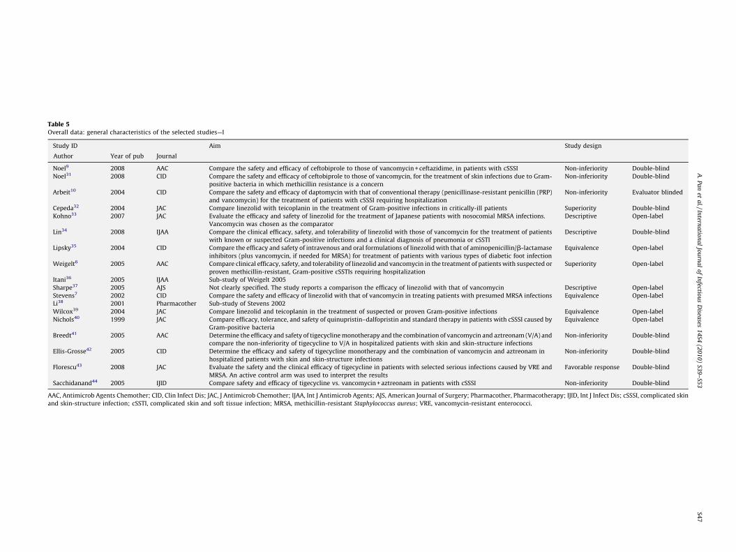
Table 5Overall data: general characteristics of the selected studies—I
Study ID Aim Study design
Author Year of pub Journal
Noel9 2008 AAC Compare the safety and efficacy of ceftobiprole to those of vancomycin + ceftazidime, in patients with cSSSI Non-inferiority Double-blind
Noel31 2008 CID Compare the safety and efficacy of ceftobiprole to those of vancomycin, for the treatment of skin infections due to Gram-
positive bacteria in which methicillin resistance is a concern
Non-inferiority Double-blind
Arbeit10 2004 CID Compare the safety and efficacy of daptomycin with that of conventional therapy (penicillinase-resistant penicillin (PRP)
and vancomycin) for the treatment of patients with cSSSI requiring hospitalization
Non-inferiority Evaluator blinded
Cepeda32 2004 JAC Compare linezolid with teicoplanin in the treatment of Gram-positive infections in critically-ill patients Superiority Double-blind
Kohno33 2007 JAC Evaluate the efficacy and safety of linezolid for the treatment of Japanese patients with nosocomial MRSA infections.
Vancomycin was chosen as the comparator
Descriptive Open-label
Lin34 2008 IJAA Compare the clinical efficacy, safety, and tolerability of linezolid with those of vancomycin for the treatment of patients
with known or suspected Gram-positive infections and a clinical diagnosis of pneumonia or cSSTI
Descriptive Double-blind
Lipsky35 2004 CID Compare the efficacy and safety of intravenous and oral formulations of linezolid with that of aminopenicillin/b-lactamase
inhibitors (plus vancomycin, if needed for MRSA) for treatment of patients with various types of diabetic foot infection
Equivalence Open-label
Weigelt6 2005 AAC Compare clinical efficacy, safety, and tolerability of linezolid and vancomycin in the treatment of patients with suspected or
Sharpe37 2005 AJS Not clearly specified. The study reports a comparison the efficacy of linezolid with that of vancomycin Descriptive Open-label
Stevens7 2002 CID Compare the safety and efficacy of linezolid with that of vancomycin in treating patients with presumed MRSA infections Equivalence Open-label
Li38 2001 Pharmacother Sub-study of Stevens 2002
Wilcox39 2004 JAC Compare linezolid and teicoplanin in the treatment of suspected or proven Gram-positive infections Equivalence Open-label
Nichols40 1999 JAC Compare efficacy, tolerance, and safety of quinupristin–dalfopristin and standard therapy in patients with cSSSI caused by
Gram-positive bacteria
Equivalence Open-label
Breedt41 2005 AAC Determine the efficacy and safety of tigecycline monotherapy and the combination of vancomycin and aztreonam (V/A) and
compare the non-inferiority of tigecycline to V/A in hospitalized patients with skin and skin-structure infections
Non-inferiority Double-blind
Ellis-Grosse42 2005 CID Determine the efficacy and safety of tigecycline monotherapy and the combination of vancomycin and aztreonam in
hospitalized patients with skin and skin-structure infections
Non-inferiority Double-blind
Florescu43 2008 JAC Evaluate the safety and the clinical efficacy of tigecycline in patients with selected serious infections caused by VRE and
MRSA. An active control arm was used to interpret the results
Favorable response Double-blind
Sacchidanand44 2005 IJID Compare safety and efficacy of tigecycline vs. vancomycin + aztreonam in patients with cSSSI Non-inferiority Double-blind
AAC, Antimicrob Agents Chemother; CID, Clin Infect Dis; JAC, J Antimicrob Chemother; IJAA, Int J Antimicrob Agents; AJS, American Journal of Surgery; Pharmacother, Pharmacotherapy; IJID, Int J Infect Dis; cSSSI, complicated skin
and skin-structure infection; cSSTI, complicated skin and soft tissue infection; MRSA, methicillin-resistant Staphylococcus aureus; VRE, vancomycin-resistant enterococci.
A.
Pa
net
al./In
terna
tion
al
Jou
rna
lo
fIn
fectiou
sD
iseases
14
S4(2
01
0)
S39
–S5
3S4
7
Table 6Overall data: general characteristics of the selected studies—II
Sacchidanand 200544 573 Tigecycline Vancomycin + aztreonam No Up to 14 Non-inferiority Double-blind
PRP, penicillinase-resistant penicillin; MRSA, methicillin-resistant Staphylococcus aureus.a The study enrolled also patients with other types of infection.
A. Pan et al. / International Journal of Infectious Diseases 14S4 (2010) S39–S53S48
was not effective against MRSA for two,52,53 no data were reportedregarding the diagnostic criteria of cSSSI for two others50,51) andsix studies were excluded after panel discussion, since theyfocused on drugs not yet registered, i.e., ceftaroline,45 dalbavan-cin,46,47 and telavancin.48–50 Of the 18 studies from which datawere extracted, two reported pharmaco-economical data of twostudies included in the analysis.36,38 All the selected studies werepublished from 1999 onwards.
Table 7Patients enrolled in the study and treated as per intention to treat (ITT), clinically and
Author Drugs ITT
Study drug Co
Study drug Study drug Cure Total Cu
Noel 20089 Ceftobiprole Vancomycin 309 397 30
Noel 200831 Ceftobiprole Vancomycin +
ceftazidime
448 547 22
Arbeit 200410 Daptomycin Vancomycin/ PRP 382 534 39
All studies evaluated both male and female adults; one studyalso enrolled patients of �13 years of age,39 and a second oneenrolled patients �16 years of age.32 The mean age of the enrolledpopulations ranged from 41.6 to 76 years. In all of the studies themajority of patients were male, with the proportion ranging from54% to 71%.35,39
microbiologically evaluable at test of cure (TOC)
Clinically evaluable Microbiologically evaluable
mparator Study drug Comparator Study drug Comparator
re Total Cure Total Cure Total Cure Total Cure Total
0 387 263 282 259 277 NR NR NR NR
7 281 292 318 149 165 NR NR NR NR
7 558 372 446 384 456 21 28 25 36
R NR NR NR NR NR 13 18 4 10
29 30 33 19 24 NR NR NR NR
120 NR NR NR NR NR NR NR NR
2 588 436 462 394 436 NR NR NR NR
R NR 29 30 13 30 29 30 23 30
108 64 99 54 87 27 30 22 30
3 117 99 106 89 102 23 32 10 18
18 23 32 10 15 7 9 3 6
3 443 197 289 193 273 5 6 3 6
5 269 200 223 201 213 25 32 25 33
4 550 365 422 364 411 NR NR NR NR
23 NR NR NR NR NR NR NR NR
3 281 165 199 163 198 16 21 17 21
Table 9Quality assessment of trials comparing the efficacy of different antibiotics in the treatment of complicated skin and skin-structure infections, following the GRADE
recommendations
Study ID Design Quality Inconsistency Directness Attrition Bias Association (RR) Dose/response Confounders Total
Noel 20089 4 0 0 0 –1 0 0 0 0 3
Noel 200831 4 �1 0 0 0 0 0 0 0 3
Arbeit 200410 4 �2 0 0 0 0 0 0 0 2
Lin 200834 4 �2 0 0 0 0 0 0 0 2
Lipsky 200435 4 �2 0 0 0 �1 0 0 0 1
Weigelt 20056 4 �2 0 0 0 0 0 0 0 2
Kohno 200733 4 �2 0 0 �1 0 0 0 0 1
Sharpe 200537 4 �2 0 0 �1 �1 0 0 0 2
Stevens 20027 4 �2 0 0 �1 0 0 0 0 1
Li 200138 4 �2 0 0 �1 0 0 0 0 1
Wilcox 200439 4 �1 0 0 0 0 0 0 0 3
Cepeda 200432 4 0 0 0 0 0 0 0 0 4
Nichols 199940 4 �1 0 0 �1 �1 0 0 0 1
Breedt 200541 4 �2 0 0 0 0 0 0 0 2
Ellis-Grosse 200542 4 �1 0 0 0 0 0 0 0 3
Florescu 200843 4 0 0 0 0 0 0 0 0 4
Sacchidanand 200544 4 0 0 0 0 0 0 0 0 4
Table 8Overall data: study design and quality score, calculated using the Jadad modified method
Study ID Study design Random Validity of
randomization
Double-blind Validity of
double-blind
Withdrawal
and/or dropouts
Total Quality
Noel 20089 Double-blind 1 1 1 0 1 4 High
Noel 200831 Double-blind 1 0 1 0 0 2 Low
Arbeit 200410 Evaluator blinded 1 0 0 0 1 2 Low
Lin 200834 Double-blind 1 0 0 0 1 2 Low
Lipsky 200354 Open-label 1 0 NA NA 0 1 Low
Itani 200536 Open-label 1 0 NA NA 1 2 Low
Weigelt 20056 (Sub-study)
Kohno 200733 Open-label 1 0 NA NA 1 2 Low
Sharpe 200537 Open-label 1 0 NA NA 0 1 Low
Stevens 20027 Open-label 1 0 NA NA 1 2 Low
Li 200138 (Sub-study)
Wilcox 200439 Open-label 1 1 NA NA 1 3 High
Cepeda 200432 Double-blind 1 1 1 1 1 5 High
Nichols 199940 Open-label 1 1 NA NA 0 2 Low
Breedt 200541 Double-blind 1 0 0 0 1 2 Low
Ellis-Grosse 200542 Double-blind 1 0 0 1 1 3 High
Florescu 200843 Double-blind 1 1 1 1 1 5 High
Sacchidanand 200544 Double-blind 1 1 1 0 1 4 High
NA, not applicable.
A. Pan et al. / International Journal of Infectious Diseases 14S4 (2010) S39–S53 S49
4.11. Interventions
The interventions evaluated in the studies identified arerepresented by an antibiotic monotherapy compared with anothermonotherapy or with a combination of two antibiotics (see Table6). The antibiotics studied were represented by: ceftobiprole,9,32
daptomycin,10 linezolid,6,7,32–49 quinupristin/dalfopristin,40 andtigecycline.41–44 The comparators are reported in Table 6.
4.12. Outcomes
The primary outcome of clinical cure of cSSSI was reported in14/16 studies on the overall population (see Table 7). Dataregarding clinical cure in MSSA infections could be retrieved in fivestudies, data on MRSA in eight studies, while data on streptococcalinfections were reported in eight papers. No study reported clinicaldata regarding enterococcal infections.
A microbiological analysis was reported in 9/16 studies. Dataregarding microbiological success for the different germs werereported as follows: MSSA: eight studies; MRSA: nine; enterococci:six; streptococci: eight.
AEs were reported in all but one study,37 while another studyreported only partial data.33 Data regarding mortality wereavailable in 12 studies, while they were not retrievable forpatients with cSSSI in four studies.7,34,39,43
Pharmaco-economic data were also retrieved: the duration ofhospital stay was reported in three papers, and the length ofintravenous therapy and the total duration of therapy werereported in 12 and five studies, respectively.
4.13. Risk of bias in included studies
Forty percent of the RCTs analyzed had a low risk of bias (6/15),while the remaining studies had a high risk of bias, based upon themodified Jadad score as reported in Table 8.19 This scoring system isbased upon an evaluation of five parameters: randomization,double-blinding, dropouts and withdrawals, generation of randomnumbers, and allocation concealment. For each of these parameters,if they were specified following the Jadad criteria, a point was given.
The attrition, i.e., the number of the initially randomizedpatients that were not clinically evaluable, was similar among thetwo study arms in all studies, in most papers below 25%, varyingfrom about 10%31,35 to over 45%.37
4.14. Global overview
A total of 8278 patients were enrolled in the 16 studiesanalyzed; of these, 8158 (98.6%) were randomized to receive eitherthe intervention drug (patient group, n = 4335) or the compara-tor(s) (n = 3823). An infection due to MRSA was diagnosed in 1698/
A. Pan et al. / International Journal of Infectious Diseases 14S4 (2010) S39–S53S50
8278 (20.5%) of the enrolled patients. The other Gram-positiveorganisms commonly reported were: MSSA (2309 patients, 27.9%),streptococci (918 patients, 11.1%), and enterococci (236 patients,2.9%).
4.15. Effects of intervention – primary outcomes
The methodological quality of the studies was analyzed throughthe GRADE system (see Table 9). Data regarding treatment successfor intention to treat (ITT) at TOC visit were available for 13 of the16 studies, while data regarding clinical efficacy at TOC visit wereretrievable from 10 papers (see Table 7). In most RCTs thecomparison was performed between the intervention drug andvancomycin or, less frequently, teicoplanin. In one case, the studycompared linezolid with a combination of penicillin and a b-lactamase inhibitor (PBLI).35
The overall efficacy was similar for study drugs and compara-tors in most studies. A significant difference was observed in threestudies, all of them comparing linezolid with vancomycin.6,35,38 Atrend towards a significant difference was observed in a furtherstudy comparing linezolid with PBLI/vancomycin.35
When the subset of patients with a microbiological diagnosis ofMRSA infection was analyzed, some studies reported data onclinical efficacy only,6,9,31,35 some on microbiology efficacyonly,10,32,33,40,42,44 and three on both.7,33,41 Two studies onlyobserved a significant difference, either clinical6 or microbiolog-ical:37 in both cases linezolid was superior to vancomycin. Of note,the absolute number of MRSA patients evaluated in tigecyclinestudies41,43 was very small, i.e., 93.
Data regarding mortality were reported in 12 studies. Thestudies that included only patients with cSSSI reported very lowmortality rates, varying between 0% and 1.5%. No difference wasobserved throughout the comparisons.
The incidence of AEs, was reported in 14/16 studies. Notably,only one study,9 reported the World Health Organization (WHO)grading system of AE, with serious AE having WHO grade>3. Threestudy drugs showed a higher incidence of AE than the comparator:linezolid, quinupristin/dalfopristin, and tigecycline. The studiescomparing linezolid with a glycopeptide/PBLI showed a signifi-cantly lower proportion of AE in the control group (36.8% vs. 42.6%for glycopeptide/PBLI and linezolid, respectively). This differencepersisted even if the patients included in the study by Lipskyet al.,35 based upon PBLI, were not considered (OR 1.23, 95% CI1.03–1.48). In these studies the most common AEs in the linezolidgroup were represented by diarrhea, nausea, anemia, thrombocy-topenia, and liver disease, while the glycopeptide-treated grouppresented more frequently renal failure and rash. Quinupristin/dalfopristin was associated with a significantly higher proportionof AEs than vancomycin/penicillinase-resistant penicillin (PRP):62.9% vs. 54%, mainly gastrointestinal problems and venousevents. Finally, tigecycline was associated with a higher incidenceof AE than vancomycin plus aztreonam: 67.8% vs. 61.3%, the mostcommon AE for tigecycline being gastrointestinal symptoms, suchas nausea (over a third of the patients) and vomiting, whilepatients on vancomycin/aztreonam complained more frequentlyof skin problems and abnormal liver function tests.
Serious AEs (SAEs) were reported in detail in 14 studies, whileone study47 reported only the total number of SAEs in the wholestudy population. No difference was observed between any studyarm.
4.16. Secondary outcomes
Microbiological cure was reported in nine of the 16 studies (seeTable 7). No significant difference was reported between theintervention drug and the comparator in all but one comparison
(linezolid) that determined a significantly better microbiologicaleradication than the comparators (OR 2.17, 95% CI 1.38–3.42).
The duration of intravenous therapy was reported by 12studies.9,10,31,34,35,37–40,42,44,45 In seven of these studies, onecomparing daptomycin with vancomycin/PRP,10 five linezolid vs.glycopeptide/PRP,35–39 and one quinupristin/dalfopristin,40 theintervention arm showed a shorter duration of intravenoustherapy.
The duration of hospital stay was analyzed in three studies, allof them comparing linezolid with vancomycin. In two of thesethree studies, a shorter duration of hospital stay was observed.Notably, two studies, one by Itani and colleagues36 and the otherby Li and colleagues,38 specifically addressed pharmaco-economicissues, and one single study37 compared the cost of linezolidtreatment with that of standard therapy, i.e., vancomycin. Theauthors calculated a significant saving of money when the patientswere treated with linezolid.
5. From the evidence to the recommendations
5.1. Question 1
‘‘What is the efficacy of topical negative pressure woundtreatment as compared to the standard of care, in the treatment ofsevere surgical site infections, i.e., deep, under the fascial andmuscle layers, due to Gram-positive microorganisms?’’
5.2. Discussion
The application of negative pressure to favor wound healingwas introduced into clinical practice in the 1960 s, but wasstandardized with the introduction of TNP/VAC in the 1990s.54 Thepossibility of maintaining a closed and clean environment, and thecontinuous drainage of necrotic and bacterial debris, couldtheoretically improve the time taken to wound cure.54 Due tothe limitation of alternative effective therapies, and to theexperience of some centers, TNP/VAC has become, in manyhospitals, the standard of care for difficult to treat chronic wounds,including post-sternotomy mediastinitis, despite the fact that itsefficacy and complications in this setting have not been fullyinvestigated.17
We analyzed 699 papers, and did not find a single RCT thataddressed the problem. A multicenter European trial on TNP/VACtreatment of post-sternotomy mediastinitis has recently beenprematurely terminated due to a lack of patient enrolment.55 Weidentified 10 comparative studies that satisfied all inclusioncriteria, with an overall medium risk of bias, that enrolled a total of562 patients (see Tables 1 and 2).21–30 It was not possible toidentify, within the selected studies, the clinical outcome ofinfections stratified by Gram-positive or Gram-negative patho-gens. The overall analysis performed showed that TNP/VAC wassignificantly more effective in 6/10 studies than standard therapyin the cure of post-sternotomy mediastinitis and of deep sternalwound infections, which, to date, represent the major indicationsof this therapeutic approach in the setting of an infection. Time towound healing was reported in two of the 10 studies23,26 and nosignificant difference was observed between the two treatments.No increased risk of complication was observed among TNP/VAC-treated patients, although one patient died of ventricular rupturedue to TNP/VAC.
Both short-term (in-hospital to 3 months) and last follow-upvisit mortality rates were significantly lower in TNP/VAC-treatedpatients than in the standard care patients, while middle-termmortality (6–12 months) was similar in the two groups.
Patients treated with TNP/VAC had a shorter duration ofhospital stay. No study compared the cost of TNP/VAC and
A. Pan et al. / International Journal of Infectious Diseases 14S4 (2010) S39–S53 S51
standard therapy, nor did any study address the quality of lifeissues.
There are several limitations to the interpretation of theseresults. First of all, the overall quality of the studies is generallylow, with no available well-designed RCTs. The studies analyzedgenerally show a medium risk of bias. Only in three cases did astudy have a GRADE score �1 (see Table 4).23,25,27 In five cases theGRADE score was zero,22,24,28–30 while in the remaining twostudies it was�1.21,29 However, when only the three higher qualitystudies were analyzed, the overall results were confirmed: asignificant difference in the effect of TNP/VAC vs. standard therapywas still observed (OR 9.19, 95% CI 2.77–30.48). These three studiesdid not show any significant difference in mortality between thetwo treatment strategies (OR 0.56, 95% CI 0.27–1.17).
Second, the TNP/VAC was not well standardized among thestudies: it was used at different pressures and the foam waschanged at different time intervals. Third, the debridement anddrainage procedures used as comparator varied significantlybetween and among centers. Fourth, in patients with post-sternotomy mediastinitis and deep surgical site infection, antibi-otic treatment is mandatory and should preferably be prescribedby an infectious disease consultant. Unfortunately, no specificinformation was reported in any study regarding the antibiotictreatment, i.e., molecule, dose, duration. Finally this limitedamount of comparative data is restricted almost exclusively toone single type of infection: post-sternotomy infections.
Recommendations
The use of TNP/VAC in patients with a post-sternotomyinfection, either mediastinitis or deep surgical site infection, is apossible alternative to the standard therapy (grade D). The cost-effectiveness of TNP/VAC should be carefully evaluated.
In the treatment of infected wounds TNP/VAC should bereserved only for patients with post-sternotomy infections,including mediastinitis (grade D).
A standardized protocol, both for the use of TNP/VAC and forthe standard care of the infected wound should be defined in
5.3. Question 2
‘‘Which are the most effective therapies in the treatment ofcomplicated skin and skin-structure infections, including surgicalsite infections?’’
5.4. Discussion
Complicated skin and skin-structure infections are caused byGram-positive cocci in the majority of cases.1 Treatment of cSSSIhas been, over the years, an area of intense investigation that haspermitted the registration of most of the novel antibiotics,particularly of those active against MRSA, such as linezolid,tigecycline, and daptomycin. With the new epidemiologicalsituation, characterized by a dramatic increase in the proportionof CA-MRSA in North America1 and by the emergence of LA-MRSAin Europe,3 with both germs frequently causing cSSSI, there is aneed to better define the potency and tolerability of the differentdrugs indicated in the treatment of these infections.
The analysis of the literature identified seven differentregistered anti-MRSA drugs for which RCTs have been publishedsince 1990. In most cases the performed studies evaluated theefficacy of a novel drug as compared to the standard of care,represented in most cases by a glycopeptide, generally vancomy-
each cardiac and thoracic surgery department to reduce intra-hospital variability (grade D).
cin and, less frequently, teicoplanin, or in a single study, by PBLI(see Table 6). Notably, as in other areas of pharmacologicalresearch, most studies aimed to demonstrate a non-inferiority ofthe newer drug as compared to the older: in the 18 studies that weanalyzed, there were only two superiority studies. Due to the highcosts of clinical research, we think that systematic reviews andmeta-analyses will represent an important tool in the future tobetter define which are the most potent and better tolerateddrugs.
We applied a methodology adapted from the GRADE WorkingGroup to assign a strength level to the recommendations. TheGRADE score of the studies analyzed was high (GRADE 4) in threeof 18 studies (17%).32,43,44 Four studies (22%) were of mediumquality (GRADE 3)9,31,39,42 and the majority, i.e., the remaining 11studies (61%) were of low quality (GRADE �2).
Comparisons between these different drugs allowed theverification that ceftobiprole, daptomycin, quinupristin/dalfopris-tin, and tigecycline are as effective as vancomycin when evaluatingthe clinical efficacy for ITT analysis. The only comparison thatpermitted the identification of a significant difference between thestudy drugs was linezolid vs. glycopeptide/PBLI, where linezolidperformed better than the comparator in three out of sevenstudies. When the analysis was performed on the population ofpatients with confirmed MRSA infections, the superiority oflinezolid vs. glycopeptide/PRP was observed in two of six studies.No other difference was observed for any other drug. No differencein mortality was observed in any comparison, as was expected dueto the low overall mortality of cSSSI.
The analysis on AEs yielded interesting results. The incidence ofSAEs was similar throughout all comparisons. The global incidenceof AEs was similar between the new cephalosporin and vancomy-cin/PRP, as well as between daptomycin and vancomycin/PRP. Allthe other newer drugs, i.e., linezolid, quinupristin/dalfopristin, andtigecycline, were tolerated significantly worse than the glycopep-tides. It is interesting to point out that vancomycin is generallyconsidered a relatively toxic and not well tolerated drug.
Data regarding duration of hospital stay were available only forthree studies, all evaluating linezolid, and showing a reducedduration of hospital stay in patients treated with this drug.Furthermore, the majority of studies reported the duration ofintravenous therapy, showing a significantly shorter duration ofintravenous therapy consistently reported in patients treated withlinezolid as compared with vancomycin/PRP. A shorter duration ofintravenous therapy was also reported in two studies comparingdaptomycin and quinupristin/dalfopristin with vancomycin/PRP.One single study evaluated the costs associated with linezolid vs.vancomycin in MRSA-infected patients, with a significant advan-tage for linezolid. Data regarding the pharmaco-economic issue arein favor of linezolid, to-date the only oral drug with anti-MRSAactivity among the newer antibiotics. Since the newer drugs havecosts that are consistently higher than vancomycin, the economicanalysis plays an important role in the choice of the antibiotic to beused. No pharmaco-economic analysis was found specificallyaddressing cSSSI, performed within an RCT.
Among the limitations to this analysis, the most important isthat most of the RCTs evaluating linezolid were open-label, thus ofreduced quality as compared with the double-blind study design.The quality score applying the modified Jadad methodology19 ofthe studies evaluating linezolid was generally low to medium (seeTable 9). Similarly, a low GRADE score was observed in moststudies. One single small study, evaluating 60 patients, reported acost-effectiveness analysis.37 No study was found that made acomparison with the efficacy of older drugs with at least a partialanti-MRSA activity, such as tetracycline, clindamycin, co-trimox-azole, and fusidic acid. RCTs have been performed with some ofthese drugs in uncomplicated skin and soft tissue infections,
A. Pan et al. / International Journal of Infectious Diseases 14S4 (2010) S39–S53S52
although, in our opinion, further investigation is needed, due alsoto the availability of oral formulations for some of these drugs.
Interestingly, in most studies analyzed, therapeutic drugmonitoring of vancomycin was not a part of the study protocol,being left to the decision of the investigator. This lack ofvancomycin therapeutic dose monitoring could have led to bothincreased toxicity due to high trough levels, as well as reducedefficacy due to low concentrations.
Finally, the studies analyzed did not enroll patients with severedisease, such as necrotizing fasciitis, gangrene, and ecthymagangrenosum, thus limiting the utility of the results, althoughsome papers did include patients with positive blood streaminfections.
Recommendations
Glycopeptides (vancomycin and teicoplanin) should beconsidered as the standard of care in patients with cSSSI dueto MRSA (grade A).
Linezolid appears to be more effective than glycopeptides(grade C). Linezolid could be an alternative treatment toglycopeptides despite the low to medium methodologicalquality of analyzed trials (grade D).
Newer drugs, tigecycline (grade B) and daptomycin (gradeC), are as effective as glycopeptides.
When choosing the therapeutic strategy, the pharmaco-economic issue should be considered, i.e., cost of the drug,duration of intravenous therapy, length of hospital stay, andearly discharge; a switch to the oral drug should be madewhenever possible (grade C).
Always carefully consider the pharmacokinetic and phar-macodynamic parameters of chosen drugs. Monitor glycopep-
Acknowledgement
The GISIG Consensus Conference was organized with supportfrom an unrestricted educational grant from Pfizer.
We are thankful to Dr Mark Wilson who provided us with dataon cSSSI retrieved from his study.
We wish to thank Tom Jefferson for sharing his expertise andideas with us.
Conflict of interest
All members of the faculty of GISIG – G. Carosi, R. Cauda, E.Concia, S. Esposito, G. Ippolito, F.N. Lauria, M. Moroni, E. Nicastri, A.Pan, G. Sganga, S. Stefani – report no other potential conflict ofinterest except as reported in the specific section.
The members of the working group have no specific conflict ofinterest to report.
Funding
For the present research, all members of the faculty of GISIGreceived a fee from the organizing secretariat of the GISIG Project.
The members of the working group have no funding to report.
Additional Conflict of interest
Conflict of interest for R. Cauda: GlaxoSmithKline, Gilead,Bristol Myers Squibb, Boehringer Ingelheim, Pfizer, Abbott, MerckSharp & Dohme, Wyeth. Funding received from GlaxoSmithKline,
tide trough levels and adapt their dosage according to theavailable guidelines (grade D).
Gilead, Bristol Myers Squibb, Boehringer Ingelheim, Pfizer, Abbott,Merck Sharp & Dohme. S. Esposito has received fees for speaking atnational and international meetings and for consulting on Advisoryboards from Pfizer, Novartis farma and Wyeth Lederle. G. Sgangahas received honoraria for speaking for Pfizer. G. Ippolito and F.N.Lauria have received expert opinion fees from Pfizer. E. Nicastri hasreceived paid expert opinion from MSD and Pfizer. A. Pan hasreceived paid expert opinion fees from Janssen.
References
1. Stevens DL, Bisno AL, Chambers HF, Everett ED, Dellinger P, Goldstein EJ, et al.Practice guidelines for the diagnosis and management of skin and soft-tissueinfections. Clin Infect Dis 2005;41:1373–406.
2. Stryjewski ME, Chambers HF. Skin and soft-tissue infections caused by com-munity-acquired methicillin-resistant Staphylococcus aureus. Clin Infect Dis2008;46(Suppl 5):S368–77.
3. Wulf M, Voss A. MRSA in livestock animals—an epidemic waiting to happen?Clin Microbiol Infect 2008;14:519–21.
4. Pan A, Battisti A, Zoncada A, Bernieri F, Boldini M, Franco A, et al. Community-acquired methicillin-resistant Staphylococcus aureus ST398 infection. ItalyEmerg Infect Dis 2009;15:845–7.
5. Smith J. Debridement of diabetic foot ulcers. Cochrane Database Syst Rev2002;(4):CD003556.
6. Weigelt J, Itani K, Stevens D, Lau W, Dryden M, Knirsch C. Linezolid versusvancomycin in treatment of complicated skin and soft tissue infections. Anti-microb Agents Chemother 2005;49:2260–6.
7. Stevens DL, Herr D, Lampiris H, Hunt JL, Batts DH, Hafkin B. Linezolid versusvancomycin for the treatment of methicillin-resistant Staphylococcus aureusinfections. Clin Infect Dis 2002;34:1481–90.
8. Stryjewski ME, Graham DR, Wilson SE, O’Riordan W, Young D, Lentnek A, et al.Telavancin versus vancomycin for the treatment of complicated skin and skin-structure infections caused by Gram-positive organisms. Clin Infect Dis2008;46:1683–93.
9. Noel GJ, Strauss RS, Amsler K, Heep M, Pypstra R, Solomkin JS. Results of adouble-blind, randomized trial of ceftobiprole treatment of complicated skinand skin structure infections caused by Gram-positive bacteria. AntimicrobAgents Chemother 2008;52:37–44.
10. Arbeit RD, Maki D, Tally FP, Campanaro E, Eisenstein BI. The safety and efficacyof daptomycin for the treatment of complicated skin and skin-structure infec-tions. Clin Infect Dis 2004;38:1673–81.
11. Singer AJ, Dagum AB. Current management of acute cutaneous wounds. N Engl JMed 2008;359:1037–46.
12. Diehr S, Hamp A, Jamieson B, Mendoza M. Clinical inquiries. Do topical anti-biotics improve wound healing? J Fam Pract 2007;56:140–4.
13. Bradley M, Cullum N, Nelson EA, Petticrew M, Sheldon T, Torgerson D. System-atic reviews of wound care management: (2). Dressings and topical agents usedin the healing of chronic wounds. Health Technol Assess 1999;3:1–35.
14. Dryburgh N, Smith F, Donaldson J, Mitchell M. Debridement for surgicalwounds. Cochrane Database Syst Rev 2008;(3):CD006214.
15. Fleischmann W, Strecker W, Bombelli M, Kinzl L. [Vacuum sealing as treatmentof soft tissue damage in open fractures]. Unfallchirurg 1993;96:488–92.
16. Argenta LC, Morykwas MJ. Vacuum-assisted closure: a new method for woundcontrol and treatment: clinical experience. Ann Plast Surg 1997;38:563–76.
17. Raja SG, Berg GA. Should vacuum-assisted closure therapy be routinely used formanagement of deep sternal wound infection after cardiac surgery? InteractCardiovasc Thorac Surg 2007;6:523–7.
18. van Rijen M, Bonten M, Wenzel R, Kluytmans J. Mupirocin ointment forpreventing Staphylococcus aureus infections in nasal carriers. Cochrane DatabaseSyst Rev 2008;(4):CD006216.
19. Mangram AJ, Horan TC, Pearson ML, Silver LC, Jarvis WR. Guideline for preven-tion of surgical site infection, 1999. Hospital Infection Control Practices Advi-sory Committee. Infect Control Hosp Epidemiol 1999;20:250–78.
20. Lauria FN, De Carli G, Nicastri E. Resistant and multi-resistant Gram-positivesevere infections: the GISIG working methodology. Int J Infect Dis 2010;14S:S13–7.
21. Berg HF, Brands WG, van Geldorp TR, Kluytmans-VandenBergh FQ, KluytmansJA. Comparison between closed drainage techniques for the treatment ofpostoperative mediastinitis. Ann Thorac Surg 2000;70:924–9.
22. Fleck TM, Koller R, Giovanoli P, Moidl R, Czerny M, Fleck M, et al. Primary ordelayed closure for the treatment of poststernotomy wound infections? AnnPlast Surg 2004;52:310–4.
23. Fuchs U, Zittermann A, Stuettgen B, Groening A, Minami K, Koerfer R. Clinicaloutcome of patients with deep sternal wound infection managed by vacuum-assisted closure compared to conventional therapy with open packing: aretrospective analysis. Ann Thorac Surg 2005;79:526–31.
24. Scholl L, Chang E, Reitz B, Chang J. Sternal osteomyelitis: use of vacuum-assistedclosure device as an adjunct to definitive closure with sternectomy and muscleflap reconstruction. J Card Surg 2004;19:453–61.
25. Simek M, Hajek R, Zalesk B, Molitor M, Lonsky V, Grulichova J, et al. Topicalnegative pressure versus conventional treatment of deep sternal wound infec-tion in cardiac surgery. European Wound Management Association Journal2008;8:17–20.
A. Pan et al. / International Journal of Infectious Diseases 14S4 (2010) S39–S53 S53
26. Song DH, Wu LC, Lohman RF, Gottlieb LJ, Franczyk M. Vacuum assisted closurefor the treatment of sternal wounds: the bridge between debridement anddefinitive closure. Plast Reconstr Surg 2003;111:92–7.
27. Catarino PA, Chamberlain MH, Wright NC, Black E, Campbell K, Robson D, et al.High-pressure suction drainage via a polyurethane foam in the management ofpoststernotomy mediastinitis. Ann Thorac Surg 2000;70:1891–5.
28. Segers P, De Jong AP, Kloek JJ, De Mol BA. Poststernotomy mediastinitis:comparison of two treatment modalities. Interact Cardiovasc Thorac Surg2005;4:555–60.
29. Sjogren J, Nilsson J, Gustafsson R, Malmsjo M, Ingemansson R. The impact ofvacuum-assisted closure on long-term survival after post-sternotomy medias-tinitis. Ann Thorac Surg 2005;80:1270–5.
30. Colwell AS, Donaldson MC, Belkin M, Orgill DP. Management of early groinvascular bypass graft infections with sartorius and rectus femoris flaps. AnnPlast Surg 2004;52:49–53.
31. Noel GJ, Bush K, Bagchi P, Ianus J, Strauss RS. A randomized, double-blind trialcomparing ceftobiprole medocaril with vancomycin plus ceftazidime for thetreatment of patients with complicated skin and skin-structure infections. ClinInfect Dis 2008;46:647–55.
32. Cepeda JA, Whitehouse T, Cooper B, Hails J, Jones K, Kwaku F, et al. Linezolidversus teicoplanin in the treatment of Gram-positive infections in the criticallyill: a randomized, double-blind, multicentre study. J Antimicrob Chemother2004;53:345–55.
33. Kohno S, Yamaguchi K, Aikawa N, Sumiyama Y, Odagiri S, Aoki N, et al. Linezolidversus vancomycin for the treatment of infections caused by methicillin-resistant Staphylococcus aureus in Japan. J Antimicrob Chemother 2007;60:1361–9.
34. Lin DF, Zhang YZ, Wu JF, Wang F, Zheng JC, Miao JZ, et al. Linezolid for thetreatment of infections caused by Gram-positive pathogens in China. Int JAntimicrob Agents 2008;32:241–9.
35. Lipsky BA, Itani K, Norden C. Treating foot infections in diabetic patients: arandomized, multicenter, open-label trial of linezolid versus ampicillin–sul-bactam/amoxicillin–clavulanate. Clin Infect Dis 2004;38:17–24.
36. Itani KM, Weigelt J, Li JZ, Duttagupta S. Linezolid reduces length of stay andduration of intravenous treatment compared with vancomycin for complicatedskin and soft tissue infections due to suspected or proven methicillin-resistantStaphylococcus aureus (MRSA). Int J Antimicrob Agents 2005;26:442–8.
37. Sharpe JN, Shively EH, Polk HC. Clinical and economic outcomes of oral linezolidversus intravenous vancomycin in the treatment of MRSA-complicated, lower-extremity skin and soft-tissue infections caused by methicillin-resistant Staph-ylococcus aureus. Am J Surg 2005;189:425–8.
38. Li Z, Willke RJ, Pinto LA, et al. Comparison of length of hospital stay for patientswith known or suspected methicilliresistant Staphylococcus species infectionstreated with linezolid or vancomycin: a randomized, multi center trial. Phar-macotherapy 2001;21:263–74.
39. Wilcox M, Nathwani D, Dryden M. Linezolid compared with teicoplanin for thetreatment of suspected or proven Gram-positive infections. J Antimicrob Che-mother 2004;53:335–44.
40. Nichols RL, Graham DR, Barriere SL, Rodgers A, Wilson SE, Zervos M, et al.Treatment of hospitalized patients with complicated Gram-positive skin andskin structure infections: two randomized, multicentre studies of quinupristin/dalfopristin versus cefazolin, oxacillin or vancomycin. Synercid Skin and SkinStructure Infection Group. J Antimicrob Chemother 1999;44:263–73.
41. Breedt J, Teras J, Gardovskis J, Maritz FJ, Vaasna T, Ross DP, et al. Safety andefficacy of tigecycline in treatment of skin and skin structure infections: results
of a double-blind phase 3 comparison study with vancomycin–aztreonam.Antimicrob Agents Chemother 2005;49:4658–66.
42. Ellis-Grosse EJ, Babinchak T, Dartois N, Rose G, Loh E. The efficacy and safety oftigecycline in the treatment of skin and skin-structure infections: results of 2double-blind phase 3 comparison studies with vancomycin–aztreonam. ClinInfect Dis 2005;41(Suppl 5):S341–53.
43. Florescu I, Beuran M, Dimov R, Razbadauskas A, Bochan M, Fichev G, et al.Efficacy and safety of tigecycline compared with vancomycin or linezolid fortreatment of serious infections with methicillin-resistant Staphylococcus aureusor vancomycin-resistant enterococci: a phase 3, multicentre, double-blind,randomized study. J Antimicrob Chemother 2008;62(Suppl 1):i17–28.
44. Sacchidanand S, Penn RL, Embil JM, Campos ME, Curcio D, Ellis Grosse E, et al.Efficacy and safety of tigecycline monotherapy compared with vancomycinplus aztreonam in patients with complicated skin and skin structure infections:results from a phase 3, randomized, double-blind trial. Int J Infect Dis2005;9:251–61.
45. Talbot GH, Thye D, Das A, Ge Y. Phase 2 study of ceftaroline versus standardtherapy in treatment of complicated skin and skin structure infections. Anti-microb Agents Chemother 2007;51:3612–6.
46. Jauregui LE, Babazadeh S, Seltzer E, Goldberg L, Krievins D, Frederick M, et al.Randomized, double-blind comparison of once-weekly dalbavancin versustwice-daily linezolid therapy for the treatment of complicated skin and skinstructure infections. Clin Infect Dis 2005;41:1407–15.
47. Seltzer E, Dorr MB, Goldstein BP, Perry M, Dowell JA, Henkel T. Once-weeklydalbavancin versus standard-of-care antimicrobial regimens for treatment ofskin and soft-tissue infections. Clin Infect Dis 2003;37:1298–303.
48. Stryjewski ME, O’Riordan WD, Lau WK, Pien FD, Dunbar LM, Vallee M, et al.Telavancin versus standard therapy for treatment of complicated skin andsoft-tissue infections due to Gram-positive bacteria. Clin Infect Dis 2005;40:1601–7.
49. Stryjewski ME, Chu VH, O’Riordan WD, Warren BL, Dunbar LM, Young DM, et al.Telavancin versus standard therapy for treatment of complicated skin and skinstructure infections caused by Gram-positive bacteria: FAST 2 study. AntimicrobAgents Chemother 2006;50:862–7.
50. Van der Auwera P, Aoun M, Meunier F. Randomized study of vancomycinversus teicoplanin for the treatment of Gram-positive bacterial infectionsin immunocompromised hosts. Antimicrob Agents Chemother 1991;35:451–7.
51. Wilcox MH, Tack KJ, Bouza E, Herr DL, Ruf BR, Ijzerman MM, et al. Complicatedskin and skin-structure infections and catheter-related bloodstream infections:noninferiority of linezolid in a phase 3 study. Clin Infect Dis 2009;48:203–12.
52. Stevens DL, Smith LG, Bruss JB, McConnell-Martin MA, Duvall SE, Todd WM,et al. Randomized comparison of linezolid (PNU-100766) versus oxacillin–dicloxacillin for treatment of complicated skin and soft tissue infections.Antimicrob Agents Chemother 2000;44:3408–13.
53. Giordano P, Song J, Pertel P, Herrington J, Kowalsky S. Sequential intravenous/oral moxifloxacin versus intravenous piperacillin–tazobactam followed by oralamoxicillin–clavulanate for the treatment of complicated skin and skin struc-ture infection. Int J Antimicrob Agents 2005;26:357–65.
54. Sjogren J, Malmsjo M, Gustafsson R, Ingemansson R. Poststernotomy medias-tinitis: a review of conventional surgical treatments, vacuum-assisted closuretherapy and presentation of the Lund University Hospital mediastinitis algo-rithm. Eur J Cardiothorac Surg 2006;30:898–905.
55. Gregor S, Maegele M, Sauerland S, Krhan JF, Peinemaa F, Lange S. Negativepressure wound therapy: a vacuum of evidence? Arch Surg 2008;143:189–96.


![Co-funded by the European Community eContentplus programme The NATURE-SDI plus and the VESTA-GIS project [G.Saio] [GISIG]](https://static.documents.pub/doc/80x56/56649d785503460f94a5b9dd/co-funded-by-the-european-community-econtentplus-programme-the-nature-sdi-plus.jpg)


