Page 1
Review Article CODEN: IJPRNK IMPACT FACTOR: 4.278 ISSN: 2277-8713 Dabhi V, IJPRBS, 2014; Volume 3(5): 468-480 IJPRBS
Available Online at www.ijprbs.com 468
OCULAR INSERTS AS CONTROLLED DRUG DELIVERY STSTEMS
DABHI V, YOGI J, BHIMANI B, PATEL U, PATEL G
Arihant School of Pharmacy & BRI, Adalaj, Gandhinagar
Accepted Date: 02/09/2014; Published Date: 27/10/2014
Abstract: Ophthalmic drug delivery is one of the most interesting and challenging endeavors facing the pharmaceutical scientist. One of the major barriers of ocular medication is to obtain and maintain a therapeutic level at the site of action for prolonged period of time. The conventional ophthalmic drug delivery systems like solution, suspensions and ointment’s show drawbacks such as increased pre-corneal elimination, high variability in efficiency and blurred vision respectively. To improve ophthalmic drug bioavailability, there are considerable efforts directed towards newer drug delivery systems for ophthalmic administration. Utilization of the principles of controlled release as embodied by ocular inserts offers an attractive approach to the problem of prolonging pre-corneal drug residence times. The article discusses soluble ocular drug insert (SODI), Ocusert, Collagen Shields, Ocufit, Minidisc with special attention to biological/clinical performances, and potential for future applications and developments.
Keywords: Ophthalmic drug delivery; Conventional formulation; soluble ocular drug insert, Ocusert, Collagen Shields, Ocufit, Minidisc.
INTERNATIONAL JOURNAL OF
PHARMACEUTICAL RESEARCH AND BIO-SCIENCE
PAPER-QR CODE
Corresponding Author: MR. VINESH DABHI
Access Online On:
www.ijprbs.com
How to Cite This Article:
Dabhi V, Yogi J, Bhimani B, Patel U, Patel G; IJPRBS, 2014; Volume 3(5):
468-480
Page 2
Review Article CODEN: IJPRNK IMPACT FACTOR: 4.278 ISSN: 2277-8713 Dabhi V, IJPRBS, 2014; Volume 3(5): 468-480 IJPRBS
Available Online at www.ijprbs.com 469
INTRODUCTION
Ocular inserts are defined as preparations with a solid or semisolid consistency, whose size and
shape are especially designed for ophthalmic application (i.e., rods or shields). These inserts are
placed in the lower fornix and, less frequently, in the upper fornix or on the cornea. They are
usually composed of a polymeric vehicle containing the drug and are mainly used for topical.
Ocular inserts are defined as preparations with a solid or semisolid consistency, whose size and
shape are especially designed for ophthalmic application (i.e., rods or shields). These inserts are
placed in the lower fornix and, less frequently, in the upper fornix or on the cornea. They are
usually composed of a polymeric vehicle containing the drug and are mainly used for topical
therapy.1
Advantages of ocular inserts
Ocular inserts offer several advantages, 2,3which can be summarized as follows:
• Increased ocular residence, hence a prolonged drug activity and a higher bioavailability with
respect to standard vehicles.
• Possibility of releasing drugs at a slow and constant rate.
• Accurate dosing contrary to eye drops that can be improperly instilled by the patient and
are partially lost after administration, each insert can be made to contain a precise dose which
is fully retained at the administration site.
• Reduction of systemic absorption (which occurs freely with eye drops via the naso lacrimal
duct and nasal mucosa).
• Better patient compliance due to reduction in frequency of administration.
• Increased shelf life with respect to aqueous solutions.
• Exclusion of preservatives, thus reducing the risk of sensitivity reactions.
• Possibility of incorporating various novel chemical/ technological approaches.
Disadvantages of ocular inserts
The disadvantages2,4 of ocular inserts are as follows
• A capital disadvantage of ocular inserts resides in their solid consistency, which means that
they are perceived by patient as a foreign body in the eye. This may constitute a formidable
physical and psychological barrier to user acceptance and compliance.
• Their movement around the eye, in rare instances, the simple removal is made more
difficult by unwanted migration of the insert to the upper fornix.
• The occasional inadvertent loss during sleep or while rubbing the eyes.
Page 3
Review Article CODEN: IJPRNK IMPACT FACTOR: 4.278 ISSN: 2277-8713 Dabhi V, IJPRBS, 2014; Volume 3(5): 468-480 IJPRBS
Available Online at www.ijprbs.com 470
• Their interference with vision.
• Difficult placement of the ocular inserts and removal for insoluble types.
1.2 MECHANISM OF DRUG RELEASE
The mechanism of controlled drug release into the eye is as follows:
A. Diffusion
B. Osmosis
C. Bioerosion.
A. Diffusion
In the diffusion mechanism, the drug is released continuously at a controlled rate through the
membrane into the tear fluid, if the insert is formed of a solid non- erodible body with pores
and dispersed drug. The release of drug can take place via diffusion through the pores.
Controlled release can be further regulated by gradual dissolution of solid dispersed drug within
this matrix as a result of inward diffusion of aqueous solutions5,6.
In a soluble device, true dissolution occurs mainly through polymer swelling. In swelling
controlled devices, the active agent is homogeneously dispersed in a glassy polymer. Since
glassy polymers are essentially drug impermeable, no diffusion through the dry matrix occurs.
When the insert is placed in the eye, water from the tear fluid begins to penetrate the matrix,
then swelling and consequently polymer chain relaxation and drug diffusion take place. The
dissolution of the matrix, which follows the swelling process, depends on polymer structure;
linear amorphous polymers dissolve much faster than cross-linked or partially crystalline
polymers. Release from these devices follows in general fickian 'square root of time' kinetics; in
some instances, however, known as case II transport, zero order kinetics has been observed.
B. Osmosis
In the osmosis mechanism, the insert comprises a transverse impermeable elastic membrane
dividing the interior of the insert into a first compartment and a second compartment; the first
compartment is bounded by a semipermeable membrane and the impermeable elastic
membrane and the second compartment is bounded by an impermeable material and the
elastic membrane. There is a drug release aperture in the impermeable wall of the insert. The
first compartment contains a solute which cannot pass through the semi-permeable membrane
and the second compartment provides a reservoir for the drug which again is in liquid or gel
form. When the insert is placed in the aqueous environment of the eye, water diffuses into the
first compartment and stretches the elastic membrane to expand the first compartment and
contract the second compartment, so that the drug is forced through the drug release
aperture.6
Page 4
Review Article CODEN: IJPRNK IMPACT FACTOR: 4.278 ISSN: 2277-8713 Dabhi V, IJPRBS, 2014; Volume 3(5): 468-480 IJPRBS
Available Online at www.ijprbs.com 471
C. Bioerosion
In the Bioerosion mechanism, the configuration of the body of the insert is constituted from a
matrix of bioerodible material in which the drug is dispersed. Contact of the insert with tear
fluid results in controlled sustained release of the drug by bioerosion of the matrix. The drug
may be dispersed uniformly throughout the matrix but it is believed a more controlled release
is obtained if the drug is superficially concentrated in the matrix. In truly erodible devices, the
rate of drug release is controlled by a chemical or enzymatic hydrolytic reaction that leads to
polymer solubilization, or degradation to smaller, water-soluble molecules. These polymers
may undergo bulk or surface hydrolysis. Erodible inserts undergoing surface hydrolysis can
display zero order release kinetics, provided that the devices maintain a constant surface
geometry and that the drug is poorly water soluble.7
CLASSIFICATION OF OCULAR INSERTS
The inserts have been classified on the basis of their physicochemical behavior, as soluble or
insoluble. Only the latter types can usually deliver drugs by a variety of methods at a controlled
and predetermined rate, but need removal from the eye when 'empty'. Soluble inserts are
monolytic polymeric devices that undergo gradual dissolution while releasing the drug and do
not need removal.8 It should be pointed out that the terms 'soluble' and 'erodible' are not
interchangeable and correspond to distinct chemical processes, even if a clear cut distinction
between the two mechanisms is sometimes difficult.9 True dissolution occurs mainly through
polymer swelling, while erosion corresponds to a chemical or enzymatic hydrolytic process.10
Hence, ocular inserts are classified as given below;
1. Insoluble
Diffusion
Osmotic
Contact lens
2. Soluble
3. Bioerodible
Page 5
Review Article CODEN: IJPRNK IMPACT FACTOR: 4.278 ISSN: 2277-8713 Dabhi V, IJPRBS, 2014; Volume 3(5): 468-480 IJPRBS
Available Online at www.ijprbs.com 472
Figure 1 Classification of ophthalmic inserts
1. Insoluble ocular inserts
Inserts made up of insoluble polymer can be classified into two categories:
• Reservoir systems
Diffusional inserts e.g. Ocuserts
Osmotic inserts
• Matrix systems
Contact lenses
Reservoir systems
Each class of inserts shows different drug release profiles. The reservoir systems can release
drug either by diffusion or by an osmotic process. It contains liquid, gel, colloid, semisolid, solid
matrix or carrier containing drug. Carriers are made of hydrophobic, hydrophilic, organic,
natural or synthetic polymers.
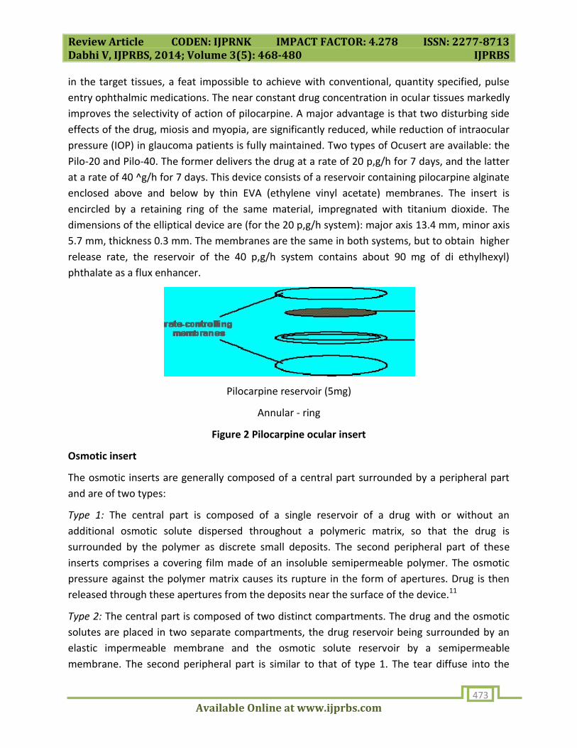
Diffusional insert or Ocuserts
Ocusert system is a novel ocular drug delivery system based on porous membrane. The release
of drug from diffusional inserts or ocuserts is based on a diffusional release mechanism. It
consists of a central reservoir of drug enclosed in specially designed microporous membrane
allowing the drug to diffuse from the reservoir at a precisely determined rate.
The ocusert pilocarpine ocular therapeutic system, developed by Alza Corporation, is notable
for several reasons. This product was the first rate controlled, rate specified pharmaceutical for
which the strength is indicated on the label by the rate of drug delivery in vivo, rather than by
the amount of contained drug. It provides predictable time independent concentrations of drug
Page 6
Review Article CODEN: IJPRNK IMPACT FACTOR: 4.278 ISSN: 2277-8713 Dabhi V, IJPRBS, 2014; Volume 3(5): 468-480 IJPRBS
Available Online at www.ijprbs.com 473
in the target tissues, a feat impossible to achieve with conventional, quantity specified, pulse
entry ophthalmic medications. The near constant drug concentration in ocular tissues markedly
improves the selectivity of action of pilocarpine. A major advantage is that two disturbing side
effects of the drug, miosis and myopia, are significantly reduced, while reduction of intraocular
pressure (IOP) in glaucoma patients is fully maintained. Two types of Ocusert are available: the
Pilo-20 and Pilo-40. The former delivers the drug at a rate of 20 p,g/h for 7 days, and the latter
at a rate of 40 ^g/h for 7 days. This device consists of a reservoir containing pilocarpine alginate
enclosed above and below by thin EVA (ethylene vinyl acetate) membranes. The insert is
encircled by a retaining ring of the same material, impregnated with titanium dioxide. The
dimensions of the elliptical device are (for the 20 p,g/h system): major axis 13.4 mm, minor axis
5.7 mm, thickness 0.3 mm. The membranes are the same in both systems, but to obtain higher
release rate, the reservoir of the 40 p,g/h system contains about 90 mg of di ethylhexyl)
phthalate as a flux enhancer.
Pilocarpine reservoir (5mg)
Annular - ring
Figure 2 Pilocarpine ocular insert
Osmotic insert
The osmotic inserts are generally composed of a central part surrounded by a peripheral part
and are of two types:
Type 1: The central part is composed of a single reservoir of a drug with or without an
additional osmotic solute dispersed throughout a polymeric matrix, so that the drug is
surrounded by the polymer as discrete small deposits. The second peripheral part of these
inserts comprises a covering film made of an insoluble semipermeable polymer. The osmotic
pressure against the polymer matrix causes its rupture in the form of apertures. Drug is then
released through these apertures from the deposits near the surface of the device.11
Type 2: The central part is composed of two distinct compartments. The drug and the osmotic
solutes are placed in two separate compartments, the drug reservoir being surrounded by an
elastic impermeable membrane and the osmotic solute reservoir by a semipermeable
membrane. The second peripheral part is similar to that of type 1. The tear diffuse into the
Page 7
Review Article CODEN: IJPRNK IMPACT FACTOR: 4.278 ISSN: 2277-8713 Dabhi V, IJPRBS, 2014; Volume 3(5): 468-480 IJPRBS
Available Online at www.ijprbs.com 474
osmotic compartment inducing an osmotic pressure that stretches the elastic membrane and
contracts the compartment including the drug, so that the active component is forced through
the single drug release aperture.11
Matrix systems
The second category, matrix system is a particular group of insoluble ophthalmic devices mainly
represented by contact lenses. It comprises of covalently cross-linked hydrophilic or
hydrophobic polymer that forms a three dimensional network or matrix capable of retaining
water, aqueous drug solution or solid components. The hydrophilic or hydrophobic polymer
swells by absorbing water. The swelling caused by the osmotic pressure of the polymer
segments is opposed by the elastic retroactive forces arising along the chains or crosslinked are
stretched until a final swelling (equilibrium) is reached.
Contact lenses
The initial use of contact lenses was for vision correction. Their use has been extended as
potential drug delivery devices by presoaking them in drug solutions. The main advantage of
this system is the possibility of correcting vision and releasing drug simultaneously. The contact
lenses have subdivided into 5 groups.
• Rigid
• Semi-rigid
• Elastomeric
• Soft hydrophilic
• Bio-polymeric
Rigid contact lenses have the disadvantage of being composed of polymers (e.g., poly methyl
methacrylic acid) hardly permeable to moisture and oxygen, a problem which has been
overcome by using gas permeable polymers such as cellulose acetate butyrate. However, these
systems are not suitable for prolonged delivery of drugs to the eye and their rigidity makes
them very uncomfortable to wear. For this reason, soft hydrophilic contact lenses were
developed for prolonged release of drugs such as pilocarpine, chloramphenicol, tetracycline,
prednisolone and sodium phosphate.12,13 The most commonly used polymer in the composition
of these types of lenses is hydroxy ethyl methyl metacrylic acid copolymerized with poly (vinyl
pyrrolidone) or ethylene glycol dimethacrylic acid (EGDM). Poly (vinyl pyrrolidone) is used for
increasing water of hydration, while EGDM is used to decrease the water of hydration. The soft
hydrophilic contact lenses are very popular because they are easy to fit and are tolerated
better. The drug incorporation into contact lenses depends on whether their structure is
hydrophilic or hydrophobic. When contact lens (including 35 to 80% water) is soaked in
Page 8
Review Article CODEN: IJPRNK IMPACT FACTOR: 4.278 ISSN: 2277-8713 Dabhi V, IJPRBS, 2014; Volume 3(5): 468-480 IJPRBS
Available Online at www.ijprbs.com 475
solution, it absorbs the drug. Drug release depends markedly on the amount of drug, the
soaking time of the contact lens and the drug concentration in the soaking solution.11
2. Soluble ocular inserts
These soluble inserts offer the advantage of being entirely soluble so that they do not need to
be removed from their site of application, thus limiting the intervention to insertion only. They
can be broadly divided into two types, the first one being based on natural polymers and the
other on synthetic or semi-synthetic polymers.
NATURAL POLYMERS
The first type of soluble inserts is based on natural polymer. Natural polymer used to produce
soluble ophthalmic inserts is preferably collagen. The therapeutic agent is preferably absorbed
by soaking the insert in a solution containing the drug. The amount of drug loaded will depend
on the amount of binding agent present, the concentration of the drug solution into which the
composite is soaked as well as the duration of the soaking. As the collagen dissolves, the drug is
gradually released from the interstics between the collagen molecules.14
SYNTHETIC AND SEMISYNTHETIC POLYMER
The second type of soluble insert is usually based on semi-synthetic polymers (e.g., cellulose
derivatives) or on synthetic polymers such as polyvinyl alcohol. A decrease of release rate can
be obtained by using eudragit, a polymer normally used for enteric coating, as a coating agent
of the insert. Eudragit coated inserts containing pilocarpine induced a miotic effect of a longer
duration, compared to the corresponding uncoated one. However, the inherent problems
encountered with these soluble inserts are the rapid penetration of the lachrymal fluid into the
device, the blurred vision caused by the solubilization of insert components and the risk of
expulsion due to the initial dry and glassy consistency of the device.15 Ethyl cellulose, a
hydrophobic polymer can be used to decrease the deformation of the insert and thus to
prevent blurred vision. As for the risk of expulsion, several authors have incorporated
carbomer, a strong but well tolerated bio-adhesive polymer. The soluble inserts offer the
additional advantage of being of a generally simple design, being based on products well
adapted for ophthalmic use and easily processed by conventional methods. The main
advantage is decreased release rate, but still controlled by diffusion.16
3. Bioerodible ocular inserts
These inserts are formed by bio-erodible polymers (e.g., cross-linked gelatin derivatives,
polyester derivatives) which undergo hydrolysis of chemical bonds and hence dissolution. The
great advantage of these bio-erodible polymers is the possibility of modulating their erosion
rate by modifying their final structure during synthesis and by addition of anionic or cationic
surfactants. A cross-linked gelatin insert was used to increase bioavailability of dexamethasone
Page 9
Review Article CODEN: IJPRNK IMPACT FACTOR: 4.278 ISSN: 2277-8713 Dabhi V, IJPRBS, 2014; Volume 3(5): 468-480 IJPRBS
Available Online at www.ijprbs.com 476
in the rabbit eye. The dexamethasone levels in the aqueous humor were found to be four-fold
greater compared to a dexamethasone suspension. However, erodible systems can have
significantly variable erosion rates based on individual patient physiology and lachrimation
patterns, while degradation products and residual solvents used during the polymer
preparation can cause inflammatory reaction.16
In the following paragraphs, some important ocular inserts are discussed which are available
commercially (SODI) or in the advanced states of development (Collagen shield, ocufit, NODS
and minidisc)
Soluble ophthalmic drug insert
Soluble ophthalmic drug insert (SODI) is a small oval wafer, which was developed by soviet
scientists for cosmonauts who could not use eye drops in weightless conditions. SODI is
together with the collagen shields, the first modern revival of the gelatin 'lamellae', which
disappeared from pharmacopoeias in the late forties. The SODI are the result of a vast
collaborative effort between eminent Russian chemists and ophthalmologists, and led
eventually (in 1976) to the development of a new soluble copolymer of acrylamide, #-
vinylpyrrolidone and ethyl acrylate (ratio 0.25: 0.25: 0.5), designated ABE. A comparison of
medicated eye films prepared with different polymers showed that ABE produced the highest
concentration of drugs in rabbit ocular tissues. After large-scale preclinical and clinical testing,
the ABE copolymer was used for the industrial manufacture of the SODI in the form of sterile
thin films of oval shape (9 x 4.5 mm, thickness 0.35 mm), weighing 15-16 mg, and color-coded
for different drugs (over 20 common ophthalmic drugs, or drug combinations). After
introduction into the upper conjunctival sac, a SODI softens in l0-15 s, conforming to the shape
of the eyeball. In the next l0-15 min the film turns into a polymer clot, which gradually dissolves
within 1 h while releasing the drug. The sensation of an 'extraneous body' in the eye disappears
in 5-15 min.17,18
Collagen shields
Collagen is the structural protein of bones, tendons, ligaments, and skin comprises more than
25% of the total body protein in mammals. This protein, which is derived from intestinal
collagen, has several biomedical applications, the main of which is probably catgut suture.
Bloomfield et al. are credited for first suggesting, in 1977 and 1978, the use of collagen inserts
as tear substitutes and as delivery systems for gentamicin. They compared the levels of
gentamicin in tears, cornea, and sclera of the rabbit eye after application of a collagen insert,
drops, an ointment or following subconjunctival administration. After 3 h, they found that the
collagen insert gave the highest concentration of gentamicin in the tear film and in the tissue.
Other treatments using collagen shields impregnated with gentamicin and dexamethasone
have been described.19 In rabbits, aqueous humor levels of dexamethasone and gentamicin
Page 10
Review Article CODEN: IJPRNK IMPACT FACTOR: 4.278 ISSN: 2277-8713 Dabhi V, IJPRBS, 2014; Volume 3(5): 468-480 IJPRBS
Available Online at www.ijprbs.com 477
achieved with collagen shields were compared to subconjunctival injections. The use of
collagen shields impregnated with gentamicin and dexamethasone was comparable to the
subconjunctival delivery of these drugs over a 10 h period.
Some drawbacks of these devices, to apply the collagen shield, the cornea is anaesthetized
while the physician uses a blunt forceps to insert the hydrated or unhydrated shield. Contrary
to medicated contact lenses, collagen shields often produce some discomfort and interfere with
vision. In rabbits, collagen shields have been found to exacerbate ulcerations of alkali-burned
corneas. A new preparation referred to as collasomes consists of small pieces (1 mm x 2 mm x
0.1 mm) of collagen suspended in a 1% methylcellulose vehicle. Kaufman and co-workers
recently reported that collasomes provide the same therapeutic advantages of the shields (high
and sustained levels of drugs and or lubricants to the cornea) while not presenting their
disadvantages.20
Ocufit
The Ocufit is a sustained release, rod shaped device made of silicone elastomer,21 patented in
1992 and currently developed by Escalon Ophthalmics Inc. (Skillman, NJ). It was designed to fit
the shape and size of the human conjunctival fornix. Accordingly, it does not exceed 1.9 mm in
diameter and 25-30 mm in length, although smaller sizes for children and newborn babies are
planned. The superiority of the cylindrical shape can be traced in an earlier paper by Katz and
Blackman. They reported the effect of the size and shape of the inserts on tolerance and
retention by human volunteers. These workers found that expulsion of rod shaped units was
significantly (P < 0.01) less frequent than expulsion of oval, flat inserts. A typical example of a
rod shaped insert is the Lacrisert (Merck and Co., Inc.), a cellulosic device used to treat dry-eye
patients. The insoluble Ocufit reportedly combines two important features, long retention and
sustained drug release. When placed in the upper fornix of volunteers, placebo devices were
retained for 2 weeks or more in 70% of the cases. Moreover, active disease (bacterial, allergic,
adenoviral conjunctivitis, trachoma, episcleritis, anterior uveitis, cornea1 ulcers or scars) did not
affect the ability of the patients to retain the inserts. Tetracycline-loaded inserts released in
vitro 45% of the drug over the 14 day period with an initial burst in the first day followed by a
constant rate over the remaining period.22
The minidisc ocular therapeutic system
This monolytic polymeric device, originally described by Bawa et al. (Bausch and Lomb,
Rochester, New York) and referred to as Minidisc ocular therapeutic system (OTS), is shaped
like a miniature (diameter 4-5 mm) contact lens, with a convex and a concave face, the latter
conforming substantially to the sclera of the eye. The particular size and shape reportedly allow
an easy placement of the device under the upper or lower lid without compromising comfort,
vision or oxygen permeability. When compared with another standard insert, the Lacrisert, the
Page 11
Review Article CODEN: IJPRNK IMPACT FACTOR: 4.278 ISSN: 2277-8713 Dabhi V, IJPRBS, 2014; Volume 3(5): 468-480 IJPRBS
Available Online at www.ijprbs.com 478
Minidisc was reported to require less time and less manual dexterity for insertion. Different
versions of the device have been evaluated, such as non-erodible hydrophilic, non-erodible
hydrophobic and erodible. In vitro tests showed that the hydrophilic OTS (based on
polyhydroxymethyl methacrylate) released sulfisoxazole for 118 h, while the hydrophobic unit
(based on a proprietary Bausch and Lomb pre-polymer) released gentamicin sulfate for more
than 320 h. Clinical trials on placebo units demonstrated that the devices were well tolerated
when placed either in the upper or lower conjunctival sac. In the eyes of healthy volunteers, the
hydrophilic OTS released sulfisoxazole continuously for 3 days. Further studies conducted on
the hydrophobic Minidisc8 showed that gentamicin sulfate was efficiently released in rabbit
eyes for 14 days.
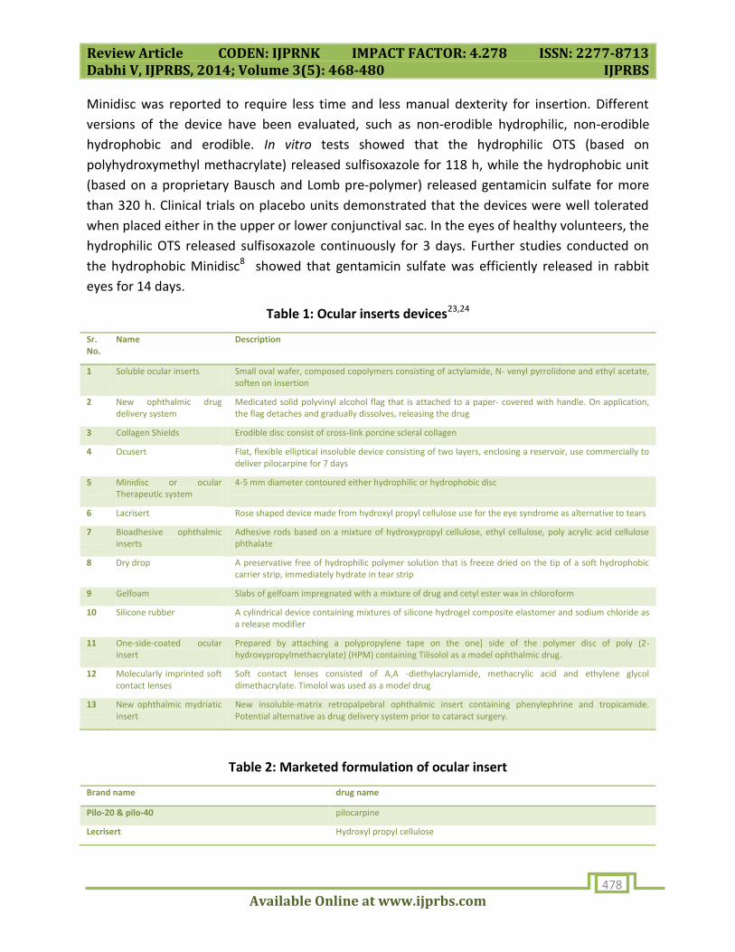
Table 1: Ocular inserts devices23,24
Sr. No.
Name Description
1 Soluble ocular inserts Small oval wafer, composed copolymers consisting of actylamide, N- venyl pyrrolidone and ethyl acetate, soften on insertion
2 New ophthalmic drug delivery system
Medicated solid polyvinyl alcohol flag that is attached to a paper- covered with handle. On application, the flag detaches and gradually dissolves, releasing the drug
3 Collagen Shields Erodible disc consist of cross-link porcine scleral collagen
4 Ocusert Flat, flexible elliptical insoluble device consisting of two layers, enclosing a reservoir, use commercially to deliver pilocarpine for 7 days
5 Minidisc or ocular Therapeutic system
4-5 mm diameter contoured either hydrophilic or hydrophobic disc
6 Lacrisert Rose shaped device made from hydroxyl propyl cellulose use for the eye syndrome as alternative to tears
7 Bioadhesive ophthalmic inserts
Adhesive rods based on a mixture of hydroxypropyl cellulose, ethyl cellulose, poly acrylic acid cellulose phthalate
8 Dry drop A preservative free of hydrophilic polymer solution that is freeze dried on the tip of a soft hydrophobic carrier strip, immediately hydrate in tear strip
9 Gelfoam Slabs of gelfoam impregnated with a mixture of drug and cetyl ester wax in chloroform
10 Silicone rubber A cylindrical device containing mixtures of silicone hydrogel composite elastomer and sodium chloride as a release modifier
11 One-side-coated ocular insert
Prepared by attaching a polypropylene tape on the one] side of the polymer disc of poly (2-hydroxypropylmethacrylate) (HPM) containing Tilisolol as a model ophthalmic drug.
12 Molecularly imprinted soft contact lenses
Soft contact lenses consisted of A,A -diethylacrylamide, methacrylic acid and ethylene glycol dimethacrylate. Timolol was used as a model drug
13 New ophthalmic mydriatic insert
New insoluble-matrix retropalpebral ophthalmic insert containing phenylephrine and tropicamide. Potential alternative as drug delivery system prior to cataract surgery.
Table 2: Marketed formulation of ocular insert
Brand name drug name
Pilo-20 & pilo-40 pilocarpine
Lecrisert Hydroxyl propyl cellulose
Page 12
Review Article CODEN: IJPRNK IMPACT FACTOR: 4.278 ISSN: 2277-8713 Dabhi V, IJPRBS, 2014; Volume 3(5): 468-480 IJPRBS
Available Online at www.ijprbs.com 479
REFERENCE
1. Maurice DM and Mishima S. Pharmacology of Eye, 1st ed.Springer-verlog, Berling 1984: 116-
9.
2. Chien YW. Novel drug delivery system. 2nd edition, revised and expanded, Marcel Dekker,
inc., New York, 1992; p. 269-300.
3. Khar RK and Vyas SP. Targeted and controlled drug delivery novel carrier systems. 1st edition.
CBS Publishers and Distributors, New Delhi, 2002.p. 384.
4. Saettone MF, Salminen L. Ocular inserts for topical delivery. Adv Drug Del Rev 1995; 16:95-
106.
5. Korsmeyer RW, Peppas NA. Macromolecular and modeling aspects of swelling- controlled
systems. In: Roseman TJ, Mansdorf SZ, editors. Controlled Release Delivery Systems. New York:
Marcel Dekker, 1983; p. 77-90
6. Darougar, Sohrab, Darougar and Dayshad, Patent literature review of ocular inserts. United
States Patent 6,264,971, Appl. No. 428967, Filed on November 4, 1999.
7. Heller J. Controlled release of biologically active compounds from bioerodible polymers.
Biomaterials 1980; 1:51-57.
8. Bawa R. Ocular inserts. In: Mitra AK, editor. Ophthalmic Drug Delivery Systems. New York:
Marcel Dekker, 1993; p. 223-259.
9. Saettone MF. Solid polymeric inserts/disks as ocular drug delivery systems. In: Edman P,
editor. Biopharmaceutics of ocular drug delivery. Boca Raton: CRC Press; 1993. p. 61-79.
10. Heller J. Controlled drug release from monolithic systems. In: Saettone MF, Bucci G, Speiser
P, editors. Ophthalmic Drug Delivery, Biopharmaceutical, Technological and Clinical Aspects,
Fidia Research Sereis. Vol. 11. Padua: Liviana Press, 1987; p. 179-189.
11. Rastogi SK, Vaya N, Mishra B. Ophthalmic inserts: An overview. The Eastern Pharmacist
1996; 2: 41-44
12. Praus R, Brettschneider I, Krejci L, Kalvodova D. Hydrophilic contact lenses as a new
therapeutic approach for the typical use of chloramphenicol and tetracycline. Ophthalmologica
1972; 165:62
Page 13
Review Article CODEN: IJPRNK IMPACT FACTOR: 4.278 ISSN: 2277-8713 Dabhi V, IJPRBS, 2014; Volume 3(5): 468-480 IJPRBS
Available Online at www.ijprbs.com 480
13. Hull DS, Edechauser HF, Hyndink RA. Ocular penetration of prednisolone and the hydrophilic
contact lense. Arch Ophthalmol 1974; 92:413.
14. Bloomfield SE, Miyata T, Dunn MW, Bueser N, Stenzel KH, Rubin AL. Soluble gentamicin
ophthalmic inserts as a drug delivery system. Arch Ophthalmol 1978; 96:885-887.
15. Saettone MF, Chetoni P, Torraca MT, Giannaccini B, Naber L, Conte U, et al. Application of
the compression technique to the manufacture of Pilocarpine inserts. Acta Pharm Technol
1990; 36:15-19
16. Attia MA, Kassem MA, Safwat SM. In vivo performance of 3-hdexamethasone ophthalmic
film delivery system in the Rabbit eye. Int J Pharm 1988; 47:21.
17. Khromow GL, Davydov AB, Maychuk YF, Tishina IF. Base for ophthalmological medicinal
preparations and an ophthalmological medicinal film. US. Patent 3, 935, 303, 1976.
18. Maichuk YF. Polymeric ophthalmic inserts with antibiotics, Proceedings of the Conference of
Ophthalmologists of the City of Moscow; 1967. p. 403-405.
19. Bloomfield SE, Miyata T, Dunn MW, Bueser N, Stenzel KH, Rubin AL. Soluble gentamicin
ophthalmic inserts as a drug delivery system. Arch Ophthalmol 1978; 96:885-887
20. Kaufman HE, Steinemann TL, Lehman E, Thompson HW, Varnell ED, Jacob- LaBarre JT, et al.
Collagen-based drug delivery and artificial tears. J Ocul Pharmacol 1994; 10:17-27.
21. Darougar S. Ocular insert for the fornix, U.S. Patent 5,147, 1992,647
22. Lamberts DW, Pavan-Langston D, Chu W. A clinical study of slow releasing artificial tears.
Ophthalmology 1978; 85:794-800.
23. Deivasigamani K, Mithun B, Pandey VP, Sonkar S. The concept of ocular inserts as drug
delivery system: An overview. Asian J. Pharm 2008; 12: 192-199.
24. Patel GM and Patel MM. Recent advances and challenges in ocular drug delivery system.
Pharma Times 2007; 39: 21-25.


![INTERNATIONAL JOURNAL OF PHARMACEUTICAL RESEARCH …ijprbs.com/issuedocs/2015/12/IJPRBS 1187.pdf · genetic modification.[7][6]. Bacteria, yeast and fungi are major producer for tannase](https://static.documents.pub/doc/80x56/6069fb864b95a814d101c24e/international-journal-of-pharmaceutical-research-1187pdf-genetic-modification76.jpg)


