Illicium verum Extracts Anti-Gastro Ulcerogenic Potential on Experimentally Rat Models Faten M. Ibrahim 1 *, Abeer Y Ibrahim 1 , EL Gohary A. E 1 , Mohamed S. Hussein 1 , Kawkab A. Ahmed 2 1 Medicinal and Aromatic Plants Research Department, Pharmaceutical and Drug Industries Division, National Research Centre, 12622 Dokki, Giza, Egypt. 2 Department of Pathology, Faculty of Veterinary Medicine, Cairo University, Giza, Egypt. Abstract: Background: The traditional herbal, medicinal plants, drugs are used in treating many diseases. The herbal treatment commonly used in treating most type of ulcers. Different extracts of Illicium verum were investigated for their anti-gastro ulcerogenic effect on rats. Aims: Evaluation of Illicium verum extracts as anti-ulcerogenic agent in rat model. Methods: plant essential oil and extracts (petroleum ether and aqueous alcoholic), were performed and then LD 50 values were determined (2500 mg/ kg b.wt. for aqueous alcoholic extract and 1250 mg/ kg for petroleum ether extract). Rats were classified into three main groups; negative control administered distilled water, positive controls administered different extracts as well as ulcerogenic sub-group and the last main group is treated animals. Results: in an in-vitro study, aqueous alcoholic extract exhibited the highest antioxidant properties and the lowest effect was recorded for petroleum ether extract. In an in-vivo experiment, aqueous alcoholic extract possessed potent anti-ulcerogenic effect as compared to famotin, reference drug. It enhanced the production of reduced glutathione and induced the glutathione reductase activity, superoxide dismutase activity and catalase activity determined in gastric mucosa with marked reduction in lipid peroxides productions in the two ulcerogenic models. Key words: Illicium verum, essential oils, antiulcer, (Alcohol & Aspirin) ulcer, antioxidants. Introduction The disordered physiological processes of PUD involve an imbalance between aggressive (acid, pepsin, and Helcobacter pylori) and defensive factors (mucin), prostaglandin, bicarbonate, nitric oxide, and growth factors). Stomach and the first few centimeters of the duodenum are the most common sites for ulcers. Acute peptic ulcers including lesions may be single or multiple and tissues down to the depth of the sub mucosa. The development of ulcers involves severe shock, illness, burns, various postsurgical complications and emotional disturbance. Chronic peptic ulcers sneak through muscle layers of the stomach wall and the epithelial. Peptic ulcers include a lot of complications such as perforation, pyloric stenosis and hemorrhage. Poor or difficult digestion and elimination, incorrect metabolism, mental and physical stresses enhance the development of ulcers 1 . There are number of drugs available for the treatment of peptic ulcers, but these drugs have a clinical evaluation of indicates high incidences of retrogression, drug interactions and side effects. Decreasing gastric lumen mucus production with suppression of mucosal blood flow by administration of ethanol encourage ulcer formation that reduces prostaglandins and glutathione concentrations through the depletion in cysteine level that's required for glutathione biosynthesis with increasing gastric vascular permeability and inducting International Journal of PharmTech Research CODEN (USA): IJPRIF, ISSN: 0974-4304, ISSN(Online): 2455-9563 Vol.9, No.5, pp 65-80, 2016
Transcript
Illicium verum Extracts Anti-Gastro Ulcerogenic Potential onExperimentally Rat Models
Faten M. Ibrahim1*, Abeer Y Ibrahim1, EL Gohary A. E1,Mohamed S. Hussein1, Kawkab A. Ahmed2
1Medicinal and Aromatic Plants Research Department, Pharmaceutical and DrugIndustries Division, National Research Centre, 12622 Dokki, Giza, Egypt. 2Department
of Pathology, Faculty of Veterinary Medicine, Cairo University, Giza, Egypt.
Abstract: Background: The traditional herbal, medicinal plants, drugs are used in treating manydiseases. The herbal treatment commonly used in treating most type of ulcers. Differentextracts of Illicium verum were investigated for their anti-gastro ulcerogenic effect on rats.Aims: Evaluation of Illicium verum extracts as anti-ulcerogenic agent in rat model.Methods: plant essential oil and extracts (petroleum ether and aqueous alcoholic), wereperformed and then LD50 values were determined (2500 mg/ kg b.wt. for aqueous alcoholicextract and 1250 mg/ kg for petroleum ether extract). Rats were classified into three maingroups; negative control administered distilled water, positive controls administered differentextracts as well as ulcerogenic sub-group and the last main group is treated animals.Results: in an in-vitro study, aqueous alcoholic extract exhibited the highest antioxidantproperties and the lowest effect was recorded for petroleum ether extract. In an in-vivoexperiment, aqueous alcoholic extract possessed potent anti-ulcerogenic effect as compared tofamotin, reference drug. It enhanced the production of reduced glutathione and induced theglutathione reductase activity, superoxide dismutase activity and catalase activity determined ingastric mucosa with marked reduction in lipid peroxides productions in the two ulcerogenicmodels.Key words: Illicium verum, essential oils, antiulcer, (Alcohol & Aspirin) ulcer, antioxidants.
Introduction
The disordered physiological processes of PUD involve an imbalance between aggressive (acid, pepsin,and Helcobacter pylori) and defensive factors (mucin), prostaglandin, bicarbonate, nitric oxide, and growthfactors). Stomach and the first few centimeters of the duodenum are the most common sites for ulcers. Acutepeptic ulcers including lesions may be single or multiple and tissues down to the depth of the sub mucosa. Thedevelopment of ulcers involves severe shock, illness, burns, various postsurgical complications and emotionaldisturbance. Chronic peptic ulcers sneak through muscle layers of the stomach wall and the epithelial. Pepticulcers include a lot of complications such as perforation, pyloric stenosis and hemorrhage. Poor or difficultdigestion and elimination, incorrect metabolism, mental and physical stresses enhance the development ofulcers1. There are number of drugs available for the treatment of peptic ulcers, but these drugs have a clinicalevaluation of indicates high incidences of retrogression, drug interactions and side effects. Decreasing gastriclumen mucus production with suppression of mucosal blood flow by administration of ethanol encourage ulcerformation that reduces prostaglandins and glutathione concentrations through the depletion in cysteine levelthat's required for glutathione biosynthesis with increasing gastric vascular permeability and inducting
International Journal of PharmTech Research CODEN (USA): IJPRIF, ISSN: 0974-4304, ISSN(Online): 2455-9563 Vol.9, No.5, pp 65-80, 2016
Faten M. Ibrahim et al /International Journal of PharmTech Research, 2016,9(5),pp 65-80. 66
leukotreines and free radicals production. These deleterious effects contribute in loss of mucosal maintenancereproduce ulceration. Free radicals strongly participate in the two ulcer types, acute and chronic cases.Therefore, agents can scavenged radicals are considered in treating gastric ulcers2. Also administration ofaspirin over than recommended dose induces oxyradicals formation and leads to erosive gastritis3. Medicinalplants considered as an alternative strategies in drug discovery and a valuable source of new molecules. Intraditional medicine there is large plants number known to possess antiulcer properties that may provide a newulcer drugs or improve existed anti-ulcer drugs specially after chemical manipulation4 Peptic ulcer disease(PUD), including gastric and duodenal ulcers, is the most prevalent gastrointestinal disorder that requires awell-targeted therapeutic strategy. These are prospective aggressive effects support belief for the developmentof new antiulcer drugs and search for natural products. Medicinal plants such as Ocimum sanctum, Azadirachtaindica, Asparagus racemosus, Musa sapientum, Centella asiatica, Bacopamonnieri and Bidenspilosa mayoffer better protection and decreasing expected relapse 5. Gastric disorder is classified in Ayurveda ancientsystem of Indian medicine, as sula, parinamsula and amlapitta which match with functional dyspepsia andclinical condition of peptic ulcers 6. Carbenoxolone, was the first drug effective against gastric ulcer discoveredas a result for research on a commonly used indigenous plant, Glycyrrhiza glabra (Leguminosceae) 7. Plantdrugs, show the antiulcer activity, constitutes the active ingredients such as flavonoid, tannins and terpenoids 8.Different classes of drugs are used in the treatment of peptic ulcer but most of these drugs exhibit serious sideeffects like arrhythmias, gynaecomastia, arthralgia, hypergastrinemia and haemopoeitic changes. Alternativeapproach in recent years is the research for medicaments from ayurvedic or traditional medicinal system. Theuse of phytoconstituents as drug therapy to treat major ailments has proved to be clinically effective and lessrelatively toxic than the existing drugs and also reduces the aggressive factors serving as a tool in the preventionof peptic ulcer. In this time, 75-80% of the world populations still use herbal medicine mainly in developingcountries for primary health care because of better cultural acceptability, better compatibility with the humanbody and lesser side effects. Illicium verum Hook. f. (Austrobaileyales: Schisandraceae), a fruit ordinarilyknown as star anise, is mainly distributed in the tropical and is native to southwest China and Vietnam andsubtropical areas of Asia. Illicium verum was considered as one of the things “both food and medicine” by theMinistry of Health of the People's Republic of China (2002), implying its low or non-toxicity to humans. Now,the research focus on I.verum has been mainly on food and medical fields 9. The essential oil of I. verum can beused as a flavoring and the fruits are commonly used as an ingredient of the traditional “five-spice” powder ofChinese cooking. The extraction from I. verum used as diuretic, stomachic, stimulant, and properties, andcarminative, is used as a pharmaceutical supplement 10. The herb is reported to be antifungal, antibacterial andantioxidant. It is able to increase production of milk new mother. The use to facilitate birth and to increase thelibido, as well as to relieve menopausal discomforts; oil is used in rheumatism as recommended by some folkremedies11. The attributed medicinal properties are carminative, stomachic, stimulant, expectorant and diuretic.In east it is used to combat colic and rheumatism. It is a common flavoring for medicinal tea, cough mixture andpastilles. In traditional system of medicines, Illicium verum fruit is used having both culinary and medicinaluses. Its seed oil is used worldwide as medicine. Due to the potential side effects of synthetic antioxidants,essential oil derived from natural products can be served as an alternative source for the further improvement ofsynthetic antioxidant.
This work focused on evaluation of Illicium verum 1) extracts as anti-ulcerogenic herbal medicine in ratmodel. 2) invitro antioxidants activity. 3) chemical composition of essential oil and finally 4) examinehistopathologically the stomach of rats treated with illicium extracts compared to reference drug.
Materials and Methods:
Plant Material:
Star anise seeds were obtained from local market and identified by Pro. Dr. Lotfy Boulis. Seeds weregrounded to small granules and then subjected to sequential soxhlet extraction with petroleum ether andaqueous ethanol (70%). The obtained petroleum ether and ethanol extracts were dried under reduced pressure tobe free from any solvent residues. The remained concentrated petroleum ether and aqueous alcoholic extractswere used in biological study.
Two methods were carried out to extract essential oil:
1. Modified (Hydrodistillation) Clevenger trap12 was used for essential oil extraction.
Faten M. Ibrahim et al /International Journal of PharmTech Research, 2016,9(5),pp 65-80. 67
2. Water and Steam: it is such as hydro-distillation but during this process, the water remains below the plantmaterial, which has been placed on a grate while the steam is introduced from outside the main still (indirectsteam). Extraction times were performed (1, 2 and 3 hour) with three replicates for both extraction methods.
GC–MS analysis of essential oil: samples were supplied into gas chromatography–mass spectrometryinstrument at Medicinal and Aromatic Plants Researches Dept., National Research Center with the followingspecifications, instrument: a TRACE GC Ultra Gas Chromatographs (THERMO Scientific Corp, USA),coupled with a THERMO mass spectrometer detector (ISQ Single Quadrupole Mass Spectrometer). The GC–MS system was equipped with a TG-WAX MS column(30 m × 0.25 mm i.d., 0.25 µm film thickness). Analyseswere carried out using helium as carrier gas at a flow rate of 1.0 ml min−1anda split ratio of 1:10 using thefollowing temperature program: 40◦C for 1 min; rising with 4.0◦C min−1–160 ◦C and held for 6 min; rising with6◦C min−1–210◦C and held for 1 min. The injector and detector were held at 210◦C. Diluted samples (1:10hexane, v/v) of 0.2µl of the mixtures were always injected. Mass spectra were obtained by electron ionization(EI) at 70 eV, using a spectral range of m/z40–450. Most of the compounds were identified using two differentanalytical methods: (a) KI, Kovats indices in reference to alkanes.
In-vitro antioxidant properties investigation
Free radical scavenging effect: The free radical scavenging activity of plant extracts was measured by 1,1-diphenyl-2-picryl-hydrazil (DPPH•)13 DPPH• (0.1 mM) was prepared in methanol and an aliquot (1mL) wasadded to each extract (3mL) prepared a concentrations of 62.5, 125, 250, 500 and 1000µg/ ml. Mixtures werevigorously shaken and allowed to stand at room temperature for 50 min before recording absorbance at 517 nm(Jasco V630 spectrophotometer). Lower absorbance of the reaction mixture indicated higher free radicalscavenging activity. The DPPH• radical concentration in the reaction medium was calculated from thefollowing equation: DPPH• scavenging effect (%) = 100 − [(A0-A1)/A0) ×100], Where A0 was the absorbance ofthe control reaction and A1 was the absorbance in the presence of the sample 14. VC was used as a positivecontrol in all in-vitro tests.
Reduction capability: Plant extract reduction capacities were determined as previously reported 15. Extractsdiluted in MeOH to 62.5, 125, 250, 500 and 1000 µg/ml (1ml) were added to phosphate buffer (2.5 ml, 0.2 M,pH 6.6) containing potassium ferricyanide [K3Fe (CN) 6] (2.5 ml, 1%). The assay mixture was incubated at 50°Cfor 20 min and the reaction stop with addition of TCA (10%, 2.5 ml) and centrifugation for 10 min at 1000 × g(MSE Mistral 2000, UK). The upper layer (ca. 2.5ml) was diluted with methanol (2.5 ml) containing FeCl3(0.5ml, 0.1%) and the absorbance was measured at 700 nm. Higher absorbance of the reaction mixture indicatedgreater reducing power.
Superoxide anion scavenging activity: Superoxide anion scavenging activity was based on the methoddescribed by 16 in which superoxide radicals are generated in a phenazinemethosulphate (PMS)–nicotinamideadenine dinucleotide (NADH) system by oxidation of NADH and assayed by the reduction ofnitrobluetetrazolium (NBT). Superoxide radicals are generated in Tris–HCl buffer (16 mM, pH 8.0, 3 ml)containing NBT (50µM, 1ml), NADH (78µM, 1 ml) and extracts to be assayed (1 ml) at differentconcentrations.
The reaction was started by adding PMS (phenazinemethosulfate) solution (10 µM, 1ml). The reactionmixture was incubated at 25ºC for 5 min and absorbance reading (560 nm) was performed. Decreasingabsorbance indicated increasing superoxide anion scavenging activity. The percentage inhibition of superoxideanion generation was calculated as: percent of inhibition = [(A0-A1)/A0] × 100; where A0 was the absorbance ofthe control (L-ascorbic acid), and A1 was the absorbance of test extracts.
Metal chelating activity: Ferrous ion chelating was estimated by the method of 17. 0.05 ml of FeCl2 solution (2mM), test extracts or V.C. was added and the reaction was initiated by an addition of ferrozine (5 mM, 0.2 ml)with vigorous shaking; after incubating at room temperature for ten minutes, absorbance at 562 nm wasrecorded. Percentage inhibition of the ferrozine-Fe2+complex was determined based on the equation: inhibition(%) = [(A0-A1)/ A0] ×100, where A0 was the absorbance of control while A1 was the absorbance in the presenceof test sample and standard.
Inhibition of lipid peroxidation: The potential of plant extracts at different concentration was determinedaccording to the method of 18 to inhibit peroxidation of linoleic acid. L-ascorbic acid was used as the reference
Faten M. Ibrahim et al /International Journal of PharmTech Research, 2016,9(5),pp 65-80. 68
compound. A pre-emulsion was prepared by mixing 175μg Tween 20, 155μL linoleic acid, and 0.04Mpotassium phosphate buffer (pH 7.0). 1mL of sample at different concentrations in 99.5% ethanol was mixedwith 4.1mL linoleic emulsion, 0.02M phosphate buffer (pH 7, 8mL) and distilled water (7.9mL). The mixedsolutions of all samples (21mL) were incubated in screw cap-tubes under dark conditions at 40˚C at certain timeintervals. To 0.1mL of this mixture was pipetted and added with 9.7mL of 75% and 0.1mL of 30% ammoniumthiocyanate sequentially. After 3 min, 0.1mL of 0.02M ferrous chloride in 3.5% hydrochloric acid was added tothe reaction mixture. The peroxide level was determined by reading daily of the absorbance at 500 nm in aspectrophotometer. All test data was the average of three replicate analyses. The inhibition of lipid peroxidationin percentage was calculated by the following equation; % Inhibition = [(A0 - A1) / A0] x 100 (1). Where A0 wasthe absorbance of the control reaction andA1 was the absorbance in the presence of extracts or standardcompounds.
Animals, Housing and Experimental Design: The acute toxicity test for plant extracts was estimated toevaluate any possible toxicity. Female albino mice (n=8) were tested by administering different extracts dosesby increasing or decreasing the dose, according to the response of animal 19.The dosing patron was 500, 1000,1500, 2000, 2500, 3000, 3500 and 4000 mg/kg body weight for alc. extract and pet ether extract, while controlgroup received only the normal saline. All groups were observed for any gross effect or mortality during 48hr.Death of half of examined animals was observed at 2500 and 1250 mg/kg b.wt for alcoholic extract and petether extract, respectively.
Anti-ulcer effect of Illicium verum extracts on rat administered alcohol induced gastric mucosal injury:The anti-ulcerogenic effect on adult female albino rats, weighing 80-100 g. Animals were obtained from animalhouse of National Research Centre. Each kedge was contained six female albino rats and they were feed onstandard diet. They maintained under standard laboratory conditions; housing temperature was 24°C; relativehumidity was 65± 5% with light/ dark cycles (12/12 h). The dose was selected on basis of acute toxicity. Plantextracts was employed at oral dose as aqueous suspension using distilled water. Three main groups includingnegative group administered distilled water (first group), second group (positive controls) to investigate anydeleterious effect caused by extract, drug or alcohol administration which include four sub-groups administeredpetroleum ether extract at 125 mg/ kg b.wt, alc. extract at 250 mg/ kg b.wt, famotin at 20mg/ kg 20 . while thethird main group was received 1 ml alcohol / 100g orally only to induce gastric ulcer 21. The third main groupincludes three sub-groups firstly rats received 1 ml alcohol with Illicium petroleum ether treatment at 125mg/kg b.wt. one hour before and after administration,22 second sub-group received 1ml alcohol orally withtreatment with Illicium EtOH at 250 mg/ kg, one hour before and after administration while famotin wasadministered as reference drug at 20mg/ kg b.wt. by the same way of extracts. Rats were anesthetized and thensacrificed; their stomachs were excised and opened along the greater curvature. Gastric mucosal tissue wastaken from the antral portion of the stomach for biochemical estimations. The gastric mucosa was scrapped witha scrapper, homogenized in ice cold phosphate buffer (pH 7.2) to prepare the mucosal homogenate.Homogenates were centrifuged at 3000 rpm for 10 min and the supernatants were used for further studies.
Anti-ulcer Effect of Illicium verum Extracts on rat injured with Aspirin Induced Gastric Mucosal Injury:The animals were fasted for 48hr 23 prior to experiment while water was permitted. Group 1 received saline,group II received Illicum pet. ether extract at 125 mg/ kg b.wt. for seven days, group III received Illicum EtOHextract at 250mg/kg b.wt. for seven days, group IV received aspirin at 400 mg/ kg suspended in 0.5% carboxymethyl cellulose, group V received aspirin with pretreatment of Illicium pet. ether ext. for seven days group VIreceived aspirin with pretreatment of Illicium EtOH extract for seven days, group VII received aspirin and thentreated with Illicium pet. ether extract for ten days, therapeutic group. However, group VIII received aspirin andthen treated with EtOH extract, therapeutic groups for ten days after aspirin administration. After theexperimental period rats were scarified after anesthesia and their stomachs were opened along the greatercurvature after four hours of aspirin administration. The gastric tissue was fixed in buffered formalin forhistopathological study while gastric mucosal tissues were taken and prepared as mentioned above forbiochemical estimations. Reduced glutathione level in the stomach tissue was determined according to themethod of 24. Gastric superoxide dismutase (SOD) activity was estimated by the method of 25. Catalase (CAT)activity was measured by following decomposition of H2O2 according to the method of 26. Glutathionereductase activity was measured spectrophotometrically at 340nm 27 and the amount of the enzyme reducing1μmol GSSG per min per mg protein was regarded one activity unit as elsewhere described. The TBARS level,an index of malondialdehyde (MDA), production was determined by the method of 28. The protein content wasdetermined by the method of Bradford 29.
Faten M. Ibrahim et al /International Journal of PharmTech Research, 2016,9(5),pp 65-80. 69
Histopathological Study: Specimens of stomach from all animals were dissected immediately after death,then opened along the greater curvature and washed thoroughly with distilled water to remove their contents toavoid digestion of upper layers of gastric mucosa by digestive enzymes. All the specimens were fixed in 10%neutral-buffered formal saline for 72 hours at least, washed in distilled water and then dehydrated in ascendinggrades of alcohol, cleared in xylene and embedded in paraffin wax. Serial sections of 6μm thick were cut andstained with Haematoxylin and eosin 30 for histopathological investigation.
Statistical Analysis: The data of biochemical assessments are expressed as mean ± SD and ANOVA followedby post hoc test (LSD) to compare the groups. The statistical analysis was carried out by the version 9.01 of theSPSS program. Values were considered statistically significant; P < 0.05.
Results
Illicium antioxidant properties
There are large number of diseases related to excessive stress of free radicals in human bodyhemorrhagic shock, Alzheimer and Parkinson’s disease, tumor promotion and carcinogenesis, AIDS andgastrointestinal dysfunctions 31. Therefore, antioxidant properties of Illicium pet. ether extract and aqueousalcoholic extract were evaluated. In all tests, the aqueous alcoholic extract showed to have the highest recordedvalues followed with pet. ether extract. Concerning radical scavenging effect, DPPH radical .
The scavenging effect was increased gradually with increasing the concentration of tested extracts.Aqueous alc. extract and VC, reference compound, reached 90% inhibition percentage at 1000 µg/ mL whilepetroleum ether produced weak effect as DPPH radical scavenger at all concentrations (Figure, 1).
Fig. 1. Free radical scavenging effect of illicium verum extracts at different concentration. All data are mean oftriplicates. Data were analyzed by ANOVA one way and presented as mean± SD, P< 0.05.
The reduction capability of alcoholic Illicium verum extract occurred to be higher than pet. etherextract. It represented the highest reduction capability at the four tested concentrations. This capability wasenhanced by increasing the concentration but they remained much lower than VC (Figure, 2).
Faten M. Ibrahim et al /International Journal of PharmTech Research, 2016,9(5),pp 65-80. 70
Fig. 2. Reduction capability of Illicium verum extracts at different concentration. All data are mean oftriplicates. Data were analyzed by ANOVA one way and presented as mean± SD, P< 0.05.
The presence of metal ions in tissue increase the oxidative stress by catalyzing cell oxidation reactions32. The two tested extracts showed to have languorous chelating effect at the four tested concentrations. Themaximum chelation percentage was 26% for alcoholic extract at 1000µg/ mL (Figure, 3). .
Fig. 3. Metal chelating effect of Illicium extracts at different concentration. All data are mean of triplicates.Data were analyzed by ANOVA one way and presented as mean± SD, P< 0.05.
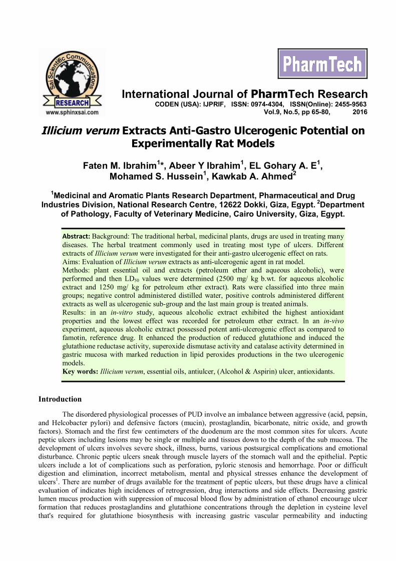
The alcoholic extract of Illicium possesed strong superoxide radical scavenging produced by phenazine.It represented 94% scavenging effect at 1000µg/ mL while pet. ether extract remained the feeble one inscavenging super radicals (Figure, 4).
Faten M. Ibrahim et al /International Journal of PharmTech Research, 2016,9(5),pp 65-80. 71
Fig. 4. Superoxide radical scavenging effect of Illicium extracts at different concentration. All data are mean oftriplicates. Data were analyzed by ANOVA one way and presented as mean± SD, P< 0.05.
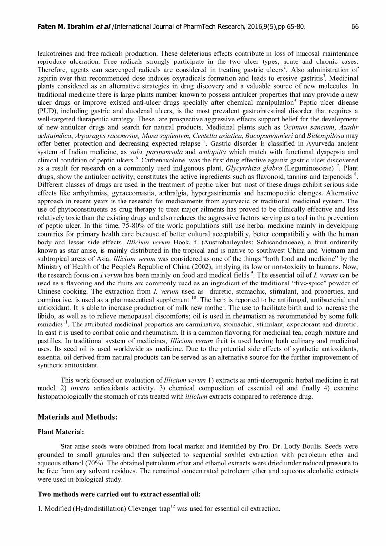
The antioxidant effect of extracts reflected on their properties as inhibitors for lipid peroxidation whenthey were tested by linoleic assay. Illicium alcoholic extract remained the best effector to inhibit lipid peroxideproduction, inhibition percentage reached 100% at 1000µg/ mL. The inhibition was increased in a concentrationdependent manner with two extracts. Alcoholic extract inhibition percentage of lipid peroxidation ranged from35 to 100% while pet. ether extract ranged from 32 to 80% (Figure, 5 ).
Fig. 5. Lipid peroxide inhibition effect of Illicium extracts at different concentration. All data are mean oftriplicates. Data were analyzed by ANOVA one way and presented as mean± SD, P< 0.05.
The recorded antioxidant properties in the in-vitro tests support the probability of Illicium verum extracts asanti-ulcerogenic agent. Animals were treated with ethanol to reproduce ulcer and the two Illicium extracts, pet.ether and aqueous alcoholic extracts, were tested as protective materials against gastric ulcers. Administrationof Table 1 revealed that Illicium extracts didn't produce any deleterious effect as observed in investigated bio-parameters. Ethanol adminstration significantly reduced the glutathione concentration in stomach wheraseaqueous alcoholic extract administration significantly induce the glutatione production in tissue to record valuemore than –ve control, administered distelled water, (19.08 and 15.46 mg/g tissue, respectively). The aqueousextract enhanced GSH production to reach 15.18 mg/g tissue. Likewise, glutathione reductase activity had thesame trend of reduced glutathione production. The activity was induced by aqueous alcoholic extractadministration (28.32μmol/ min/ mg protein) more than pet. ether extract (24.32μmol/ min/ mg protein) toameliorate the status of injured tissue (11.16μmol/ min/ mg protein, for ethanol induced group). As well,administration of Illicium aqueous alcoholic extract possessed a plausible ameliorative effect on superoxide
Faten M. Ibrahim et al /International Journal of PharmTech Research, 2016,9(5),pp 65-80. 72
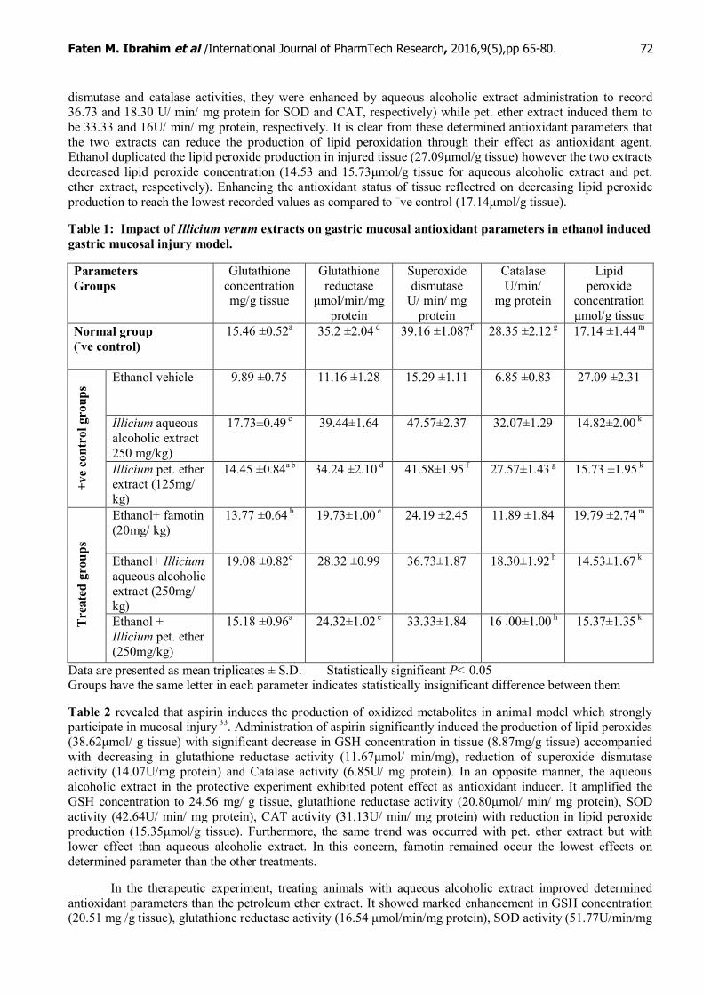
dismutase and catalase activities, they were enhanced by aqueous alcoholic extract administration to record36.73 and 18.30 U/ min/ mg protein for SOD and CAT, respectively) while pet. ether extract induced them tobe 33.33 and 16U/ min/ mg protein, respectively. It is clear from these determined antioxidant parameters thatthe two extracts can reduce the production of lipid peroxidation through their effect as antioxidant agent.Ethanol duplicated the lipid peroxide production in injured tissue (27.09μmol/g tissue) however the two extractsdecreased lipid peroxide concentration (14.53 and 15.73μmol/g tissue for aqueous alcoholic extract and pet.ether extract, respectively). Enhancing the antioxidant status of tissue reflectred on decreasing lipid peroxideproduction to reach the lowest recorded values as compared to –ve control (17.14μmol/g tissue).
Table 1: Impact of Illicium verum extracts on gastric mucosal antioxidant parameters in ethanol inducedgastric mucosal injury model.
ParametersGroups
Glutathioneconcentrationmg/g tissue
Glutathionereductase
μmol/min/mgprotein
Superoxidedismutase
U/ min/ mgprotein
CatalaseU/min/
mg protein
Lipidperoxide
concentrationμmol/g tissue
Normal group(-ve control)
15.46 ±0.52a 35.2 ±2.04 d 39.16 ±1.087f 28.35 ±2.12 g 17.14 ±1.44 m
19.08 ±0.82c 28.32 ±0.99 36.73±1.87 18.30±1.92 h 14.53±1.67 k
Tre
ated
gro
ups
Ethanol +Illicium pet. ether(250mg/kg)
15.18 ±0.96a 24.32±1.02 e 33.33±1.84 16 .00±1.00 h 15.37±1.35 k
Data are presented as mean triplicates ± S.D. Statistically significant P< 0.05Groups have the same letter in each parameter indicates statistically insignificant difference between them
Table 2 revealed that aspirin induces the production of oxidized metabolites in animal model which stronglyparticipate in mucosal injury 33. Administration of aspirin significantly induced the production of lipid peroxides(38.62μmol/ g tissue) with significant decrease in GSH concentration in tissue (8.87mg/g tissue) accompaniedwith decreasing in glutathione reductase activity (11.67μmol/ min/mg), reduction of superoxide dismutaseactivity (14.07U/mg protein) and Catalase activity (6.85U/ mg protein). In an opposite manner, the aqueousalcoholic extract in the protective experiment exhibited potent effect as antioxidant inducer. It amplified theGSH concentration to 24.56 mg/ g tissue, glutathione reductase activity (20.80μmol/ min/ mg protein), SODactivity (42.64U/ min/ mg protein), CAT activity (31.13U/ min/ mg protein) with reduction in lipid peroxideproduction (15.35μmol/g tissue). Furthermore, the same trend was occurred with pet. ether extract but withlower effect than aqueous alcoholic extract. In this concern, famotin remained occur the lowest effects ondetermined parameter than the other treatments.
In the therapeutic experiment, treating animals with aqueous alcoholic extract improved determinedantioxidant parameters than the petroleum ether extract. It showed marked enhancement in GSH concentration(20.51 mg /g tissue), glutathione reductase activity (16.54 μmol/min/mg protein), SOD activity (51.77U/min/mg
Faten M. Ibrahim et al /International Journal of PharmTech Research, 2016,9(5),pp 65-80. 73
protein) and CAT activity (32.77U/min/mg protein) with decreasing lipid peroxide production (14.24μmol/ gtissue). However, the pet. ether extract produced lower effect than aqueous alcoholic extract. It increased GSHconcentrations (14.48 mg/ g tissue) with increasing the activity of G. reductase (12.24μmol/min/mg protein),SOD (48.27 U/min/mg protein) and CAT (29.03U/min/mg protein) with reduction in lipid peroxide production(16.73μmol/ g tissue) .
It is clear from the mentioned data in Table 2 that petroleum ether extract of Illicium verum exhibitedprotective effect equals to its therapeutic effect in most cases whereas protective effect of aqueous alcoholicextract was better than its therapeutic effect.
Table 2: Protective and therapeutic potential of illicium verum extracts on the gastric antioxidantparameters in aspirin ulcer model
Glutathioneconcentration
mg/g tissue
Glutathionereductaseμmol/min/mg
protein
SuperoxidedismutaseU/ min/ mg
protein
CatalaseU/min/
mg protein
Lipidperoxideμmol/gtissue
Parameter
Group
-ve Control 14.95±1.43 d 35.55±1.37 38.61±2.19 28.35±2.43k 17.14±1.23 p
Illicium aqueousalcoholic extract(250 mg/ kg)
39.17±2.63 24.90±1.96 53.97±2.89 e 56.47±2.51 14.75±1.09 t
Illicium pet.ether extract (125mg/ kg)
30.99±1.82 21.47±2.16 b 44.73±1.97 f 49.30±2.61 15.99±1.25 t
Aspirin(400 mg/kg)
11.67±0.95 8.87±0.98 a 14.07±1.42 6.85±1.00 38.62±2.36
+ ve c
ontr
ol g
roup
s
Aspirin+famotin(20mg/ kg)
15.19±1.17 h 10.89±1.64 a 28.88±1.94 13.89±1.13 14.94±1.38 t
Aspirin +Illicium aqueousalcoholic extract( 250mg/ kg)
24.56±1.48 20.80±1.85 b 42.64±2.38 f 31.13±1.45m 15.35±1.39 t
Prot
ectiv
etr
eatm
ents
Aspirin +Illicium pet.ether extract(125mg/ kg)
20.31±1.31 g 13.07±1.07 c 34.29±2.41 27.40±2.14k 17.71±1.41 p
Aspirin +Illicium aqueousalcoholic extract(250 mg/ kg)
20.51±1.43 g 16.54±1.11d 51.77±1.99 e 32.77±2.47m 14.24±1.27 t
The
rape
utic
trea
tmen
ts
Aspirin +Illicium pet.ether extract( 125 mg/ kg)
14.48±1.11 h 12.24±1.36 c 48.27±2.18 29.03±1.29k 16.73±1.19 p
Data are presented as mean triplicates ± S.D. Statistically significant P< 0.05Groups have the same letter in each parameter indicates insignificant difference between them
Analysis of Illicium verum essential oil
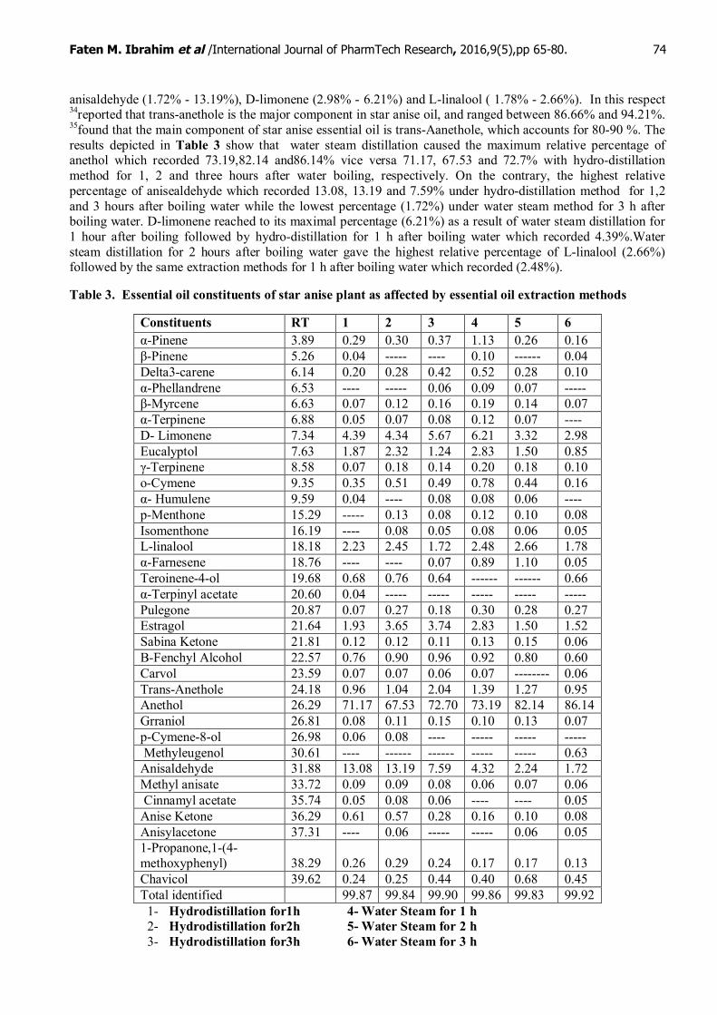
Table 3 summarized the effect of both extraction methods and the boiling time on essential oil constituents.Thirty five components were identified in the essential oil of A. cerefolium L. underwent at different treatmentsthat represented 99.83–99.92% of the oils. The major components were anethol (67.53 % - 73.19%),
Faten M. Ibrahim et al /International Journal of PharmTech Research, 2016,9(5),pp 65-80. 74
anisaldehyde (1.72% - 13.19%), D-limonene (2.98% - 6.21%) and L-linalool ( 1.78% - 2.66%). In this respect34reported that trans-anethole is the major component in star anise oil, and ranged between 86.66% and 94.21%.35found that the main component of star anise essential oil is trans-Aanethole, which accounts for 80-90 %. Theresults depicted in Table 3 show that water steam distillation caused the maximum relative percentage ofanethol which recorded 73.19,82.14 and86.14% vice versa 71.17, 67.53 and 72.7% with hydro-distillationmethod for 1, 2 and three hours after water boiling, respectively. On the contrary, the highest relativepercentage of anisealdehyde which recorded 13.08, 13.19 and 7.59% under hydro-distillation method for 1,2and 3 hours after boiling water while the lowest percentage (1.72%) under water steam method for 3 h afterboiling water. D-limonene reached to its maximal percentage (6.21%) as a result of water steam distillation for1 hour after boiling followed by hydro-distillation for 1 h after boiling water which recorded 4.39%.Watersteam distillation for 2 hours after boiling water gave the highest relative percentage of L-linalool (2.66%)followed by the same extraction methods for 1 h after boiling water which recorded (2.48%).
Table 3. Essential oil constituents of star anise plant as affected by essential oil extraction methods
Faten M. Ibrahim et al /International Journal of PharmTech Research, 2016,9(5),pp 65-80. 75
Histopathological results:
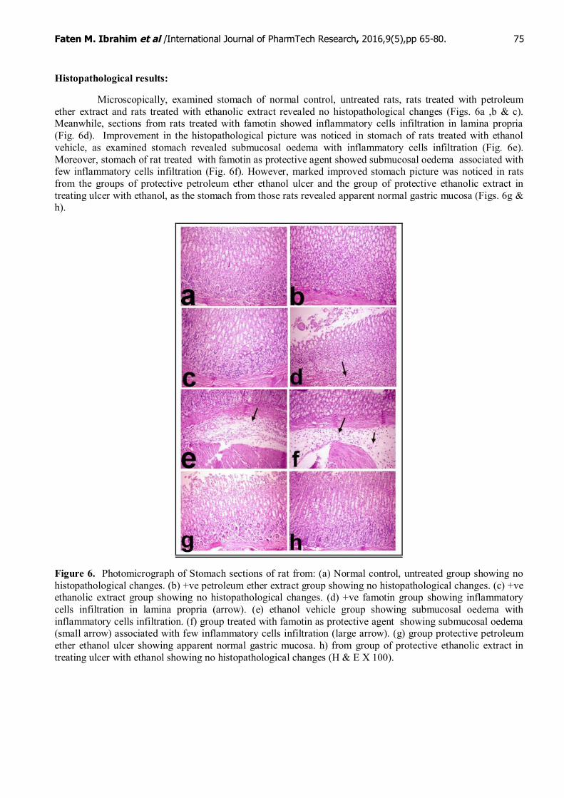
Microscopically, examined stomach of normal control, untreated rats, rats treated with petroleumether extract and rats treated with ethanolic extract revealed no histopathological changes (Figs. 6a ,b & c).Meanwhile, sections from rats treated with famotin showed inflammatory cells infiltration in lamina propria(Fig. 6d). Improvement in the histopathological picture was noticed in stomach of rats treated with ethanolvehicle, as examined stomach revealed submucosal oedema with inflammatory cells infiltration (Fig. 6e).Moreover, stomach of rat treated with famotin as protective agent showed submucosal oedema associated withfew inflammatory cells infiltration (Fig. 6f). However, marked improved stomach picture was noticed in ratsfrom the groups of protective petroleum ether ethanol ulcer and the group of protective ethanolic extract intreating ulcer with ethanol, as the stomach from those rats revealed apparent normal gastric mucosa (Figs. 6g &h).
Figure 6. Photomicrograph of Stomach sections of rat from: (a) Normal control, untreated group showing nohistopathological changes. (b) +ve petroleum ether extract group showing no histopathological changes. (c) +veethanolic extract group showing no histopathological changes. (d) +ve famotin group showing inflammatorycells infiltration in lamina propria (arrow). (e) ethanol vehicle group showing submucosal oedema withinflammatory cells infiltration. (f) group treated with famotin as protective agent showing submucosal oedema(small arrow) associated with few inflammatory cells infiltration (large arrow). (g) group protective petroleumether ethanol ulcer showing apparent normal gastric mucosa. h) from group of protective ethanolic extract intreating ulcer with ethanol showing no histopathological changes (H & E X 100).
Faten M. Ibrahim et al /International Journal of PharmTech Research, 2016,9(5),pp 65-80. 76
Histopathological results of asprin ulcer
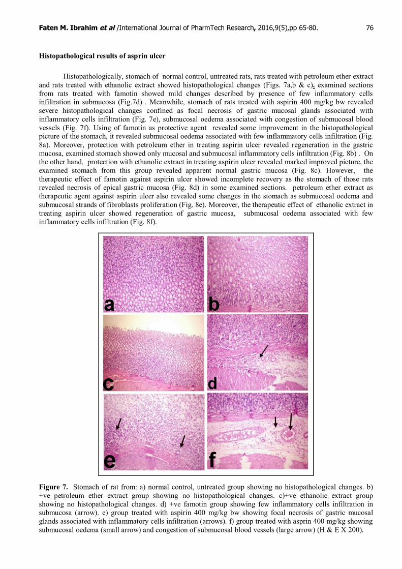
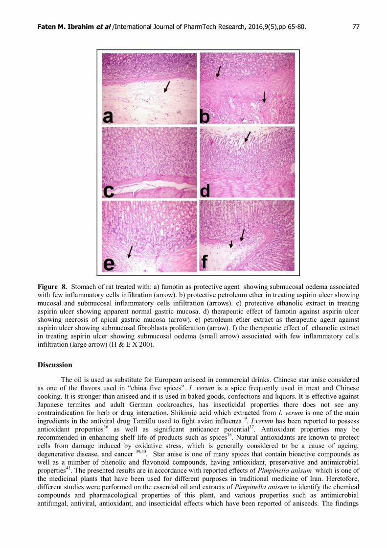
Histopathologically, stomach of normal control, untreated rats, rats treated with petroleum ether extractand rats treated with ethanolic extract showed histopathological changes (Figs. 7a,b & c). examined sectionsfrom rats treated with famotin showed mild changes described by presence of few inflammatory cellsinfiltration in submucosa (Fig.7d) . Meanwhile, stomach of rats treated with aspirin 400 mg/kg bw revealedsevere histopathological changes confined as focal necrosis of gastric mucosal glands associated withinflammatory cells infiltration (Fig. 7e), submucosal oedema associated with congestion of submucosal bloodvessels (Fig. 7f). Using of famotin as protective agent revealed some improvement in the histopathologicalpicture of the stomach, it revealed submucosal oedema associated with few inflammatory cells infiltration (Fig.8a). Moreover, protection with petroleum ether in treating aspirin ulcer revealed regeneration in the gastricmucosa, examined stomach showed only mucosal and submucosal inflammatory cells infiltration (Fig. 8b) . Onthe other hand, protection with ethanolic extract in treating aspirin ulcer revealed marked improved picture, theexamined stomach from this group revealed apparent normal gastric mucosa (Fig. 8c). However, thetherapeutic effect of famotin against aspirin ulcer showed incomplete recovery as the stomach of those ratsrevealed necrosis of epical gastric mucosa (Fig. 8d) in some examined sections. petroleum ether extract astherapeutic agent against aspirin ulcer also revealed some changes in the stomach as submucosal oedema andsubmucosal strands of fibroblasts proliferation (Fig. 8e). Moreover, the therapeutic effect of ethanolic extract intreating aspirin ulcer showed regeneration of gastric mucosa, submucosal oedema associated with fewinflammatory cells infiltration (Fig. 8f).
Figure 7. Stomach of rat from: a) normal control, untreated group showing no histopathological changes. b)+ve petroleum ether extract group showing no histopathological changes. c)+ve ethanolic extract groupshowing no histopathological changes. d) +ve famotin group showing few inflammatory cells infiltration insubmucosa (arrow). e) group treated with aspirin 400 mg/kg bw showing focal necrosis of gastric mucosalglands associated with inflammatory cells infiltration (arrows). f) group treated with asprin 400 mg/kg showingsubmucosal oedema (small arrow) and congestion of submucosal blood vessels (large arrow) (H & E X 200).
Faten M. Ibrahim et al /International Journal of PharmTech Research, 2016,9(5),pp 65-80. 77
Figure 8. Stomach of rat treated with: a) famotin as protective agent showing submucosal oedema associatedwith few inflammatory cells infiltration (arrow). b) protective petroleum ether in treating aspirin ulcer showingmucosal and submucosal inflammatory cells infiltration (arrows). c) protective ethanolic extract in treatingaspirin ulcer showing apparent normal gastric mucosa. d) therapeutic effect of famotin against aspirin ulcershowing necrosis of apical gastric mucosa (arrow). e) petroleum ether extract as therapeutic agent againstaspirin ulcer showing submucosal fibroblasts proliferation (arrow). f) the therapeutic effect of ethanolic extractin treating aspirin ulcer showing submucosal oedema (small arrow) associated with few inflammatory cellsinfiltration (large arrow) (H & E X 200).
Discussion
The oil is used as substitute for European aniseed in commercial drinks. Chinese star anise consideredas one of the flavors used in “china five spices”. I. verum is a spice frequently used in meat and Chinesecooking. It is stronger than aniseed and it is used in baked goods, confections and liquors. It is effective againstJapanese termites and adult German cockroaches, has insecticidal properties there does not see anycontraindication for herb or drug interaction. Shikimic acid which extracted from I. verum is one of the mainingredients in the antiviral drug Tamiflu used to fight avian influenza 9. I.verum has been reported to possessantioxidant properties36 as well as significant anticancer potential37. Antioxidant properties may berecommended in enhancing shelf life of products such as spices38. Natural antioxidants are known to protectcells from damage induced by oxidative stress, which is generally considered to be a cause of ageing,degenerative disease, and cancer 39,40. Star anise is one of many spices that contain bioactive compounds aswell as a number of phenolic and flavonoid compounds, having antioxidant, preservative and antimicrobialproperties41. The presented results are in accordance with reported effects of Pimpinella anisum which is one ofthe medicinal plants that have been used for different purposes in traditional medicine of Iran. Heretofore,different studies were performed on the essential oil and extracts of Pimpinella anisum to identify the chemicalcompounds and pharmacological properties of this plant, and various properties such as antimicrobialantifungal, antiviral, antioxidant, and insecticidal effects which have been reported of aniseeds. The findings
Faten M. Ibrahim et al /International Journal of PharmTech Research, 2016,9(5),pp 65-80. 78
revealed that aniseeds can cause gastric protection, muscle relaxant, and affect digestive system. These findingsare in agreement with anisum has hypolipidemic and hypoglycemic effects and reduces lipid peroxidation indiabetic patients. In addition to, aniseeds showed anticonvulsant impact, reduced morphine dependence, andinduced conditioned place aversion in mice. Aniseed also has beneficial effects on menopausal anddysmenorrhea hot flashes in women. The recorded activities in this work for Illicium verum are attributed to itscomponents. Especially its essential oil content. The plant essential oil is traditionally obtained by hydro-distillation. This technique has been controversial for subsequent determination of the oil chemical compositionbecause of the possible transformation of aroma-active compounds by heat, steam, and pH 42. Steam distillationprocess, in which the combination of the relatively high temperature of steam and the hydrolytic influence ofwater may cause the degradation of essential oil components 43,44. It was reported that the most importantcompounds of aniseeds essential oils were estragole, trans-anethole, γ-hymachalen, panisaldehyde and methylchavicol. Due to broad spectrum of very few clinical studies and pharmacological effects of this plant,performed on this plant, more clinical trials are recommended to evaluate the beneficial effects of Pimpinellaanisum in human models and identification of active compounds of plant which can lead to synthesis of newdrugs from the active ingredients in future. Our results are in agreement with that anise significantly inhibitedgastric mucosal damage induced by necrotizing agents and indomethacin. The antiulcer effect was furtherconfirmed histologically45.
Conclusions:
Our findings show that star anise fruits could be considered as a good source of natural compoundswith significant antioxidant and anti gastric ulcer activities (ethanol and aspirin) which can be attributed to thehigh percentage of the main constituents (phenolic compounds) or to synergy among the different oilconstituents.
References
1. Ghangale GR, Mahale Tushar, Jadhav ND. Evaluation of antiulcer activity of Ocimum sanctum inrats.Veterinary World 2009; 2;12: 465-466.
2. Salim AWS. Use of scavenging oxygen derived free radicals to protect rat against aspirin and ethanol-induced erosive gastritis. J. Pharmaceutical Sci., 1992; 81: 9.
3. Adams JD. Burgers Medicinal Chemistry and Drug Discovery. 5th Edition, H. Ewolff (John Wiley andSons, New York), 1996; pp: 261.
4. Zakaria ZA, Hisamb E, Rofieeb MS, Norhafizahc M, Somchita MN, Tehd LK, Salleh MZ. In vivoantiulcer activity of the aqueous extract of Bauhinia purpurea leaf. J., Ethnopharmacol. 2011;137:1047– 1054.
5. Vasudeva N, Sethi P, Sharma SK, Kumar S, Sharma S. Antiulcer Potential of the Ethanolic Extract ofAervaPersica Merrill Root in Rats. JAcupunct Meridian Stud. 2012; 5(2):80-86.
6. Sharma PV. Treatment of parinamasula. In: Cakradutta: a treatise on principles and practices ofAyurvedic Medicine. Varanasi: Chaukhambha Orientalia, 1994b; pp. 249–258.
7. Brown HM, Christie AB, Colin Jones E. Glycyrrhetinic acid hydrogen succinate (disodium) salt, a newanti-inflammatory compound. Lancet 1959; 2: 492–496.
8. Gadekar R, Singaur PK, Chaurasia PK, Pawar RS, Patil UK. A potential of some medicinal plants asan antiulcer agents. Pharmacogn Rev; 2010; 4(8): 136–146.
9. Ohira H, Torii N, Aida TM, Watanabe M, Smith RLJ. Rapid separation of shikimic acid fromChinese star anise Illicium verum (Hook f) with hot water extraction. Separation and PurificationTechnology. 2009; 69:102–108.
10. Lee SW, Li G, Lee KS, Jung JS, Xu ML, Seo CS, Chang HW, Kim SK, Song DK Son JK. Preventiveagents against sepsis and new phenylpropanoid glucosides from the fruits of Illicium verum. PlantaMedica. 2003; 69:861–864.
11. http;//www.digherbs.com/star-anise.html.12. ASTA. Official analytical methods of the American Spice Trade Association. ASTA, Englewood
Cliffs, N.J. p. 1968; 8-11.
Faten M. Ibrahim et al /International Journal of PharmTech Research, 2016,9(5),pp 65-80. 79
13. Yamaguchi T, Takamura H, Matoba T, Terao J. HPLC method for evaluation of the free radicalscavenging activity of foods by using 1,1, diphenyl-2-picrylhydrazyl. Biosci. Biotechnol. Biochem.1998; 62: 1201-1204.
14. Oktay M, Gülçin I, Küfrevio ˇglu ÖI. Determination of in vitro antioxidant activity of fennel(Foeniculumvulgare) seed extracts. Lebensmittel-Wissenchaft und Technologie. 2003; 36: 263–271.
15. Oyaizu M. Studies on products of browning reaction: Antioxidative activities of products of browningreaction prepared from glucosamine. Jpn J Nutr. 1986; 44: 307–315.
16. Gülçin I, Büyükokuroˇglu ME, Küfrevioˇglu ÖI. Metal chelating and hydrogen peroxide scavengingeffects of melatonin. Journal of Pineal Research 2003a; 34: 278–281.
17. Dinis TCP, Madeira VMC, Almeida LM. Action of phenolic derivatives (acetaminophen, salicylate,and 5-aminosalicylate) as inhibitors of membrane lipid peroxidation and as peroxyl radical scavengers.Arch Biochem Biophys. 1994; 315: 161 – 169.
18. Gulcin I, Sat I G, Beydemir S, Elmastas M, Kufrevioglu OI. Comparison of antioxidant activity ofclove (Eugenia caryophylataThunb) buds and lavender (Lavandulastoechas L.). Food Chem., 2004; 87:393-400.
19. Bruce RD. An up-and-down procedure for acute toxicity testing. Fundamental and Applied Toxicology.1985; 5: 151-157.
20. Bharti S, Wahane VD, Kumar VL. Protective effect of Calotropis procera latex extracts onexperimentally induced gastric ulcers in rat. Journal of Ethnopharmacology. 2010; 127: 440-444.
21. Robert A. Cytoprotection by prostaglandins. Gastroenterology. 1979; 77: 761-767.22. Mahmood AA, Mariod AA, Al-Bayaty F, Abdel-Wahab SI. Antiulcerogenic activity of Gynura
procumbens leaf extract against experimentally-induced gastric lesions in rats. J. Med. Plants Res.,2010; 4(8): 685-691.
23. Malltka Jainu, Shyamala Devi CS. Antioxidant effect of methanolic Solanum nigrum berries on aspirininduced gastric mucosal injury. Indian Journal of Clinical Biochemistry. 2004; 19(1): 57- 61.
24. Ellman GL. Tissue sulfhydryl groups. Archives of Biochemistry and Biophysics. 1959; 82: 70-77.25. Kakkar P, Das B, Viswanathan PN. A modifie spectrophotometric assay of superoxide dismutase.
Indian Journal of Biochemistry&, Biophysics. 1984; 21: 130-132.26. Beers RF, Sizer IW. A spectrophotometric method for measuring the breakdown of H2 O2 by catalase.
258-265, Verlog Chemie, Deerfield Beach, Fl.28. Ohkawa H, Ohishi N, Yagi K. Assay for lipid peroxides in animal tissues by thiobarbituric acid
reaction. Anal. Biochem. 1979; 95: 351-358.29. Bradford MM. A rapid and sensitive method for the quantitation of microgram quantities of protein
utilizing the principle of protein-dye binding. Analytical Biochemistry. 1976;72: 248-254.30. Drury RAB,Wallington FA. Carleton’s Histological Technique 4th Ed. Oxford, New York, Toronto,
Oxford university press. 1980.31. Mac-Manus JPA, Cason JE. Carbohydrate histochemistry studies by a cetylation technique. Periodic
acid method. J. Exp. Med. 1950;91: 651-668.32. El Gengaihi S, Faten M. Aboul Ella, Abeer Y. Ibrahim, Doha H. Abou Baker. Antioxidant Activities of
Selected Grape Wastes from Egypt. Ijppr. Human, 2015; 4 (1): 212-229.33. Abdelaaty AS, Abeer Y. Ibrahim, Essam E, Mansour SA. Acetylcholinesterase Inhibition And
Antioxidant Activity Of Some Medicinal Plants For Treating Neuro Degenarative Disease., Afr J TraditComplement Altern Med. 2015; 12(3):97-103.
34. Abeer Y. Ibrahim, Nermeen M. Shaffie. Protective Effect of Solanum indicum Var. Distichum Extracton Experimentally Induced Gastric Ulcers in Rat. Global Journal of Pharmacology. 2013;7 (3): 325-332.
35. Padmashree A, Roopa N, Semwal AD, Sharma GK, Agathian G, Bawa AS. Star anise (Illicium verum)and black caraway (Carum nigrum) as natural antioxidants. Food Chemistry. 2007; 104: 59–66.
36. Chempakam B, Balaji S. Star Anise. United Kingdom: CAB International. 2008; 319-330.37. Shu X, Liu XM, Fu CL, Liang QX. Extraction, characterization and antitumor effect of the
polysaccharides from star anise Illicium verum (Hook f). Journal of Medicinal Plants Research. 2010;4: 2666–2673.
Faten M. Ibrahim et al /International Journal of PharmTech Research, 2016,9(5),pp 65-80. 80
38. Prakash B, Shukla R, Singh P., Mishra PK, Dubey NK, Kharwar RN. Efficacy of chemicallycharacterized Ocimum gratissmum L. essential oil as an antioxidant and a safe plant based antimicrobialagainst fungal and aflatoxin B1 contamination of spices. Food Res. Int. 2011;44:385–390.
39. Ringman M, Frautschy A, Cole M, Masterman L, Cummings L. Potential role of the curry spicecurcumin in Alzheimer’s disease. Curr. Alzheimer’s Res. 2005; 2:131–136.
40. Viuda-Martos M, Ruiz-Navajas Y, Sa´ nchez-Zapata E, Ferna´ ndez- Lo´ pez, J, Pe rez-Alvarez JA.Antioxidant activity of essential oils of five spice plants widely used in Mediterranean diet. Flav. Fragr.J. 2010; 25: 13–19.
41. Shobana S, Naidu KA. Antioxidant activity of selected Indian spices. Prostaglandins Leukot. Essent.Fatty Acids. 2000; 6:107–110.
42. Jiménez-Carmona MM, Ubera JL, Luque de Castro MD. Comparison of continuous subcritical waterextraction and hydrodistillation of marjoram essential oil. Journal of Chromatography A. 1999; 855:625.
43. Katsiotis ST. Study of Different Parameters Influencing the Composition of Hydrodistilled SweetFennel Oil [J]. Flavour Fragrance Journal. 1988; 4: 221-224.
44. Yan Jianhui, Xiao Xuxian, Huang Kelong. Component Analysis of Volatile Oil from Illicium VerumHook. f. [J]. Journal of Central South University of Technology.2002; 9(3):173-176(Ch).
45. Al Mofleh IA, Alhalder AA, Mossa JS, Al- Soohalbani MO Rafatullah S. “Aqueous suspension ofanise “Pimpinella anisum” protects rats against chemically induced gastric ulcers,” World Journal ofGastroenterology. 2007; 13, 7:1112–1118.