Eur. J. Biochem. 104,289-296 (1980) Interpretation of the Mossbauer Spectra of the High-Potential Iron Protein from Chromatium Peter MIDDLETON, Dominic P. E. DICKSON, Charles E. JOHNSON, and James D. RUSH Department of Physics, University of Liverpool (Received July 19, 1979) The Mossbauer spectra of both reduced and oxidized high-potential iron protein (Hipip) from Chromatium have been analysed using computer fits to theoretical spectra derived from a spin Hamiltonian. Fits to spectra obtained over a range of temperatures between 4.2 and 195 K and in applied magnetic fields up to 10.0 T lead to a consistent set of hyperfine parameters. These results are interpreted in terms of a model of the four-iron four-sulphide active centre which is consistent with its electronic and magnetic properties in both redox states. In the model for the reduced centre all four iron atoms have essentially the same valence, intermediate between ferric and ferrous, with the spins being coupled antiferromagnetically to give the centre zero net spin. The oxidized centre has one less electron which at low temperatures appears to have come predominantly from one pair of iron atoms which thus become ferric with the other pair remaining substantially unchanged. It is clear from the Mossbauer hyperfine parameters obtained from the computer fits to the low-temperature spectra that a larger magnetic moment is associated with the ferric/ferrous pair of iron atoms than with the ferric pair of iron atoms. This also explains the g values with an average of greater than 2 which are observed in electron paramagnetic resonance (EPR) measurements. At higher temperatures the differences between the electron charge density at the different iron atoms in the oxidized centre appear to become smeared out. The high-potential iron protein (Hipip) from Chro- matium belongs to the class of iron-sulphur proteins which contain one four-iron four-sulphide centre per molecule. X-ray crystallographic studies on this pro- tein show that the active centre consists of four iron atoms, each in a distorted tetrahedral environment of three labile sulphur atoms and a fourth sulphur ligand from a cysteine residue of the amino acid chain [l]. The four-iron and eight-iron ferredoxins also con- tain active centres of this type. It has been proposed that the centre can exist in three oxidation states [2], which may be designated C3+, C2+ and C1+. C2+ corresponds to the equivalent states of oxidized fer- redoxin and reduced Hipip, with C3 corresponding to oxidized Hipip and C'+ to reduced ferredoxin. Differences in the surrounding protein configuration are thought to be responsible for the different redox behaviour of the active centre in the different proteins. Mossbauer spectroscopy on all three states has assigned formal valences of 3 Fe3 + 1 Fez+, 2 Fe3 + 2 Fe2+ Abbreviations. EPR, electron paramagnetic resonance; Hipip high-potential iron protein; ENDOR, electron nuclear double resonance. and 1 Fe3 + 3 Fe2 for the C3 +, C2 + and C1 + states respectively [3]. While a considerable amount of information can be obtained from a qualitative interpretation of the Mossbauer spectra of these proteins [3,4], the present work represents the next stage with computer-fitting of the Mossbauer spectra. The aim is to provide data relevant to an understanding of both the magnetic properties of the iron atoms within the active centre and of the distribution of electron charge between them. The procedures adopted for the computer analysis of the Mossbauer spectra of Chromatium Hipip are closely similar to those used in analyzing the spectra of the four-iron ferredoxin from Bacillus stearothermo- philus [5]. The program used is a form of that of Lang and Dale [6], modified to fit a spectrum with up to four components, each resulting from a separate spin Hamiltonian of the form:
Transcript
Eur. J. Biochem. 104,289-296 (1980)
Interpretation of the Mossbauer Spectra of the High-Potential Iron Protein from Chromatium Peter MIDDLETON, Dominic P. E. DICKSON, Charles E. JOHNSON, and James D. RUSH
Department of Physics, University of Liverpool
(Received July 19, 1979)
The Mossbauer spectra of both reduced and oxidized high-potential iron protein (Hipip) from Chromatium have been analysed using computer fits to theoretical spectra derived from a spin Hamiltonian. Fits to spectra obtained over a range of temperatures between 4.2 and 195 K and in applied magnetic fields up to 10.0 T lead to a consistent set of hyperfine parameters.
These results are interpreted in terms of a model of the four-iron four-sulphide active centre which is consistent with its electronic and magnetic properties in both redox states. In the model for the reduced centre all four iron atoms have essentially the same valence, intermediate between ferric and ferrous, with the spins being coupled antiferromagnetically to give the centre zero net spin. The oxidized centre has one less electron which at low temperatures appears to have come predominantly from one pair of iron atoms which thus become ferric with the other pair remaining substantially unchanged. It is clear from the Mossbauer hyperfine parameters obtained from the computer fits to the low-temperature spectra that a larger magnetic moment is associated with the ferric/ferrous pair of iron atoms than with the ferric pair of iron atoms. This also explains the g values with an average of greater than 2 which are observed in electron paramagnetic resonance (EPR) measurements. At higher temperatures the differences between the electron charge density at the different iron atoms in the oxidized centre appear to become smeared out.
The high-potential iron protein (Hipip) from Chro- matium belongs to the class of iron-sulphur proteins which contain one four-iron four-sulphide centre per molecule. X-ray crystallographic studies on this pro- tein show that the active centre consists of four iron atoms, each in a distorted tetrahedral environment of three labile sulphur atoms and a fourth sulphur ligand from a cysteine residue of the amino acid chain [l]. The four-iron and eight-iron ferredoxins also con- tain active centres of this type. It has been proposed that the centre can exist in three oxidation states [2], which may be designated C3+, C2+ a nd C1+. C 2 + corresponds to the equivalent states of oxidized fer- redoxin and reduced Hipip, with C3 + corresponding to oxidized Hipip and C'+ to reduced ferredoxin. Differences in the surrounding protein configuration are thought to be responsible for the different redox behaviour of the active centre in the different proteins. Mossbauer spectroscopy on all three states has assigned formal valences of 3 Fe3 + + 1 Fez+, 2 Fe3 + + 2 Fe2+
Abbreviations. EPR, electron paramagnetic resonance; Hipip high-potential iron protein; ENDOR, electron nuclear double resonance.
and 1 Fe3 + + 3 Fe2 + for the C3 +, C2 + and C1 + states respectively [3].
While a considerable amount of information can be obtained from a qualitative interpretation of the Mossbauer spectra of these proteins [3,4], the present work represents the next stage with computer-fitting of the Mossbauer spectra. The aim is to provide data relevant to an understanding of both the magnetic properties of the iron atoms within the active centre and of the distribution of electron charge between them.
The procedures adopted for the computer analysis of the Mossbauer spectra of Chromatium Hipip are closely similar to those used in analyzing the spectra of the four-iron ferredoxin from Bacillus stearothermo- philus [ 5 ] . The program used is a form of that of Lang and Dale [6], modified to fit a spectrum with up to four components, each resulting from a separate spin Hamiltonian of the form:
290 Interpretation of the Mossbauer Spectra of Chromatiurn Hipip
where H is the applied magnetic field, S and I are the electronic and nuclear spins, Q is the nuclear quadru- pole moment, V,, and q are the principal component and asymmetry parameter of the electric field gradient tensor, A is the magnetic hyperfine coupling tensor, and p, p,, g and g , are the Bohr magneton, nuclear magneton, electronic g tensor and nuclear g factor respectively.
The details of the sample preparation and Moss- bauer spectroscopy have been given previously [4].
Table 1. Mean Mossbauer parameters for reduced Chromatium Hlpip The chemical shifts 6 (relative to the centre of the iron metal spectrum at room temperature) and the quadrupole splittings LIE, = 1/2r2QVz. (1 - y2/3)"2 are given. y is the asymmetry parameter
Temperature 6 AEQ
K mm SKI
195 0.38 0.97 -
I 1 0.42 1.11 -
4.2" 0.43 + 2.14 0.8
RESULTS
Reduced Hipip The previous Mossbauer data [4] and the lack of
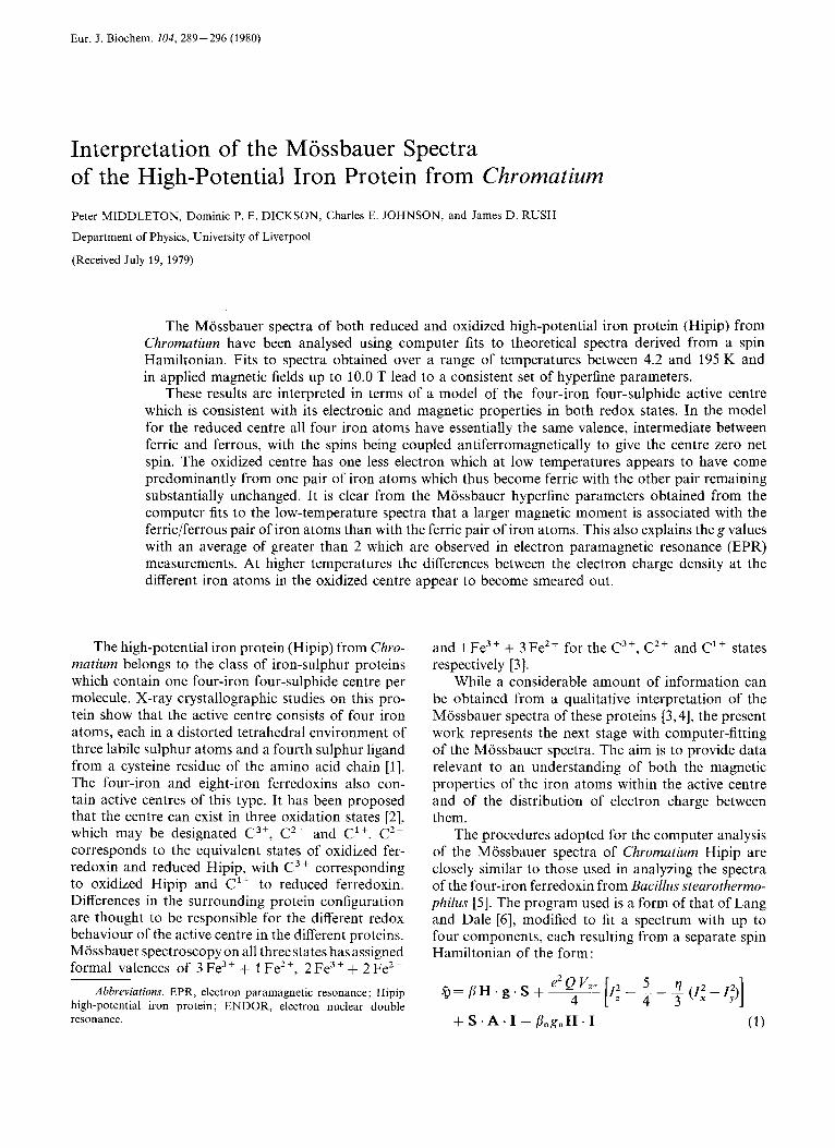
any EPR signal clearly indicates that in reduced Hipip the total spin of the centre S equals zero. The pro- jections of the individual spins along the total spin are thus also zero and the first and third terms of Eqn (1) vanish. Although the spectra do not indicate large differences between the iron atoms, completely satis- factory fits could not be obtained using only one or two components ; therefore four components were used in the fitting, with each subspectrum being ob- tained using the second and fourth terms of Eqn (1) together with a chemical shift term. The use of four components is in keeping with there being four iron atoms in the active centre but as it is impossible to be sure that the fits are unique only the mean values of the Mossbauer parameters are given in Table 1. Some representative spectra, together with the computer- fitted components, are shown in Fig. 1. The values of the chemical shifts are consistent with the formal valence assignment 2 Fe3 + and 2 Fe2+ and are very close to those obtained from the four-iron and eight- iron ferredoxins in the oxidized (i.e. C2+) state [3]. The computer fits give no evidence of any hyperfine field, which confirms that the centre has zero net spin.
Oxidized Hip ip
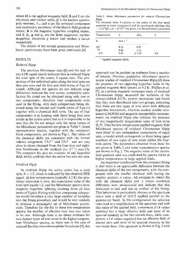
In oxidized Hipip the active centre has a total spin, S = 112, which is indicated by the observed EPR signal. At low temperatures (typically 4.2 K) the spin- lattice relaxation is slow, the expectation value of the total spin equals 112, and the Mossbauer spectra show magnetic hyperfine splitting resulting from all four terms of Eqn(1). Fitting with four component subspec- tra would introduce a vary large number of variables into the fitting procedure and would be very unlikely to produce a meaningful set of Mossbauer param- eters. Therefore for the fits to these low-temperature spectra the number of effective iron sites was taken to be two. Although there is no direct evidence for two distinct types of iron atom in the higher-tempera- ture Mossbauer spectra, as there was in the case of reduced Bacillus stearothermophilus ferredoxin [ 5 ] , this
a Applied magnetic fields.
approach can be justified on evidence from a number of sources. Previous qualitative Mossbauer spectro- scopic studies of oxidized Chromatium Hipip [4] show the presence of two opposing hyperfine fields in the applied magnetic field spectra at 4.2 K. Phillips et al. [7], in a proton magnetic resonance study of oxidized Chromutium Hipip, measured the variation of the contact-shifted P-CH2 proton resonances and found that they were distributed into two groups, indicating that there are two types of iron atom with different hyperfine interactions. Electron nuclear double reso- nance (ENDOR) and magnetic susceptibility measure- ments on oxidized Hipip also indicate the presence of two magnetically inequivalent types of iron atom [8,9]. Thus the low-temperature applied magnetic field Mossbauer spectra of oxidized Chromatium Hipip were fitted to two independent components of equal area, a model which corresponds to there being equal numbers of two types of iron atom within the four- iron centre. The parameters obtained from these fits are given in Table 2 and some representative spectra are shown in Fig. 2. The negative value of the electric field gradient used was confirmed by spectra taken at higher temperatures in large applied fields.
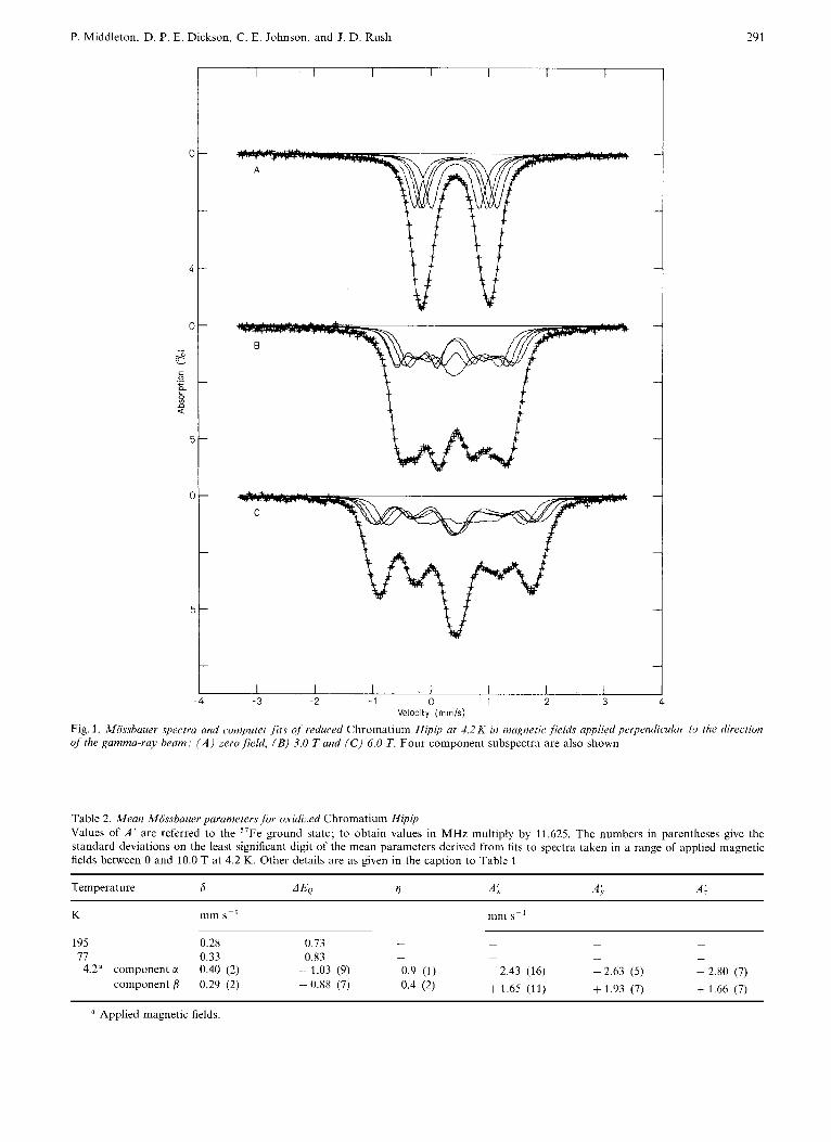
An important conclusion from the computer fitting is that there is an appreciable difference between the chemical shifts of the two components, with the com- ponent with the smaller chemical shift having the smaller, positive A values. All attempts to obtain fits with the chemical shifts and A values combined differently were unsuccessful and indicate that this behaviour is real and not an artifact of the fitting. This behaviour is particularly obvious in the spectrum taken with a field of 10.0 T applied parallel to the gamma-ray beam. In this configuration the selection rules lead to a simplification of the spectrum and with this value of the applied field, component P ( A values positive) has a large effective field and most of its spectral intensity in the two outside lines, while com- ponent CI ( A values negative) has an effective field of almost zero and most of its spectral intensity in the two inside lines. This spectrum is shown in Fig. 3 with
P. Middleton, D. P. E. Dickson, C. E. Johnson, and J. D. Rush
c -
-
5 -
-
291
I I I I I I I I - 4 -3 -2 - 1 0 1 2 3 4
Velocity (mrnls) Fig. I . Mossbauer spectra and computer fits of reduced Chromatium Hipip at 4.2 K in magnetic. ,field.s upplied perpendicular to the direction of'the gamma-ray beam: ( A ) zerofield, (B) 3.0 Tund (C) 6.0 T. Four component subspectra are also shown
Table 2. Mean Mossbauer parameters for oxidized Chromatium Hipip Values of A' are referred to the 57Fe ground statc; to obtain values in MHz multiply by 11.625. The numbers in parentheses give the standard deviations on the least significant digit of the mean parameters derived from fits to spectra taken in a range of applied magnetic fields between 0 and 10.0 T at 4.2 K. Other details are as given in the caption to Table 1
292 Interpretation of the Mossbauer Spectra of Chromatium Hipip
0
2
0
--. 0'-02
g $ 0
E .- c
2
0
3
I I I I I
' . .. ..... ...
+
B
C
I I I I I -6 -4 -2 0 2 4 6
Velocity (mmls) Fig. 2. Mossbauer spectra and computer fits of oxidized Chromatium Hipip at 4.2 K in magnetic fields applied perpendicular to the direction of the gamma-ray beam: ( A ) 0.1 T, ( B ) 3.0 T, ( C ) 6.0 T and ( D ) 10.0 T. The two component subspectra are also shown. The dotted line corresponds to component c( and the continuous line to component j
both the computer fit and a stick diagram inter- pretation.
The Mossbauer chemical shift is a measure of the valence state, being lower for Fe3+ than for Fe2+.
Therefore, as the formal valence assignment of the centre is 3 Fe3+ + 1 Fez+, the simplest interpretation of the chemical shift data is in terms of component p arising from a pair of Fe3+ atoms and component CI
P. Middleton, D. P. E. Dickson, C. E. Johnson. and J. D. Rush 293
I I I I I
+ -+
1 ++ + +
+ + +
+ $ + + + t
w # + ++ #
-4 -2 0 2 4 6 Velocity (mrn/s)
Fig. 3. Mossbauer spectrum of oxidized Chromatiurn Hipip at 4.2 K in a magnetic field of 10.0 T applied parallel to the direction of the gamma-ray beam: ( A ) with the two components a and fi indicated by stick spectra, and ( B ) showing the two computer-ftted components, a (dotted line) and p (continuous line)
arising from a pair of iron atoms with valence inter- mediate between Fe3+ and Fe2+. The chemical shift values of 0.29 and 0.40mm s-' at 4.2 K are consistent with this assignment [3]. Component also has the larger quadrupole splitting that would be expected for more ferrous iron atoms.
The magnitude of the A values depends on the size of the atomic magnetic moment. In general a high- spin ferric atom will have a larger magnetic moment than a high-spin ferrous atom. The sign of the A values depends on whether this magnetic moment is aligned parallel or antiparallel to the applied magnetic field. The quoted A values are relative to the 57Fe ground- state nuclear magnetic moment and are thus negative if the atomic magnetic moment is parallel to the applied field and positive if the atomic magnetic moment is antiparallel to the applied magnetic field. A single isolated atomic magnetic moment aligns parallel to the applied field giving negative A values. Positive A
values occur when there are a number of atomic magnetic moments aligned antiparallel to each other. Therefore the interpretation of the A value data is that component a arises from a pair of iron atoms with the larger magnetic moment that aligns parallel to the applied field and component p arises from a pair of atoms with the smaller magnetic moment that aligns antiparallel to the applied field.
Taking the interpretation of the chemical shift and A value data together it appears that the more ferrous pair of iron atoms has a larger magnetic moment than the ferric pair. This is an unusual and interesting result that is discussed further in the next section.
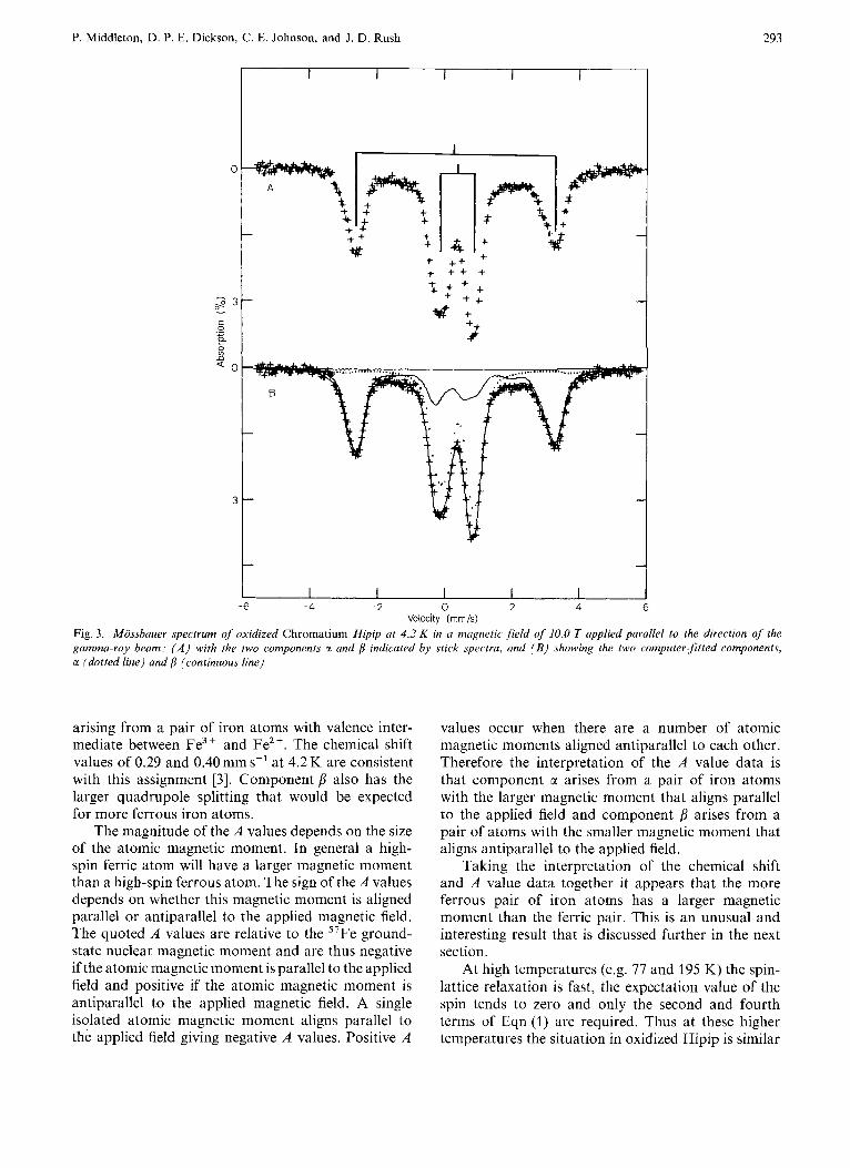
At high temperatures (e.g. 77 and 195 K) the spin- lattice relaxation is fast, the expectation value of the spin tends to zero and only the second and fourth terms of Eqn (1) are required. Thus at these higher temperatures the situation in oxidized Hipip is similar
294 Interpretation of the Mossbauer Spectra of Chromatiurn Hipip
I I I I I
0
- 1.5
L
a ._ 1
8 n Q O
1
2
I I I I I -3 -2 -1 0 1 2
Velocity (rnrnis)
Fig. 4. Mossbauer spectra and computerfits of oxidized Chromatium Hipip at ( A ) 195 K and ( B ) 77 K . Four component subspectra are also shown
to that in the reduced protein and four component subspectra were used in the computer fitting. Again there is the problem of the uniqueness of the fits and only the mean values of the Mossbauer parameters are given in Table 2. However the best set of fits was obtained with very similar chemical shifts for all four components. This is in keeping with the overall sym- metry of these doublet spectra, as seen in Fig. 4. It is not possible to obtain satisfactory fits to the 77 and 195 K spectra using only two component subspectra. Thus although the analysis of the low-temperature spectra implies that the four iron atoms within the centre can be treated as two pairs, this treatment does not appear to be appropriate at the higher temperatures. This point will be considered again in the next section.
DISCUSSION
In agreement with the three-state hypothesis, the Mossbauer parameters of reduced Hipip (Table 1) are
closely similar to those of oxidized Bucilfus stearo- thermophilus ferredoxin [ 5 ] and other oxidized ferre- doxins with 4Fe-4S centres [3]. The data are consistent with a C2+ centre consisting of two (Fe3+ + Fe2+) pairs with the 3d electrons partially delocalized over each pair. In this model the magnetic moments of the two iron atoms within each pair are parallel to each other and the magnetic moments associated with the two pairs are antiferromagnetically coupled so that the centre has zero net spin [3].
On oxidation the centre gives up one electron and the data indicate that in the ground state (i.e. at low temperatures, e.g. 4.2 K) of the oxidized Hipip centre this extra electron appears to have been given up by one of the Fe3+ + Fez+ pairs which becomes a pair of Fe3+ iron atoms, although the Mossbauer param- eters suggest that the other pair is also affected, but only to a considerably lesser extent. At higher tem- peratures it seems that the change in the electronic structure following the loss of one electron on oxida- tion affects all of the iron atoms approximately equally.
P. Middleton, D. P. E. Dickson, C. E. Johnson, and J. D. Rush 295
The ENDOR measurements of Anderson et al. [8] provide two sets of A values which, after converting to the same energy units as those used for the Moss- bauerA values, areAll=2.56mmsp',Al= 2.75mms-' (corresponding to pair a) and All = 1.82 mm s- l , Al = 1.96 mm s-' (corresponding to pair p). These are consistent with the present Mossbauer A values. The values obtained by the two techniques are re- ferred to different axis systems (the principal axis system of the electric field gradient tensor in the case of the Mossbauer measurements and the principal axis system of the g tensor in the case of ENDOR) and the ENDOR measurements give no information on the sign of the A values.
Using the information on the size and orientation of the magnetic moments, the model for the ground state of the C3+ centre can be developed to include details of the spin of the iron atoms. The pair of Fe3+ + Fez+ atoms (component M ) might be expected to have spins aligned parallel because they share at least one electron (the sharing of valence electrons between iron atoms with opposite spins is inhibited by the Pauli exclusion principle). The spins of the atoms of the other pair would then be mutually parallel but antiparallel to the spins of the Fe3+ + Fez+ pair. This could occur either because of strong anti- ferromagnetic couplings between the atoms of the two pairs or because of the net effect of a relatively strong ferromagnetic coupling of the atoms within each pair and a weaker antiferromagnetic coupling between the total spins of the two pairs. Although this pairwise model may correspond to a considerable oversimplification of the actual situation, it does represent an approach to the problem of four coupled spins that appears to have a reasonable physical justification. This approach was also used in analyzing the Mossbauer data on reduced B. stearothermophilus ferredoxin [5] and is similar to that of Johnson et al. [lo] for the case of the reduced two-iron ferredoxins which have one ferric atom and one ferrous atom coupled together antiferromagnetically [l 1,121.
In the pairwise model the four iron atoms with individual spins S1, S2, S3 and S4 form two pairs CI
and such that :
s, = s1 + s2, s, = s3 + sq, s = s, + s, = 112 (2)
where S, and S, are the spins of the two pairs and S is the total spin of the centre. Following the previous work [5,10] and using the Mossbauer result that the iron atoms within each pair are essentially equivalent, one obtains:
and
where A i ,2 ,3 ,4 are the A values of iron atoms 1, 2, 3 or 4, related to the spin of the whole centre (as are measured by Mossbauer spectroscopy) and A1,2,3,4
are the A values of iron atoms 1, 2, 3 or 4, related to their individual spins, g' is the whole-centre g value (as is measured by EPR) and g,and go are the g values of the pairs of iron atoms. For the model under con- sideration the second terms in brackets in Eqns (3) and (4) are positive and, as the single-atom A values are always negative, the Mossbauer A' value data indicate that the first term in brackets in Eqn (3) must be positive (pair M ) while the first term in brackets in Eqn(4) must be negative (pair p). One can now go on to consider the implications of this for theg' values measured by EPR. Pair consists of atoms of an es- sentially ferric character and thus one would expect gp to have g = 2 while pair a consists of atoms with a more ferrous character and thus g, might be expected to have g,, > 2. As the first term in brackets in Eqn(5) is positive while the second is negative, it can be shown that this leads to giv > 2, which is consistent with the g ' values observed in EPR measurements [ 3 ] .
Thus in the case of oxidized Chromatium Hipip both the Mossbauer and EPR data indicate, via the sign of the factors in the equations considered above, that the iron atoms of more ferrous character have associated with them a larger magnetic moment than the iron atoms of ferric character. The reasons for the magnetic moments associated with the ferric iron atoms being smaller than those associated with more ferrous iron atoms is open to speculation but it may be due to a large orbital contribution to the magnetic moment of the more ferrous pair. Although there is no direct evidence, it is tempting to think that this behaviour may be related to the anomalous redox properties of Hipip compared with the ferredoxins which have the same active centres. It is possible that both aspects may be due to the effects on the centre of the surrounding protein chain.
The authors wish to thank Professor D. 0. Hall and Drs R. Cammack and K. K. Rao of King's College, University of London for preparing the 57Fe-enriched protein samples used in the present work. This work is supported by the Science Research Council.
REFERENCES
1. Carter, C. W., Kraut, J., Freer, S. T., Alden, R. A,, Sieker, L. C., Adman, E. T. & Jensen, L. H. (1972) Proc. Nut/ Acud. Sci. U.S .A. 69, 3526-3529.
2. Carter, C. W. (1977) in Iron-Sulfur Proteins (Lovenberg, W., ed.) vol. 3 , pp. 157-204, Academic Press, New York.
296 P. Middleton et al. : Interpretation of the Mossbauer Spectra of Chromatium Hipip
3. Cammack, R., Dickson, D. P. E. & Johnson, C. E. (1977) in Iron-Sulfur Proteins (Lovenberg, W., ed.) vol. 3, pp. 283- 330, Academic Press, New York.
4. Dickson, D. P. E., Johnson, C. E., Cammack, R., Evans, M. C. W., Hall, D. 0. & Rao, K. K. (1974) Biochem. J . 139,
5. Middleton, P., Dickson, D. P. E., Johnson, C. E. & Rush, J. D.
6. Lang, G. & Dale, B. W. (1974) Nucl. Instrum. Methods, 116,
7. Phillips, W. D., Poe, M., McDonald, C. E. & Bartsch, R. G.
105- 108.
(1978) Eur. J . Biochem. 88, 135-141.
567 - 571.
(1970) Proc. NatlAcad. Sci. U.S.A. 67,682-687.
8. Anderson, R. E., Anger, G., Petersson, L., Ehrenberg, A., Cammack, R., Hall, D. O., Mullinger, R. & Rao, K. K. (1975) Biochim. Biophys. Acta, 376, 63-71.
9. Antanaitis, B. C. & Moss, T. H. (1975) Biochim. Biophys. Acta,
10. Johnson,C. E., Cammack, R., Ra0,K.K. & Hal1,D.O. (1971) Biochem. Biophys. Res. Commun. 43, 564-571.
11. Gibson, J. F., Hall, D. O., Thornley, J. H. M. & Whatley, F. R. (1966) Proc. Natl Acad. Sci. U.S.A. 56, 987-991.
12. Thornley, J. H. M., Gibson, J. F., Whatley, F. R. & Hall, D. 0. (1966) Biochem. Biophys. Res. Commun. 24, 877- 886.
405, 262 - 279.
P. Middleton, D. P. E. Dickson, C. E. Johnson, and J. D. Rush, Department of Physics, University of Liverpool, Liverpool, Great Britain, L69 3BX