82
Interstitial Lung Disease: A Practical Approach to CT Diagnosis Heber MacMahon The University of Chicago Department of Radiology Venice 2017
Interstitial Lung Disease: A Practical Approach to CT Diagnosis
Heber MacMahon
The University of Chicago
Department of Radiology
Venice
2017

Disclosures
Consultant for Riverain Medical Minor stockholder in Hologic, Inc. Consultant for GE Healthcare Research Support from Philips Healthcare License and royalty fees from University of Chicago (UC Tech)
Educational site of the Radiological Society of the NetherlandsRobin Smithuis MDwww.radiologyassistant.nl
Credits

Interstitial Lung Disease
CT Technique for ILD
CT patterns in ILD
Classification of Interstitial pneumonias
Integrating imaging findings with patient history

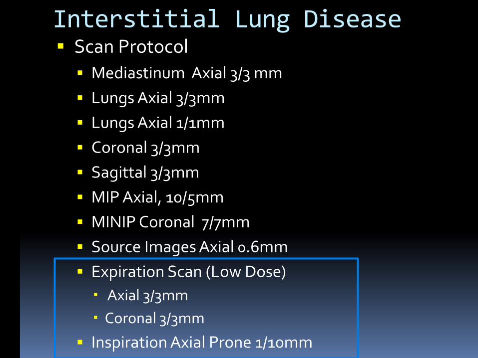
Interstitial Lung Disease Scan Protocol
Mediastinum Axial 3/3 mm
Lungs Axial 3/3mm
Lungs Axial 1/1mm
Coronal 3/3mm
Sagittal 3/3mm
MIP Axial, 10/5mm
MINIP Coronal 7/7mm
Source Images Axial 0.6mm
Expiration Scan (Low Dose)
Axial 3/3mm
Coronal 3/3mm
Inspiration Axial Prone 1/10mm
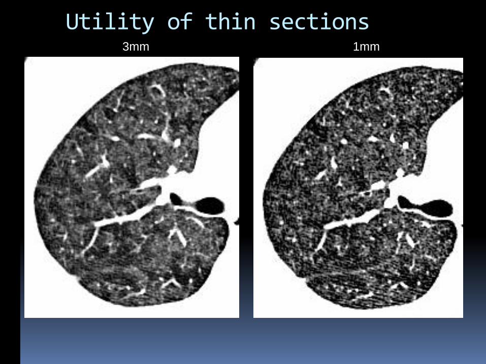
3mm 1mm
Utility of thin sections

Utility of thin sections
1mm
3mm 1mm
Interstitial Lung Disease Scan Protocol
Mediastinum Axial 3/3 mm
Lungs Axial 3/3mm
Lungs Axial 1/1mm
Coronal 3/3mm
Sagittal 3/3mm
MIP Axial, 10/5mm
MINIP Coronal 7/7mm
Source Images Axial 0.6mm
Expiration Scan (Low Dose)
Axial 3/3mm
Coronal 3/3mm
Inspiration Axial Prone 1/10mm
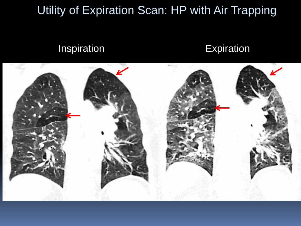
Utility of Expiration Scan: HP with Air Trapping
Inspiration Expiration

Inspiration Expiration
Evaluating Expiratory Effort
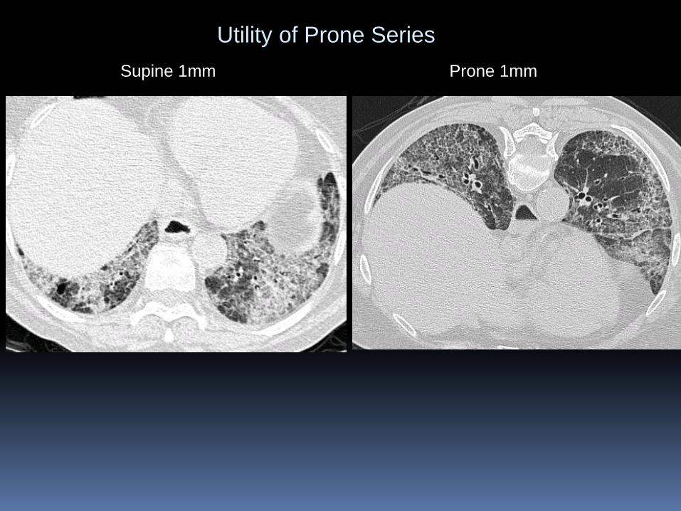
Supine 1mm Prone 1mm
Utility of Prone Series

Slab MINIP
MINIP – Minimum Intensity Projection
Axial Slab Coronal MINIP – Mosaic perfusion
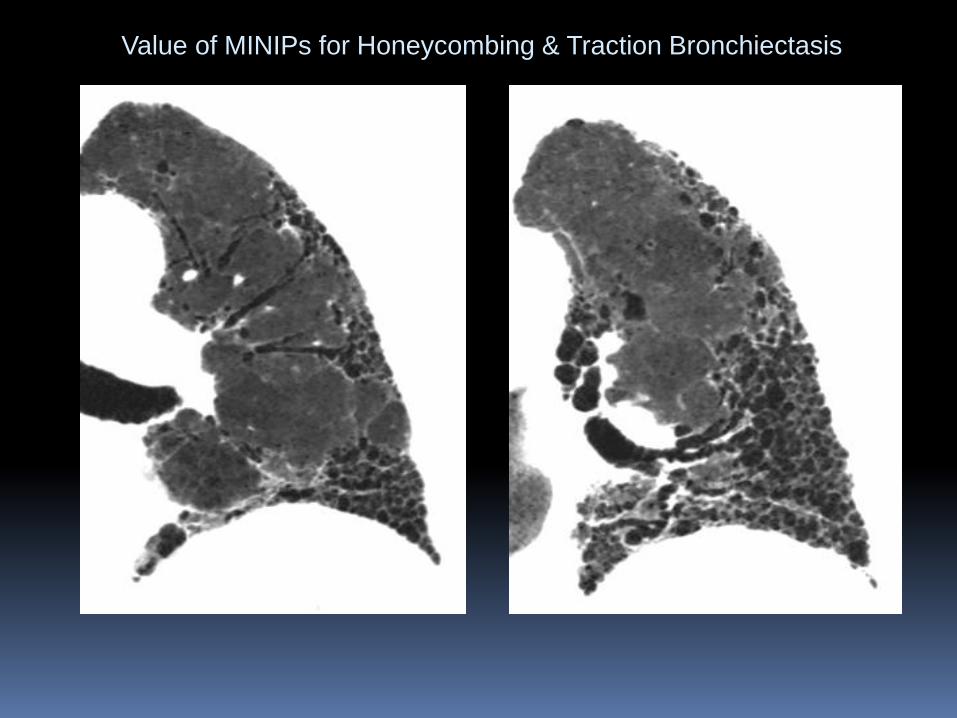
Value of MINIPs for Honeycombing & Traction Bronchiectasis
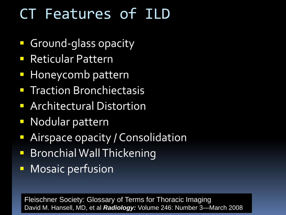
C CT Features of ILD
Ground-glass opacity
Reticular Pattern
Honeycomb pattern
Traction Bronchiectasis
Architectural Distortion
Nodular pattern
Airspace opacity / Consolidation
Bronchial Wall Thickening
Mosaic perfusion
Fleischner Society: Glossary of Terms for Thoracic ImagingDavid M. Hansell, MD, et al Radiology: Volume 246: Number 3—March 2008 �

Ground-Glass Opacity
Features:
Hazy lung opacity
Preservation of the bronchial and vascular margins
Less opaque than consolidation
Caused by:
Partial filling of airspaces
Interstitial thickening
Partial collapse of alveoli
1mm section

31 y/o male with increasing dyspnea and fever
• Infections
• Edema
• Drug related
• Hemorrhage

Pneumocystis Pneumonia
31 y/o male with increasing dyspnea and fever

Predominantly Ground-glass Opacity (GGO)
Pneumocystis or viral pneumonia
Acute Drug/Hypersensitivity Reactions
Pulmonary Edema
Hemorrhage
Acute Aspiration
AIP, NSIP, DIP
Mucinous adenocarcinoma
Classification of ILD by Predominant Pattern

Drug ToxicityCase #1 Case #2
Imaging Findings:
•Pulmonary Edema
•Diffuse Alveolar Damage (DAD)
•Pulmonary hemorrhage
•Organizing Pneumonia
•Eosinophilic Pneumonia
•UIP/NSIP

3mm 1mm
3mm Coronal
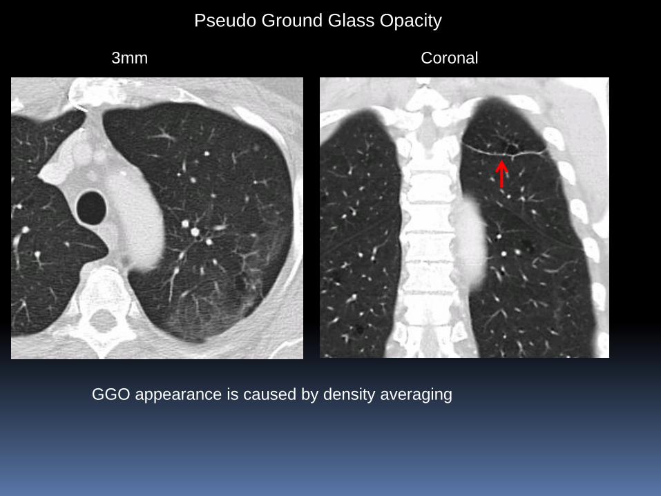
Pseudo Ground Glass Opacity
GGO appearance is caused by density averaging

Coronal
Pseudo Ground Glass Opacity
GGO appearance is caused by density averaging
Axial

Reticular Pattern
Features:
• Netlike pattern, linear opacities
Cause:
• Infiltration of the interstitial framework of the secondary pulmonary lobule

Classification of ILD by Predominant Pattern
Predominantly Linear or Reticular Pattern
Edema
Lymhangitic Mets
UIP (Usual Interstitial Pneumonia)
NSIP (Non-specific Interstitial Pneumonia)
HP (Hypersensitivity Pneumonia)
Asbestosis

Acute Pulmonary Edema
Smooth thickening of interlobular septa
Ground glass and airspace opacity
Pleural effusions

Thickened axial interstitium
Irregular thickening of interlobular septae
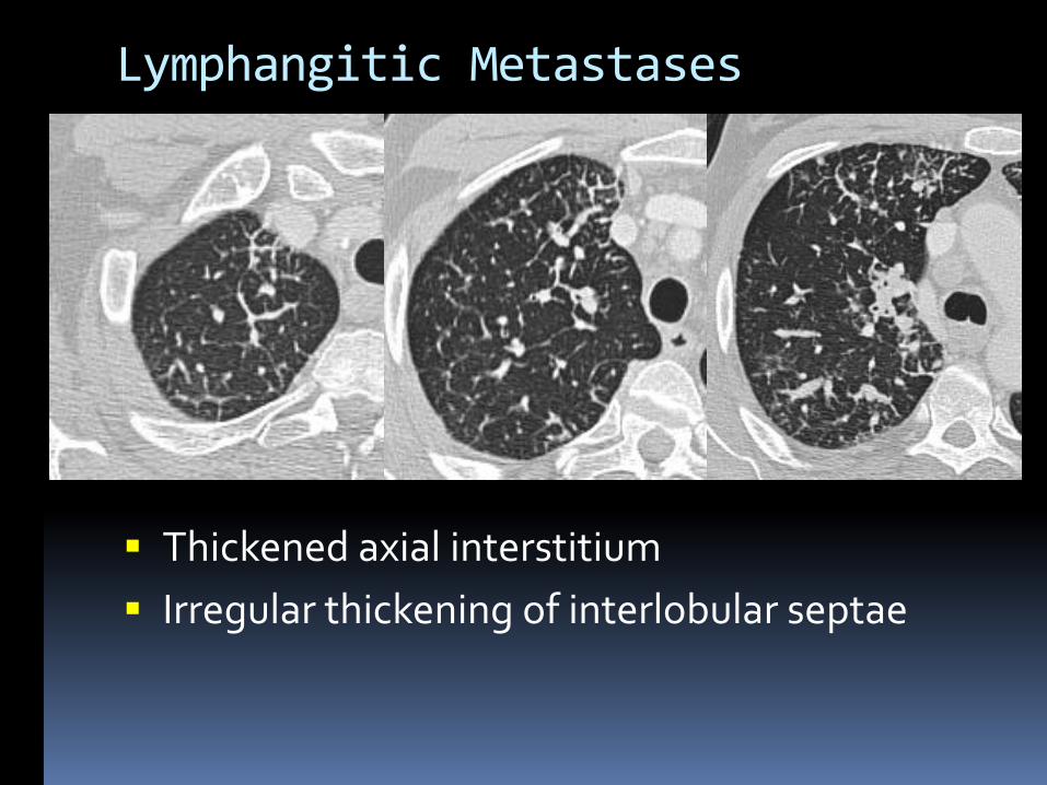
Lymphangitic Metastases
Thickened axial interstitium
Irregular thickening of interlobular septae

Honeycomb Pattern
Features:
• Clustered cystic airspaces ~2-10mm
• Subpleural
• Well-defined walls
Indicates:
• Late stage fibrosis
• Destroyed fibrotic lung tissue with numerous cystic airspaces with thick fibrous walls

Mild Subpleural Microcystic Honeycomb Pattern

Traction Bronchiectasis
Irregular bronchial dilatation caused by surrounding retractile pulmonary fibrosis
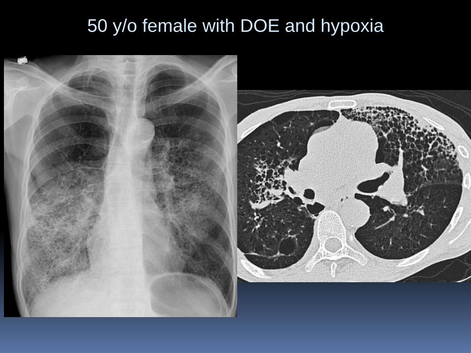
50 y/o female with DOE and hypoxia
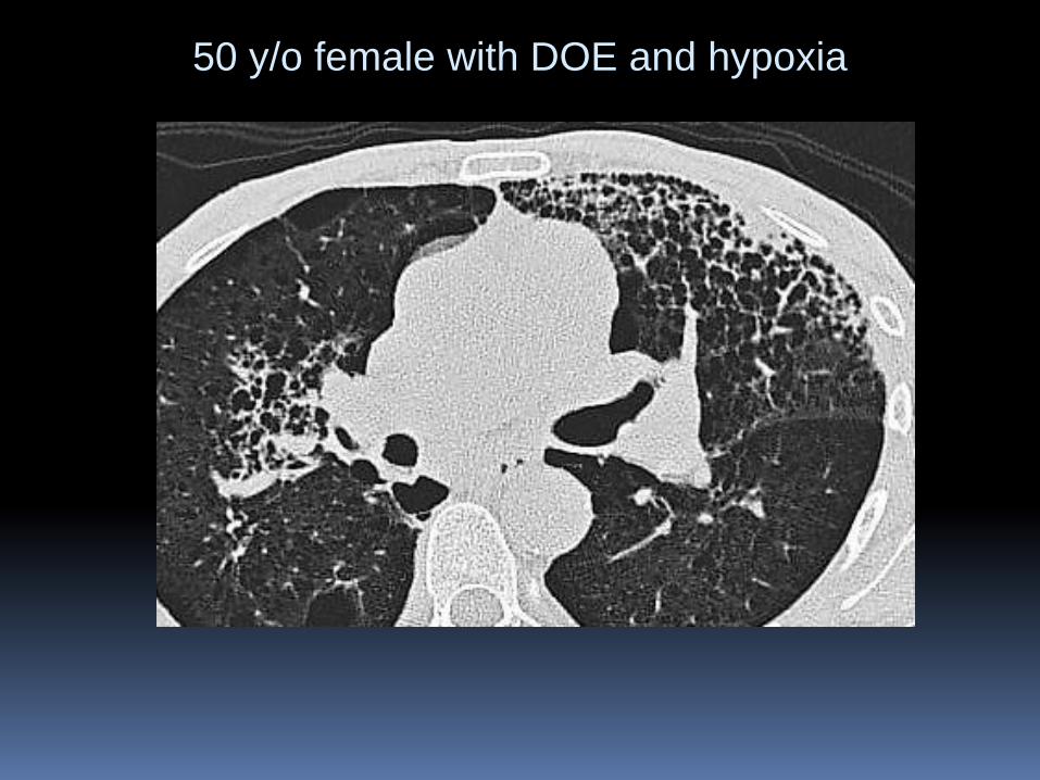
50 y/o female with DOE and hypoxia

50 y/o female with DOE and hypoxia

Follow up CXR after antibiotic RX
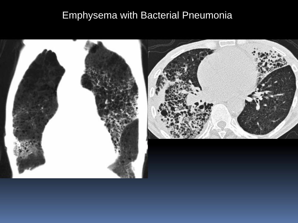
Emphysema with Bacterial Pneumonia

Airspace Opacity/Consolidation
Air space filling process
Obscures vascular structures
Air bronchograms

Consolidation
Acute
Infection, edema, hemorrhage
Chronic COP (Cryptogenic Organizing Pneumonia)
Chronic infections
Chronic Eosinophilic Pneumonia
Lipoid Pneumonia
Mucinous adenocarcinoma “BAC”
Lymphoma
Classification of ILD by Predominant Pattern

78 y/o female w cough and low grade fever: organizing pneumonia

Cryptogenic Organizing Pneumonia (COP)
Clinical Features
Mean age 55 years
Nonsmokers > smokers 2:1
Dyspnea, cough, fever
Respond ++ to steroid rx
Good prognosis
CT
Multifocal
Lower lobes more frequently involved
Opacities vary from ground glass to consolidation

Cryptogenic Organizing Pneumonia (COP)
Clinical Features
Mean age 55 years
Nonsmokers > smokers 2:1
Dyspnea, cough, fever
Respond ++ to steroid rx
Good prognosis
CT
Multifocal
Lower lobes more frequently involved
Opacities vary from ground glass to consolidation
• Subpleural arcuate opacities
• Atoll sign
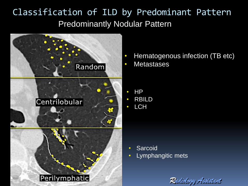
Classification of ILD by Predominant PatternPredominantly Nodular Pattern
• Hematogenous infection (TB etc)
• Metastases
• HP
• RBILD
• LCH
• Sarcoid
• Lymphangitic mets
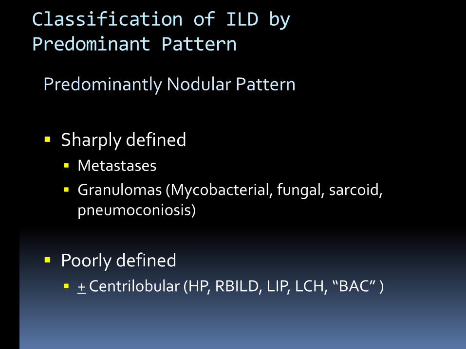
Predominantly Nodular Pattern
Sharply defined
Metastases
Granulomas (Mycobacterial, fungal, sarcoid, pneumoconiosis)
Poorly defined
+ Centrilobular (HP, RBILD, LIP, LCH, “BAC” )
Classification of ILD by Predominant Pattern

Nodular PatternSharply defined, Random, Uniform

Nodular PatternMiliary MTB
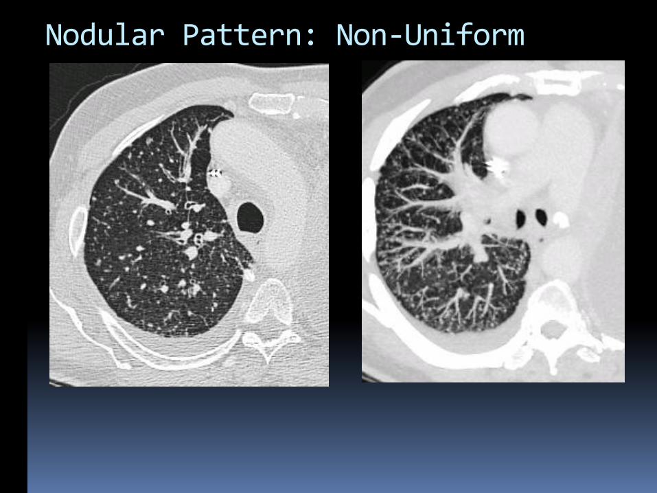
Nodular Pattern: Non-Uniform

Metastatic Lung Cancer
Nodular Pattern: Non-Uniform

Diffuse small sharp solid nodules
Metastases Thyroid , lung, GI etc
Granulomas Miliary infection:TB, Histo etc
Sarcoid
Silicosis etc

Sarcoidosis
Perilymphatic/ Peribronchovascular Distribution

Inflammation of the terminal bronchiole and lobular pulmonary artery
Small round opacity in the center of a secondary pulmonary lobule
Centrilobular Nodules
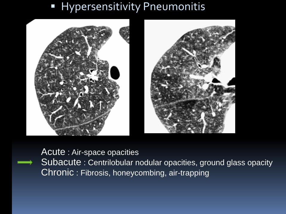
Hypersensitivity Pneumonitis
Acute : Air-space opacities
Subacute : Centrilobular nodular opacities, ground glass opacity
Chronic : Fibrosis, honeycombing, air-trapping

Hypersensitivity Pneumonitis
Acute : Air-space opacities
Subacute : Centrilobular nodular opacities, ground glass opacity
Chronic : Fibrosis, honeycombing, air-trapping

Centrilobular Nodules
Respiratory Bronchiolitis ILD
Tobacco smoke produces mild immunosuppression.
Smokers get RB/RBILD, but not get HP

Aspiration Bronchiolitis
• Centrilobular nodules
• Tree-in-bud opacities
• Airspace opacities

Cystic Patterns
Emphysema
LAM (Lymphangioleiomyomatosis)
LCH (Langerhans Cell Histiocytosis)
Honeycomb pattern in UIP, Sarcoid etc.
Chronic PJP
Lymphocytic Interstitial pneumonia (LIP)
Classification of ILD by Predominant Pattern

Emphysema
Centrilobular
Paraseptal
Panlobular

Centrilobular Emphysema - Coronal MINIP

Upper lobe emphysema, basilar fibrosis

Almost all male
Predominant paraseptal emphysema
PFTs ( Lung Volumes, FEV1) less abnormal than expected
DLCO very abnormal
High incidence of PAH
Prognosis intermediate between COPD and UIP
Combined Pulmonary Fibrosis and Emphysema (CPFE)
Cottin et al.Eur Respir J 2005; 26: 586–593

CPFE
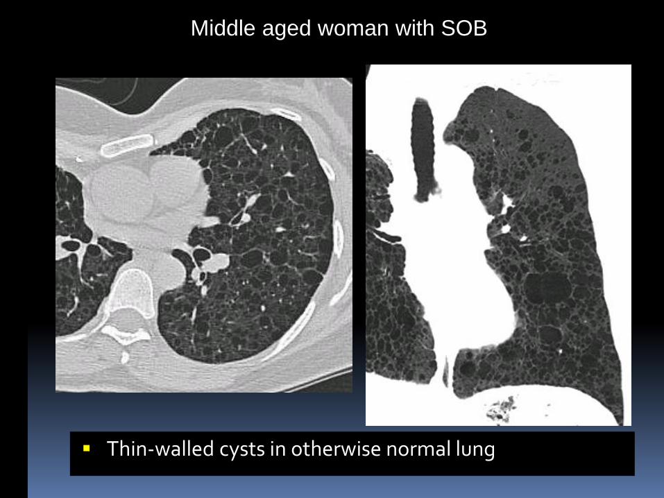
Thin-walled cysts in otherwise normal lung
Middle aged woman with SOB

Lymphangioleiomyomatosis (LAM)
Thin-walled cysts in otherwise normal lung

Women of child bearing age or older
Smooth muscle proliferation
Can present with hemoptysis, pneumothorax or chylous effusion
Pulmonary cysts with thin walls, + nodules
Related to TSC (Tuberous Sclerosis Complex)
+ Renal angiomyolipomas
Lymphangioleiomyomatosis (LAM)

Example #1: 48 y/o female smoker with PA hypertension

• Irregularly shaped cysts
• Small nodules
• Upper lobe predominant
• Sparing of CP angles
Case #2: 41 y/o smoker

• Irregularly shaped cysts
• Small nodules
• Upper lobe predominant
• Sparing of CP angles
Langerhans Cell Histiocytosis (LCH)

Smoking Related Lung Disease
Respiratory Bronchiolitis (RB) Usually asymptomatic – all smokers have it.
RB-ILD 5-10% of smokers have clinically significant lung disease in
association with RB
DIP More diffuse and severe than RB-ILD
LCH (Langerhans Cell Histiocytosis) 90-100% are cigarette smokers
< 40 yrs old
Cysts, nodules
UL predominance, sparing of CP angles

50 y/o man with fever, SOB and hypoxia

Pneumocystis Jiroveci Pneumonia (PJP)
Clinical: AIDS : CD4 < 200
Organ transplant
Lymphoma / leukemia
CT:
Hazy interstitial opacity (GGO)
Usually diffuse
Thin-walled cysts >UL < 38%

Interstitial Pneumonias: Definition and Classification
Definition Idiopathic: Uncertain etiology
Interstitial Pneumonia: interstitial lung inflammation and fibrosis which also affects airspaces, peripheral airways, and vessels
Classification Standardized by American Thoracic Society and European
Respiratory Society in 2001
Based on clinical information, radiology, and pathology.

Histologic
Pattern
Distribution Typical CT Findings Smoking
Related?
UIP Peripheral,
Subpleural,
Apicobasal
Gradient
Reticular, Honeycombing,
Traction Bronchiectasis,
Architectural Distortion
Usually
NSIP Peripheral,
Subpleural,
Basal,
Symmetric
Ground Glass,
Reticular,
Consolidation
NO
OP Subpleural,
Peribronchial
Patchy Consolidation and/or Nodules NO
DAD Diffuse Consolidation, Ground Glass, Lobular
Sparing, Traction Bronchiectasis
NO
RB Diffuse,
Upper lobe
Bronchial Wall Thickening, Centrilobular
Nodules, Patchy Ground Glass
YES
DIP Lower Zone,
Peripheral
Ground Glass, Reticular Lines YES
LIP Diffuse Centrilobular Nodules, Ground Glass,
Septal and Bronchovascular
Thickening, Thin-walled Cysts
NO

UIP (Usual Interstitial Pneumonia)
NSIP (Non-Specific Interstitial Pneumonia)
HP (Hypersensitivity Pneumonitis)
Sarcoid
Common Causes of Fibrosing ILD

Pathological Diagnosis Etiology
UIP IPF (Idiopathic Pul. Fibrosis)
Collagen Vascular Disease
End stage HP
Asbestosis, etc
NSIP Primary NSIP
Collagen Vascular Disease
Hypersensitivity (HP)
Drug reactions

UIP
Etiology
IPF (Idiopathic Pul. Fibrosis)
Collagen Vascular Disease
End stage HP
Asbestosis, etc
Histology Temporal heterogeneity
Different stages in the evolution of fibrosis
Scattered fibroblastic foci
Patchy lung involvement with subpleural and basal
Honeycombing

UIP
CT Findings Apicobasal gradient
Most extensive in the most basal section
Subpleural reticular opacities
Honeycombing
Traction bronchiectasis
Heterogeneous Fibrosis with areas of normal lung
Limited ground-glass opacity

UIP
Traction Bronchiectasis
Honeycombing

Idiopathic Pulmonary Fibrosis (IPF)
Typical Features
Age > 50 years
Progressive dyspnea and nonproductive cough
Symptoms 3 months or greater
Fatigue and weight loss
Does not respond to high-dose corticosteroid therapy
Mean survival 2-4 years
Histological pattern is UIP
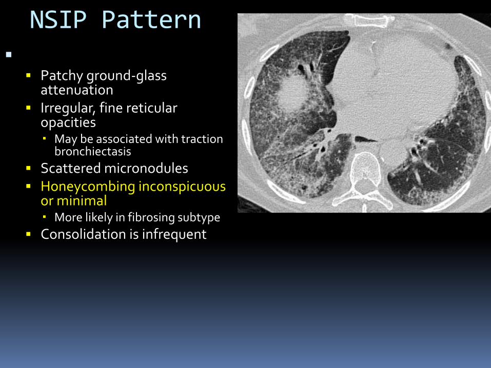
NSIP Pattern CT Findings (cont.)
Patchy ground-glass attenuation
Irregular, fine reticular opacities May be associated with traction
bronchiectasis
Scattered micronodules Honeycombing inconspicuous
or minimal More likely in fibrosing subtype
Consolidation is infrequent

Nonspecific Interstitial Pneumonia (NSIP)
Typical Features
40-50 years, decade younger than IPF
Worsening dyspnea over several months
Milder than IPF
Fatigue and weight loss
Treatment with corticosteroids and cyclosporin
Prognosis better than IPF
Correlates with the extent of fibrosis
Histological pattern is NSIP

Uncommon Interstitial Pneumonias
AIP (Acute IP): Diffuse Alveolar Damage (DAD)
Only idiopathic interstitial pneumonia with acute onset of symptoms
Occurs in healthy individuals with prior illness suggestive of a viral upper respiratory infection
Severe dyspnea with need for mechanical ventilation in less than 1-2 weeks
DIP (Desquamative IP):
LIP: Lymphocytic Interstitial Pneumonia

Multidisciplinary Approach to ILD
Clinical
Age, sex, Occupation
Clinical presentation (Acute or Chronic)
Immune status, drug exposures, RT
Imaging
Lung volumes
Disease distribution
CT Pattern
Lymphadenopathy, effusions, CM, PAH

Exposures
Tobacco:
+ LCH, RBILD, DIP, UIP, Eosinophilic Pna
- HP
Drugs
Chemotherapy
Immunotherapy
Illicit drugs (Cocaine, Heroin etc)
Dust
Silica, Coal dust, Asbestos, Berylium
Clinical History
Summary
Define key features including dominant pattern and distribution
Consider clinical context, including age, gender, immune status, drug and occupational exposures
Compare with previous scans and CXRs
Multidisciplinary approach works best
Venice
2017


