109
Interstitial lung diseases Restrictive lung diseases Lung fibrosis ”small sized” lungs Finn Rasmussen

Interstitial lung diseases
Restrictive lung diseases
Lung fibrosis
”small sized” lungs
Finn Rasmussen

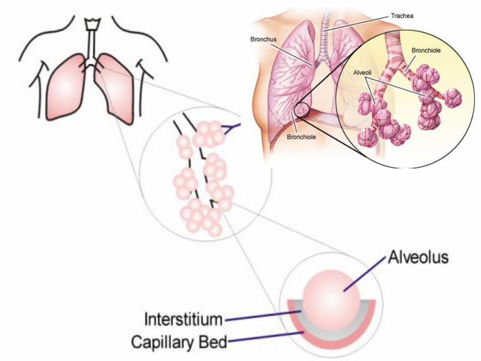
The interstitium of the lung is not normally visible radiographic-
ally; it becomes visible only when disease (e.g., edema,
fibrosis, tumor) increases its volume and attenuation.
The interstitial space is defined as continuum of loose
connective tissue throughout the lung composed of three
subdivisions:
(i) the bronchovascular (axial), surrounding the bronchi,
arteries, and veins from the lung root to the level of the
respiratory bronchiole
(ii) the parenchymal (acinar), situated between the alveolar
and capillary basement membranes
(iii) the subpleural, situated beneath the pleura, as well as in
the interlobular septae.
The Lung Interstitium
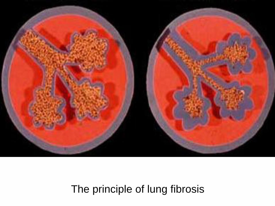
The principle of lung fibrosis

Important -Restriktive lung function
Also seen
without
problems in
the lung
parenkyma

In principle there
is are ”acute”
restriktive
disorders
Pleuraexsudate,
pneumonia
Atelektasis etc..
Disease localized in the paremkyma
What is lost is forever lost …..
…if treatment is not started in time…
However. Normally in lungfibrosis
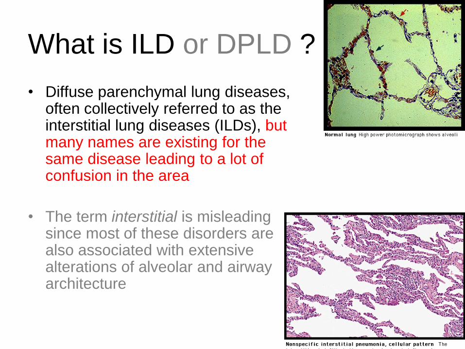
What is ILD or DPLD ?
• Diffuse parenchymal lung diseases, often collectively referred to as the interstitial lung diseases (ILDs), but many names are existing for the same disease leading to a lot of confusion in the area
• The term interstitial is misleading since most of these disorders are also associated with extensive alterations of alveolar and airway architecture

Symptoms
• Signs
• Slowly progession– But attracts is prevalent
• Breathlessness– At first at activity
– Later all the time
• cough– Non- productive
• Findings cyanosis
– Low saturation
• low lung function– TLC
– RV
– dlco
• Dromstikfingers
• Velcro sound at stethoscopy
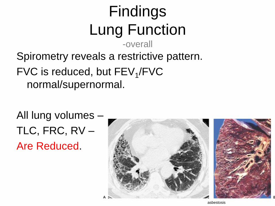
Findings
Lung Function-overall
Spirometry reveals a restrictive pattern.
FVC is reduced, but FEV1/FVC
normal/supernormal.
All lung volumes –
TLC, FRC, RV –
Are Reduced.
asbestosis
CASE1975, operatted for retentio testis. .
1989, operated inqvinal hernia right side. .
47 year old male: The Pt. s symptoms started 7 month ago with dry
cough, initially treated by a doctor with Salbuvent mixture, but as no
effect . Never smoker.
He then stopped the treatment after 3 days due to heart beat and
tremor. Cough is mostly dry but intermittent yellow. Had has som pain
in the joints without swelling.
The patient has a complain of increasing dyspnea especially the last
month. Whezzing has been observed a few times. Some joint pain
especially in both shoulders.
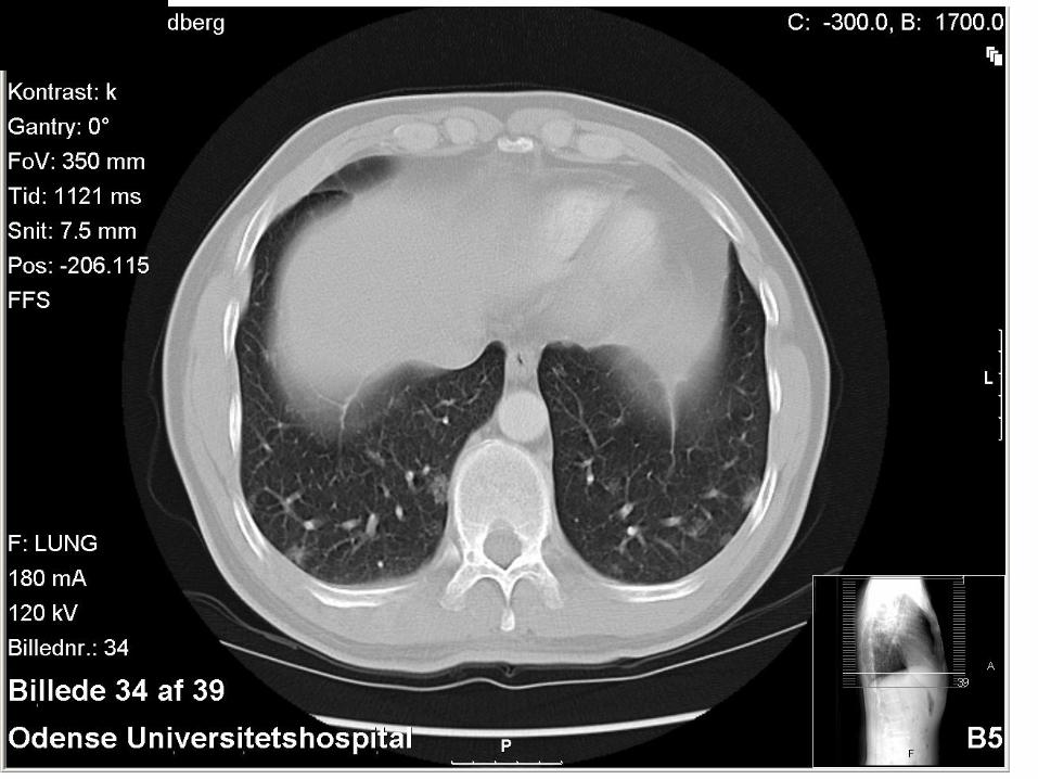
Returns to his doctor after 7 month and the doctor due a X-RAY:
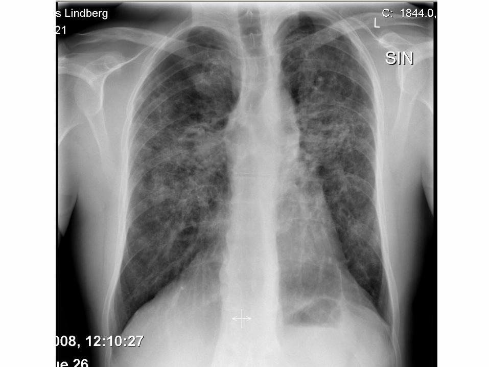

CASEX-RAY showing bilat. Infiltrats and increased size of
lympnodes at hilus.
The doctor says pneumonia and gives Klacid 500mg x2
Do you agree ??

How do we approach this patient ?
• Tentative diagnoses– Asthma
– Infection
– Diffus parenkymatøs lung disease• In particular sarcoidosis
– Cancer• lymphoma
• Tests – CT- thorax + upper abdomen
– Bloodtest
– Lung function incl reversibility
– Bronkoskopi


ACE= 94 og ANA,ANCA;Anti-CCP: normal; IgG,IgM,IgA,IgE normal
EKG= SINUS RYTME no ischemia
LF is technically ok performed; FEV1 =3,14 (65% of predicted), FVC= 4,51
(73% of predicted) A ratio of 70 ?
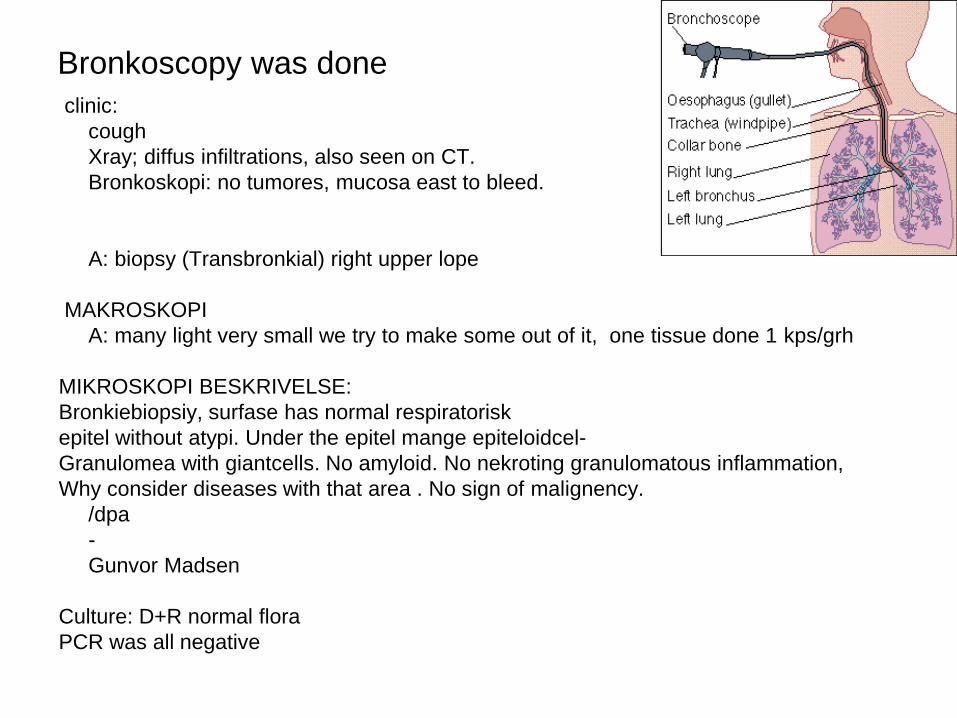
clinic:
cough
Xray; diffus infiltrations, also seen on CT.
Bronkoskopi: no tumores, mucosa east to bleed.
A: biopsy (Transbronkial) right upper lope
MAKROSKOPI
A: many light very small we try to make some out of it, one tissue done 1 kps/grh
MIKROSKOPI BESKRIVELSE:
Bronkiebiopsiy, surfase has normal respiratorisk
epitel without atypi. Under the epitel mange epiteloidcel-
Granulomea with giantcells. No amyloid. No nekroting granulomatous inflammation,
Why consider diseases with that area . No sign of malignency.
/dpa
-
Gunvor Madsen
Culture: D+R normal flora
PCR was all negative
Bronkoscopy was done

1. COPD
2. Asthma
3. Idiopatic lungefibrosis
4. Sarcoidose
5. Allergic alveolitis
6. Lymfoma
7. Tuberculose
8. Wegeners granulomatosis
TEST: Among the diseases mentioned below where
do you see Non-caceating granulomas inflammation
???

1. COPD
2. Asthma
3. Idiopatic lung fibrosis
4. Sarcoidose
5. Allergic alveolitis
6. Lymfoma
7. Tuberculosis
8. Wegeners granulomatosis
Among the diseases mentioned below where do NOT see non-caceating
granulomas inflammation ??? (casuistik not included)
4, 5, 6 og 8 are the right one
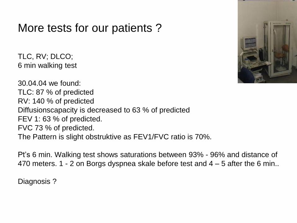
More tests for our patients ?
TLC, RV; DLCO;
6 min walking test
30.04.04 we found:
TLC: 87 % of predicted
RV: 140 % of predicted
Diffusionscapacity is decreased to 63 % of predicted
FEV 1: 63 % of predicted.
FVC 73 % of predicted.
The Pattern is slight obstruktive as FEV1/FVC ratio is 70%.
Pt’s 6 min. Walking test shows saturations between 93% - 96% and distance of
470 meters. 1 - 2 on Borgs dyspnea skale before test and 4 – 5 after the 6 min..
Diagnosis ?
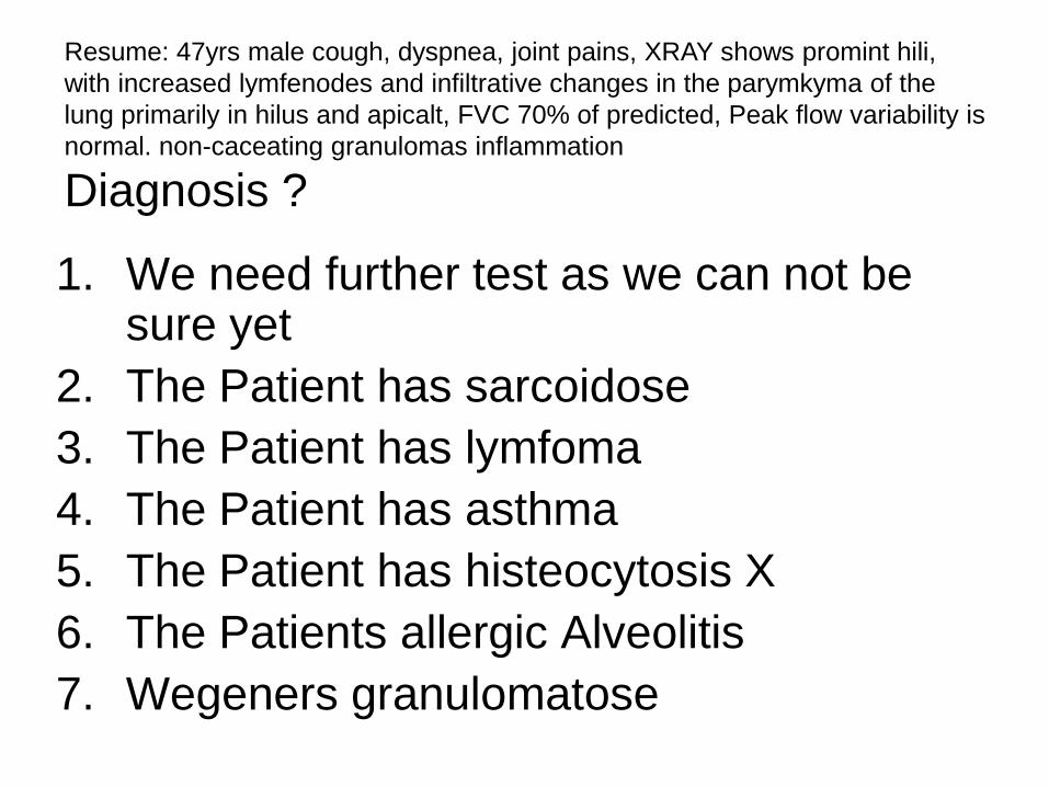
1. We need further test as we can not be sure yet
2. The Patient has sarcoidose
3. The Patient has lymfoma
4. The Patient has asthma
5. The Patient has histeocytosis X
6. The Patients allergic Alveolitis
7. Wegeners granulomatose
Resume: 47yrs male cough, dyspnea, joint pains, XRAY shows promint hili,
with increased lymfenodes and infiltrative changes in the parymkyma of the
lung primarily in hilus and apicalt, FVC 70% of predicted, Peak flow variability is
normal. non-caceating granulomas inflammation
Diagnosis ?
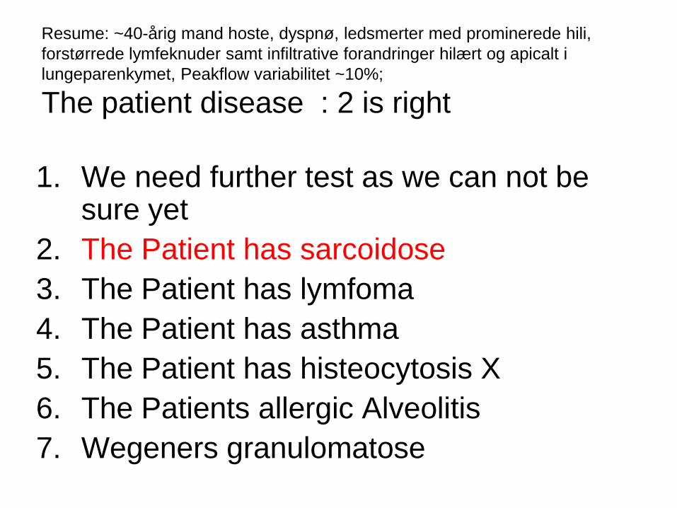
Resume: ~40-årig mand hoste, dyspnø, ledsmerter med prominerede hili,
forstørrede lymfeknuder samt infiltrative forandringer hilært og apicalt i
lungeparenkymet, Peakflow variabilitet ~10%;
The patient disease : 2 is right
1. We need further test as we can not besure yet
2. The Patient has sarcoidose
3. The Patient has lymfoma
4. The Patient has asthma
5. The Patient has histeocytosis X
6. The Patients allergic Alveolitis
7. Wegeners granulomatose

Diseases
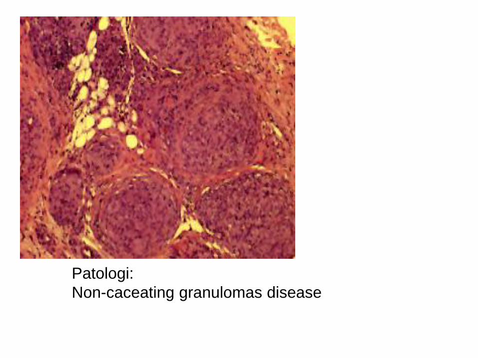
Patologi:
Non-caceating granulomas disease

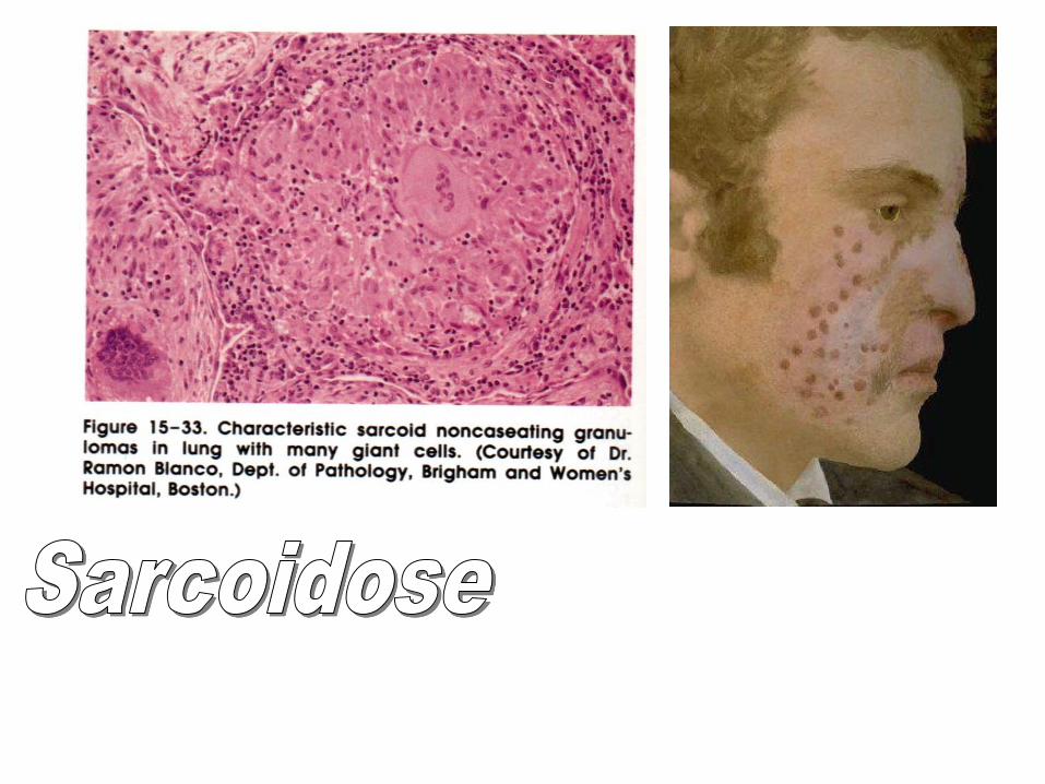
Who gets Sarcoidose??
Most often between 25-40 yrs
More women than men
More negro than white
Livs risiko for at få sygdommen er 0.0085

Is Sarcoidose infectious?
NO !
Familiar cases seen !
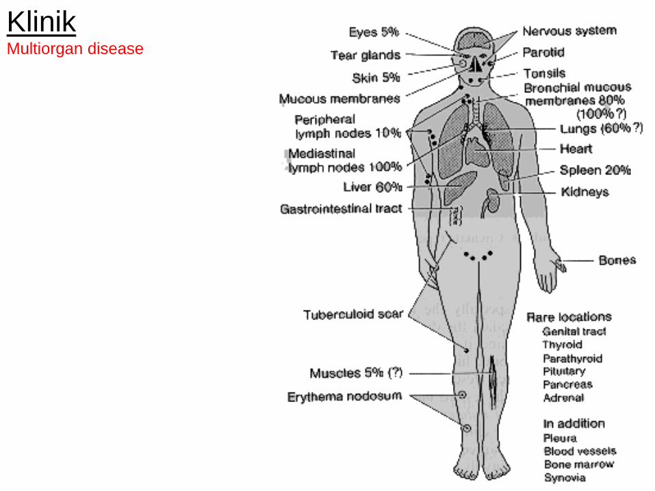
KlinikMultiorgan disease

Lung symptoms
>90%
Cough
Dyspnoe
Chest pain
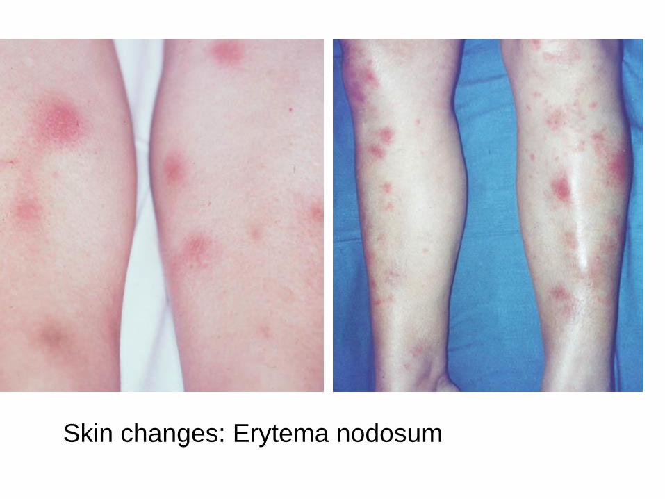
Skin changes: Erytema nodosum
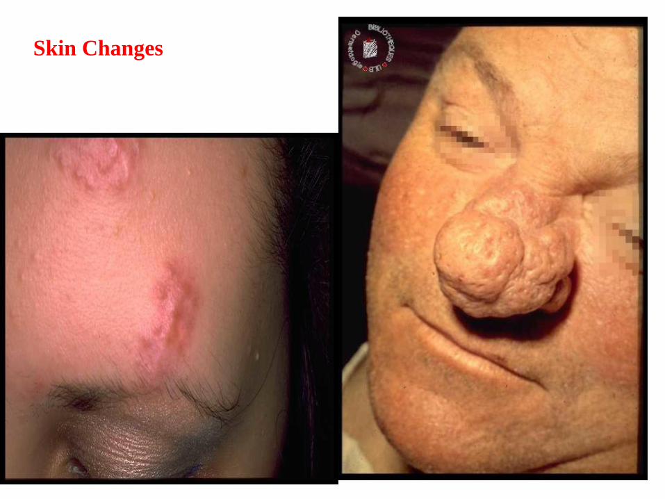
Skin Changes

CNS manifestations
10% a debut of parese
Often perifer Fasialis parese

Eye symptoms
Send to eye doctor

Sarcoidose
treatment
SteroidsEffect on symptoms +++++
Effect on lung function +++
Effect on prognose +?
Steroid
• Systemic steroids
• Lokal – lokal steroid dependent on organ
• HypercalciemaMore rare treatments:
Andre Immun modulerende stoffer :anti-malaria midler,
Metrotrexat, Azathiprine, Infliximab – TNF α blokker

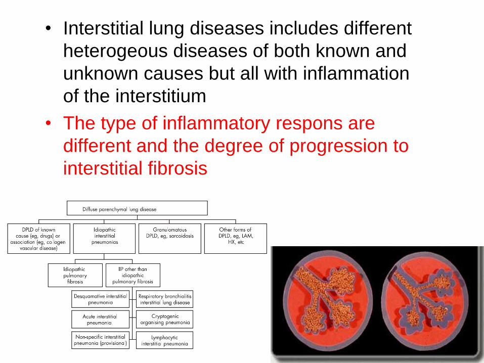
• Interstitial lung diseases includes different
heterogeous diseases of both known and
unknown causes but all with inflammation
of the interstitium
• The type of inflammatory respons are
different and the degree of progression to
interstitial fibrosis

Case60 yrs old man .The Pt symptoms started for 1,5-2 yrs ago,
where he for the first time experienced to get severedyspnea while running. Dry cough . Still working as leader in and institution for disabled. While work works at least 10 km a day. Ex-smoker, for 20 yrs 20 cigarets a day stopped 15 yrs ago.
Bloodtests showed increased IgG, marginally increasedALT, normal IgM reumafaktor. Normal ACE, anti-CCP, ANCA ,but weakly positive ANA.
Obj: clubbing,, vencro sound on stetocopy

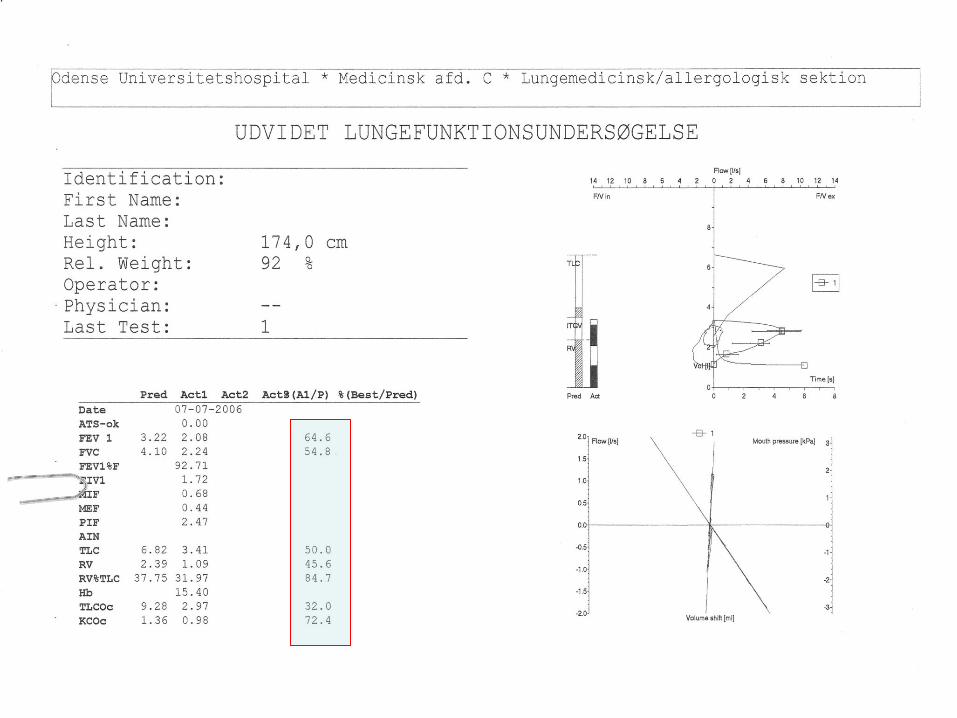

HRCT skan


Bronchial lavage for flow cytometri is done and
shows total cellenumber of 26 mio., distribution
is 66% makrofags, 2% lymfocyttes, 32%
granulocyttes and of those 28% is neutrofile og
4% er eosinofile. .
Biospsy is suboptimal material very few parts of
interstitielt lung tissue, nor enough to make a
diagnosis . But signs of few active cells and
some fibrosis. .

1. Patient has restriktive lung disease
2. Patient has DPLD but the excact diagnose
is uncertain
3. Patient has allergic alveolis
4. Patient has sarcoidosis
5. Patienten has asthma
6. Patienten has COPD
Resume:61 årig man, subjektive progression over 2 years, clubbing and vencrosound on
stetoscopy; ANA positiv, 6 min walk sat. fra 91-78%, TLC 50%, diffusion: 32%; bronko. Shows
inflammation with fibrosis og HRCT honny combing , tractions bronkieectasiers primary in the
lowest lung part
Diagnosis ?

1. Patient has restriktive lung disease
2. Patient has DPLD but the excact diagnose
is uncertain
3. Patienten has allergic alveolis
4. Patient has sarcoidosis
5. Patienten has asthma
6. Patienten has COPD


• Patienten is offered VATS mhp lung
biospy to gain a more specific diagnosis
He refuses initially !!!!

Diffuse Parenchymal Lung Disease (DPLD)
Incidence: 1 in ~ 3500
– pulmonary practice ~ 15% of patients
Usually a subacute or chronic clinical presentation with a slowly progressive course
– Exertional dyspnea
– Nonproductive cough
– Hypoxemia
– Restrictive pulmonary function
Many heterogeneous clinicopathologic entities
– Etiology may be known (1/3) or unknown (2/3)

What do we see ?
A-gas
• On exercise PaO2 decreases dramatically.
• Arterial PaO2 are reduced, pH normal.
• Physiologic dead space and physiologic shunt and VQ mismatch are increased.
• Diffuse impairment contributes to hypoxemia on exercise.
• There is marked reduction in diffusing capacity due to thickening of blood gas barrier and VQ mismatch.

Clinical presentation
Subacute or chronic onset, Exertional dyspnea,
nonproductive cough,
Constitutional Symptoms
Tachypnea, digital clubbing
Inspiratory “velcro” crackles
Hypoxemia,
cor pulmonaleAbnormal chest x-ray
– Reticular, reticulo-nodular patterns
– Distribution (bases, periphery)
– Honeycombing
– Ground-glass pattern (HRCT criteria, not on CXR)
Pulmonary symptoms associated with another disease, such as a connective tissue
disease
Lung function abnormalities

– Reticular, reticulo-nodular patterns
– Distribution (bases, periphery)


• One to see !

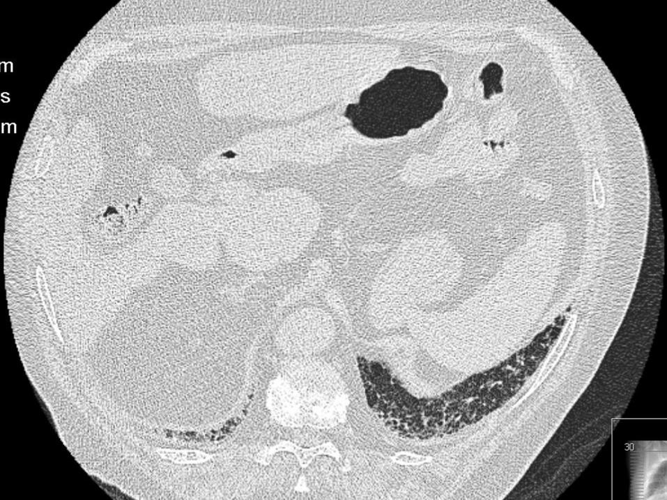


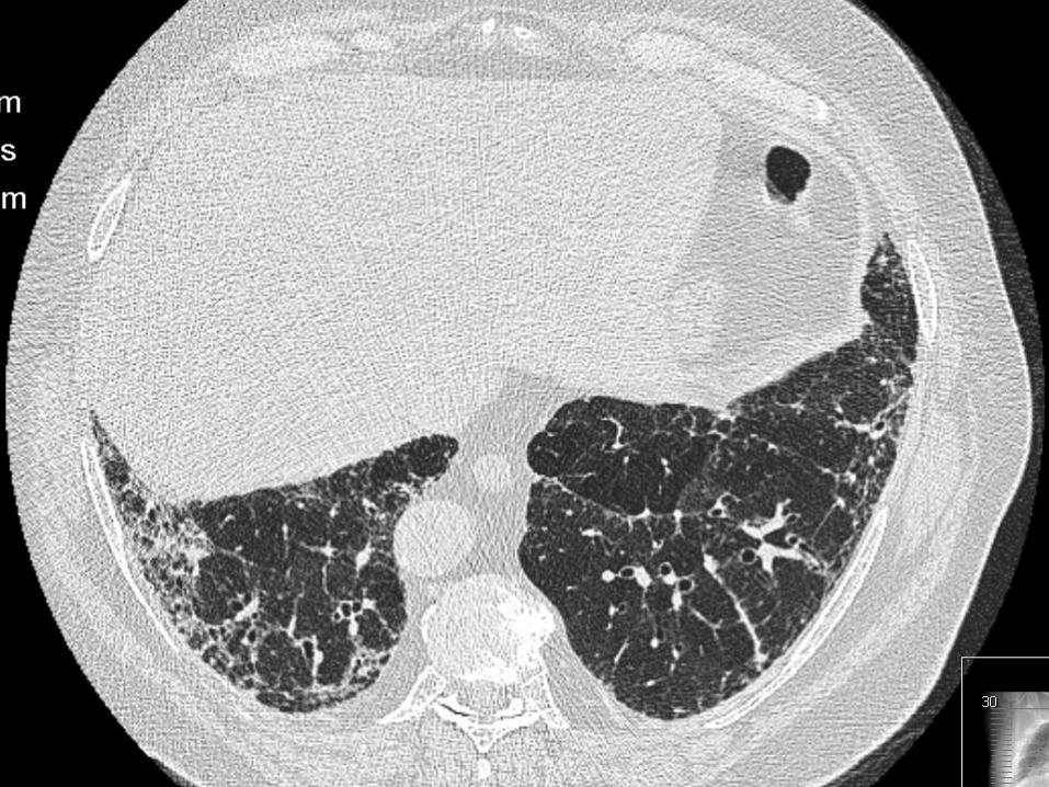
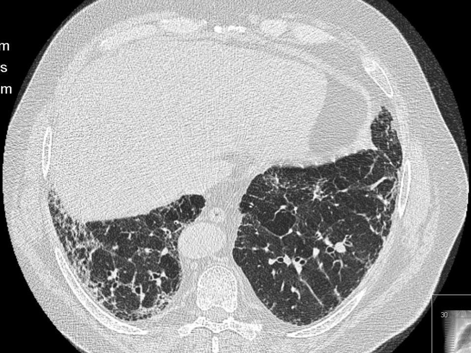

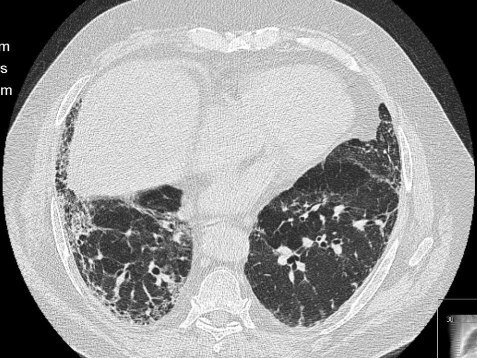
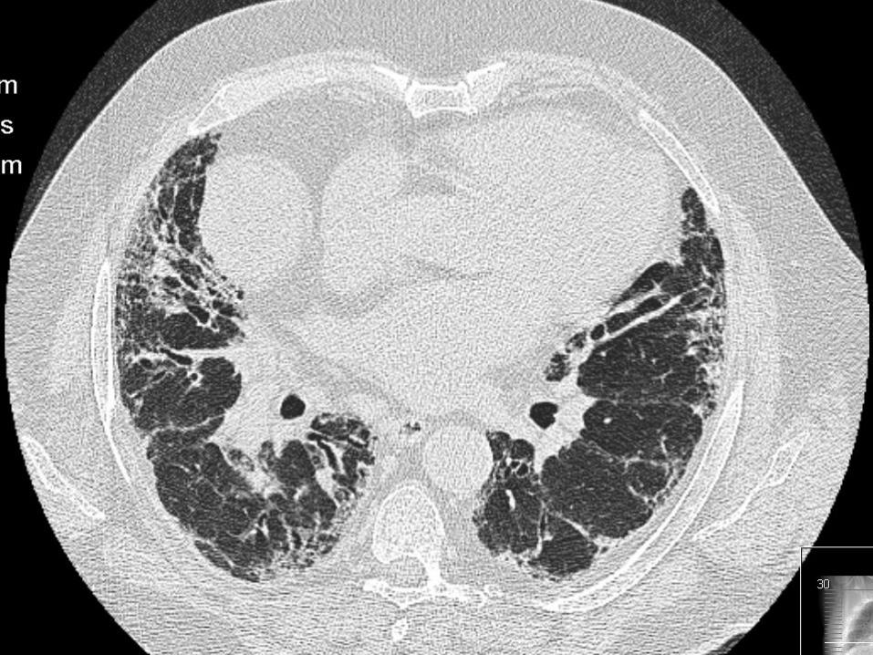

• 03.04.08
• HRCT shows bronkieektasies in both
sides from apex to basis. Lower
subpleurale cysts are seen and clasic
honney combing seen in idiopatisk lung
fibrosis. Apikalt findes øget interstitiel
tegning.
• RD: Lung fibrosis with bronkieektasies

•
• biopsi was done : B: firekantet perfusionsfikseret lungeresektat med stapler på
• to kanten. Resektatet måler 48 x 33 x 8 mm. Vævet fremtræder
• lidt uregelmæssigt med diskrete fibrotiske områder.
• Alt med i 6 kaps. undtagen den staplede kant./jyc
• Undersøger: KEO/pke
•
• MIKROSKOPI BESKRIVELSE:
• A-B: Alt materialet er indstøbt og undersøgt i histologiske
• snit, hvor der ses heterogent lungevæv. I samme synsfelt
• forekommer normalt lungevæv, fibrose med cystedannelser
• samt område med kronisk inflammation. Der er desuden en
• del sekretstagnation til dilaterede luftveje.
• De inflammatoriske områder viser beskeden inflammatorisk
• aktivitet, overvejende med lymfocytter, men også spredt
• forekomst af eosinofile granulocytter. Der er regeneration
•
• PATIENT: 280639-0359
• med fibroblastiske foci. I perifere luftveje ses bronkial
• metaplasi. I områder med forandringer af mere kronisk
• karakter ses bindevæv samt glat muskelcellehyperplasi.
• Der er ikke granulomer, amyloid eller malignitet.
• Der er en del tykvæggede kar, hvilket opfattes som sekundært
• til lungeforandringerne.
•
• conclusion :
• good material shows fibroserende alveolitis (UIP). /dpa
Important
The process of achieving a diagnosis in a
patient with interstitial lung diseases is
dynamic, requiring close communication
between clinician, radiologist, and
pathologist.
2002 Jan 15;165(2):277-304
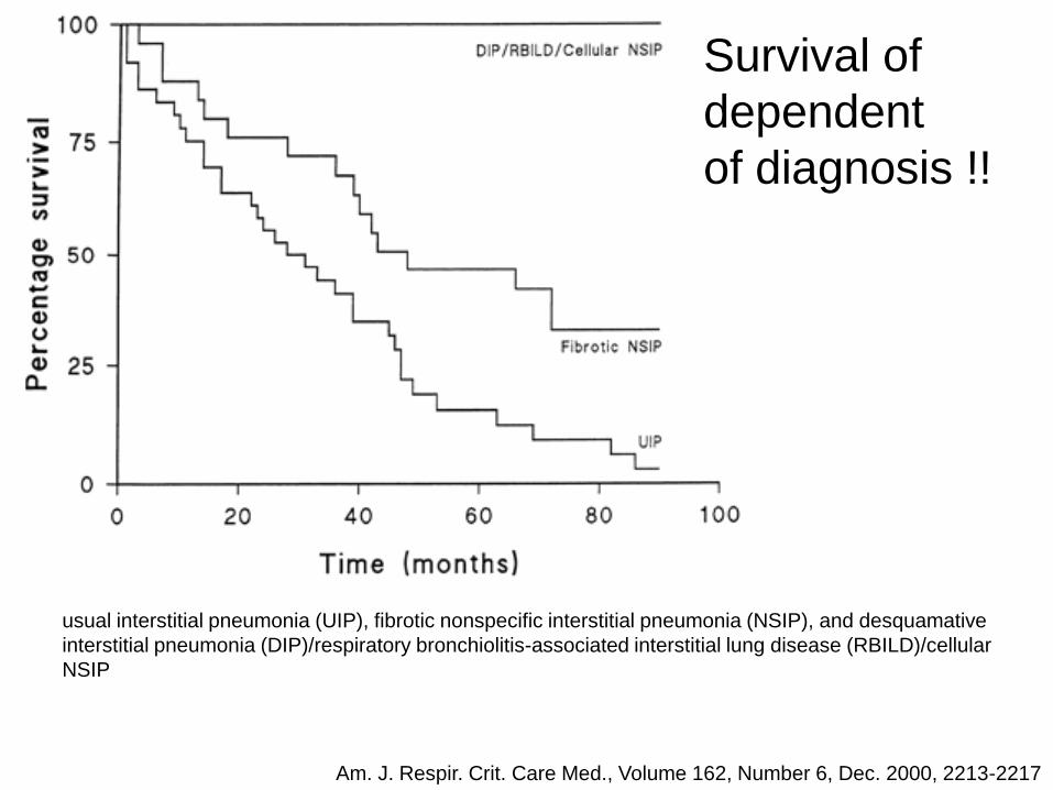
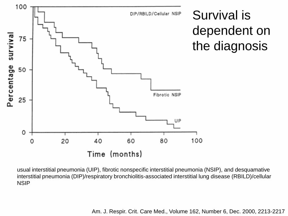
usual interstitial pneumonia (UIP), fibrotic nonspecific interstitial pneumonia (NSIP), and desquamative
interstitial pneumonia (DIP)/respiratory bronchiolitis-associated interstitial lung disease (RBILD)/cellular
NSIP
Survival of
dependent
of diagnosis !!
Am. J. Respir. Crit. Care Med., Volume 162, Number 6, Dec. 2000, 2213-2217

Prevalence of subgroup of interstitial pneumonia i 4 studies
UIP
NSIP
Cel
ular NSIP
Fibro
tic N
SIP
DIP
/RBIL
D
BOOP/C
OP
0
20
40
60
80
100
Diagnosis
Am. J. Respir. Crit. Care Med., Volume 162, Number 6, Dec. 2000, 2213-2217

Remember !
Restrictive lung diseases are defined by reduced total lung capacity, vital capacity
and functional residual capacity, but with preserved air flow
Alteration in lung parenchyma, diseases of the pleura, chest wall or neuromuscular
Lung parenchyma:
Exertional dyspnea, nonproductive cough,Tachypnea, digital clubbing, Inspiratory “velcro”
crackles, Hypoxemia, cor pulmonale
Does the patient have UIP ? – important due to treatment and prognosis.
Myths !
• Smoking does NOT normally cause lung
fibrosis
• However nothing without exceptions …..
Very rare lung diseases ……
• Smoking gives lungfibrosis…..???!!!


Langerhan cell histiocytosis.
This 50-year-old man had a
30 pack-year history of
cigarette smoking.
A: PA chest radiograph
shows hyperinflation of the
lungs and fine bilateral
reticular ILD.
B: CT scan shows multiple
cysts (solid arrow) and
nodules (dashed arrow).

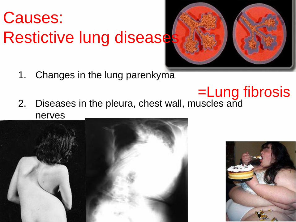
Causes:
Restictive lung diseases
1. Changes in the lung parenkyma
2. Diseases in the pleura, chest wall, muscles and
nerves
=Lung fibrosis
What happens in
lung fibrosis ??
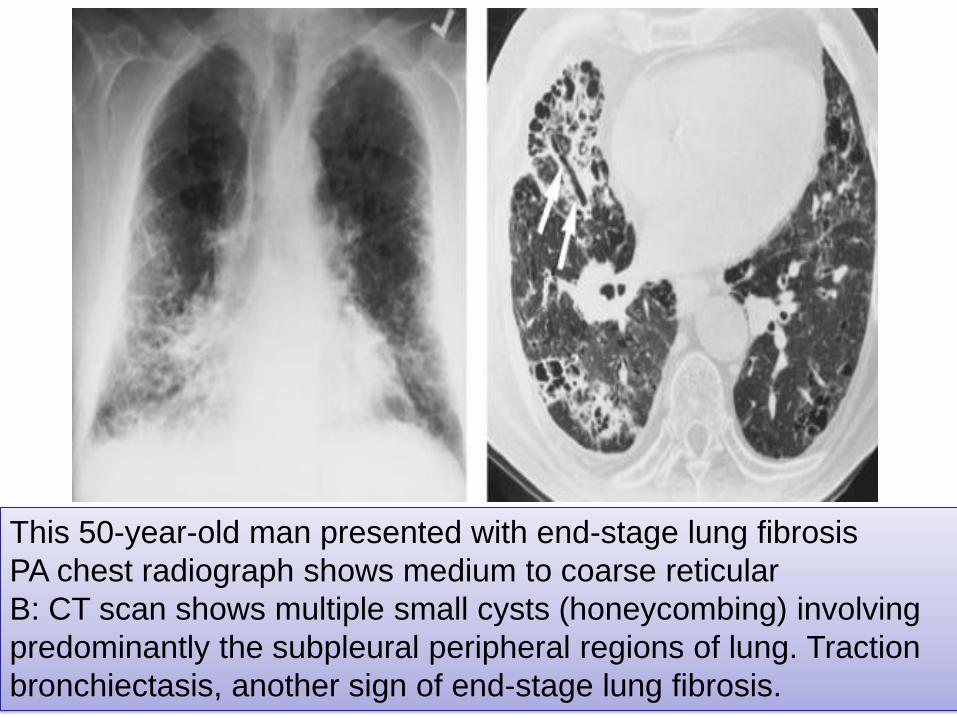
This 50-year-old man presented with end-stage lung fibrosis
PA chest radiograph shows medium to coarse reticular
B: CT scan shows multiple small cysts (honeycombing) involving
predominantly the subpleural peripheral regions of lung. Traction
bronchiectasis, another sign of end-stage lung fibrosis.


Lung fibrosis
decreased ability
of oxygen optake
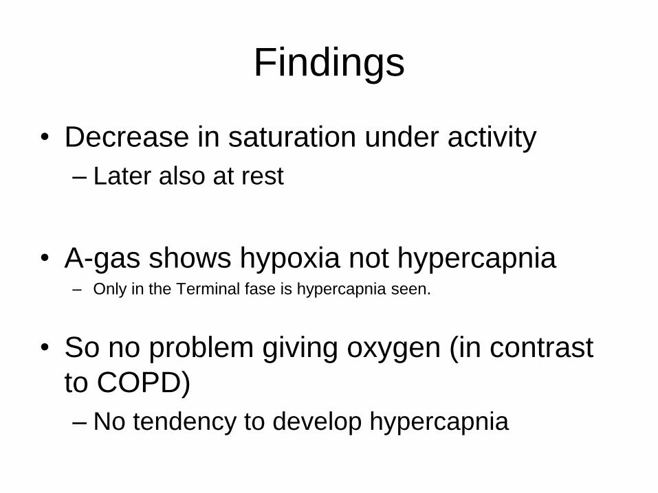
Findings
• Decrease in saturation under activity
– Later also at rest
• A-gas shows hypoxia not hypercapnia– Only in the Terminal fase is hypercapnia seen.
• So no problem giving oxygen (in contrast
to COPD)
– No tendency to develop hypercapnia

What do we have to measure in
patients with lung fibosis??


There is a slight difference in diagnosing
and monitoring the disease
• Lung function– Forced volumen
– TLC, RV and DLCO
• Anatomic changes– Bronkoscopy
– HRCT scan
– X-RTG Thorax
– Ekko/hjertekat
– Lungebiopsy
– Dexa scanning
• Serological changes – Blood tests
• Activity – 6 min walking test

Always initially do TLC;RV and DLCO

TLC: How big a the lungs ?
TLCO: How ”good” are the
lungs to oxygen optake ?

6-min Walking
test
– How far?
– Desaturation?
– Symptoms severe ?
– The test accesses the physical ability?
– Degree of severeness
– Disease development
– Guidance to when transplantation
should be considered
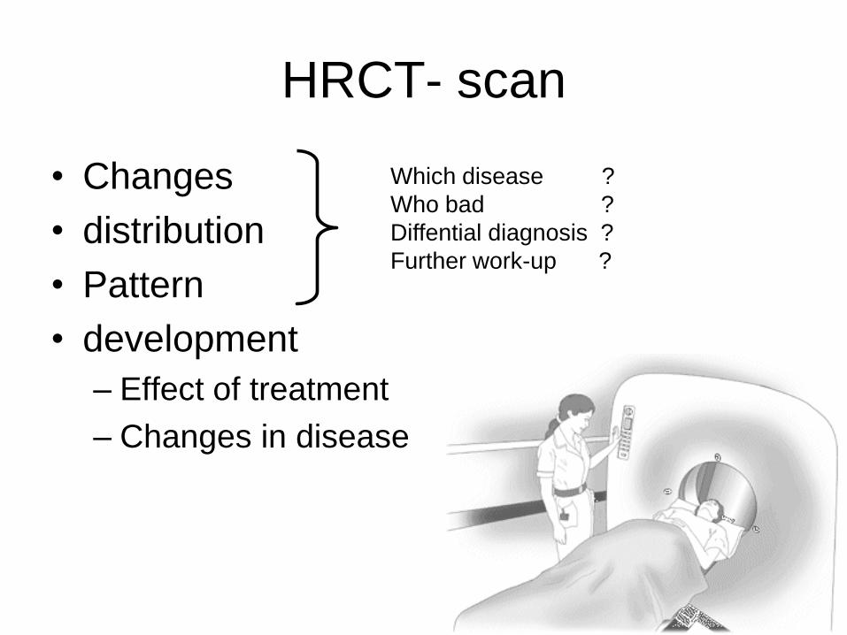
HRCT- scan
• Changes
• distribution
• Pattern
• development
– Effect of treatment
– Changes in disease
Which disease ?
Who bad ?
Diffential diagnosis ?
Further work-up ?

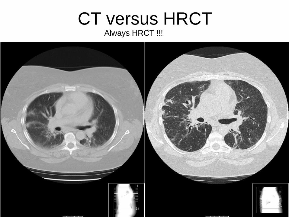
CT versus HRCTAlways HRCT !!!
usual interstitial pneumonia (UIP), fibrotic nonspecific interstitial pneumonia (NSIP), and desquamative
interstitial pneumonia (DIP)/respiratory bronchiolitis-associated interstitial lung disease (RBILD)/cellular
NSIP
Survival is
dependent on
the diagnosis
Am. J. Respir. Crit. Care Med., Volume 162, Number 6, Dec. 2000, 2213-2217

Restrictive diseases
Intrinsic lung diseases
– Interstitial lung diseases
» Arthritis related (SLE, RA, scleroderma)
» “Ideopatic” (ex UIP)
- “smoke related” (ex Histeocytosis X)
– Asbestosis/silicosis
– Allergic (allergic alveolitis)
– Pleura (debris-exsudat)
– Medicine (nitrofurantoin, amiodarone, bleomycin).
– Pneumonia
– radiation
Extrinsic diseases (extra-parenchymale diseases)
– Non-neuromuskular
• Deformities
• Heart disease
• ARDS
– Neuromuscular
• Poliomyelitis, Guillain-Barre syndrome, ALS, myasthenia gravis, muscular
dystrophies
Inflammation and/or scarring of lung tissue
Fill airspaces exudat/debris (pneumonnitis)
reduced space or muscular power
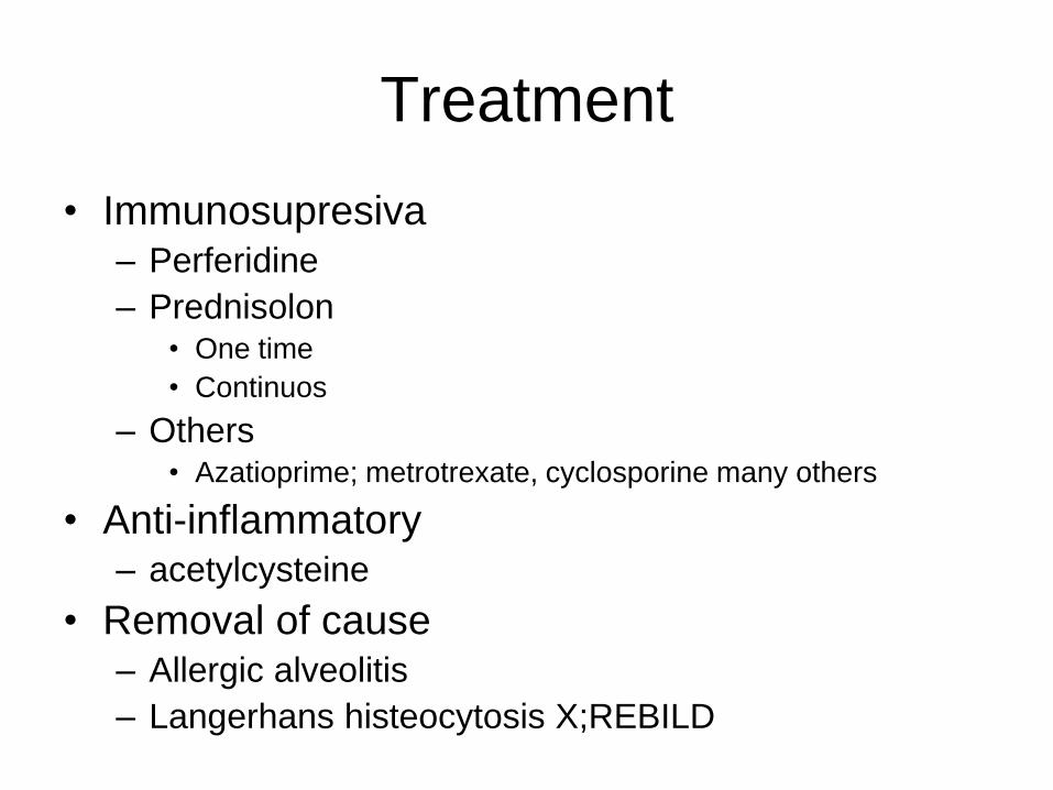
Treatment
• Immunosupresiva– Perferidine
– Prednisolon• One time
• Continuos
– Others • Azatioprime; metrotrexate, cyclosporine many others
• Anti-inflammatory – acetylcysteine
• Removal of cause– Allergic alveolitis
– Langerhans histeocytosis X;REBILD

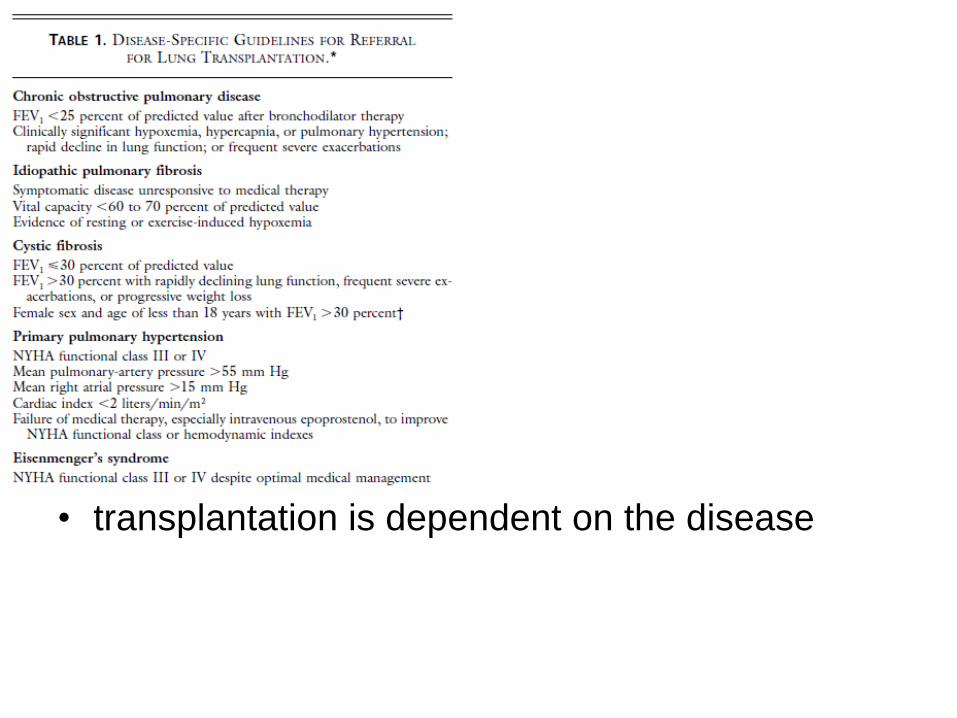
• transplantation is dependent on the disease

CM-104
0
200
400
600
800
1000
1200
1400
1600
1800
85 86 87 88 89 90 91 92 93 94 95 96 97 98 99 00 01 02 03
Year
Double lung
Single lung
Worldwide Lung Transplantation Numbers
Source: International Society of Heart and Lung Transplantation (ISHLT); UNOS
Lung transplants performed worldwide, by year
Emphysema/COPD Idiopathic pulmonary fibrosis Cystic fibrosis Alpha-1 antitrypsin deficiency Primary pulmonary hypertension Sarcoidosis Retransplant/graft failure Other
1.8% 2.6%
4.2%
39.0%
10.4%
17.0%16.0%
9.0%
Primary diagnosis, 01/1995 - 06/2003

CM-106
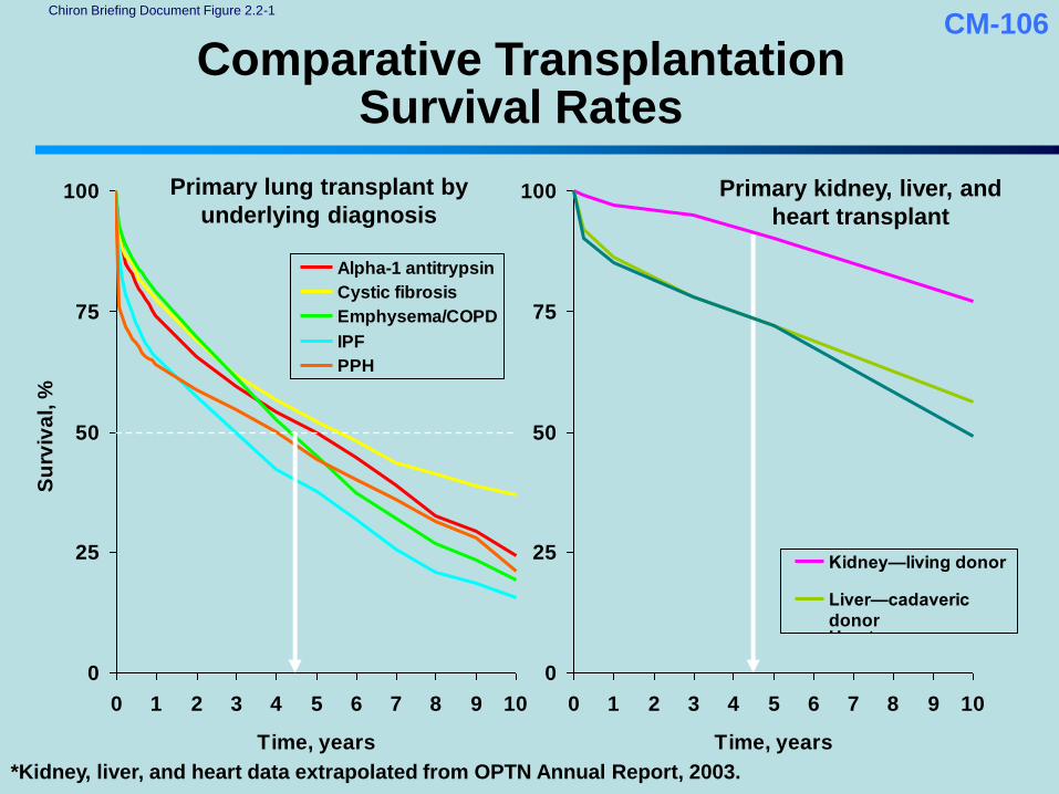
Comparative Transplantation Survival Rates
0
25
50
75
100
0 1 2 3 4 5 6 7 8 9 10
Time, years
Su
rviv
al, %
Alpha-1 antitrypsin
Cystic fibrosis
Emphysema/COPD
IPF
PPH
0
25
50
75
100
0 1 2 3 4 5 6 7 8 9 10
Time, years
Kidney—living donor
Liver—cadaveric
donorHeart
Primary lung transplant by
underlying diagnosisPrimary kidney, liver, and
heart transplant
*Kidney, liver, and heart data extrapolated from OPTN Annual Report, 2003.
Chiron Briefing Document Figure 2.2-1
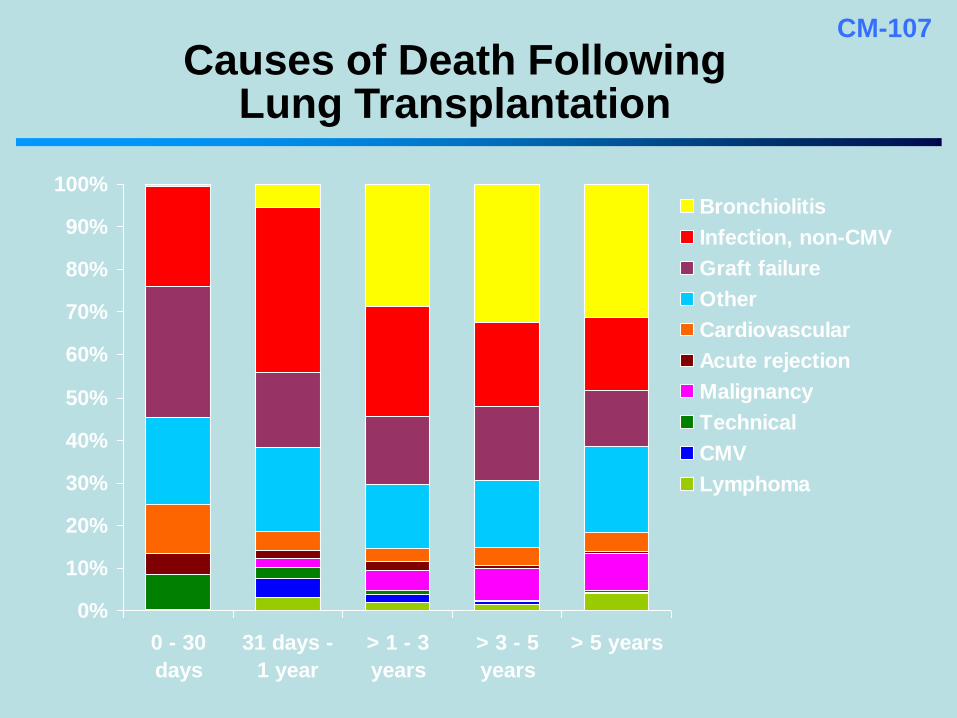
CM-107
Causes of Death Following Lung Transplantation
0%
10%
20%
30%
40%
50%
60%
70%
80%
90%
100%
0 - 30
days
31 days -
1 year
> 1 - 3
years
> 3 - 5
years
> 5 years
Bronchiolitis
Infection, non-CMV
Graft failure
Other
Cardiovascular
Acute rejection
Malignancy
Technical
CMV
Lymphoma

Status forLung transplantation
Survivel —50% died after 5 years
Bronchiolitis obliterans main reason for a bad
survivel rate
Main aim to treat and prevent bronchiolitis
obliterans

More time???


