Page 1
Functional Foods in Health and Disease 2013; 3(8):344-366
Page 344 of 366
Review Open Access
Intravenous ascorbic acid protocol for cancer patients: scientific
rationale, pharmacology, and clinical experience
Mikirova NA, Casciari JJ, Hunninghake RE, Riordan NH
Riordan Clinic, 3100 N Hillside, Wichita, KS
Corresponding Author: Nina Mikirova, Riordan Clinic, 3100 N Hillside, Wichita, KS, USA
Submission date: May 15, 2013; Acceptance date: August 23, 2013; Publication date: August 26,
2013
ABSTRACT:
Background: Ascorbic acid (vitamin C, ascorbate) has been shown to protect cells against
various types of oxidant injury at physiologically relevant concentrations. Vitamin C has been
suggested as having both a preventative and therapeutic role in a number of pathologies when
administered at much higher-than-recommended dietary allowance levels. This article reviews
the scientific rational for intravenous vitamin C as a potential treatment for cancer.
Many mechanisms of action for ascorbate efficacy against cancer have been proposed over
the years. Cancer patients are often deficient in vitamin C, and require large doses to replenish
depleted stores. It has been demonstrated in vitro and in animal studies that vitamin C is
preferentially toxic to tumor cells at millimolar concentrations; moreover, pharmacokinetic data
suggest that these concentrations are clinically achievable when ascorbate is administered
intravenously. Data suggests that ascorbate may serve as a biological response modifier,
affecting inflammation and angiogenesis as well as improving immune function parameters.
While Phase II clinical trials using ascorbate in cancer therapy are under way, vitamin C is
not subject to the regulations that synthetic drugs are and therefore has been used clinically for
decades to treat cancer patients. This clinical experience suggests the therapy is safe, and may be
effective in some instances. Attached to this article is the Riordan IVC Protocol, which details
an intravenous vitamin C protocol that can be safely administered to cancer patients.
Keywords: Cancer, inflammation, C-reactive protein, inflammatory cytokines, high-dose
vitamin C
INTRODUCTION:
Vitamin C is a major water-soluble antioxidant that increases extracellular collagen production
and is important for proper immune cell functioning [1, 2]. It plays key roles in L-carnitine
synthesis, cholesterol metabolism, cytochrome P-450 activity, and neurotransmitter synthesis [3].
Page 2
Functional Foods in Health and Disease 2013; 3(8):344-366
Page 345 of 366
Intravenous vitamin C (IVC) use has increased recently among integrative and orthomolecular
medicine practitioners. A survey of roughly 300 practitioners conducted between 2006 and 2008
indicated that roughly ten thousand patients received IVC, at an average dose of 0.5 g/kg,
without significant ill effects [4, 5]. While IVC may have a variety of possible applications, such
as combating infections [6], treating rheumatoid arthritis [7], or treating fatigue, it has generated
the most interest for its potential use in adjunctive cancer care.
Vitamin C was first suggested as a tool for cancer treatment in the 1950’s; its role in
collagen production and protection led scientists to hypothesize that ascorbate replenishment
would protect normal tissue from tumor invasiveness and metastasis [2, 8]. Also, since cancer
patients are often depleted of vitamin C [1, 9], replenishment may improve immune system
function and enhance patient health and well-being [10]. Cameron and Pauling observed
fourfold survival times in terminal cancer patients treated with intravenous ascorbate infusions
followed by oral supplementation [11]. However, two randomized clinical trials with oral
ascorbate alone conducted by the Mayo clinic showed no benefit [12, 13]. Most research from
that point is focused on intravenous ascorbate. The rationales for using intravenous ascorbate
infusions (IVC) to treat cancer, which are discussed in detail below, can be summarized as
follows:
At millimolar concentrations, ascorbate is preferentially toxic to cancer cells in vitro and
is able to inhibit angiogenesis in vitro and in vivo.
Vitamin C can accumulate in tumors, with significant tumor growth inhibition seen
(study with guinea pigs) at intra-tumor concentrations of 1 mM or higher.
Plasma ascorbate concentrations in the millimolar range can be safely achieved with IVC
infusions. Phase I clinical studies indicate that IVC can be administered safely with
relatively few adverse effects.
Published case studies report anti-cancer efficacy, improved patient well-being, and
decreases in markers of inflammation and tumor growth.
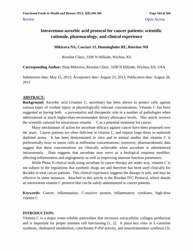
The Riordan Clinic has treated hundreds of cancer patients (Figure 1) using the developed
protocol.
We have been researching the potential of intravenous vitamin C therapy for over thirty
years. Our efforts have included in vitro studies, animal studies, pharmacokinetic analyses, and
clinical trial. The Riordan IVC protocol, along with the research results that have motivated its
use, is described below.
Pharmacokinetics: Vitamin C is water-soluble, and is limited in how well it can be absorbed
when given orally. While ascorbate tends to accumulate in adrenal glands, the brain, and in
some white blood cell types, plasma levels stay relatively low [14-16]. According to the study
[17], the plasma levels in healthy adults stayed below 100 µM, even if 2.5 grams were taken
when administered once daily by the oral route.
Cancer patients tend to be depleted of vitamin C: fourteen out of twenty-two terminal
cancer patients in our phase I study were depleted of vitamin C, with ten of those having zero
detectable ascorbate in their plasma [9]. In a study of cancer patients in hospice care, thirty
Page 3
Functional Foods in Health and Disease 2013; 3(8):344-366
Page 346 of 366
percent of the subjects were deficient in vitamin C [18]. Deficiency (below 10 µM) was
correlated with elevated inflammation marker C-reactive protein (CRP) and shorter survival
times. Given the role of vitamin C in collagen production, immune system functioning, and
antioxidant protection, it is not surprising that subjects depleted of ascorbate would fare poorly in
mounting defenses against cancer. This also suggests that supplementation to replenish vitamin
C stores might serve as adjunctive therapy for these patients.
Figure 1. Types of cancers treated with IVC by the Riordan Clinic. Cancer patients from the database
of patients for whom plasma ascorbic acid levels before and after treatment were available along with laboratory
tests of inflammation marker CRP and cancer markers.
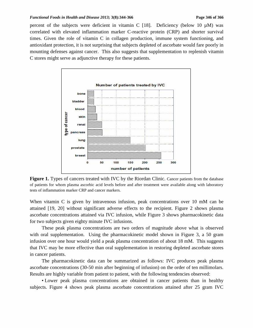
When vitamin C is given by intravenous infusion, peak concentrations over 10 mM can be
attained [19, 20] without significant adverse effects to the recipient. Figure 2 shows plasma
ascorbate concentrations attained via IVC infusion, while Figure 3 shows pharmacokinetic data
for two subjects given eighty minute IVC infusions.
These peak plasma concentrations are two orders of magnitude above what is observed
with oral supplementation. Using the pharmacokinetic model shown in Figure 3, a 50 gram
infusion over one hour would yield a peak plasma concentration of about 18 mM. This suggests
that IVC may be more effective than oral supplementation in restoring depleted ascorbate stores
in cancer patients.
The pharmacokinetic data can be summarized as follows: IVC produces peak plasma
ascorbate concentrations (30-50 min after beginning of infusion) on the order of ten millimolars.
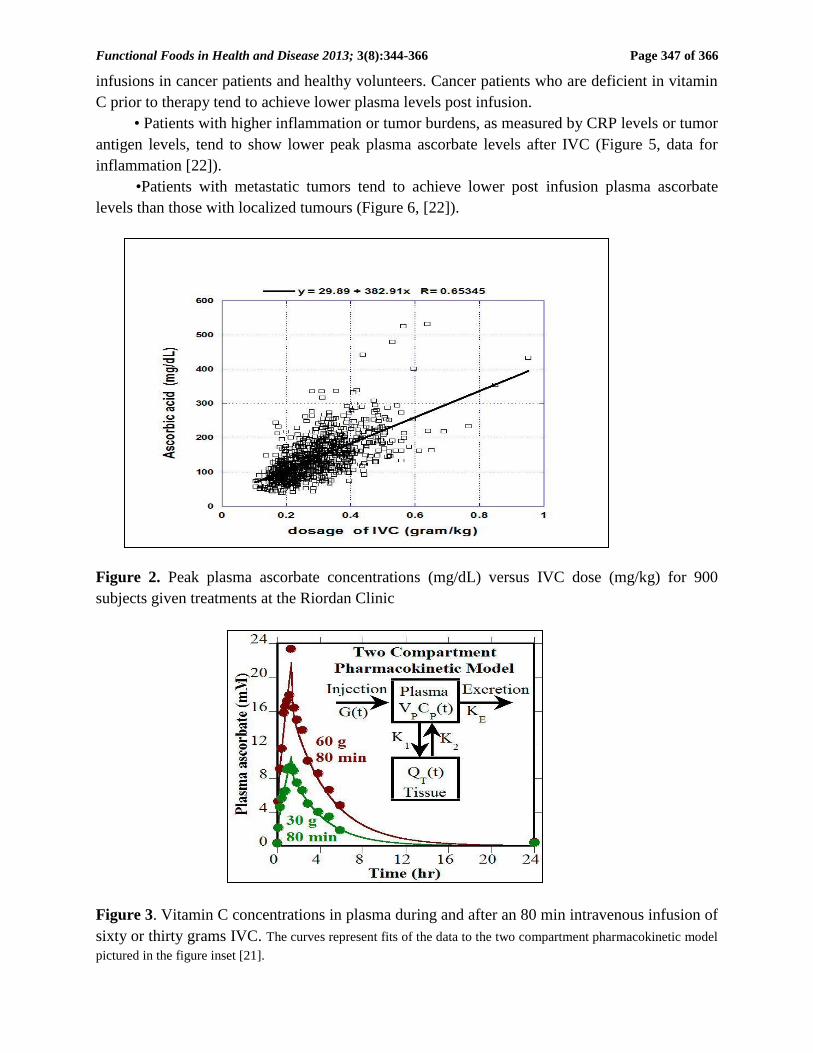
Results are highly variable from patient to patient, with the following tendencies observed:
• Lower peak plasma concentrations are obtained in cancer patients than in healthy
subjects. Figure 4 shows peak plasma ascorbate concentrations attained after 25 gram IVC
Page 4
Functional Foods in Health and Disease 2013; 3(8):344-366
Page 347 of 366
infusions in cancer patients and healthy volunteers. Cancer patients who are deficient in vitamin
C prior to therapy tend to achieve lower plasma levels post infusion.
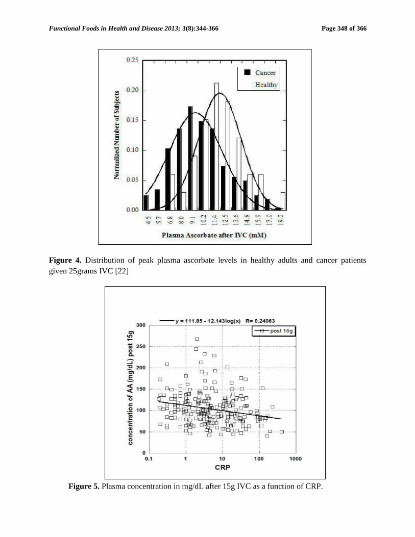
• Patients with higher inflammation or tumor burdens, as measured by CRP levels or tumor
antigen levels, tend to show lower peak plasma ascorbate levels after IVC (Figure 5, data for
inflammation [22]).
•Patients with metastatic tumors tend to achieve lower post infusion plasma ascorbate
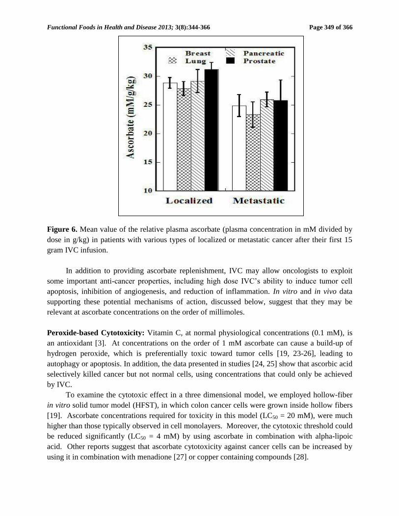
levels than those with localized tumours (Figure 6, [22]).
Figure 2. Peak plasma ascorbate concentrations (mg/dL) versus IVC dose (mg/kg) for 900
subjects given treatments at the Riordan Clinic
Figure 3. Vitamin C concentrations in plasma during and after an 80 min intravenous infusion of
sixty or thirty grams IVC. The curves represent fits of the data to the two compartment pharmacokinetic model
pictured in the figure inset [21].
Page 5
Functional Foods in Health and Disease 2013; 3(8):344-366
Page 348 of 366
Figure 4. Distribution of peak plasma ascorbate levels in healthy adults and cancer patients
given 25grams IVC [22]
Figure 5. Plasma concentration in mg/dL after 15g IVC as a function of CRP.
Page 6
Functional Foods in Health and Disease 2013; 3(8):344-366
Page 349 of 366
Figure 6. Mean value of the relative plasma ascorbate (plasma concentration in mM divided by
dose in g/kg) in patients with various types of localized or metastatic cancer after their first 15
gram IVC infusion.
In addition to providing ascorbate replenishment, IVC may allow oncologists to exploit
some important anti-cancer properties, including high dose IVC’s ability to induce tumor cell
apoptosis, inhibition of angiogenesis, and reduction of inflammation. In vitro and in vivo data
supporting these potential mechanisms of action, discussed below, suggest that they may be
relevant at ascorbate concentrations on the order of millimoles.
Peroxide-based Cytotoxicity: Vitamin C, at normal physiological concentrations (0.1 mM), is
an antioxidant [3]. At concentrations on the order of 1 mM ascorbate can cause a build-up of
hydrogen peroxide, which is preferentially toxic toward tumor cells [19, 23-26], leading to
autophagy or apoptosis. In addition, the data presented in studies [24, 25] show that ascorbic acid
selectively killed cancer but not normal cells, using concentrations that could only be achieved
by IVC.
To examine the cytotoxic effect in a three dimensional model, we employed hollow-fiber
in vitro solid tumor model (HFST), in which colon cancer cells were grown inside hollow fibers
[19]. Ascorbate concentrations required for toxicity in this model (LC50 = 20 mM), were much
higher than those typically observed in cell monolayers. Moreover, the cytotoxic threshold could
be reduced significantly (LC50 = 4 mM) by using ascorbate in combination with alpha-lipoic
acid. Other reports suggest that ascorbate cytotoxicity against cancer cells can be increased by
using it in combination with menadione [27] or copper containing compounds [28].
Page 7
Functional Foods in Health and Disease 2013; 3(8):344-366
Page 350 of 366
Studies from many laboratories in a variety animal models, using hepatoma, pancreatic
cancer, colon cancer, sarcoma, leukemia, prostate cancer, and mesothelioma, confirm that
ascorbate concentrations sufficient for its cytotoxicity can be attained in vivo, and that treatments
can reduce tumor growth [29-34].
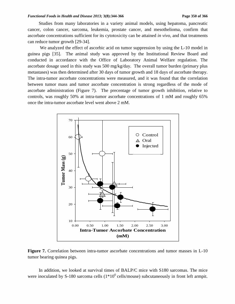
We analyzed the effect of ascorbic acid on tumor suppression by using the L-10 model in
guinea pigs [35]. The animal study was approved by the Institutional Review Board and
conducted in accordance with the Office of Laboratory Animal Welfare regulation. The
ascorbate dosage used in this study was 500 mg/kg/day. The overall tumor burden (primary plus
metastases) was then determined after 30 days of tumor growth and 18 days of ascorbate therapy.
The intra-tumor ascorbate concentrations were measured, and it was found that the correlation
between tumor mass and tumor ascorbate concentration is strong regardless of the mode of
ascorbate administration (Figure 7). The precentage of tumor growth inhibition, relative to
controls, was roughly 50% at intra-tumor ascorbate concentrations of 1 mM and roughly 65%
once the intra-tumor ascorbate level went above 2 mM.
Figure 7. Correlation between intra-tumor ascorbate concentrations and tumor masses in L-10
tumor bearing quinea pigs.
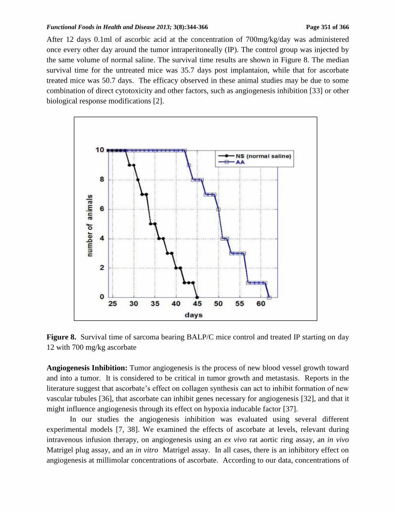
In addition, we looked at survival times of BALP/C mice with S180 sarcomas. The mice
were inoculated by S-180 sarcoma cells (1*106
cells/mouse) subcutaneously in front left armpit.
10
20
30
40
50
60
70
0.00 0.50 1.00 1.50 2.00 2.50 3.00
Intra-Tumor Ascorbate Concentration
(mM)
Tu
mor
Mas
s (g
)
Control
Oral
Injected
Data Fit
Page 8
Functional Foods in Health and Disease 2013; 3(8):344-366
Page 351 of 366
After 12 days 0.1ml of ascorbic acid at the concentration of 700mg/kg/day was administered
once every other day around the tumor intraperitoneally (IP). The control group was injected by
the same volume of normal saline. The survival time results are shown in Figure 8. The median
survival time for the untreated mice was 35.7 days post implantaion, while that for ascorbate
treated mice was 50.7 days. The efficacy observed in these animal studies may be due to some
combination of direct cytotoxicity and other factors, such as angiogenesis inhibition [33] or other
biological response modifications [2].
Figure 8. Survival time of sarcoma bearing BALP/C mice control and treated IP starting on day
12 with 700 mg/kg ascorbate
Angiogenesis Inhibition: Tumor angiogenesis is the process of new blood vessel growth toward
and into a tumor. It is considered to be critical in tumor growth and metastasis. Reports in the
literature suggest that ascorbate’s effect on collagen synthesis can act to inhibit formation of new
vascular tubules [36], that ascorbate can inhibit genes necessary for angiogenesis [32], and that it
might influence angiogenesis through its effect on hypoxia inducable factor [37].
In our studies the angiogenesis inhibition was evaluated using several different
experimental models [7, 38]. We examined the effects of ascorbate at levels, relevant during
intravenous infusion therapy, on angiogenesis using an ex vivo rat aortic ring assay, an in vivo
Matrigel plug assay, and an in vitro Matrigel assay. In all cases, there is an inhibitory effect on
angiogenesis at millimolar concentrations of ascorbate. According to our data, concentrations of
Page 9
Functional Foods in Health and Disease 2013; 3(8):344-366
Page 352 of 366
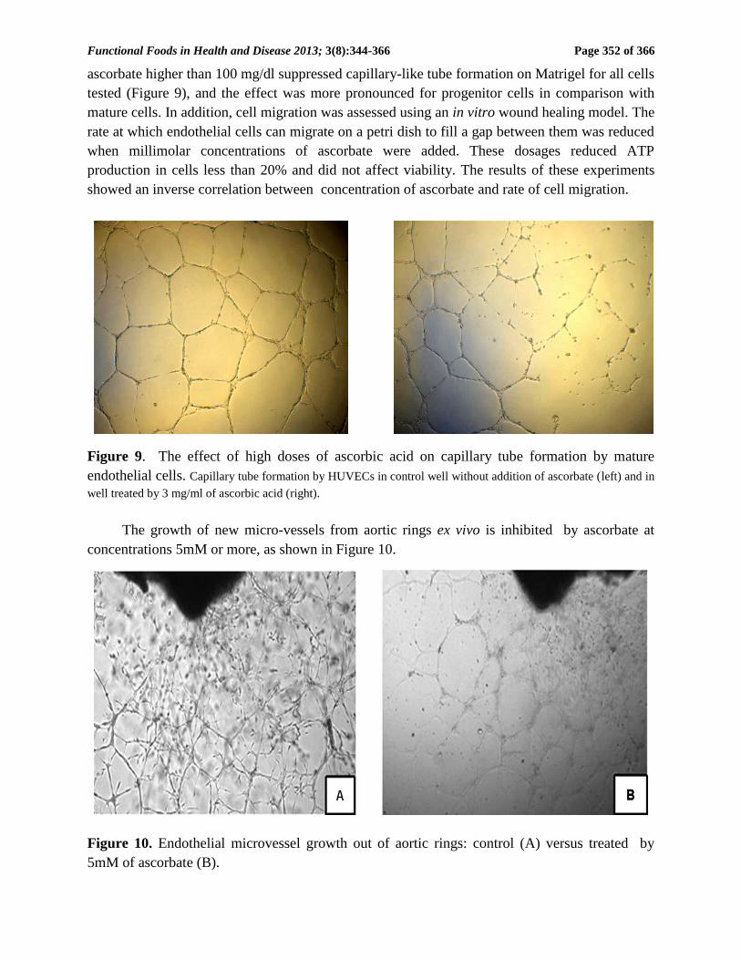
ascorbate higher than 100 mg/dl suppressed capillary-like tube formation on Matrigel for all cells
tested (Figure 9), and the effect was more pronounced for progenitor cells in comparison with
mature cells. In addition, cell migration was assessed using an in vitro wound healing model. The
rate at which endothelial cells can migrate on a petri dish to fill a gap between them was reduced
when millimolar concentrations of ascorbate were added. These dosages reduced ATP
production in cells less than 20% and did not affect viability. The results of these experiments
showed an inverse correlation between concentration of ascorbate and rate of cell migration.
Figure 9. The effect of high doses of ascorbic acid on capillary tube formation by mature
endothelial cells. Capillary tube formation by HUVECs in control well without addition of ascorbate (left) and in
well treated by 3 mg/ml of ascorbic acid (right).
The growth of new micro-vessels from aortic rings ex vivo is inhibited by ascorbate at
concentrations 5mM or more, as shown in Figure 10.
Figure 10. Endothelial microvessel growth out of aortic rings: control (A) versus treated by
5mM of ascorbate (B).
Page 10
Functional Foods in Health and Disease 2013; 3(8):344-366
Page 353 of 366
For Matrigel plugs implanted subcutaneously in mice, the micro-vessel densities were
significantly lower in mice treated with 400 mg/kg every other day for two weeks; numbers of
vessels and vessel densities in plugs from treated mice were roughly 30% less than those in plugs
from untreated mice.
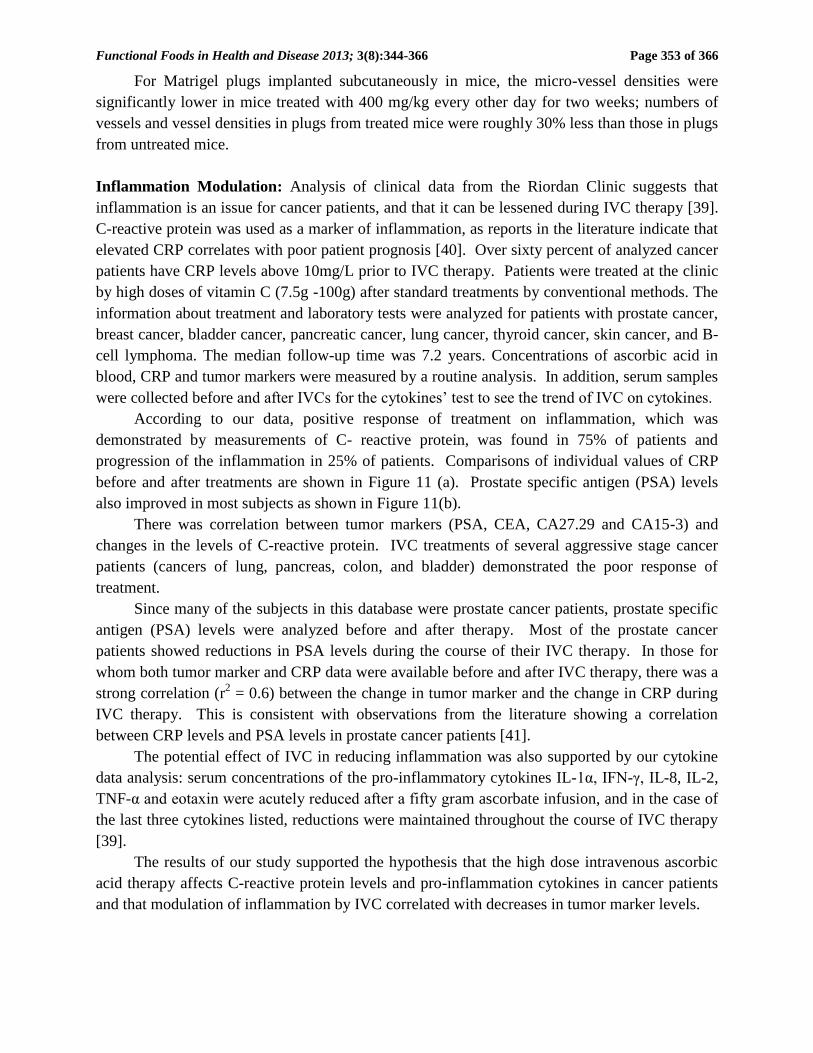
Inflammation Modulation: Analysis of clinical data from the Riordan Clinic suggests that
inflammation is an issue for cancer patients, and that it can be lessened during IVC therapy [39].
C-reactive protein was used as a marker of inflammation, as reports in the literature indicate that
elevated CRP correlates with poor patient prognosis [40]. Over sixty percent of analyzed cancer
patients have CRP levels above 10mg/L prior to IVC therapy. Patients were treated at the clinic
by high doses of vitamin C (7.5g -100g) after standard treatments by conventional methods. The
information about treatment and laboratory tests were analyzed for patients with prostate cancer,
breast cancer, bladder cancer, pancreatic cancer, lung cancer, thyroid cancer, skin cancer, and B-
cell lymphoma. The median follow-up time was 7.2 years. Concentrations of ascorbic acid in
blood, CRP and tumor markers were measured by a routine analysis. In addition, serum samples
were collected before and after IVCs for the cytokines’ test to see the trend of IVC on cytokines.
According to our data, positive response of treatment on inflammation, which was
demonstrated by measurements of C- reactive protein, was found in 75% of patients and
progression of the inflammation in 25% of patients. Comparisons of individual values of CRP
before and after treatments are shown in Figure 11 (a). Prostate specific antigen (PSA) levels
also improved in most subjects as shown in Figure 11(b).
There was correlation between tumor markers (PSA, CEA, CA27.29 and CA15-3) and
changes in the levels of C-reactive protein. IVC treatments of several aggressive stage cancer
patients (cancers of lung, pancreas, colon, and bladder) demonstrated the poor response of
treatment.
Since many of the subjects in this database were prostate cancer patients, prostate specific
antigen (PSA) levels were analyzed before and after therapy. Most of the prostate cancer
patients showed reductions in PSA levels during the course of their IVC therapy. In those for
whom both tumor marker and CRP data were available before and after IVC therapy, there was a
strong correlation (r2 = 0.6) between the change in tumor marker and the change in CRP during
IVC therapy. This is consistent with observations from the literature showing a correlation
between CRP levels and PSA levels in prostate cancer patients [41].
The potential effect of IVC in reducing inflammation was also supported by our cytokine
data analysis: serum concentrations of the pro-inflammatory cytokines IL-1α, IFN-γ, IL-8, IL-2,
TNF-α and eotaxin were acutely reduced after a fifty gram ascorbate infusion, and in the case of
the last three cytokines listed, reductions were maintained throughout the course of IVC therapy
[39].
The results of our study supported the hypothesis that the high dose intravenous ascorbic
acid therapy affects C-reactive protein levels and pro-inflammation cytokines in cancer patients
and that modulation of inflammation by IVC correlated with decreases in tumor marker levels.
Page 11
Functional Foods in Health and Disease 2013; 3(8):344-366
Page 354 of 366
Figure 11. Changes in inflammation and PSA markers for cancer patients treated at the Riordan
Clinic after IVC therapy.
Page 12
Functional Foods in Health and Disease 2013; 3(8):344-366
Page 355 of 366
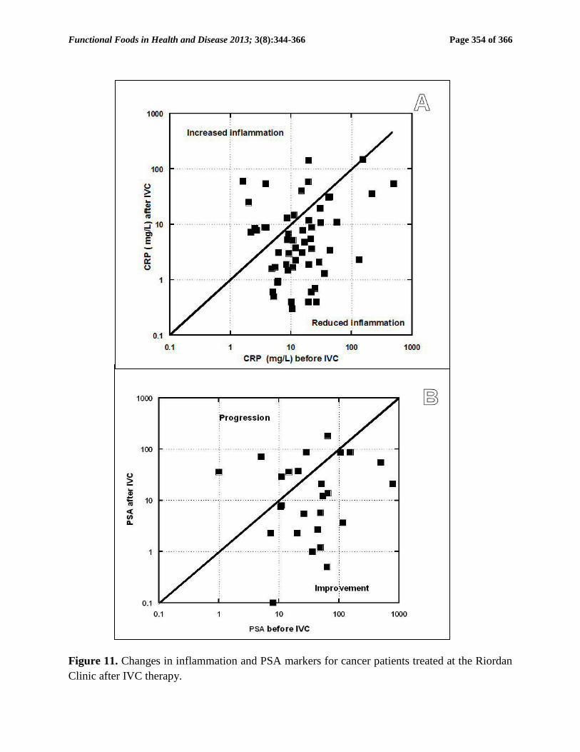
In addition, we analyzed the effect of ascorbic acid on transcriptional factor NK-kB.
When stimulated by tumor necrosis factor (TNF-), cancer cells up-regulate production of NF-
B, a molecule that is the key to inflammation and is thought to promote tumor angiogenesis and
tumor invasiveness. Our experiments, using a HeLa cancer cell line transfected with the
bioluminescence marker luciferase added to the promoter region of the gene for NF-B, indicate
that vitamin C might lessen the up-regulation of this inflammation marker. The effect of the
reduced form of ascorbic acid and its oxidized form, dehydroascorbate (DHA), on NF-B
production in HeLa cell in the presence of TNF- is shown in Figure 12.
Figure 12. Vitamin C inhibits NF-kB activation by TNF-α in confluent monolayers of HeLa
cells pretreated with 2 mM or 5 mM of ascorbate or DHA before stimulation by 10 ng/ml TNF-
α. Control cells were left un-stimulated.
The data suggest that addition of DHA in medium inhibited cellular NF-B production in a
concentration dependent fashion. The greater efficacy of the oxidized form of ascorbate (DHA)
may be related to a preference of HeLa cells toward uptake of DHA, as the most cells
accumulate ascorbic acid (AA) intracellularly by transporting the oxidized form of the vitamin,
dehydroascorbic acid (DHA). The DHA-loaded cells showed significantly decreased TNF alpha-
induced nuclear translocation of NF-kB.
0
1 104
2 104
3 104
4 104
5 104
control TNF TNF+2mM TNF+5mM
Effect of AA and DHA on suppression of NF-kB
activation during TNF-alpha stumulation
AA
DHA
lum
inesce
nt
co
un
t
Page 13
Functional Foods in Health and Disease 2013; 3(8):344-366
Page 356 of 366
Chemotherapy Controversy: The observations that ascorbate is an antioxidant and that it
preferentially accumulates in tumors [42] have raised fears that ascorbate supplementation would
compromise the efficacy of chemotherapy [43]. In support of this, Heaney and coworkers found
that tumor cells in vitro and xenografts in mice were more resistant to a variety of anticancer
agents when the tumor cells were pretreated with dehydroascorbic acid [44]. Questions have
been raised, however, whether the experimental conditions used in this study are clinically or
biochemically relevant, considering, among other issues, that dehydroascorbic acid rather than
ascorbic acid was used [45].
A variety of laboratory studies suggest that, at high concentrations, ascorbate does not
interfere with chemotherapy or irradiation and may enhance efficacy in some situations [46-52].
This is supported by meta-analyses of clinical studies involving cancer and vitamins; these
studies conclude that antioxidant supplementation does not interfere with the toxicity of
chemotherapeutic regiments [53, 54].
CLINICAL DATA:
Case Studies: The situation with intravenous ascorbate therapy is different from that with new
chemotherapeutic agents in that FDA approval was not strictly required in order for physicians to
administer IVC. As a result, clinical investigations tended to run concurrently with laboratory
research. Two early studies indicated that intravenous ascorbate therapy could increase survival
times beyond expectations in cancer patients [11, 55]. There have been several case studies
published by the Riordan Clinic team [56-58] and collaborators [59, 60]. While these case
studies do not represent conclusive evidence in the same way that a well-designed Phase III
study would, they are nonetheless of interest for comparing methodologies and motivating future
research, in addition to being of monumental importance to the individuals who were their
subjects.
Several other clinical studies looked into the effect of vitamin C on quality of life in cancer
patients. In a Korean study, IVC therapy significantly improved global quality of life scores,
with benefits including less fatigue, reduction in nausea and vomiting, and improved appetite
[60]. In a recent German study, breast cancer patients receiving IVC along with standard therapy
were compared to subjects receiving standard therapy alone [61]. Patients given IVC benefited
from less fatigue, reduction in nausea, improved appetite, reductions in depression and fewer
sleep disorders. Overall intensity scores of symptoms during therapy and aftercare was twice as
high in the control group as the IVC group. No side effects due to ascorbate were observed, nor
were changes in tumor status compared to controls reported.
Phase I Clinical Trials: The safety of intravenous ascorbate has been addressed in recently
published Phase I clinical studies [9, 63, 64]. The first Phase I study was conducted with twenty-
four terminal cancer patients (mostly liver and colorectal cancers) [9]. The study used doses up
to 710 mg/kg/day. Blood chemistry parameters were monitored during treatment. Of particular
interest were those associated with renal function: BUN, creatinine, and glucose levels in urine
remained stable during treatment, while uric acid levels decreased over time. This is significant
since they would be expected to rise during treatment if ascorbate was having an acute
Page 14
Functional Foods in Health and Disease 2013; 3(8):344-366
Page 357 of 366
detrimental effect on renal function. Blood chemistries suggested no compromise in renal
function, and one patient showed stable disease, continuing treatment for an additional 48 weeks.
Adverse effects reported were mostly minor (nausea, edema, dry mouth or skin). Two grade
three adverse events “possibly related” to the agent were reported: a kidney stone in a patient
with a history of renal calculus and a patient who experienced hypokalemia. These patients were
generally vitamin C deficient at the start of treatment, and plasma ascorbate concentrations did
not exceed 3.8 mM.
In the study by Hoffer and coworkers [63], twenty-four subjects with advanced cancer or
hematologic malignancy not amenable to standard therapy were given IVC at doses of 0.4 g/kg
to 1.5 g/kg (equivalent to a range of 28 to 125 grams in a 70 kg adult) three times weekly. In this
study, peak plasma concentrations in excess of 10 mM were obtained, and no serious side effects
were reported. Subjects at higher doses maintained physical quality of life, but no objective anti-
cancer response was reported. The study by Monti and coworkers [64], fourteen patients
received IVC in addition to nucleoside analogue gemcitabine and the tyrosine kinase inhibitor
erlotinib. Observed adverse events were attributable to the chemotherapeutic agents, but not to
the ascorbate, and no added efficacy due to the ascorbate was observed.
Thus far, Phase I studies indicate that IVC can be safely administered to terminal cancer
patients at high doses (10 to 100 grams or more), but anti-cancer efficacy of the sort reported in
case studies has not yet been observed. Of course, the terminal subjects used in Phase I studies
would be expected to be the most difficult to treat. Phase II studies, with longer durations, are
needed at this point.
Safety Issues Reported In Literature: Evidence indicates that patients who show no prior signs
or history of renal malfunction are unlikely to suffer ill effects to their renal systems as a result of
intravenous ascorbate [9]. In cases where there are preexisting renal problems, however, caution
is advised. In addition to a kidney stone forming in one patient with a history of stone formation
[9], a patient with bilateral urethral obstruction and renal insufficiency suffered acute oxalate
neuropathy [65]. A full blood chemistry and urinalysis work-up is thus recommended prior to
the onset of intravenous ascorbate therapy.
Campbell and Jack [66] reported that one patient died due to massive tumor necrosis and
hemorrhaging following an initial dose of intravenous ascorbate. It is thus recommended that
treatment start at a low dose and be carried out using slow drip infusion. Fatal hemolysis can
occur if a patient has glucose-6-phosphate dehydrogenase deficiency. It is thus recommended
that G6PD levels be assessed prior to the onset of therapy. The treatment is contra-indicated in
situations where increased fluids, sodium, or chelating may cause serious problems. These
situations include congestive heart failure, edema, ascites, chronic hemodialysis, unusual iron
overload, and inadequate hydration or urine void volume [67].
CONCLUSIONS:
Vitamin C can be safely administered by intravenous infusion at maximum doses of one-hundred
grams or less, provided the precautions outlined in this report are taken. At these doses, peak
plasma ascorbate concentrations can exceed 20 mM.
Page 15
Functional Foods in Health and Disease 2013; 3(8):344-366
Page 358 of 366
There are several potential benefits to giving IVC to cancer patients that make it an ideal
adjunctive care choice:
Cancer patients are often depleted of vitamin C, and IVC provides an efficient means of
restoring tissue stores.
IVC has been shown to improve quality of life in cancer patients by a variety of metrics.
IVC reduces inflammation (as measured by C-reactive protein levels) and reduces the
production of pro-inflammatory cytokines.
At high concentrations, ascorbate is preferentially toxic to tumor cells and is an
angiogenesis inhibitor.
The next key step in researching the use of IVC for cancer would be Phase II studies, some
of which are currently underway. IVC may also have a variety of other applications, such as
combating infections, treating rheumatoid arthritis, and treating ADHD and other mental
illnesses where inflammation may play a role.
APPENDIX:
The Riordan IVC Protocol: Inclusion Criteria and Candidates
1) Candidates include those who have failed standard treatment regimens; those seeking to
improve the effectiveness of their standard cancer therapy; those seeking to decrease
the severity and carcinogenicity of side effects from standard cancer therapy; those
attempting to prolong their remission with health-enhancing strategies; those declining
standard treatment, yet wishing to pursue primary, alternative treatment.
2) Patient (guardian or legally recognized care-giver) must sign a consent-to-treat or release
form for the IVC treatment. Patient should have no significant psychiatric disorder,
end-stage CHF, or other uncontrolled co-morbid conditions.
3) Obtain baseline and screening laboratory:
a) Serum chemistry profile with electrolytes
b) Complete blood count (CBC) with differential
c) Red blood cell G6PD (must be normal)
d) Complete urinalysis
4) In order to properly assess the patient’s response to IVC therapy, obtain
complete patient record information prior to beginning IVC therapy:
a) Tumor type and staging, including operative reports, pathology reports,
special procedure reports, and other staging information. (Re-staging may
be necessary if relapse and symptom progression has occurred since
diagnosis.)
b) Appropriate tumor markers, CT, MRI, PET scans, bone scans, and x-ray
imaging.
c) Prior cancer treatments, the patient’s response to each treatment type,
including side effects.
d) The patient’s functional status with an ECOG Performance Score.
e) Patient weight.
Page 16
Functional Foods in Health and Disease 2013; 3(8):344-366
Page 359 of 366
Precautions and Side Effects: The side-effects of high-dose IVC are rare. However, there are
precautions and potential side-effects to consider.
1) The danger of diabetics on insulin incorrectly interpreting their glucometer finger stick has
been found. It is important to give notice to health care workers using this protocol for the
treatment of cancer in patients who are also diabetic: high dose intravenous vitamin C
(IVC) at levels 15 grams and higher will cause a false positive on finger-stick blood
glucose strips (electrochemical method) read on various glucometers [68].
2) Tumor necrosis or tumor lysis syndrome has been reported in one patient after high-dose
IVC [66]. For this reason, the protocol always begins with a small 15 gram dose.
3) Acute oxalate nephropathy (kidney stones) was reported in one patient with renal
insufficiency who received a 60 gram IVC. Adequate renal function, hydration, and urine
voiding capacity must be documented prior to starting high-dose IVC therapy. In our
experience, however, the incidence of calcium oxalate stones during or following IVC is
negligible [9].
4) Hemolysis has been reported in patients with G6PD deficiency when given high-dose
IVC. The G6PD level should be assessed before beginning IVC.
5) IV site irritation may occur at the infusion site when given in a vein and not a port. This
can be caused by an infusion rate exceeding 1.0 gram/minute. The protocol suggests
adding magnesium to reduce the incidence of vein irritation and spasm.
6) Due to the chelating effect of IVC, some patients may complain of shakiness due to low
calcium or magnesium. An additional 1.0 mL of MgCl added to the IVC solution will
usually resolve this. If severe, it can be treated with an IV push of 10 mL’s of calcium
gluconate, 1.0 mL per minute.
7) Eating before the IVC infusion is recommended to help reduce blood sugar fluctuations.
8) Given the amount of fluid used as a vehicle for the IVC, any condition that could be
adversely affected by fluid or sodium overload (the IV ascorbate is buffered with sodium
hydroxide and bicarbonate) is a relative contraindication; i.e. congestive heart failure,
ascites, edema, etc.
9) There have been some reports of iron overload with vitamin C therapy.
10) As with any I.V. infusion, infiltration at the site is possible. This is usually not a problem
with ports.
11) IVC should only be given by slow intravenous drip at a rate of 0.5 grams per minute.
Rates up to 1.0 gram/minute are generally tolerable, but close observation is warranted.
Patients can develop nausea, shakes, and chills.
12) It should never be given as an IV push, as the osmolality at high doses may cause
sclerosing of peripheral veins, nor should it be given intramuscularly or subcutaneously.
The accompanying table lists the calculated osmolality of various amounts of fluid
volume. Our experience has found that an osmolality of less than 1000 mOsm/kg H2O is
tolerated by most patients. A low infusion rate (0.5 grams IVC per minute) also reduces
the tonicity, although up to 1.0 grams per minute can be used in order to achieve higher
Page 17
Functional Foods in Health and Disease 2013; 3(8):344-366
Page 360 of 366
post IVC saturation levels. (Pre and post serum osmolality measurements are advisable at
this dose.)
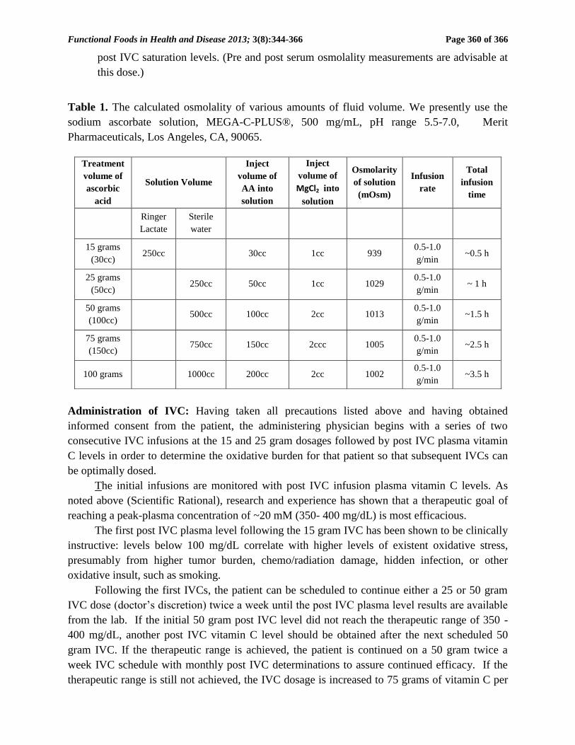
Table 1. The calculated osmolality of various amounts of fluid volume. We presently use the
sodium ascorbate solution, MEGA-C-PLUS®, 500 mg/mL, pH range 5.5-7.0, Merit
Pharmaceuticals, Los Angeles, CA, 90065.
Administration of IVC: Having taken all precautions listed above and having obtained
informed consent from the patient, the administering physician begins with a series of two
consecutive IVC infusions at the 15 and 25 gram dosages followed by post IVC plasma vitamin
C levels in order to determine the oxidative burden for that patient so that subsequent IVCs can
be optimally dosed.
The initial infusions are monitored with post IVC infusion plasma vitamin C levels. As
noted above (Scientific Rational), research and experience has shown that a therapeutic goal of
reaching a peak-plasma concentration of ~20 mM (350- 400 mg/dL) is most efficacious.
The first post IVC plasma level following the 15 gram IVC has been shown to be clinically
instructive: levels below 100 mg/dL correlate with higher levels of existent oxidative stress,
presumably from higher tumor burden, chemo/radiation damage, hidden infection, or other
oxidative insult, such as smoking.
Following the first IVCs, the patient can be scheduled to continue either a 25 or 50 gram
IVC dose (doctor’s discretion) twice a week until the post IVC plasma level results are available
from the lab. If the initial 50 gram post IVC level did not reach the therapeutic range of 350 -
400 mg/dL, another post IVC vitamin C level should be obtained after the next scheduled 50
gram IVC. If the therapeutic range is achieved, the patient is continued on a 50 gram twice a
week IVC schedule with monthly post IVC determinations to assure continued efficacy. If the
therapeutic range is still not achieved, the IVC dosage is increased to 75 grams of vitamin C per
Treatment
volume of
ascorbic
acid
Solution Volume
Inject
volume of
AA into
solution
Inject
volume of
MgCl2 into
solution
Osmolarity
of solution
(mOsm)
Infusion
rate
Total
infusion
time
Ringer
Lactate
Sterile
water
15 grams
(30cc) 250cc 30cc 1cc 939
0.5-1.0
g/min ~0.5 h
25 grams
(50cc) 250cc 50cc 1cc 1029
0.5-1.0
g/min ~ 1 h
50 grams
(100cc) 500cc 100cc 2cc 1013
0.5-1.0
g/min ~1.5 h
75 grams
(150cc) 750cc 150cc 2ccc 1005
0.5-1.0
g/min ~2.5 h
100 grams 1000cc 200cc 2cc 1002 0.5-1.0
g/min ~3.5 h
Page 18
Functional Foods in Health and Disease 2013; 3(8):344-366
Page 361 of 366
infusion for four infusions, at which time a subsequent post IVC plasma level is obtained. If the
patient remains in a sub-therapeutic range, the IVC dosage is increased to the 100 gram level.
If after four infusions the post IVC dosage remains sub-therapeutic, the patient may have
an occult infection, may be secretly smoking, or may have tumor progression. While these
possibilities are being addressed, the clinician can elect to increase the 100 gram IVC frequency
to three times per week. Higher infusion doses beyond 100 grams are not recommended without
serum osmolality testing before and after infusions in order to properly adjust the infusion rate to
maintain a near physiologic osmolality range.
If higher dosages are not tolerated, or there is tumor progression in spite of achieving the
therapeutic range, lower dosages can still augment the biological benefits of IVC, including
enhanced immune response, reduction in pain, increased appetite, and a greater sense of well-
being.
Very small patients, such as children, and very large obese patients need special dosing.
Small patients < 110 lbs. with small tumor burdens and without infection may only require 25
gram vitamin C infusions 2x/week to maintain therapeutic range. Large patients > 220 lbs. or
patients with large tumor burdens or infection are more likely to require 100 grams IVC
infusions 3x/week. Post IVC plasma levels serve as an excellent clinical guide to this special
dosing.
In our experience, the majority of cancer patients require 50 gram IVC infusions 2-
3x/week to maintain therapeutic IVC plasma levels. All patients reaching therapeutic range
should still be monitored monthly with post IVC plasma levels to ensure that these levels are
maintained long term. We advise patients to orally supplement with at least 4 grams of vitamin
C daily, especially on the days when no infusions are given, to help prevent a possible vitamin C
“rebound effect.” Oral alpha lipoic acid is also recommended on a case by case basis.
Acknowledgements: The study was supported by Flossie E West Memorial Trust and Allan P
Markin.
REFERENCES:
1. Hoffman F. Micronutrient requirements of cancer patients. Cancer 1985; 55(Supl.1): 145-
50.
2. Cameron E, Pauling L, Leibovitz B. Ascorbic acid and cancer, a review. Cancer Res 1979;
39: 663-681.
3. Geeraert L. Intravenous high-dose vitamin C. CAM-Cancer Consortium 2012. [Online]
Available at: http://www.cam-cancer.ort/CAM-Summaries/Other-CAM/Intravenous-high-
dose-vitamin-C
4. Riordan HD, Hunninghake RB, Riordan NH, Jackson JJ, Meng X, Taylor P, Casciari JJ,
Gonzalez MJ, Miranda-Massari JR, Mora EM, Rosario N Rivera A. Intravenous Ascorbic
Acid: Protocol for its Application and Use. P R Health Sci J 2003; 22: 225-32.
Page 19
Functional Foods in Health and Disease 2013; 3(8):344-366
Page 362 of 366
5. Padayatty SJ, Sun AY, Chen Q, Espey MG, Drisko J, Levine M. Vitamin C: intravenous
use by complementary and alternative medicine practitioners and adverse effects. PLoS
ONE 2010; 5(7): 11414.
6. Ayami Furuya, Misao Uozuki, Hisashi Yamasaki, Tsutomu Arakawa, Mikio Ariata, A.
Hajime. Antiviral effects of ascorbic and dehydroascorbic acids in vitro. International
journal of molecular medicine 22: 541-545, 2008.
7. Mikirova N, Casciari J, Riordan N. Ascorbate inhibition of angiogenesis in aortic rings ex
vivo and subcutaneous Matrigel plugs in vivo. J Angiogenesis Res 2012; 2: 2-6.
8. McCormick, W. Cancer: a collagen disease, secondary to nutrition deficiency. Arch.
Pediatr 1959; 76: 166-171.
9. Riordan HD, Casciari JJ, González MJ, Riordan NH, Miranda-Massari JR, Taylor P,
Jackson JA. A pilot clinical study of continuous intravenous ascorbate in terminal cancer
patients. P R Health Sci J 2005; 24:269–276.
10. Henson D, Block G, Levine M. Ascorbic acid: biological functions and relation to cancer.
JNCI 1991; 83: 547-50.
11. Cameron E, Pauling L. Supplemental ascorbate in the supportive treatment of cancer:
Prolongation of survival times in terminal human cancer. PNAS USA 1976; 73: 3685-9.
12. Creagan ET, Moertel CG, O'Fallon JR, Schutt AJ, O'Connell MJ, Rubin J, Frytak S. Failure
of high-dose vitamin C (ascorbic acid) therapy to benefit patients with advanced cancer. A
controlled trial. N Engl J Med 1979; 301: 687-90.
13. Moertel CG, Fleming TR, Creagan ET, Rubin J, O'Connell MJ, Ames MM. High-dose
vitamin C versus placebo in the treatment of patients with advanced cancer who have had
no prior chemotherapy. A randomized double- blind comparison. N Engl J Med 1985; 312:
137-41.
14. Hornig D. Distribution of ascorbic acid metabolites and analogues in man and animals.
Ann NY Acad Sci 1975;258: 103-18.
15. Keith M, Pelletier O. Ascorbic acid concentrations in leukocytes and selected organs of
guinea pigs in response to increasing ascorbic acid intake. Am J Clin Nutr 1974; (27): 368-
72.
16. Ginter E, Bobeck P, Vargova D. Tissue levels and optimal dosage of vitamin C in guinea
pigs. Nutr Metab1979; (27):217-26.
17. Levine M, Conry-Cantilena C, Yang Y, Welch RW, Washko PW, Dhariwal KR, Park JB,
Lazarev A, Graumlich JF, King J, Cantilena LR. Vitamin C pharmacokinetics in healthy
volunteers: evidence for a recommended dietary allowance. PNAS USA1996;(93): 3704-9.
18. Mayland CR, Bennett MI, Allan K. Vitamin C deficiency in cancer patients. Palliat Med
2005; 19:17-20.
19. Casciari JJ, Riordan NH, Schmidt TS, Meng XL, Jackson JA, Riordan HD. Cytotoxicity of
ascorbate, lipoic acid, and other antioxidants in hollow fiber in vitro tumors. Br J Cancer
2001; 84: 1544-1550.
20. Padayatti SJ, Sun H, Wang Y, Riordan HD, Hewitt SM, Katz A, Wesley RA. Vitamin C
pharmacokinetics: implications for oral and intravenous use. Ann Intern Med 2004; 140:
533-537.
Page 20
Functional Foods in Health and Disease 2013; 3(8):344-366
Page 363 of 366
21. Riordan NH, Rioredan HD, Casciari JJ. Clinical and experimental experiences with
intravenous vitamin C. Journal of Orthomolecular medicine, 2000, 15, 201-213.
22. Mikirova N, Casciari J, Hunninghake R, Riordan N. Clinical experience with intravenous
administration of ascorbic acid: achievable levels in blood for different states of
inflammation and disease in cancer patients. Journal of translational medicine, 2013, 11 (in
press).
23. Benade L, Howard, T, Burk, D., 1969. Synergistic killing of Ehrlich ascites carcinoma
cells by ascorbate and 3-amino-1,2,4-triazole. Oncology 1969; (23): 33-43.
24. Riordan NH, Riordan HRD, Meng X, Li Y, Jackson JA. Intravenous ascorbate as a tumor
cytotoxic chemotherapeutic agent. Medical hypothesis 1995; 44:207-213.
25. Chen Q, Espey MG, Krishna MC, Mitchell JB, Corpe CP, Buettner GR, Shacter E, Levine
M. Pharmacologic ascorbic acid concentrations selectively kill cancer cells: action as a pro-
drug to deliver hydrogen peroxide to tissues. Proc. Natl Acad. Sci. U. S. A. 2005;
102:13604–13609.
26. Frei B, Lawson S. Vitamin C and cancer revisited. PNAC USA 2008; 105: 11037-8.
27. Verrax J, Cadrobbi J, Marques C, Taper H, Habraken Y, Piette J, Calderon PB. Ascorbate
potentiates the cytotoxicity of menadione leading to an oxidative stress that kills cancer
cells by a non-apoptotic capsase-3 independent form of cell death. Apoptosis 2004; 9: 223-
33.
28. Gonzalez M, Mora EM, Miranda-Massari JR, Matta J, Riordan HD, Riordan NH.
Inhibition of human breast cancer carcinoma cell proliferation by ascorbate and copper.
PRHSJ 2002; 21: 21-3.
29. Chen Q, Espey MG, Sun AY, Pooput C, Kirk KL, Krishna MC, Khosh DB, Drisko J,
Levine M. Pharmacologic doses of ascorbate act as a prooxidant and decrease growth of
aggressive tumor xenografts in mice. Proc. Natl Acad. Sci. U. S. A. 2008; 105:11105–
11109.
30. Verrax J, Calderon P. Pharmacologic concentrations of ascorbate are achieved by
parenteral administration and exhibit antitumoral effects. Free Radic Biol Med 2009;
47:32-40.
31. Du J, Martin SM, Levine M, Wagner BA, Buettner GR, Wang SH, Taghiyev AF, Du C,
Knudson CM, Cullen JJ. Mechanisms of ascorbate-induced cytotoxicity in pancreatic
cancer. Clin. Cancer Res. 2010; 16:509–520.
32. Belin S, Kaya F, Duisit G, Giacomett S, Ciccoloni J, Fortes M. Antiproliferative effect of
ascorbic acid is associated with the inhibition of genes necessary to cell cycle progression.
PLoS ONE 2009; 4(2):E44-0.
33. Yeom CH, Lee G, Park JH, Yu J, Park S, Yi SY, Lee HR, Hong YS, Lee S. High dose
concentration administration of ascorbic acid inhibits tumor growth in BALB/C mice
implanted with sarcoma 180 cancer cells via the restriction of angiogenesis. J Transl Med
2009; 7: 70.
34. Pollard H, Levine M, Eidelman O, Pollard M. Pharmacological ascorbic acid supresses
syngenic tumor growth and metastases in hormone-refractory prostate cancer. In Vivo
2010; 24(3): 249-55.
Page 21
Functional Foods in Health and Disease 2013; 3(8):344-366
Page 364 of 366
35. Casciari JJ, Riordan HD, Miranda-Massari JR, Gonzalez MJ. Effect of high dose ascorbate
administration on L-10 tumor grows in guinea pigs. PRHSJ, 2005, 24, 145-150.
36. Ashino H, Shimamura M, Nakajima M, Kawanaka S, Oikawa T, Iwaguchi T, Kawashima
S. Novel function of ascorbic acid as an angiostatic factor. Angiogenesis 2003; 6:259-269.
37. Page EL, Chan DA, Giaccia AJ, Levine M, Richard DE. Hypoxia-inducible factor-1
(alpha) stabilization in nonhypoxic conditions: role of oxidation and intracellular ascorbate
depletion. Mol Biol Cell 2007; 19:86-94.
38. Mikirova N, Ichim T, Riordan N. Anti-angiogenic effect of high doses of ascorbic acid.
Journal of Translational Medicine 2008; 6:50.
39. Mikirova Nina, Casciari Joseph, Taylor Paul, Rogers Andrea, Effect of high-dose
intravenous vitamin C on inflammation in cancer patients. Journal of Translational
Medicine.2012, 10:189.
40. St. Sauver JL, Sarma AV, McGree DJ, Lieber MM, Girman CJ, Nehra A, Jacobsen SJ.
Associations betweeen c-reactive protein and benigh prosaic hyperplasia lower urinary
tract outcomes in a population based cohort. Am J Epidemiol 2009; 169: 1281-90.
41. Lin A, Chen K, Chung H, Chang S. The significance of plasma c-reactive protein in
patients with elevated serum prostate-specific antigen levels. Urological Sci 2010; 21: 88-
92.
42. Agus DB, Vera JC, Golde DW. Stromal cell oxidation: a mechanism by which tumors
obtain vitamin C. Cancer Res 1999; 59(18):4555–8.
43. Raloff J. Antioxidants may help cancers thrive. Science News 2000; 157: 5.
44. Heaney ML, Gardner JR, KarasawasN, Golde DW, Scheinberg DA, Smith EA, O’Connor
OA. Vitamin C antagonizes the cytotoxic effects of antineoplastic drugs. Cancer Research
2008; 68: 8031-8038.
45. Espey M, Chen Q, Levine M. Comment re: vitamin C antagonizes the cytotoxic effects of
chemotherapy. Cancer Research 2009; 69:8830.
46. Fujita K, Shinpo K, Yamada K, Sato K, Niimi H, Shamoto M, Nagatsu T, Takeuchi T,
Umezawa H. Reduction of adriamycin toxicity by ascorbate in mice and guinea pigs.
Cancer Res 1982; 309-16: 42.
47. Okunieff P, Suit H. Toxicity, radiation sensitivity modification, and combined drug effects
of ascorbic acid with misonidazole in vivo on FSaII murine firbosarcomas. JNCI 1987; 79:
377-81
48. Kurbacher CM, Wagner U, Kolster B, Andreotti PE, Krebs D, Bruckner HW. Ascorbic
acid (vitamin C) improves the antineoplastic activity of doxorubicin, cisplatin, and
paclitaxel in human breast carcinoma cells in vitro. Cancer Lett 1996; 103: 183-9.
49. Taper H, Keyeux A, Roberfroid M. Potentiation of radiotherapy by nontoxic pretreatment
with combined vitamins C and K3 in mice bearing solid transplantable tumor. Anticancer
Res 1996; 16: 499-503.
50. Fromberg A, Gutsch D, Schulze D, Vollbracht C, Weiss G, Czubayko F, Aigner A.
Ascorbate exerts anti-proliferative effects through cell cycle inhibition and sensitizes tumor
cells toward cytostatic drugs. Cancer Chemother Pharmacol 2011; 67: 1157-66.
Page 22
Functional Foods in Health and Disease 2013; 3(8):344-366
Page 365 of 366
51. Shinozaki K, Hosokawa Y, Hazawa M, Kashiwakura I, Okumura K, Kaku T, Nakayama E.
Ascorbic acid enhances radiation-induced apoptosis in an HL60 human leukemia cell line.
J Ratiat Res 2011; 52: 229-237.
52. Espey M, Chen P, Chalmers B, Drisko J, Sun AY, Levine M, Chen Q. Pharmacologic
ascorbate synergizes with gemcitabine in preclinical models of pancreatic cancer. Free
Radic Biol Med 2011; 50: 1610-1619.
53. Simone CB 2nd, Simone NL, Simone V, Simone CB. Antioxidants and other nutrients do
not inferfere with chemotherapy or radiation therapy and can increase survival, part 1.
Atlern Ther Health Med 2007; 13: 22-28.
54. Block K, Koch AC, Mead MN, Tothy PK, Newman RA, Gyllenhaal C. Impact of
antioxidant supplementaion on chemotherapeutic toxicity: a systematic review of the
evidence from randomized controlled trials. Int J Cancer 2008; 123: 1227-39.
55. Murata A, Morishige F, Yamaguchi H. Prolongation of survival times of terminal cancer
patients by administration of large doses of ascorbate. Int J Vitam Res Suppl 1982; 23:
103-13.
56. Jackson J, Riordan H, Hunninghauke R, Riordan N. High dose intravenous vitamin C and
long time survival of a patient with cancer of the head and pancreas. J Ortho Med 1995;
10:87-88.
57. Riordan H, Jackson J, Riordan N, Schultz M. High-dose intravenous vitamin C in the
treatment of a patient with renal cell carcinoma of the kidney. J Ortho Med 1998; 13:72-73.
58. Riordan N, Jackson J, Riordan H. Intravenous vitamin C in a terminal cancer patient. J
Ortho Med 1996; 11:80-82.
59. Padayatty SJ, Riordan HD, Hewitt SM, Katz A, Hoffer LJ, Levine M. Intravenous vitamin
C as a cancer therapy: three cases. CMAJ 2006; 174(7):937-42.
60. Drisko J, Chapman J, Hunter V. The use of antioxidants with first-line chemotherapy in
two cases of ovarian cancer. Am J Coll Nutr 2003; 22:118-23.
61. Yeom C, Jung G, Song K. Changes of terminal cancer patietns health related qualtiy of life
after high dose vitamin C administration. Korean Med Sci 2007; 22: 7-11.
62. Vollbracht C, Schneider B, Leendert V, Weiss G, Auerbach L, Beuth J. Vollbracht, C.
Intravenous vitamin C administration improves quality of life in breast cancer patients
during chemo-radiotherapy and aftercare: results of a retrospective, multicentre,
epidemiological cohort study in Germany. In Vivo 2011; 82:983-90.
63. Hoffer LJ, Levine M, Assouline S, Melnychuk D, Padayatty SJ, Rosadiuk K, Rousseau C,
Robitaille L, Miller WH Jr. Phase I clinical trial of i.v. ascorbic acid in advanced
malignancy. Ann Oncol 2008; 19(12)-2095
64. Monti D, Mitchell E, Bazzan AJ, Littman S, Zabrecky G, Yeo CJ, Pillai MV, Newberg AB,
Deshmukh S, Levine M. Phase I evaluation of intravenous ascorbic acid in combination
with gemcitabine and erlotinib in patients with metastatic pancreatic cancer. PLoS One
2012; 7: e29794.
65. Wong K, Thomson C, Bailey RR, McDiarmid S, Gardner J. Acute oxalate nephropathy
after a massive intravenous dose of vitamin C. Aust N Z J Med 1994; 24: 410-411.
Page 23
Functional Foods in Health and Disease 2013; 3(8):344-366
Page 366 of 366
66. Campbell A, Jack T. Acute reactions to mega ascorbic acid therapy in malignant disease.
Scott Med J 1979; 24: 151.
67. Rivers J. Safety of high-level vitamin C ingestion. In: Third Conference on Ascorbic Acid.
Ann NY Acad Sci 1987; 489: 95-102.
68. Jackson J, Hunninghake R. False positive blood glucose readings after high-dose
intravenous vitamin C. J Ortho Med 2006; 21: 188-90.