CELLULAR IMMUNOLOGY 123,294-306 (1989) Intravenous Endotoxin Recruits a Distinct Subset of Human Neutrophils, Defined by Monoclonal Antibody 31 D8, from Bone Marrow to the Peripheral Circulation’ CHRISTOPHER C. BROWN, HARRY L. MALECH, AND JOHN I. GALLING Bacterial Diseases Section, Laboratory of Clinical Investigation, National Institute ofAllergy and Infectious Diseases, National Institutes of Health, Bethesda, Maryland 20892 Received February 23, 1989; accepted June 19, 1989 Human neutrophils label with fluorochrome-labeled monoclonal antibody 3 1 D8 as bright or dull. We determined the source and fate of 3 1 D8 dull neutrophils by studying volunteers in- jected with endotoxin, epinephrine, or hydrocortisone, by examining bone marrow, and by examining skin blister exudate. We find that 31D8 dull neutrophils are normally not present in significant numbers in the circulation, are present in large numbers in normal marrow, and are recruited from the marrow by endotoxin, to a lesserextent by steroid, but not at all by epineph- rine. 3 1 D8 dull pattern correlates with morphologic immaturity in postendotoxin peripheral blood and bone marrow; however, blister exudate neutrophils contain only morphologically mature neutrophils, of which a significant number are 3 1 D8 dull. We conclude that 3 1 D8 dull neutrophils reside primarily in bone marrow and are released by agents which enhance bone marrow release of neutrophils. Their accumulation in skin blister exudate is unexplained, but SUggeStS a Special role in the i&MnIIIa~Ofy process. 0 1989 Academic Press, Inc. INTRODUCTION Peripheral blood neutrophils are commonly regarded as homogeneous in structure and function. Over 50 years ago, Sabin first suggested that neutrophils were heteroge- neous with regard to motility (l), and recent studies have confirmed these observa- tions (3). Neutrophils may be heterogeneous with regard to other functions aswell (2), including motile response to chemoattractants (25), IgG- and &A-mediated rosette formation (4-6), protein synthesis (7), membrane depolarization (8), and oxidative metabolism (9, 12). Neutrophils may also differ in their density (11) and alkaline phosphatase content ( 10). More recently, a number of anti-neutrophil monoclonal antibodies have been used to define subsets of neutrophils (9, 13- 15). One of these, 3 1 D8 (mouse IgG,), labels neutrophils heterogeneously (9). Greater than 95% of circulating neutrophils in nor- mal adults label intensely with fluorochrome-linked 3 1 D8 (3 lD8 bright neutrophils), while the remainder also label with 3 I D8, yet less intensely (3 1 D8 dull neutrophils). ’ Data were presented in part at the 7 1 st annual meeting of the Federation of American Society of Experimental Biologists, Washington, DC, 29 March-2 April 1987. ’ To whom correspondence should be addressed. 294 0008-8749189 $3.00 Copyright 0 1989 by Academic Press, Inc. All rights of reproduction in any form reserved.
Transcript
CELLULAR IMMUNOLOGY 123,294-306 (1989)
Intravenous Endotoxin Recruits a Distinct Subset of Human Neutrophils, Defined by Monoclonal Antibody 31 D8,
from Bone Marrow to the Peripheral Circulation’
CHRISTOPHER C. BROWN, HARRY L. MALECH, AND JOHN I. GALLING
Bacterial Diseases Section, Laboratory of Clinical Investigation, National Institute ofAllergy and Infectious Diseases, National Institutes of Health, Bethesda, Maryland 20892
Received February 23, 1989; accepted June 19, 1989
Human neutrophils label with fluorochrome-labeled monoclonal antibody 3 1 D8 as bright or dull. We determined the source and fate of 3 1 D8 dull neutrophils by studying volunteers in- jected with endotoxin, epinephrine, or hydrocortisone, by examining bone marrow, and by examining skin blister exudate. We find that 31D8 dull neutrophils are normally not present in significant numbers in the circulation, are present in large numbers in normal marrow, and are recruited from the marrow by endotoxin, to a lesser extent by steroid, but not at all by epineph- rine. 3 1 D8 dull pattern correlates with morphologic immaturity in postendotoxin peripheral blood and bone marrow; however, blister exudate neutrophils contain only morphologically mature neutrophils, of which a significant number are 3 1 D8 dull. We conclude that 3 1 D8 dull neutrophils reside primarily in bone marrow and are released by agents which enhance bone marrow release of neutrophils. Their accumulation in skin blister exudate is unexplained, but SUggeStS a Special role in the i&MnIIIa~Ofy process. 0 1989 Academic Press, Inc.
INTRODUCTION
Peripheral blood neutrophils are commonly regarded as homogeneous in structure and function. Over 50 years ago, Sabin first suggested that neutrophils were heteroge- neous with regard to motility (l), and recent studies have confirmed these observa- tions (3). Neutrophils may be heterogeneous with regard to other functions as well (2), including motile response to chemoattractants (25), IgG- and &A-mediated rosette formation (4-6), protein synthesis (7), membrane depolarization (8), and oxidative metabolism (9, 12). Neutrophils may also differ in their density (11) and alkaline phosphatase content ( 10).
More recently, a number of anti-neutrophil monoclonal antibodies have been used to define subsets of neutrophils (9, 13- 15). One of these, 3 1 D8 (mouse IgG,), labels neutrophils heterogeneously (9). Greater than 95% of circulating neutrophils in nor- mal adults label intensely with fluorochrome-linked 3 1 D8 (3 lD8 bright neutrophils), while the remainder also label with 3 I D8, yet less intensely (3 1 D8 dull neutrophils).
’ Data were presented in part at the 7 1 st annual meeting of the Federation of American Society of Experimental Biologists, Washington, DC, 29 March-2 April 1987.
’ To whom correspondence should be addressed.
294
0008-8749189 $3.00 Copyright 0 1989 by Academic Press, Inc. All rights of reproduction in any form reserved.
NEUTROPHIL HETEROGENEITY 295
The distinction between these populations in the normal adult is often technically difficult to demonstrate because of the small number of dull cells and because of the small difference in mean fluorescence between the two populations. With normal neonates and patients with blunt trauma, there is a marked increase in 3 1 D8 dull neutrophils, and the resolution of two distinct peaks of 3 1 D8 fluorescence is often easily demonstrated ( 16, 17). Other studies have demonstrated functional differences between 3 1 D8 bright and dull neutrophils, indicating that the bright cells were more responsive with regard to chemotaxis, membrane depolarization, NBT reduction, and superoxide production using formyl peptide as the stimulant (9,22).
Studies of neonates and trauma patients suggested that there was an association between the increased numbers of circulating band form neutrophils and the appear- ance of increased numbers of circulating 3 lD8 dull neutrophils. This raised the ques- tion of whether 3 1 D8 dull neutrophils were “immature” and/or whether 3 1 D8 bright neutrophils could acquire a “dull” labeling pattern because of activation. In the pres- ent studies, we examined the source and kinetics of 31D8 dull neutrophils by chal- lenging normal volunteers with intravenous steroids, epinephrine, or endotoxin and examining changes in 3 1 D8 labeling of circulating neutrophils. We also examined 3 1 D8 labeling of normal bone marrow myeloid cells and of blister fluid exudate neu- trophils.
MATERIALS AND METHODS
Study population. The study population, for injection studies, bone marrow aspi- rate, or blister exudate analysis consisted of 15 normal volunteers (8 men and 7 women, age 19-38). NIH protocols included 74-I-99 (bone marrow aspiration), 76- 1-349 (steroid administration), 77-I- 185 (skin blister exudate), 79-I-9 1 (endotoxin ad- ministration), and 80-I-96 (epinephrine administration). Other volunteers were used as a source of venous peripheral blood for in vitro activation studies of normal periph- eral blood neutrophils.
Study design of injection studies. Three normal volunteers were injected intrave- nously with 0.1 mg epinephrine. Venous blood was collected before epinephrine in- jection, immediately after injection, at 5 min, and at 1 h. Three normal volunteers were injected intravenously with 200 mg hydrocortisone. Venous blood was collected before steroid injection and at 1,2,4, and 6 hr after injection. Six normal volunteers were injected with 3 rig/kg Escherichia coli RE-2 endotoxin (federal reference stan- dard; FDA BB-IND No. 1309). Venous blood was collected before injection and at 0.5, 1, 1.5,2,4,6, 8, and 24 hr.
Study design for blister exudate analysis. Three normal volunteers had an 8-well blister suction unit (Neuro Probe, Inc., Bethesda, MD) applied to one forearm and blisters raised using -360 mm Hg vacuum over 1 hr and 70% autologous serum added to each blister as previously described (26). Blister exudate was sampled at 12 hr. This procedure generally yielded between 20 and 40 million cells, of which >98% were mature neutrophils. After washing, ~95% of cells excluded trypan blue.
Cellpreparation. Neutrophils were separated from heparinized venous blood using dextran sedimentation alone or by Ficoli-Hypaque gradient centrifugation followed by dextran sedimentation (2 1). The latter method resulted in >95% neutrophils. Leu- kocytes were separated from bone marrow aspirate by dextran sedimentation (2 1). Where multiple samples from a volunteer were obtained over time, processing of each
296 BROWN, MALECH, AND GALLIN
individual blood sample (neutrophil purification, antibody labeling, cell fixation) was started immediately after the sample was drawn. Processing was completed within 4 hr for each sample.
Antibody preparation and cell labeling. Monoclonal antibody 3 1 D8 either was ob- tained as culture medium supemate from growing hybridoma cells or was purified by ammonium sulfate precipitation from mouse ascites (9). Control antibody was polyclonal mouse Ig anti-horse spleen ferritin (Jackson Immunoresearch Labora- tories, Avondale, PA). For indirect labeling, fluorescein-conjugated goat Fal$ anti- mouse IgG (Cooper Biomedical, Malvem, PA) was used as the second antibody. In some studies, purified 3 1 D8 or control mouse antibody was conjugated with fluores- cein and used to directly label cells (Sigma, St. Louis, MO). Freshly prepared live cells (106/ml) were labeled at 4°C for 30 min in phosphate-buffered saline (PBS) containing 1% bovine serum albumin, 1 mg/ml human IgG, and hybridoma media supematant, control antibody, or directly conjugated antibodies. For indirect labeling, cells labeled with the first antibody were washed and resuspended in the same buffer, additives, and conditions. This was followed by the addition of fluorescein goat FabZ anti-mouse IgG for 30 min. Labeled cells were washed, immediately fixed, and stored in the dark in PBS containing 1% paraformaldehyde. Control studies indicated that fixation and storage in the dark after labeling did not alter the fluorescence pattern even over a period of several days before analysis.
Fluorescence-activated cell analysis and sorting (FACS). Samples were analyzed with a fluorescence-activated cell sorter (FACS II, Becton-Dickinson, Sunnyvale, CA), an Epics 753, or a Profile (both from Coulter, Hialeah, FL). The Epics 753 was also used for sorting. Forward and 90” light scatters were used to distinguish neutro- phils (gating) from monocytes and lymphocytes (verified by sorting). Gating did not distinguish eosinophils or basophils from neutrophils, but 3 1 D8 does not label eosin- ophils or basophils (9). Sorting studies (see below) indicated that neither 3 ID8 bright nor 3 1 D8 dull neutrophil peaks contained more than 1% eosinophils plus basophils. For each sample analyzed, 20,000 cells (gated to count neutrophils) were counted. For some studies, the gating parameters for forward light scatter or side light scatter were altered to examine portions of the neutrophil population which had different light scatter characteristics.
Data analysis. Data were displayed with the number of cells on the vertical axis and the logarithm of fluorescence intensity on the horizontal axis. Negative fluores- cence was defined by analyzing cells labeled with control antibody and using a cutoff at 2% of the upper limit of this curve. A computer program (VSP/DSP created by Dr. T. Chused, National Institutes of Health) was used to generate a symmetric curve using the mode of the brightest peak and defining these as 3 1 D8 bright cells. The 3 ID8 dull cells were then defined by subtracting the integral of the computer-gener- ated 3 lD8 bright peak from the total integral of cells more fluorescent than the upper 2% ofthe negative control fluorescence peak. Since 3 1 D8 staining represents a contin- uum from no (negative) staining to intense (bright) staining, we used the integral subtraction method to arbitrarily define “negative,” “dull,” and “bright” staining populations. This approach made it possible to quantitate changes in the relative numbers of 3 1 D&stained cells of different labeling intensity.
For some studies, the data from each patient were expressed as the number of total circulating neutrophils (white blood count multiplied by the percentage of neutro- phils plus percentage bands), total circulating bands (white blood count multiplied
NEUTROPHIL HETEROGENEITY 297
by the percentage of bands), or total circulating 3 ID8 bright or dull neutrophils (neu- trophils plus bands multiplied by the percentage of 3 1 D8 bright or dull, derived from fluorescence analysis). The Student t test was used to determine the significance of changes in the 3 lD8-defined subsets at different time points.
Fluorescence-activated sorting. Live peripheral blood neutrophils or marrow leu- kocytes, labeled with 3 1 D8, were sorted in an Epics 753 within 3 hr of obtaining the sample from the volunteer. Gates for the sort of 3 1 D8 bright cells were set to include only cells with fluorescence intensity above the mode of the bright peak in order to minimize any contribution of 3 1 D8 dull cells. Gates for the sort of 3 1 D8 dull cells were set to include cells above the upper limit of negative control labeling and well below the lower limit of the symmetrical curve estimated for 3 1 D8 bright fluores- cence. These limits were chosen to ensure minimal overlap of negative, 3 1 D8 dull, or 3 1 D8 bright populations. A minimum of one million cells was sorted from each gated region, spun down, resuspended in buffer to a concentration of 100,000 cells/ ml, and distributed on slides with a cytospin (Shandon-Southern). The cytospins were then stained with a modified Giemsa stain and 200 cell differential counts were per- formed by two separate observers. A x2 analysis was used to determine significance of any correlation between bands and 3 1 D8 dull labeling pattern.
In vitro activation or aging of neutrophils. For activation studies, purified neutro- phils were suspended at a concentration of 100,000 cells/ml in Hanks’ buffer with magnesium and calcium to which a variety of activators had already been added (phorbol myristate acetate 10 and 20 &ml; f-Met-Leu-Phe 1 nM, 100 nM, and 10 PM; RE-2 E. coli endotoxin 10 pg/ml; 10% zymosan-activated serum). The samples were incubated for 20 min in a shaking water bath at 37°C washed at 4°C and labeled with 3 1 D8 as described above.
For aging studies, heparinized whole blood was collected and incubated under ster- ile conditions overnight in Teflon-coated beakers. Neutrophils from these samples were then purified and antibody labeled as previously described.
RESULTS
Changes in 3108 labeling of neutrophils after administration of RE-2 E. coli endo- toxin to normal volunteers. A representative series of FACS analyses of 3 1 D8-labeled neutrophils obtained from one individual at successive time intervals after endotoxin injection is shown in Fig. 1. The Time 0 curve principally comprises bright cells as defined under Materials and Methods. Over the first 1.5 hr, a progressively greater proportion of less fluorescent (dull) cells appears, such that at 1.5 and 2 hr most of the cells obtained from this individual are 3 ID8 dull. At 4 to 8 hr, there is a progres- sive increase in the proportion of 3 ID8 bright cells, and the two population modes are clearly visible. By 24 hr, the labeling pattern approaches that of the Time 0 curve.
Table 1 shows the relative percentage of 3 1 D8 bright and 3 1 D8 dull neutrophils from six individuals at time intervals after injection of 3 rig/kg endotoxin. Starting at 1 hr, there is a significant increase in the percentage of 3 1 D8 dull neutrophils which peaked at 1.5 hr. The mean relative percentage of 3 1 D8 dull neutrophils is back to normal at 24 hr although rare patients still have elevated dull cells at 24 hr. In Fig. 2, the same data are expressed in terms of the number of total circulating peripheral blood neutrophils, the number of circulating neutrophils that are bands, and the cal- culated number of circulating neutrophils which are 3 lD8 bright or 3 ID8 dull (see
298 BROWN, MALECH, AND GALLIN
h
FLUORESCENCE (LOG) -
FIG. I. 3 I D8 labeling patterns at time intervals after intravenous endotoxin injection in a representative normal volunteer. The hatched area represents 3 1 D8 dull labeling before endotoxin injection (A), 0.5 hr after injection (B), 1 hr (C), 2 hr (D), 4 hr (E), 6 hr (F), 8 hr (G), and 24 hr after endotoxin injection (H). Each figure includes only those cells staining with fluorescence intensity above 2% of the upper limit of negative control staining.
Materials and Methods). A decrease in total neutrophil count, reaching a nadir at 1 hr, is followed by a dramatic increase which peaks at 8 hr. For the first 1.5 hr after endotoxin injection, 3 1 D8 bright cells decrease dramatically. In contrast, 3 ID8 dull cells remain relatively unchanged for the first hour. At 1.5 hr when 3 1 D8 bright cells reach a nadir, there is already a significant rise in circulating 3 1 D8 dull cells. It is important to appreciate that, although the relative percentage of 3 1 D8 dull cells peaks at 1.5 hr, this appears to be as much a result of a marked loss of absolute numbers of 3 lD8 bright cells as it is an increase in 3 1 D8 dull cells. From 2 to 6 hr, there is a large,
NEUTROPHIL HETEROGENEITY 299
TABLE 1
Changes in Percentage of 3 lD8 Dull Neutrophils over Time after Injection of Intravenous Endotoxin in Normal Volunteers
Time after endotoxin injection (hr) % 3 1 D8 dull neutrophils P*
* Significance of difference vs 0 time, Student’s t test, six subjects.
almost linear, increase in both 3 1 D8 bright and 3 1 D8 dull neutrophils. Between 6 and 8 hr, 3 1 D8 bright cells continue a linear increase, while the number of 3 1 D8 dull cells has reached a plateau. By 24 hr, the total number of 3 1 D8 bright cells is still elevated, but the number of 3 1 D8 dull cells has decreased dramatically, although not quite to preinjection levels.
It is of note that changes in numbers of band form neutrophils parallel those of 3 1 D8 dull cells, but the total number of bands at most time points is only about 80% of the total number of 3 1 D8 dull cells. Sorting studies were performed as outlined below in order to examine this correlation.
Changes in 3108 labeling of neutrophils after administration of hydrocortisone to normal volunteers. Table 2 shows the relative percentage of 3 1 D8 bright and 3 lD8 dull neutrophils from three individuals at time intervals after injection of 200 mg hydrocortisone. By 4 hr after steroid administration, there is a small yet statistically significant increase in the relative percentage of 3 1 D8 dull cells. The one individual with the greatest increase in circulating neutrophil count (3.3-fold) also showed the greatest increase of 3 1 D8 dull neutrophils (3% at 0 time to 20% at 4 hr).
Changes in 3108 labeling after epinephrine injection in normal volunteers. Table 2 shows the relative percentage of 3 1 D8 bright and 3 1 D8 dull neutrophils from three individuals at time intervals after injection of 0.5 mg epinephrine. The peak increase in circulating neutrophil count occurred within 5 min after injection and averaged 1.6-fold higher than baseline. No changes in the percentage of 3 lD8 dull neutrophils were detected at any time point in any of the three subjects studied. One of the indi- viduals had a 2.1 -fold increase in circulating neutrophil count immediately after in- jection, but was not accompanied by any detectable change in the 3 1 D8 labeling curve at this time point.
Sorting of 3108 bright and dull neutrophils from peripheral blood and bone marrow and relationship to neutrophil maturity. Previously published studies of neonates and trauma patients suggested a positive correlation between the increased circulating band form neutrophils and an increase in the percentage of 3 1 D8 dull neutrophils. Since the endotoxin studies noted above indicated a similar correlation (Fig. 2) we
FIG. 2. Total numbers of circulating 3 1 D8 bright and dull neutrophils, total neutrophils, and bands after intravenous endotoxin from the six subjects shown in Table 1.
sorted 3 lD%labeled peripheral blood neutrophils obtained at 4 hr after endotoxin injection. The gating used for defining 3 lD8 bright and dull populations was as indi- cated under Materials and Methods. The results of this analysis are shown in the upper panel of Table 3. There was a highly significant correlation between morpho-
TABLE 2
Changes in Percentage of 3 I D8 Dull Neutrophils over Time after Intravenous Injection of Hydrocortisone or Epinephrine
% 3 1 D8 dull cells P*
Time after hydrocortisone injection (hr) 0 4
Time after epinephrine injection (min) 0 5
5.6 f 2.2 - 16.1 k 3.0 -co.05
1.0 * 4.2 - 8.3 + 5.2 >0.5
* Significance of difference vs 0 time, Student’s t test, three subjects in each group.
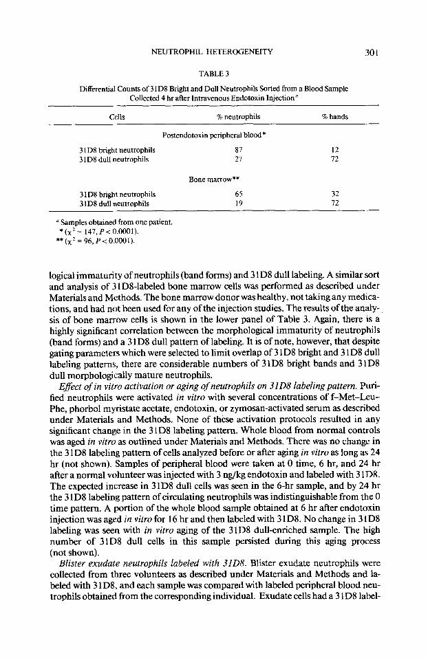
NEUTROPHIL HETEROGENEITY 301
TABLE 3
Differential Counts of 3 1 D8 Bright and Dull Neutrophils Sorted from a Blood Sample Collected 4 hr after Intravenous Endotoxin Injection”
0 Samples obtained from one patient. *(x2= 147,P<0.0001).
**(~*=96,P<O.O001).
logical immaturity of neutrophils (band forms) and 3 1 D8 dull labeling. A similar sort and analysis of 3 1 DS-labeled bone marrow cells was performed as described under Materials and Methods. The bone marrow donor was healthy, not taking any medica- tions, and had not been used for any of the injection studies. The results of the analy- sis of bone marrow cells is shown in the lower panel of Table 3. Again, there is a highly significant correlation between the morphological immaturity of neutrophils (band forms) and a 3 1 D8 dull pattern of labeling. It is of note, however, that despite gating parameters which were selected to limit overlap of 3 1 D8 bright and 3 1 D8 dull labeling patterns, there are considerable numbers of 3 lD8 bright bands and 3 ID8 dull morphologically mature neutrophils.
Eflect of in vitro activation or aging of neutrophils on 3108 labeling pattern. Puti- fied neutrophils were activated in vitro with several concentrations of f-Met-Leu- Phe, phorbol myristate acetate, endotoxin, or zymosan-activated serum as described under Materials and Methods. None of these activation protocols resulted in any significant change in the 3 ID8 labeling pattern. Whole blood from normal controls was aged in vitro as outlined under Materials and Methods. There was no change in the 3 1 D8 labeling pattern of cells analyzed before or after aging in vitro as long as 24 hr (not shown). Samples of peripheral blood were taken at 0 time, 6 hr, and 24 hr after a normal volunteer was injected with 3 rig/kg endotoxin and labeled with 3 1 D8. The expected increase in 31D8 dull cells was seen in the 6-hr sample, and by 24 hr the 3 1 D8 labeling pattern of circulating neutrophils was indistinguishable from the 0 time pattern. A portion of the whole blood sample obtained at 6 hr after endotoxin injection was aged in vitro for 16 hr and then labeled with 3 1 D8. No change in 3 1 D8 labeling was seen with in vitro aging of the 3 1 D8 dull-enriched sample. The high number of 3 ID8 dull cells in this sample persisted during this aging process (not shown).
Blister exudate neutrophils labeled with 3108. Blister exudate neutrophils were collected from three volunteers as described under Materials and Methods and la- beled with 3 1 D8, and each sample was compared with labeled peripheral blood neu- trophils obtained from the corresponding individual. Exudate cells had a 3 1 D8 label-
302 BROWN, MALECH, AND GALLIN
FLUORESCENCE (LOG)
FIG. 3. 3 ID8 labeling pattern of circulating neutrophils (-) and blister exudate neutrophils (---),
ing pattern which differed substantially from that of circulating neutrophils. Blister exudate neutrophils from three normal volunteers consisted of 46.3 f 11.9% 3 1 D8 dull cells, compared with 7.7 + 3.8% 3 1 D8 dull cells found in simultaneously sampled peripheral blood (P < 0.05). Paradoxically, there was a slight increase in the mode of the 3 1 D8 bright peak in the exudate cells, despite the simultaneous increase in 3 1 D8 dull cells compared to circulating peripheral blood neutrophils (Fig. 3). In all three volunteers, no bands were seen in differential counts of the exudate neutrophils.
Light scatter characteristics of 3108 dull neutrophils. Forward light scatter and right angle light scatter (side scatter) are sensitive indicators of cell size and internal light scattering characteristics, respectively. These light scatter parameters are rou- tinely used to gate on different types of cells in a mixed population during fluores- cence-activated cell analysis. For all of the studies noted above, the gating parameters were set to include almost all neutrophils and to exclude lymphocytes, monocytes, and erythrocytes. For the studies shown in Fig. 4, we examined different portions of the neutrophil gating region, as defined by forward and side light scatters in order to look for possible differences in light scatter between 3 1 D8 bright and dull neutrophils. We found that gating on those neutrophils with the lowest side scatter resulted in an enrichment of 3 1 D8 dull cells, while gating on those neutrophils with the highest side scatter resulted in a preferential increase in 3 1 D8 bright cells. This correlation was observed in samples of peripheral blood obtained after endotoxin injection as well as in samples of blister exudate neutrophils (Fig. 4). Sampling the high or low forward scatter portions of the neutrophil gating region did not result in any preferential en- richment of 3 lD8 dull or bright neutrophils (not shown).
DISCUSSION
Clearly defined functional and antigenic subsets of human lymphocytes have been documented for many years; however, reports of heterogeneity among human neu- trophils are few in number and of unclear significance (2). With regard to antigenic heterogeneity, a few murine monoclonal antibodies have been reported which iden-
NEUTROPHIL HETEROGENEITY 303
SIDE SCATTER+ FLUORESCENCE (LOG)r
FIG. 4. Forward and side scatter characteristics of 3 1 D8 bright and dull neutrophils from skin blister exudate. (A) The typical forward and side scatter gatings, which include most live granulocytes and ex- cludes mononuclear cells. With the gates set as in A, the resulting histogram of 3 1 D8 fluorescence is ob- tained as shown in B. (C) The gating is changed to collect only neutrophils with the lowest side scatter resulting in a histogram of 3 1 D8 fluorescence as obtained in D, indicating an enrichment of 3 1 D8 dull cells. When gating is set to collect only those neutrophils with the highest side scatter (E), the resulting 3 1 D8 fluorescence histogram (F) indicates an enrichment for 3 lD8 bright cells.
tify distinct neutrophil subsets (9, 13-15). One of these antibodies, 3 ID8 identifies subsets of neutrophils which differ in functional responses to chemotactic stimuli (9).
Almost all neutrophils bind 3 1 D8 antibody to some degree. The binding is hetero- geneous, with most circulating neutrophils in normal adults labeling intensely with fluorochrome-labeled antibody, a pattern referred to in this and previous reports as 3 1 D8 bright. These 3 1 D8 bright neutrophils demonstrate a greater functional capac- ity than those labeling less intensely (3 1 D8 dull). Studies of 3 1 D8 labeling patterns in neonates and trauma patients suggested a correlation between the appearance of circulating band form neutrophils and the increased numbers of 3 1 D8 dull neutro- phils ( 16, 17). The present study was undertaken in order to more clearly document the source of 3 1 D8 dull cells. Since the two clinical situations associated with in- creased numbers of circulating 3 1 D8 dull cells were related to stress (16-20) the present study was designed to test the effects of endotoxin, steroids, and epinephrine as models of infectious or noninfectious stress states. We also examined bone marrow and blister fluid neutrophils in order to determine the source and fate of 3 1 D8 bright and dull neutrophils.
While epinephrine injection resulted in no changes in the relative number of 3 1 D8 bright and dull neutrophils in the peripheral circulation, intravenous injection with steroid led to a small yet significant increase in 3 1 D8 dull cells relative to 3 1 D8 bright cells. Intravenous injection with endotoxin led to dramatic increases in both 3 1 D8 dull and 3 1 D8 bright cells, peaking at 6 hr after injection. 3 1 D8 dull cells began to increase in number between 1 and 2 hr, coincident with a decrease in the number of
304 BROWN, MALECH, AND GALLIN
3 1 D8 bright cells. After this time, both 3 I D8 bright and dull neutrophils demon- strated a dramatic increase in numbers. We conclude that 3 1 D8 dull cells are located primarily within the bone marrow and that a mild stimulus such as intravenous ste- roid, which has been reported to recruit only mature marrow neutrophils (24), also recruits some 3 1 D8 dull neutrophils.
Endotoxin is a strong stimulus for recruitment of both 3 1 D8 bright and dull cells, and these include a large number of band form neutrophils. The correlation between bands and 3 1 D8 dull cells is close, although not coincident, since sorting studies show that band form neutrophils and morphologically mature-appearing neutrophils can label in both a 3 ID8 dull and a 31D8 bright pattern. Also, within the first 90 min after endotoxin, 3 1 D8 bright cells decreased precipitously, probably due to margina- tion. In contrast, the 3 1 D8 dull cells actually begin to increase by 1 hr, even though 31D8 bright cells continue to decrease until 90 min. A hypothesis consistent with these kinetics is that 3 1 D8 bright cells marginate more readily than the 3 1 D8 dull cells. This would also be consistent with the observation that 3 1 D8 bright cells are more functionally responsive to chemotactic factors (9). Both 3 ID8 bright and dull neutrophils participate in the endotoxin-induced leukocytosis. At the peak of the leukocytosis, the contribution in number of neutrophils from both 3 1 D8-defined la- beling patterns is equivalent.
Of note is that the rise in 3 1 D8 bright cells persists after the rise in 3 lD8 dull cells ceases. It is possible that the marrow is becoming preferentially depleted of 3 1 D8 dull cells. Alternatively, 3 1 D8 dull cells may be converting to 3 1 D8 bright, a transforma- tion not supported by the in vitro studies. The third, and most likely, explanation is that 31D8 dull cells have a higher threshold for recruitment from the marrow to peripheral circulation, and that by 6 hr the stimulus provided by the original endo- toxin injection has begun to wane.
By 24 hr after endotoxin injection, the total number of circulating neutrophils has declined toward preinjection levels. Of note is that the number of 3 1 D8 dull neutro- phils has decreased far out of proportion to the decrease in 3 1 D8 bright neutrophils, resulting in a ratio approaching preinjection conditions. Our studies do not show where the newly recruited 3 ID8 bright and dull cells go. As noted above, the in vitro studies suggest that 31D8 dull cells do not become bright although the results from the “aging” studies do not preclude the evolution of a different 3 1 D8 phenotype with time. The studies of blister exudate neutrophils suggest that 3 lD8 dull cells do appear at sites of inflammation. The return toward preinjection numbers and ratio of 3 1 D8 bright and dull neutrophils at 24 hr after endotoxin injection are probably results of continuing loss of both 3 ID8 bright and dull neutrophils from the circulation associ- ated with a declining output of, by that time, predominantly 3 1 D8 bright neutrophils from the bone marrow.
To confirm that 3 1 D8 dull neutrophils come from bone marrow, we labeled and examined normal human bone marrow. We found that a correlation of morphologic immaturity and 3 1 D8 dull labeling exists; however, the correlation is not associated with an absolute coincidence of bands and 3 1 D8 dull pattern of labeling. Band form neutrophils with a 3 1 D8 bright labeling pattern, as well as morphologically mature neutrophils with a 3 1 D8 dull pattern of labeling, were seen in the bone marrow. It is possible that 3 1 D8 dull neutrophils become 3 1 D8 bright as they mature, that this transformation is dysynchronous with transformation of the nucleus from a band
NEUTROPHIL HETEROGENEITY 305
form to a multilobed morphology, and that this transformation can only occur in marrow (23).
The data support the conclusion that there is a relative restriction of egress of 3 1 D8 dull cells from bone marrow to peripheral circulation in normal individuals regard- less of nuclear morphology. The antigen detected by 3 lD8 might be involved in what- ever process determines when a neutrophil is ready to exit from the bone marrow in a normal unstressed individual. In addition, 3 ID8 labeling may be a more accurate measure of neutrophil maturity than nuclear morphology. 3 lD8 labeling of circulat- ing neutrophils is a useful means for investigating mechanisms controlling egress of neutrophils from human bone marrow and may explain some aspects of the appear- ance of functional subsets of neutrophils in a variety of pathologic states (22).
The unique side scatter characteristics of 3 1 D8 dull cells are identical regardless of the source of the 3 lD8 dull cells (e.g., gating on the low side scatter region of neutro- phils results in an enrichment of 3 1 D8 dull cells, regardless of whether the neutrophils are derived from normal peripheral blood, postendotoxin peripheral blood, or blister fluid exudate). This suggests that 3 1 D8 dull cells from blister fluid and peripheral blood have identical physical characteristics. The significance of differences in side scatter between 3 1 D8 bright and dull neutrophils is not known, although it may relate to differences in cell granularity or surface characteristics. It should also be noted that a choice of gating parameters which unintentionally leaves out low side scatter neutrophils would adversely affect the ability to detect 3 1 D8 dull neutrophils.
It is surprising that there is such a large percentage of 3 1 D8 dull neutrophils in blister exudate relative to the simultaneous circulating neutrophil labeling pattern. All of the blister neutrophils are morphologically mature. We have no explanation for this phenomenon; however, the enrichment of 3 1 D8 dull cells in blister exudate might result from a conversion of 3 1 D8 bright labeling pattern to a dull labeling pattern, a selective egress of 3 1 D8 dull cells to blisters, or a longer life span of 3 1 D8 dull cells in the blister fluid relative to 3 1 D8 bright cells. A conversion of 3 1 D8 bright to dull is not seen either with in vitro aging or with activation. The kinetics of the endotoxin studies argues against selective egress of 3 1 D8 dull cells and, in fact, sug- gests that 3 1 D8 dull neutrophils may marginate less readily than 3 1 D8 bright neu- trophils.
As discussed above, the analysis of bone marrow suggests that 3 1 D8 dull cells are probably less mature. Such cells may be longer-lived. Thus, we suggest that there exists a persistent low level egress of morphologically mature 3 1 D8 dull cells from bone marrow into the peripheral circulation and, subsequently, into sites of inflam- mation such as blister fluid. The presence in the peripheral circulation of these mor- phologically mature 3 1 D8 dull neutrophils may result from the nonspecific and ran- dom escape from the bone marrow of immature cells which are not ready for release. This does not, however, obviate a special role for these functionally unique neutro- phils at sites of inflammation.
REFERENCES
1. Sabin, F., Bull. Jbhns. Hopk. H5sp. 34,211, 1923. 2. Gallin, J. I., BIoud63,977, 1984. 3. Howard, T. H. E., Blood 59,946, 1982. 4. Wong, L., and Wilson, J. D., J. Immunol. Methods 7,69, 1975. 5. Klempner, M. S., and Gallin, J. I., Blood 51,659, 1978.
306 BROWN, MALECH, AND GALLIN
6. Fanger, M. W., Shen, L., and Pug, J., Proc. Nat/. Acad. Sci. USA 77,3640, 1980. 7. Crane&Pipemo, A., Vassalli, J. D., and Reich, E., J. Exp. Med. 146, 1693, 1977. 8. Seligmann, B., Chused, T. M., and Gallin, J. I., J. Clin. Invest. 68, 1125, 198 1. 9. Seligmann, B., Malech, H. L., Melnick, D. A., and Gallin, J. I., J. Immunol. 135,2647, 1985.
10. Fehr, J., and Grossman, H., Amer. J. Hematol. 7,369, 1979. 11. Pember, S. O., Barnes, K. C., and Brandt, S. J., Blood61, 1105, 1983. 12. Bass, D. A., Olbrantz, P., and Szejda, P., J. Immunol. 136,860, 1986. 13. Ball, E. D., Graziano, R. F., Shen, L., and Fanger, M. W., Proc. Nat/. Acad. Sci. USA 79,5374, 1984. 14. Clement, L. T., Lehmeryer, J. E., and Gartland, G. L., Blood61,326, 1983. 15. Brown, C. C., Malech, H. L., Shrimpton, C. F., Beverly, P. C., Segal, T., and Gallin, J. I., Chin. Res.
35,42la, 1987. [Abstract] 16. Krause, P. J., Malech, H. L., Kristie, J., Kosciol, C. M., Herson, V. C., Eisenfeld, V., Pastuszak, W. T.,
Kraus, A., and Seligmann, B., Blood 68,200, 1986. 17. Krause, P. J., Maderazo, E. G., Bannon, P., Kosciol, C. M., and Malech, H. L., Clin. Rex 35,48Oa,
1987. [Abstract] 18. Yurt, R. W., and Shires, G. T., In “Principles and Practice of Infectious Diseases” (G. Mandell, R.
Douglas, and J. Bennett, Eds.), pp. 624-628. Wiley, New York, 1985. 19. Balch, H. H., Ann. Surg. 142, 145, 1955. 20. Miller, M. E., Pediatr. Clin. North Amer. 24,4 13, 1977. 21. Zimmerli, W., Seligmann, B., and Gallin, J. I., J. Clin. Invest. 77,925, 1986. 22. Gallin, J. I., Jacobson, R. J., Seligmann, B. E., Metcalf, J. A., McKay, J. H., Sacher, R. A., and Malech,
H. L., Blood68,343, 1986. 23. Rundles, R. W., In “Hematology” (W. J. Williams, E. Beutler, and A. Erslev, Eds.), pp. 624-627.
McGraw-Hill, New York, 1982. 24. Dale, D. C., Fauci, A. S., Guerry, D. P., and Wolff, S. M., J. Clin. Invest. 56,808, 1975. 25. Harvarth, L., Surv. Immunol. Res. 2, 145, 1983. 26. Zimmerli, W., and Gallin, J. I., J. Immunol. Methods 96, 11, 1987.