48
Adaptations

Adaptations
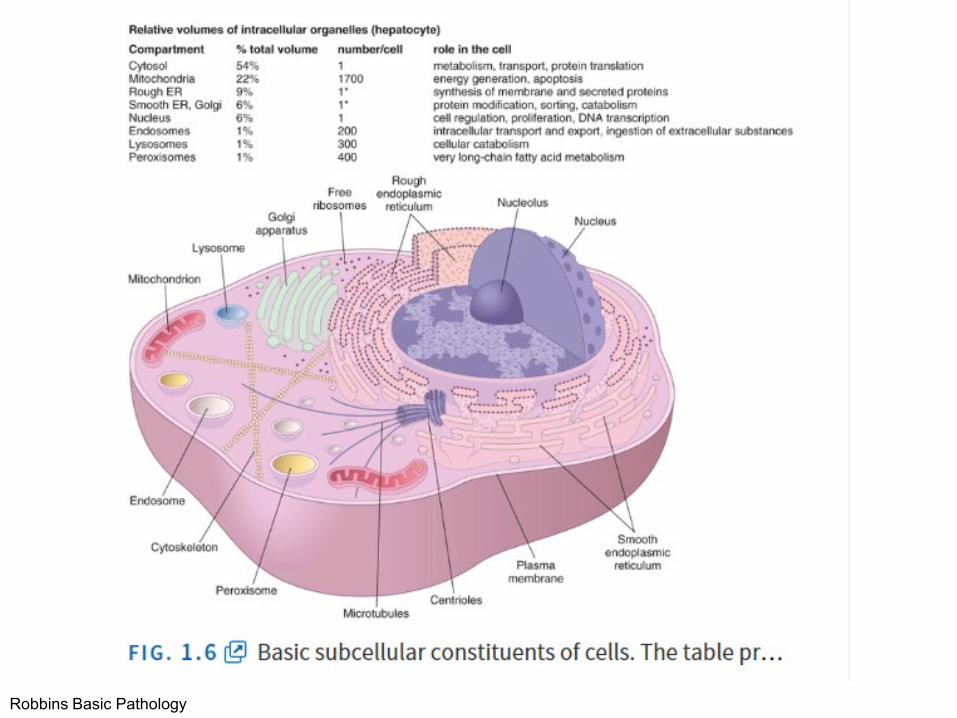
Robbins Basic Pathology

Robbins Basic Pathology

Robbins Basic Pathology

Robbins Basic Pathology
Coagulation

Robbins Basic Pathology
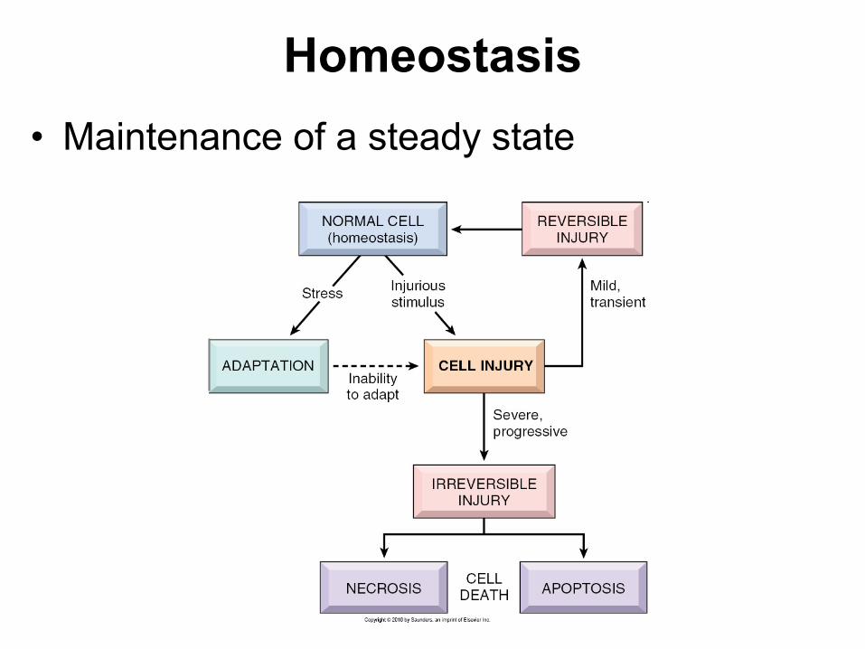
Homeostasis• Maintenance of a steady state

Adaptations• Reversible functional and structural
responses to physiologic stress and some pathogenic stimuli
• New altered “steady state” is achieved
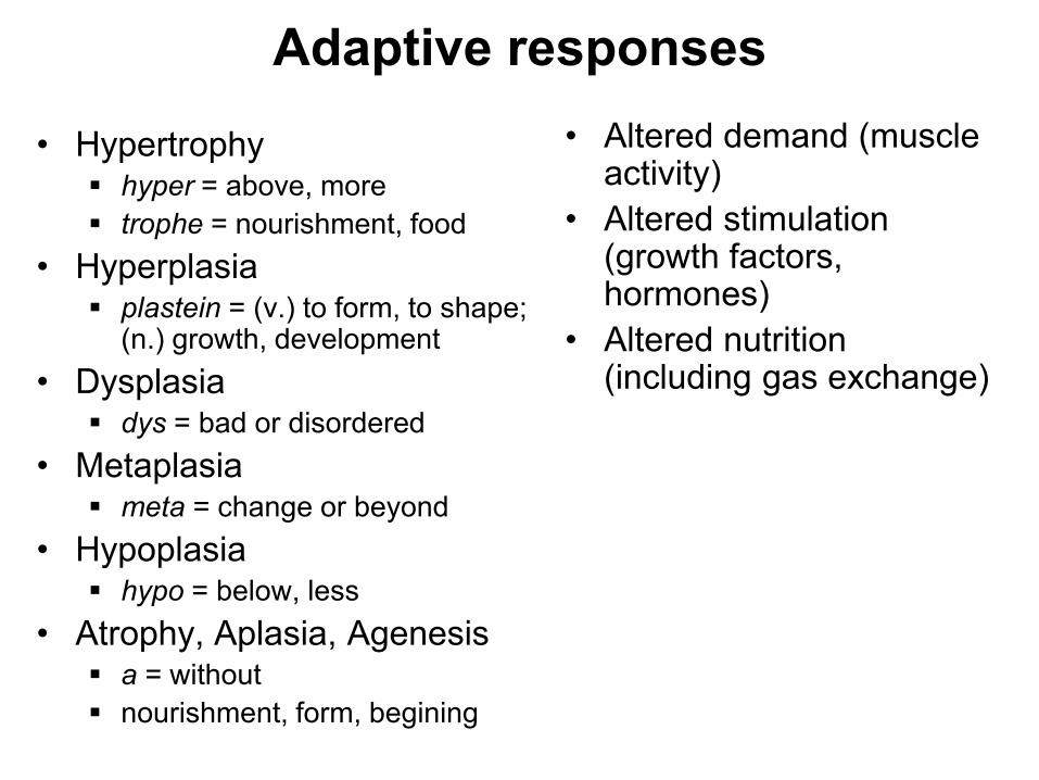
Adaptive responses• Hypertrophy
hyper = above, more trophe = nourishment, food
• Hyperplasia plastein = (v.) to form, to shape;
(n.) growth, development• Dysplasia
dys = bad or disordered• Metaplasia
meta = change or beyond• Hypoplasia
hypo = below, less• Atrophy, Aplasia, Agenesis
a = without nourishment, form, begining
• Altered demand (muscle activity)
• Altered stimulation (growth factors, hormones)
• Altered nutrition (including gas exchange)
Robbins Basic Pathology

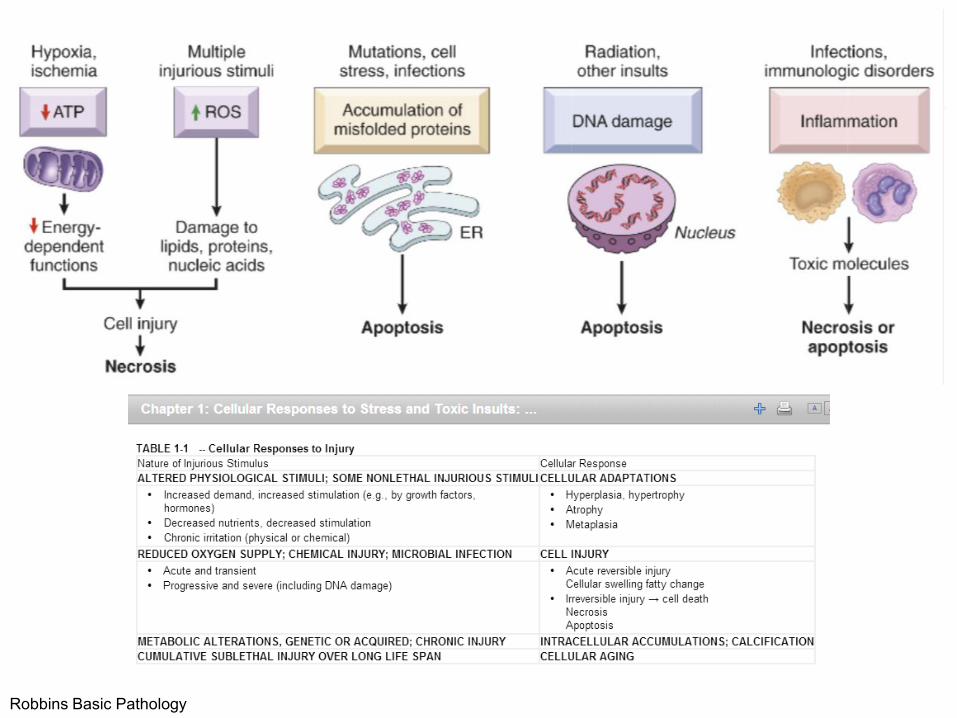
Cell death, the end result of progressive cell injury, is one of the most crucial events in the evolution of disease in any tissue or organ. It results from diverse causes, including ischemia (reduced blood flow), infection, and toxins. Cell death is also a normal and essential process in embryogenesis, the development of organs, and the maintenance of homeostasis.
Two principal pathways of cell death, necrosis and apoptosis.
Nutrient deprivation triggers an adaptive cellular response called autophagy that may also culminate in cell death.

Adaptations• Hypertrophy• Hyperplasia• Atrophy• Metaplasia

Hypertrophy refers to an increase in the size of cells, resulting in an increase in the size of the organ
No new cells, just larger cells. The increased size of the cells is due to the synthesis of more structural components of the cells usually proteins.
Cells capable of division may respond to stress by undergoing both hyperrtophy and hyperplasia
Non-dividing cell increased tissue mass is due to hypertrophy.
HYPERTROPHY

Physiological hypertrophy—normal?
http://jcsm.info/documents/0311/The%20role%20of%20myostatin%20in%20muscle%20wasting-Dateien/13539_2011_35_Fig2_HTML.gif
http://thevoiceofnetizen.blogspot.com/2012/04/genetic-factors-myostatin-and-size-of.html

Exercise hypertrophy
Microscopic views of muscle hypertrophy. Enlarged type 2 (fast twitch) fibers stain dark with ATPase at pH 9.4. Enlarged fast twitch fibers stain pale with cytochrome c oxidase. Hypertrophied fibers with less sarcoplasmic reticulum relative to fiber proteins stain paler with the trichrome stain.
http://neuromuscular.wustl.edu/pathol/hypertrophy.htm
http://www.expert-nutrition.com/image-files/hypertrophy.png
http://neuromuscular.wustl.edu/pathol/hypertrophy.htm
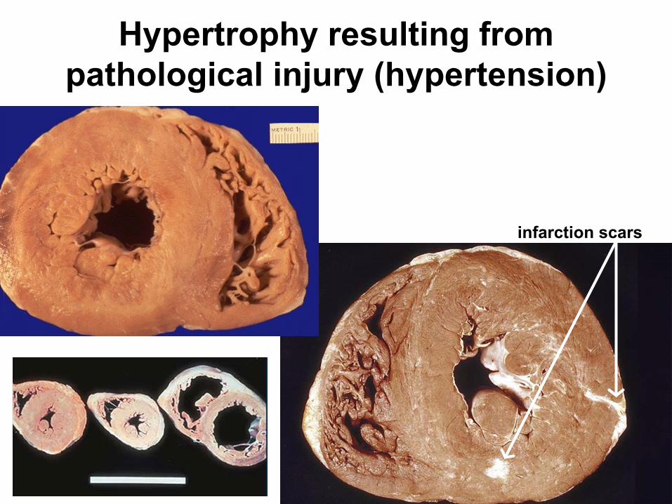
Hypertrophy resulting from pathological injury (hypertension)
infarction scars

Circulation as a Circut
http://www.bing.com/images/search?q=circulation+as+a+circuit&view=detailv2&qpvt=circulation+as+a+circuit&id=B21CEBB7A9FFF9827EBDC962BC427E7C33BA9D85&selectedIndex=5&ccid=MEAKfgQu&simid=608015281329144869&thid=OIP.M30400a7e042e8fc86bd3a2cfd4a3c89aH0&ajaxhist=0

VH
https://www.bing.com/images/search?q=hypertension+and+cardiac+hypertrophy&view=detailv2&&id=AABA9F337D501E1B4B1976767CB4597641C3414B&selectedIndex=14&ccid=XcWZE%2bif&simid=608049924542303816&thid=OIP.M5dc59913e89f4909761570e989f8d132o0&ajaxhist=0
https://www.bing.com/images/search?q=hypertension+and+cardiac+hypertrophy&view=detailv2&&id=C2A390B5E541DE9EE23884852AB9CF8150D8D7FE&selectedIndex=56&ccid=ge%2ftspqQ&simid=608040351063936810&thid=OIP.M81efedb29a90bf54dbef49a423ec3706H0&ajaxhist=0
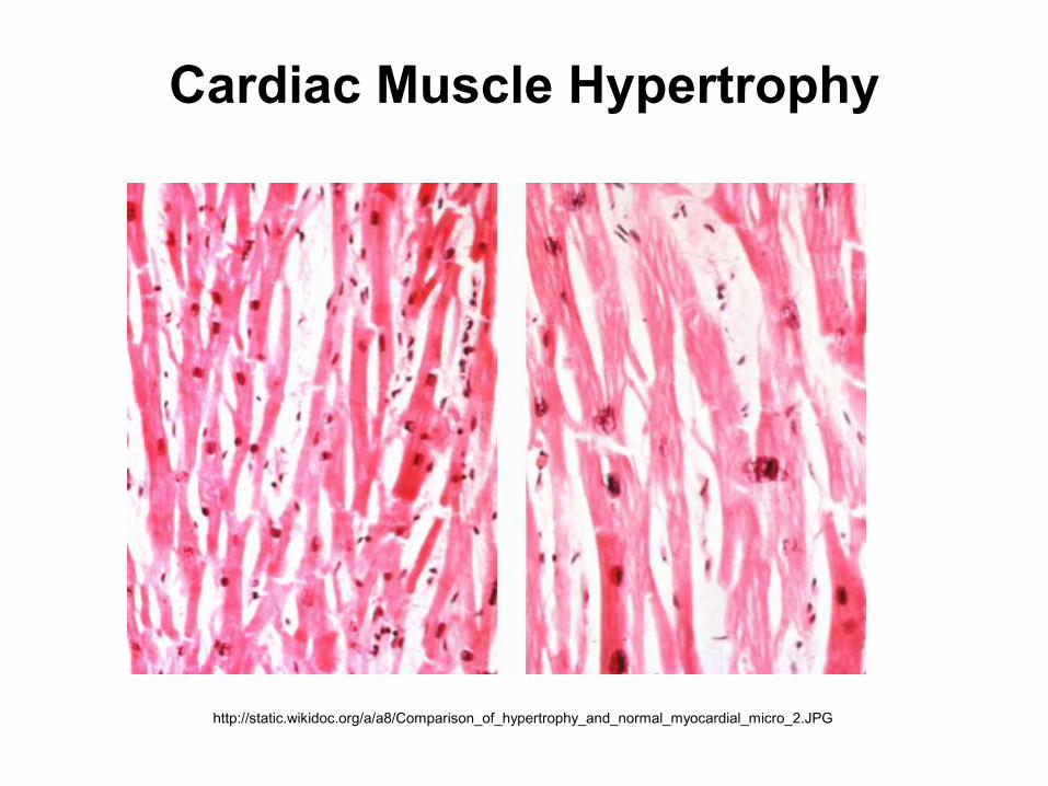
http://static.wikidoc.org/a/a8/Comparison_of_hypertrophy_and_normal_myocardial_micro_2.JPG
Cardiac Muscle Hypertrophy

Physiologic adaptation vs. pathology

Scarred necrosis
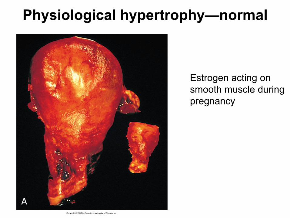
Physiological hypertrophy—normal
Estrogen acting on smooth muscle during pregnancy

Microscopic physiological hypertrophy
NORMAL GRAVID

Mechanisms of muscle hypertrophy
• Increased protein synthesis increased cell size increased organ size
• Nondividing cells produce more protein and membrane without division
• Mechanosensors, PI3K /Aktsignaling pathway important in exercise-induced growth
• Growth factors, vasoactive agents, hormones mediate stress-induced response
• Unrelieved stress eventually results in irreversible injury
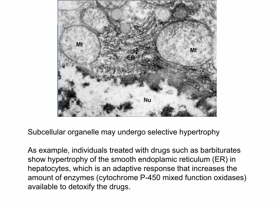
Subcellular organelle may undergo selective hypertrophy
As example, individuals treated with drugs such as barbiturates show hypertrophy of the smooth endoplamic reticulum (ER) in hepatocytes, which is an adaptive response that increases the amount of enzymes (cytochrome P-450 mixed function oxidases) available to detoxify the drugs.

Hyperplasia is an increase in the number of cells in an organ or tissue, usually resulting in increased mass of the organ or tissue
Hyperplasia and hypertrophy are distinct processes but frequently occur together
Both can triggered by the same external stimulus
Hyperplasia takes place if the cell population is capable of dividing resulting in increased cell numbers
HYPERPLASIA

Physiologic vs Pathologic
Physiologichormonal vs compensatory
HYPERPLASIA
Artrenewal.org

Physiological, hormonal hyperplasia
https://c1.staticflickr.com/3/2840/11359744405_50a0c0ce4a_b.jpg
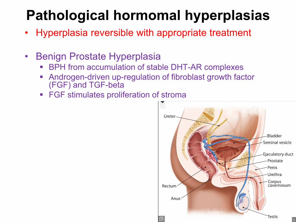
Pathological hormomal hyperplasias• Hyperplasia reversible with appropriate treatment
• Benign Prostate Hyperplasia BPH from accumulation of stable DHT-AR complexes Androgen-driven up-regulation of fibroblast growth factor
(FGF) and TGF-beta FGF stimulates proliferation of stroma

Prostate normal vs. hyperplasia
Benign prostatic hyperplasia androgens
Hyperplasia regresses if the hormonal stimulation is eliminated
Hyperplasia is distinct from cancer, but cancerous proliferation may arise

HYPERTROPHIC
Prostate
Atrophy is reduced size of an organ or tissue resulting from a decrease in cell size and number
Physiologic or Pathologic.
Physiologic atrophy is common during normal development.
Some embryonic structures, such as the notochord and thyroglossal duct, undergo atrophy during fetal development.
The uterus decreases in size shortly after parturition.
ATROPHY

When a fractured bone is immobilized in a plaster cast or when a patient is restricted to complete bedrest, skeletal muscle atrophy ensues
The initial decrease in cell size is reversible once activity is resumed., leading to osteoporosis of disuse
Some of these skeletal muscle fibers here show atrophy, compared to normal fibers. The number of cells is the same as before the atrophy occurred, but the size of some fibers is reduced. This is a response to injury by "downsizing" to conserve the cell. In this case, innervation to the small, atrophic fibers was lost. (This is a trichrome stain.)
Decreased workload (atrophy of disuse)Pathologic
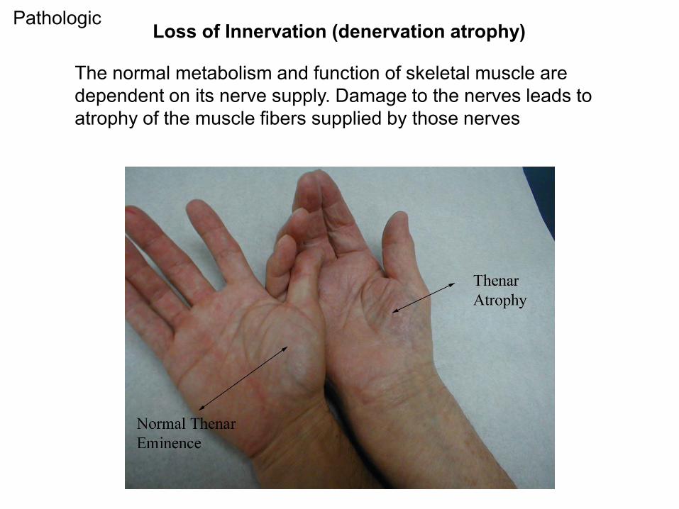
The normal metabolism and function of skeletal muscle are dependent on its nerve supply. Damage to the nerves leads to atrophy of the muscle fibers supplied by those nerves
Loss of Innervation (denervation atrophy)Pathologic
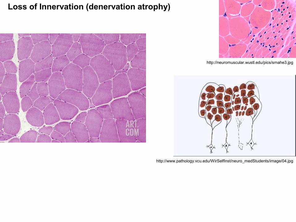
http://neuromuscular.wustl.edu/pics/smahe3.jpg
http://www.pathology.vcu.edu/WirSelfInst/neuro_medStudents/image/04.jpg
Loss of Innervation (denervation atrophy)

Normal Lung
http://www.meddean.luc.edu/lumen/bbs/p/pulpathi/pulpath3.jpeg
http://www.microscopy-uk.org.uk/mag/imgsep08/Apocap4.jpg
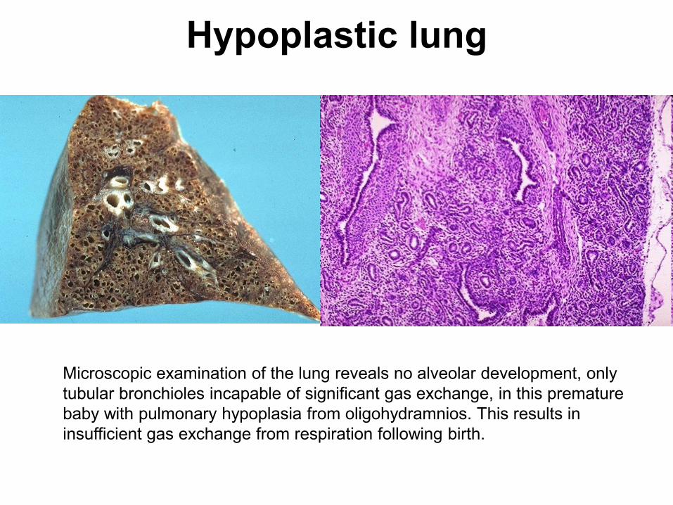
Hypoplastic lung
Microscopic examination of the lung reveals no alveolar development, only tubular bronchioles incapable of significant gas exchange, in this premature baby with pulmonary hypoplasia from oligohydramnios. This results in insufficient gas exchange from respiration following birth.

A decrease in blood supply such as ischemia
The brain may undergo progressive atrophy, mainly because of reduced blood supply as a result of atherosclerosis
This is called senile atrophy; it also affects the heart
Diminished Blood Supply Pathologic
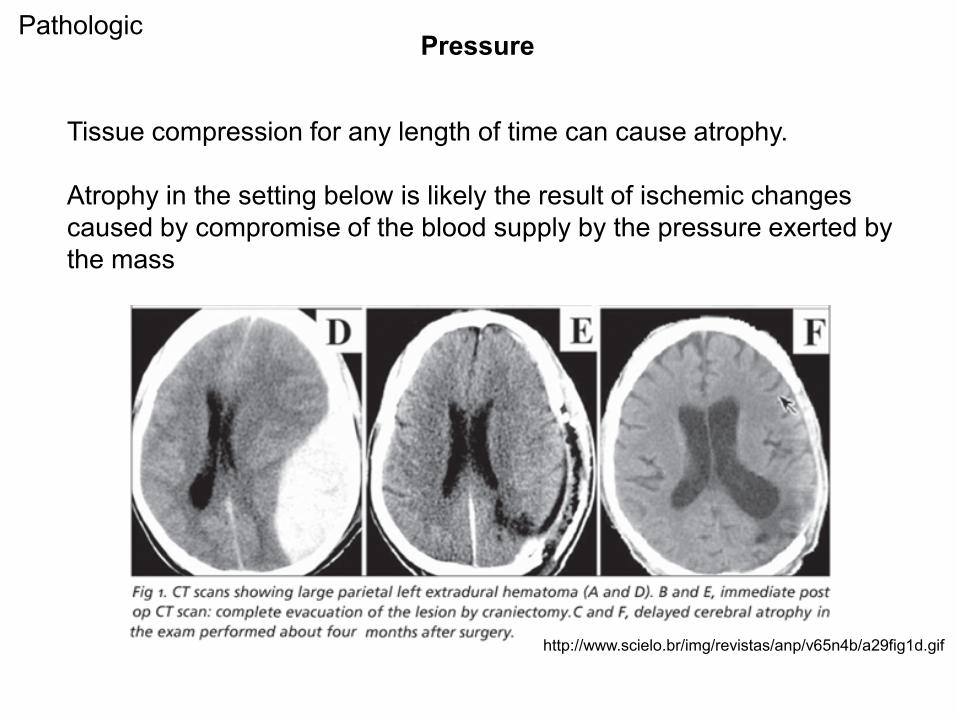
Tissue compression for any length of time can cause atrophy.
Atrophy in the setting below is likely the result of ischemic changes caused by compromise of the blood supply by the pressure exerted by the mass
Pressure
http://www.scielo.br/img/revistas/anp/v65n4b/a29fig1d.gif
Pathologic
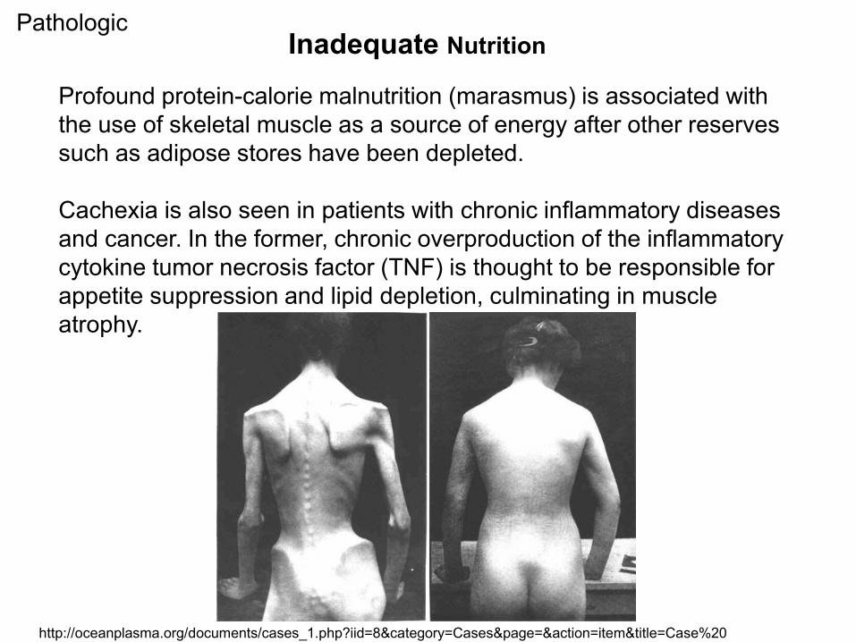
Profound protein-calorie malnutrition (marasmus) is associated with the use of skeletal muscle as a source of energy after other reserves such as adipose stores have been depleted.
Cachexia is also seen in patients with chronic inflammatory diseases and cancer. In the former, chronic overproduction of the inflammatory cytokine tumor necrosis factor (TNF) is thought to be responsible for appetite suppression and lipid depletion, culminating in muscle atrophy.
Inadequate NutritionPathologic
http://oceanplasma.org/documents/cases_1.php?iid=8&category=Cases&page=&action=item&title=Case%20

Many hormone-responsive tissues, such as the breast and reproductive organs, are dependent on endocrine stimulation for normal metabolism and function
Loss of Endocrine Stimulation
testicular atrophy
Pathologic

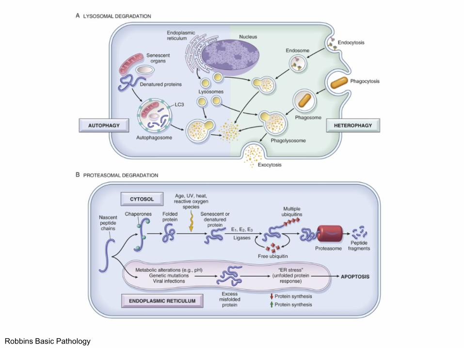
Decreased protein synthesis and increased protein degradation in cells because of reduced metabolic activity.
The degradation of cellular proteins occurs mainly by the ubiquitin-proteasome pathway.
MECHANISMS of ATROPHY

Atrophy can be accompanied by increased autophagy
Autophagy (“self eating”) is the process in which the starved cell eats its own components
Autophagic vacuoles are membrane-bound vacuoles that contain fragments of cell components
The vacuoles ultimately fuse with lysosomes, and their contents are digested by lysosomal enzymes
Autophagy
Robbins Basic Pathology

Reversible change in which one differentiated cell type is replaced by another cell type.
It may represent an adaptive substitution of cells that are sensitive to stress by cell types better able to withstand the adverse environment
METAPLASIA

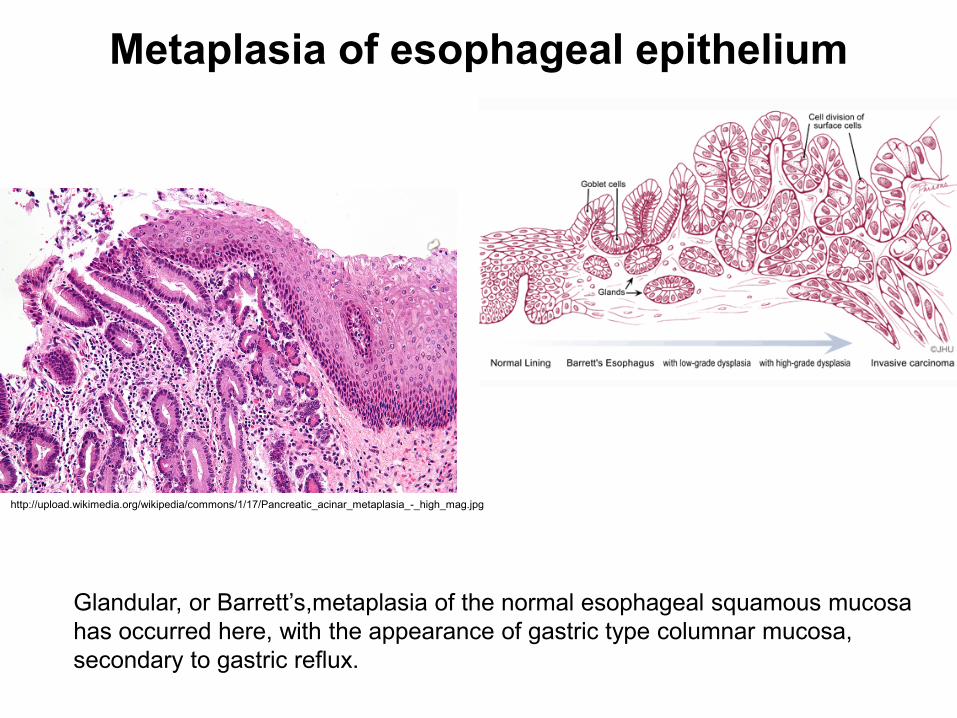
Metaplasia of esophageal epithelium
Glandular, or Barrett’s,metaplasia of the normal esophageal squamous mucosa has occurred here, with the appearance of gastric type columnar mucosa, secondary to gastric reflux.
http://upload.wikimedia.org/wikipedia/commons/1/17/Pancreatic_acinar_metaplasia_-_high_mag.jpg

Result of a reprogramming of stem cells that are known to exist in normal tissues, or of undifferentiated mesenchymal cells present in connective tissue
Precursor cells differentiate along a new pathway
The differentiation of stem cells to a particular lineage is brought about by signals generated by cytokines,growth factors, and extracellular matrix components in the cells' environment
Mechanisms of Metaplasia
http://mahendrasinghphd.blogspot.com/2011/07/molecular-dissection-of-barretts.html


