9256 Biochemistry 1989, 28, 9256-9263 Introduction of a Cysteine Protease Active Site into Trypsin? Jeffrey N. Higaki,$ Luke B. Evnin,§ and Charles S. Craik*,$,s Department of Pharmaceutical Chemistry and Department of Biochemistry and Biophysics, University of California, San Francisco, California 941 43-0446 Received May 15, 1989; Revised Manuscript Received July 27, I989 ABSTRACT: Active site serine 195 of rat anionic trypsin was replaced with a cysteine by site-specific mutagenesis in order to determine if a thiol group could function as the catalytic nucleophile in a serine protease active site environment. Two genetically modified rat thiol trypsins were generated; the first variant contained a single substitution of Ser195 with Cys (trypsin S195C) while the second variant contained the Ser195 to Cys as well as an Asp102 to Asn substitution (trypsin D102N,S195C) that more fully mimics the putative catalytic triad of papain. Both variants were expressed as his J signal peptide-trypsin fusion proteins to high levels under the control of the tac promoter. The mature forms of both variants were secreted into the periplasmic space of Escherichia coli. Trypsin S195C shows a low level of activity toward the activated ester substrate Z-Lys-pNP, while both trypsin S195C and trypsin D102N,S195C were active toward the fluorogenic tripeptide substrate Z-GPR-AMC. Esterase and peptidase activities of both thiol trypsin variants were inhibited by known Cys protease inhibitors as well as by specific trypsin inhibitors. The k,,, of trypsin S195C was reduced by a factor of 6.4 X lo5 relative to that of trypsin while the k,,, of trypsin D102N,S195C was lowered by a factor of 3.4 X lo7 with Z-GPR-AMC as substrate. K, values were unaffected. The loss of activity of trypsin D102N,S195C was partially attributed to an inappropriate Asn102-His57 interaction that precludes the formation of the catalytically competent His57-Cys195 ion pair although loss of the negative charge of D102 at the active site probably contributes to diminished activity. These results indicate that the presence of the active site residues Asp(Asn)102-His57-Cys195 in rat trypsin are required but not sufficient for this enzyme to function as an effective thiol protease. Structural and electronic factors that contribute to the higher catalytic activity of naturally occurring thiol proteases may not be present or operational in these thiol trypsin variants. Eukaryotic serine and cysteine proteases are structurally distinct families of proteolytic enzymes that carry out analo- gous reactions. Although the overall three-dimensional structures of the two classes of enzymes are very different, features of the serine protease catalytic triad (Ser195, His57, and Aspl02) can also be observed in the cysteine proteases. The cysteine protease papain features an essential nucleophilic thiol group of Cys25 (Light et al., 1964) and the imidazole of His159 (Husain & Lowe, 1968; Drenth et al., 1970) whose N61 nitrogen is within hydrogen-bonding distance of the thiol group. These two amino acid residues play roles analogous to Ser195 and His57 in eukaryotic Ser proteases. In addition, since the Ne2 nitrogen of His159 in papain can hydrogen bond to the side chain of Am175 (Kamphuis et al., 1984; Baker & Drenth, 1987), Asn175 has been implicated as an essential active site residue in the Cys proteases analogous to the Asp102 in the Ser proteases. Further, Asn175 is highly conserved among Cys proteases (Brocklehurst, 1987). The three residues, Cys25, His159, and Asn175, constitute the putative catalytic triad of papain, and they can be superimposed on the catalytic triad of the serine proteases with a root-mean-square difference of less than 1.0 A (Garavito et al., 1977). This conservation in the active site geometry of serine and cysteine proteases has This work was supported by NSF Grant GM 8608086 to C.S.C. J.N.H. was a recipient of NIH Postdoctoral Fellowship (National Re- search Service Award) GM 11598. L.B.E. was supported by NIH Training Grant T32-GM 07810 and the Lucille P. Markey Charitable Trust. The Mass Spectrometry Facility, University of California, was supported by the NIH Division of Research Resources, Grant RR 01614. *Address correspondence to this author at the Department of Phar- maceutical Chemistry. *Department of Pharmaceutical Chemistry. Department of Biochemistry and Biophysics. led to the suggestion that the active sites of these enzymes have evolved through a process of convergent evolution (Garavito et al., 1977). In order to examine the extent of active site similarities in Ser versus Cys proteases, the nucleophilic oxygen of the active site Ser was previously chemically converted to the more nu- cleophilic sulfur atom in subtilisin (Polgar & Bender, 1966; Neet & Koshland, 1966) and in Streptomyces griseus trypsin (Yokosawa et al., 1977). Since oxygen and sulfur belong to the 6A chemical group of periodic elements and possess similar chemical properties, the resulting thiol enzymes were predicted to behave as efficient proteases; however, these predictions were not confirmed in experiments. Thiol subtilisin and thiol trypsin were found to be inactive toward normal ester and amide substrates (Polgar & Bender, 1967; Neet & Koshland, 1966; Yokosawa et al., 1977). Low levels of activity toward activated ester substrates led to the conclusion that these modified en- zymes had reduced deacylation rates in the hydrolysis of ac- tivated esters but grossly deficient acylation rates in the hy- drolysis of normal ester and amide substrates. The chemistry employed to carry out the original conversion was limited to the active site hydroxyl group of the small subset of Ser proteases containing few or no disulfide bonds. Using current methods of molecular genetics, it is possible to avoid the limitations and potential artifacts resulting from nonspecific and incomplete modification of the enzyme. Analysis of en- gineered variants can now be extended to proteins that are not amenable to chemical modification due to inherent instabilities or amino acid compositions that are incompatible with the chemical reaction conditions employed. To determine whether the putative catalytic triad of the cysteine protease papain can be reconstructed within the structurally distinct serine protease trypsin, two recombinant 0006-2960/89/0428-9256$01.50/0 0 1989 American Chemical Society
Transcript
9256 Biochemistry 1989, 28, 9256-9263
Introduction of a Cysteine Protease Active Site into Trypsin? Jeffrey N. Higaki,$ Luke B. Evnin,§ and Charles S. Craik*,$,s
Department of Pharmaceutical Chemistry and Department of Biochemistry and Biophysics, University of California, San Francisco, California 941 43-0446
Received May 15, 1989; Revised Manuscript Received July 27, I989
ABSTRACT: Active site serine 195 of rat anionic trypsin was replaced with a cysteine by site-specific mutagenesis in order to determine if a thiol group could function as the catalytic nucleophile in a serine protease active site environment. Two genetically modified rat thiol trypsins were generated; the first variant contained a single substitution of Ser195 with Cys (trypsin S195C) while the second variant contained the Ser195 to Cys as well as an Asp102 to Asn substitution (trypsin D102N,S195C) that more fully mimics the putative catalytic triad of papain. Both variants were expressed as his J signal peptide-trypsin fusion proteins to high levels under the control of the tac promoter. The mature forms of both variants were secreted into the periplasmic space of Escherichia coli. Trypsin S195C shows a low level of activity toward the activated ester substrate Z-Lys-pNP, while both trypsin S195C and trypsin D102N,S195C were active toward the fluorogenic tripeptide substrate Z-GPR-AMC. Esterase and peptidase activities of both thiol trypsin variants were inhibited by known Cys protease inhibitors as well as by specific trypsin inhibitors. The k,,, of trypsin S195C was reduced by a factor of 6.4 X lo5 relative to that of trypsin while the k,,, of trypsin D102N,S195C was lowered by a factor of 3.4 X lo7 with Z-GPR-AMC as substrate. K , values were unaffected. The loss of activity of trypsin D102N,S195C was partially attributed to an inappropriate Asn102-His57 interaction that precludes the formation of the catalytically competent His57-Cys195 ion pair although loss of the negative charge of D102 at the active site probably contributes to diminished activity. These results indicate that the presence of the active site residues Asp(Asn)102-His57-Cys195 in rat trypsin are required but not sufficient for this enzyme to function as an effective thiol protease. Structural and electronic factors that contribute to the higher catalytic activity of naturally occurring thiol proteases may not be present or operational in these thiol trypsin variants.
Euka ryo t i c serine and cysteine proteases are structurally distinct families of proteolytic enzymes that carry out analo- gous reactions. Although the overall three-dimensional structures of the two classes of enzymes are very different, features of the serine protease catalytic triad (Ser195, His57, and Aspl02) can also be observed in the cysteine proteases. The cysteine protease papain features an essential nucleophilic thiol group of Cys25 (Light et al., 1964) and the imidazole of His159 (Husain & Lowe, 1968; Drenth et al., 1970) whose N61 nitrogen is within hydrogen-bonding distance of the thiol group. These two amino acid residues play roles analogous to Ser195 and His57 in eukaryotic Ser proteases. In addition, since the Ne2 nitrogen of His159 in papain can hydrogen bond to the side chain of Am175 (Kamphuis et al., 1984; Baker & Drenth, 1987), Asn175 has been implicated as an essential active site residue in the Cys proteases analogous to the Asp102 in the Ser proteases. Further, Asn175 is highly conserved among Cys proteases (Brocklehurst, 1987). The three residues, Cys25, His159, and Asn175, constitute the putative catalytic triad of papain, and they can be superimposed on the catalytic triad of the serine proteases with a root-mean-square difference of less than 1.0 A (Garavito et al., 1977). This conservation in the active site geometry of serine and cysteine proteases has
This work was supported by NSF Grant GM 8608086 to C.S.C. J.N.H. was a recipient of NIH Postdoctoral Fellowship (National Re- search Service Award) GM 11598. L.B.E. was supported by NIH Training Grant T32-GM 07810 and the Lucille P. Markey Charitable Trust. The Mass Spectrometry Facility, University of California, was supported by the NIH Division of Research Resources, Grant RR 01614.
*Address correspondence to this author at the Department of Phar- maceutical Chemistry.
*Department of Pharmaceutical Chemistry. Department of Biochemistry and Biophysics.
led to the suggestion that the active sites of these enzymes have evolved through a process of convergent evolution (Garavito et al., 1977).
In order to examine the extent of active site similarities in Ser versus Cys proteases, the nucleophilic oxygen of the active site Ser was previously chemically converted to the more nu- cleophilic sulfur atom in subtilisin (Polgar & Bender, 1966; Neet & Koshland, 1966) and in Streptomyces griseus trypsin (Yokosawa et al., 1977). Since oxygen and sulfur belong to the 6A chemical group of periodic elements and possess similar chemical properties, the resulting thiol enzymes were predicted to behave as efficient proteases; however, these predictions were not confirmed in experiments. Thiol subtilisin and thiol trypsin were found to be inactive toward normal ester and amide substrates (Polgar & Bender, 1967; Neet & Koshland, 1966; Yokosawa et al., 1977). Low levels of activity toward activated ester substrates led to the conclusion that these modified en- zymes had reduced deacylation rates in the hydrolysis of ac- tivated esters but grossly deficient acylation rates in the hy- drolysis of normal ester and amide substrates.
The chemistry employed to carry out the original conversion was limited to the active site hydroxyl group of the small subset of Ser proteases containing few or no disulfide bonds. Using current methods of molecular genetics, it is possible to avoid the limitations and potential artifacts resulting from nonspecific and incomplete modification of the enzyme. Analysis of en- gineered variants can now be extended to proteins that are not amenable to chemical modification due to inherent instabilities or amino acid compositions that are incompatible with the chemical reaction conditions employed.
To determine whether the putative catalytic triad of the cysteine protease papain can be reconstructed within the structurally distinct serine protease trypsin, two recombinant
0006-2960/89/0428-9256$01.50/0 0 1989 American Chemical Society
Cysteine Protease Active Site in Trypsin
rat thiol trypsins were produced by site-specific mutagenesis. The first of these (trypsin S195C) was generated in order to test whether the active site Ser195 of pancreatic trypsin can be replaced with a Cys. The second thiol trypsin (trypsin D 102N,S195C) more fully mimicked the putative catalytic triad of papain.
Both variants were overexpressed as active enzymes in bacteria and have been purified to homogeneity and crystal- linity. Both thiol trypsin variants are catalytically active al- though the levels of activity are significantly below that of trypsin. Using the sensitive fluorogenic peptide substrate Z-GPR-AMC,' a kinetic analysis of these variants was per- formed. The results of these experiments are reported here, and plausible explanations for the reduced activities observed for trypsin S195C and trypsin D102N,S195C are discussed.
MATERIALS AND METHODS Materials. Plasmid pFA54 containing the tac promoter,
the Salmonella typhimurium his J coding sequence, and the flanking termination signal from the 5 s RNA gene was a generous gift of Dr. G. F.-L. Ames. Escherichia coli strain X90 [F' lac IQ, lac ZY, pro AB/A(lac-pro), ara, nul A, argEam, thi, ri'] was obtained from Dr. A. Vershon. [3H]- TLCK was a gift of Dr. C.-T. Peng (supported by NIH CA 33537). The restriction enzymes BstXI, StyI, XbaI, SalI, PuuII, SspI, BamHI, EcoRI, and EcoRV, Klenow DNA po- lymerase, and T4 DNA ligase were purchased from New England Biolabs, Inc. Modified T7 polymerase was from US. Biochemical Corp. DEAE-Sepharose Fast Flow and CM- Sepharose Fast Flow were from Pharmacia. Immobilized p-aminobenzamidine-agarose was obtained from Pierce. Isopropyl @-D-thiogalactopyranoside (IPTG), TAME, Z- Lys-pNP, BAPNA, DTNB, PMSF, TLCK, and IAA were purchased from Sigma Chemical Co. pNPGB was obtained from Vega Biotechnologies. Z-Arg-AMC, D-VLR-AFC, and Z-GPR-AMC were the products of Bachem Bioscience, Inc. BAEE was from the Chemical Dynamics Corp. pCMB was purchased from ICN Pharmaceuticals, Inc., and tBOC-FPR- ald was a gift from Dr. L. Graf. APPA was purchased from Aldrich Chemical Co.
Site-Specific Mutagenesis and Expression Plasmid Con- strucrion. Oligonucleotide-directed site-specific mutagenesis was performed as described previously (Higaki et al., 1987). Trypsin S195C and trypsin D102N,S195C were initially ex- pressed with the vector pTRAP (Graf et al., 1987; Higaki et al., 1987).
Although fair yields of trypsin were obtained with the al- kaline phosphatase promotor and signal peptide of the pTRAP vector, we anticipated that the stronger tac promoter and his J signal peptide combination would work more efficiently than the pTRAP (Graf et al., 1987) and pBsTn2 (Evnin & Craik, 1988) systems. Furthermore, this inducible promoter-signal peptide combination would provide greater flexibility for op-
timizing the expression of trypsin in various strains of E. coli. Plasmid pT3 was constructed from plasmids pFA54 and
pBsTn2. pFA54 contains the tac promoter and all of the S . typhimurium his J coding sequences including the putative Shine-Dalgarno ribosome binding site (Higgins & Ames, 1981). When cells carrying the pFA54 plasmid are induced with 1 mM IPTG and osmotically shocked, approximately 25% of the resulting periplasmic fraction is the his J protein (Nikaido and Ames, personal communication). A 460 base pair BamHI-EcoRV fragment from pFA54 encoding the tac promoter, his J signal peptide, and the first 30 amino-terminal residues of the mature his J coding sequence was blunt-end ligated into pBsTn2 at the unique BamHI site located 5' to the alkaline phosphatase promoter (Evnin & Craik, 1988). The trpA transcriptional terminator was located at the 3' end of the trypsin coding region in pBsTn2. The 431 base pair region separating the end of the his J signal peptide and the first codon of mature trypsin (Ile16) was then deleted by oligonucleotide-directed mutagenesis, precisely fusing the his J signal peptide sequence to the mature trypsin sequence. The final step in the construction of pT3 was to excise the SspI- SalI DNA fragment encoding the tac promoter, the his J signal peptide, and trypsin sequences along with the trpA terminator from the pUC-based pBsTn2 vector and ligate it into pBR322. We anticipated that transcription from the tac promoter would be more tightly controlled in a lower copy number plasmid. To achieve this, the SspI-Sal1 DNA frag- ment of pBsTn2 was ligated into the compatible ends of PuuII-SalI-digested pBR322. The resulting plasmid pT3 (T3 = tac, trypsin, terminator) was used to transform several lacIQ strains of E. coli to identify a strain that provided the highest levels of expression. Strain X90 was found to be the most efficient strain for the expression of mature trypsin. High-level expression of active trypsin in the periplasmic space of transformed X-90 cells did not appear to be detrimental to their growth.
To introduce the DNA sequences encoding trypsin S195C and trypsin D102N,S195C into pT3, pTRAP-trypsin S195C or pTRAP-trypsin D102N,S195C (Higaki et al., 1987) was digested with BstXI and StyI. The 554-bp BstXI-Sty1 DNA fragment encoding amino acids 40-229 was then ligated into pT3D189E digested with BstXI and StyI. pT3 S195C and pT3 D102N,S195C plasmid DNA was then used to transform X90 cells. Positive clones were easily identified since they lacked a unique XbaI site present in the trypsin coding se- quence of the pT3D189E vector. Unambiguous determination of the appropriate mutation was accomplished by dideoxy sequence analysis.
Expression and Purification. The expression and purifi- cation of trypsin and thiol trypsins were carried out as de- scribed previously (Higaki et al., 1987) with the following modifications. Since trypsin expression was regulated by the tac promoter, X90 E. coli cells harboring either pT3 S195C or pT3 D102N,S195C were induced with 1 mM IPTG. Two-liter shake flasks were inoculated with 0.5 mL of a saturated culture and grown in the presence of 1 mM IPTG and 50 pg/mL ampicillin. The X90 strain provided better yields when trypsin was expressed constitutively during growth rather than being induced at log-phase growth. A typical preparation began with saturated 12-L cultures of cells that had been grown previously for 12-15 h at 37 "C with vigorous shaking.
The cells were centrifuged at 13000g for 10 min and sub- jected to osmotic shock treatment (Neu & Heppel, 1965). The treated cells were centrifuged again at 13000g for 15 min, and
9258
the supernatant containing the contents of the periplasmic space was adjusted to pH 6.0 with 10 mM MES. The sample was then subjected to both anionic and cationic exchange chromatography as described previously (Higaki et al., 1987). The final step of purification using an immobilized p-amino- benzamidine affinity column was also performed as described; however, since mature trypsin was expressed, it was not nec- essary to activate the sample with enterokinase prior to loading onto the affinity column. No reducing agents were added at any step of this purification.
Sulfhydryl Determinations. A quantitative determination of the free sulfhydryl groups for each of the trypsin variants was performed with 5,5'-dithiobis(2-nitrobenzoic acid) (DTNB) (Ellman, 1959). A 50-pL aliquot of the appropriate enzyme solution was added to 940 pL of 0.1 M Tris-HC1, pH 8.0, containing 0.02M CaC1,. A IO-pL aliquot of 0.01 M DTNB in dimethylformamide was then added to the protein sample and allowed to react at room temperature for 5 min. The absorbance of the solution was measured at 412 nm, and the sulfhydryl concentration was calculated from the con- centration of the thionitrobenzoate anion with a molar ex- tinction coefficient of 13 600 M-' cm-'.
Active Site Titrations. The concentration of trypsin active sites was determined by the method of Chase and Shaw (1967). A 60-pL aliquot of enzyme solution was added to 935 pL of 0.1 M veronal, pH 8.3, 25 OC. The absorbance at 410 nm was then monitored in a double-beam spectrophotometer for approximately 30 s. Subsequently, 5 pL of 0.012 M pNPGB in DMF-acetonitrile (1:4 v/v) was added, and the mixture was stirred quickly. The absorbance at 410 nm was recorded immediately for several minutes. A blank containing no protein was run simultaneously to determine nonenzymatic hydrolysis of pNPGB. The initial burst in absorbance at 410 nm was determined by extrapolating the linear portion of the resulting profile back to the point corresponding to the instant that pNPGB was added. The molar concentration of active sites was calculated using a molar extinction coefficient of 1.66 X lo4 M-' cm-' for p-nitrophenol (Chase & Shaw, 1967).
An alternate method of quantifying the active sites of trypsin and thiol trypsins made use of the highly reactive and tritiated form of the chloromethyl ketone derivative of lysine, [3H]- TLCK. This compound reacts specifically and stoichiomet- rically with the active site of trypsin and was used originally to demonstrate the presence of the essential His57 in the active site (Shaw et a]., 1965). A 30 pM sample of trypsin, trypsin S195C, or trypsin D102N,S195C was reacted with 78 pM [3H]TLCK in 0.1 M veronal, pH 8.3, for 30 min at 37 "C. Unincorporated [3H]TLCK was removed by reversed-phase high-performance liquid chromatography (HPLC) using a Vydac C18 column (4.5 mm X 25 cm) equilibrated in 20% acetonitrile in 0.1% trifluoroacetic acid with a flow rate of 1.0 mL/min. [3H] trypsin eluted during an acetonitrile gradient (0-80%, 25 min). Trypsin-containing fractions were collected and monitored for incorporated 3H by scintillation counting. The quantity of [3H]TLCK incorporated into protein was then compared to the amount of trypsin or thiol trypsin present in the same fractions on the basis of absorbance at 280 nm. The number of active sites determined by [3H]TLCK incorporation was in good agreement with those determined by titration with pNPGB (data not shown).
Enzyme Assays. Trypsin assays using either the ester substrates TAME (1 mM) and Z-Lys-pNP (20 pM) or the amide substrate BAPNA (500 pM) were performed spectro- photometrically in 0.1 M veronal or 50 mM Tris-HC1, pH 8.0, and i n 50 mM MES, pH 6.0. All reactions contained 10 mM
Biochemistry, Vol. 28, No. 24, 1989 Higaki et al.
CaCl, and were carried out at 25 "C. Base-line absorbance changes were monitored for a brief period prior to the addition of 4.0 X lo-' nmol of enzyme. The absorbance at 410 nm was then monitored to follow the release of either p-nitrophenol or free p-nitroaniline. The absorbance at 247 nm was mon- itored to follow the hydrolysis of TAME. Thiol trypsin ac- tivities using these substrates were negligible even after 10-15 min.
Fluorescence assays were performed by following the release of free (aminomethy1)coumarin from Z-Arg-AMC or Z- GPR-AMC. A 950-pL volume of 0.05 M Tris-HC1, pH 8.0, containing 10 mM CaC1, and 3.9-391 pM AMC substrate was equilibrated to 25 OC. Fluorescence measurements were carried out with an excitation wavelength of 380 nm and an emission wavelength of 460 nm. Base-line fluorescence was monitored for 1 min after which time a 50-pL aliquot of the appropriate enzyme was added (0.15-1 5.0 nmol of thiol trypsin or 1.4 pmol of trypsin). The reactions were followed for 5 min, and the initial rates were determined from the linear portion of the assay.
Papain assays with Z-GPR-AMC were performed in the same fashion as trypsin assays, except that the papain sample was first activated by mild reduction under anaerobic con- ditions (Arnon, 1970). The concentration of active papain was determined by the standard BAEE assay method (Bender et al., 1966), and the fluorescence assays were performed in the presence of 3 1-8 15 pM Z-GPR-AMC.
Inhibition Studies. In order to test the effect of known Cys and Ser protease inhibitors on the amidase activities observed for trypsin and both thiol trypsins, 10-200 pM samples of each enzyme were first incubated at 25 "C with either 2.5 mM pCMB, 7.5 mM DTNB (initially solubilized in DMF), 30 mM PMSF (initially solubilized in acetone), or 0.5 mM APPA (initially dissolved in 0.1 M HCl, 80% DMF). After a 1.5-h incubation period, the treated samples were assayed as usual with 20 pM Z-GPR-AMC (or 4.0 pM in the case of APPA- treated samples). Extended incubations were not carried out since control samples autolyzed significantly with time.
The effect of various Cys and Ser protease inhibitors on the esterase activities observed with pNPGB was also examined. A stock solution of trypsin, trypsin S195C, or trypsin D102N,S195C was diluted from 40 to 140 pM in 50 mM veronal, pH 8.0, in the presence of 2.0 mM TLCK, 2.0 mM IAA, or 4.0 mM tBOC-FPR-ald (initially dissolved in DMF). The samples were incubated at 25 "C for 30 min after which time standard pNPGB assays were performed.
RESULTS To relate the mechanistic and structural features of serine
and thiol proteases, two modified forms of rat anionic trypsin containing a Cys at position 195 were created by site-directed mutagenesis. The first variant, trypsin S195C, contained a single amino acid substitution at position 195 (Hartley, 1964) such that only the catalytic Ser195 was changed to a Cys. This variant was generated to determine whether a thiol group could serve as the nucleophilic catalyst in a trypsin-like active site. The second variant, trypsin D102N,S195C, while also con- taining a Cys at position 195, more closely mimicked the putative catalytic triad of papain by introducing a D102N mutation. By placing the altered trypsin sequences under control of the alkaline phosphatase promoter and 3' to the alkaline phosphatase signal peptide (Vasquez et al., 1989), expression of both variants was obtained. Furthermore, the addition of the signal peptide directed the expression of trypsin and trypsin variants into the periplasmic space of E . coli (Higaki et al., 1987).
Cysteine Protease Active Site in Trypsin Biochemistry, Vol. 28, No. 24, 1989 9259
A .
sal I (3209)
Pvurvssp (2489)
colEl origin of I
i replication PT3 (3864bp) I EcoRI
B .
..... GCA TTT GCG ATC GTT GGA ..... ...... Ala Phe Ala Ile Val Gly ......
his J signal processil peptide trypsin Site amino terminus
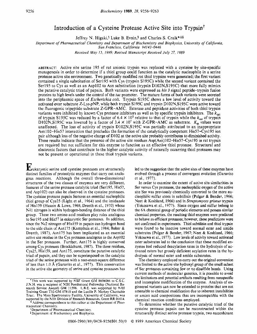
FIGURE 1 : Schematic representation of the bacterial pT3 expression plasmid. (A) A schematic representation of the pT3 expression vector used to express trypsin, trypsin S195C, and trypsin D102N,S195C. The tac promoter-his J signal peptide coding sequence was inserted upstream (5') and inframe with the coding sequence of mature trypsin. The trpA transcriptional termination signal was located on the 3' side of the trypsin coding sequence. Expression was under the control of the tac promoter so that E . coli strain X90 transformed with this plasmid was induced with 1 mM IPTG. The area outlined in this figure shows the his J signal peptide-trypsin junction. The precise site of processing by endogenous signal peptidase at this junction is illustrated in more detail in (B). (B) The his J signal peptide-trypsin junction outlined above is diagrammed here. Trypsin expressed in E . coli is directed through the inner membrane by the his J signal peptide and secreted into the periplasmic space. Endogenous signal peptidase cleaves the his J-trypsin polypeptide on the C-terminal side of the his J Ala-Phe-Ala sequence, liberating mature trypsin containing the critical N-terminal Ile residue.
Although this expression system (pTRAP) provided fair levels of expression of selected variants, improved levels of expression were obtained with the vector pT3 (Figure 1A) which utilizes the tac promoter (deBoer et al., 1983) and E . coli strain X90. Expression of trypsin and trypsin variants from pT3 was induced by the addition of 1 mM IPTG to the culture media. Replacement of the alkaline phosphatase signal peptide with the his J signal peptide still allowed expressed trypsin and trypsin variants to be localized in the periplasmic space of the host. Additionally, processing of the his J signal peptide by the endogenous signal peptidase liberated free, mature trypsin lacking the activation hexapeptide (Davie & Neurath, 1955; Walsh & Wilcox, 1970). Cleavage after Ala(-1) of the his J signal peptide (Figure 1B) liberated trypsin containing the amino-terminal Ile residue critical for activity (Huber & Bode, 1978). Active trypsin can be purified from cells harboring this construction, confirming that the correct processing of trypsin had occurred. Therefore, in contrast to the pTRAP system, subsequent activation of trypsin was not necessary. Trypsin expressed in this fashion was purified to homogeneity and shown to possess kinetic constants that were indistinguishable from those of rat trypsin.
The pT3 plasmid directed the synthesis of a 7-fold increase of trypsin S195C and a 14-fold increase of trypsin D102N,- S195C relative to the previous pTRAP system. A 12-L culture typically yielded approximately 4 mg of pure thiol trypsin. The addition of IPTG to the culture medium at the time of inoc-
-0.02 I c B A;D I I I '
0.0 1.0 2.0 3.0
Time (minutes)
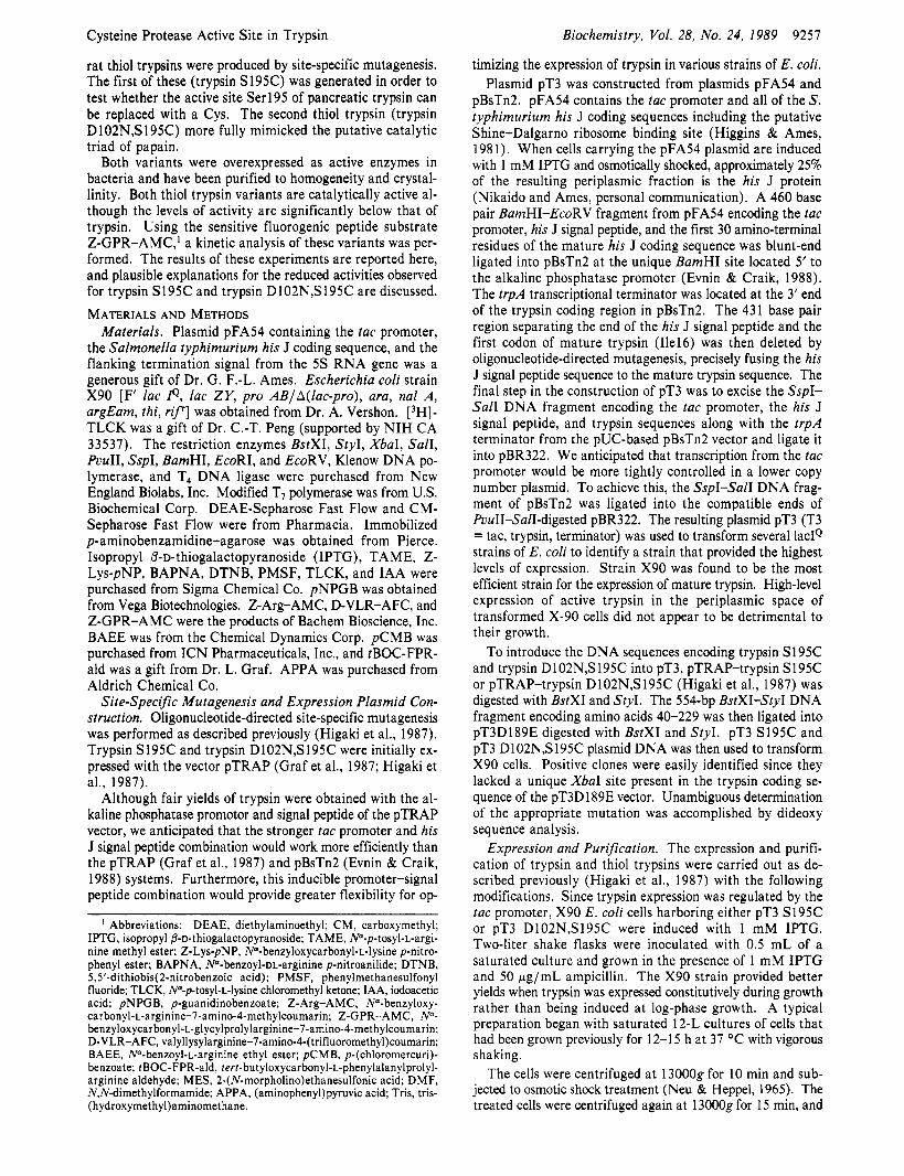
FIGURE 2: Active site titrations of trypsin and thiol trypsins. Active site titrations of trypsin, trypsin S195C, and trypsin D102N,S195C with pNPGB are shown. A 60-pL aliquot of enzyme solution was added to 935 pL of 0.1 M veronal, pH 8.3, 25 "C. The absorbance at 410 nm was monitored for approximately 30 s after which time 5 pL of 0.01 2 M pNPGB in DMF-acetonitrile (1:4 v/v) was added, and the absorbance at 410 nm was monitored further. The arrows indicate the time of pNPGB addition to the respective assay. (A) trypsin; (B) trypsin D102N,S195C; (C) trypsin S195C; (D) blank (no enzyme).
ulation resulted in the constitutive expression of trypsin and thiol trypsin variants which apparently was not deleterious to the cells. Constitutive expression was used instead of induction since slightly higher yields of trypsin resulted. The thiol trypsins were purified to homogeneity by a series of ion ex- change columns and an affinity column of immobilized p- aminobenzamidine (Higaki et al., 1987). Both variants be- haved electrophoretically and chromatographically identical with trypsin suggesting that the amino acid substitutions did not grossly alter the folding of trypsin. These thiol trypsin variants bound tightly to the affinity matrix, suggesting that the introduction of an additional Cys at position 195 with either an Asp or an Asn at position 102 did not produce conforma- tional alterations that resulted in a significantly decreased affinity for benzamidine. Both mutations were confirmed at the nucleic acid level by DNA sequencing and at the amino acid sequence level by mass spectrometry (Higaki et al., 1987). The presence of a single free sulfhydryl group in both trypsin S195C and trypsin D102N,S195C expressed from the pT3 plasmid was confirmed by treatment with DTNB (Ellman, 1959). A 1:l stoichiometry of sulfhydryl to protein was de- termined for both trypsin S 195C and trypsin D 102N,S 195C. Thus, with no prior treatment of the two thiol trypsins with reducing agent, the single sulfhydryl group substituted at position 195 remained in the reduced state throughout the purification.
To determine the molar concentration of active enzyme in each of the purified samples, both trypsin S195C and trypsin D102N,S195C were subjected to active site titrations with the activated ester pNPGB. pNPGB reacts stoichiometrically with trypsin to form an acylated enzyme intermediate and p- nitrophenol and was used as an active site titrant for the native enzyme (Chase & Shaw, 1969; Kezdy & Kaiser, 1970). Like trypsin, both trypsin S195C and trypsin D102N,S195C reacted very rapidly with this reagent to liberate free nitrophenol (Figure 2). Active site concentrations were calculated on the basis of the magnitude of the initial burst of p-nitrophenol from three independent determinations and are listed in Table I. These concentrations were compared to the molar concen- trations calculated by UV absorbance (e1% = 14.4; Davie & Neurath, 1955)./ The lower concentration determined by pNPGB assays rklative to those determined by UV absorbance reflects the presence of inactive enzyme in these samples. Although the actual rates of the initial bursts were not de- termined for any of the enzymes, it is apparent that the two
9260 Biochemistry, Vol. 28, No. 24, 1989
Table I: Active Site Determinations of Trypsin and Thiol Trypsins
Higaki et al.
concn by OD concn by 7% active
enzyme ( P M ) ~ NPGB (pM) sitesb trypsin 15.0 7.0 47
(I Enzyme concentrations were determined at 280 nm with an cl% = 14.4 (Davie & Neurath, 1955). bPercent active sites calculated from pNPGB assays are relative to the total amount of enzyme determined bv uv sDectroDhotometrv.
thiol variants are capable of reacting very rapidly with pNPGB. This suggests that the acylation step for the reaction of this activated ester with thiol trypsins is not dramatically reduced and is consistent with previous results using chemically generated S. griseus thiol trypsin (Yokosawa et al., 1977).
After the initial burst, p-nitrophenol was liberated by the variant enzymes at a slow rate that was similar to the postburst rate observed for trypsin. This may be due to a very slow rate of hydrolysis (deacylation) of the acyl-enzyme or due to nonspecific turnover of the titrant (Kezdy & Kaiser, 1970). It appears that this postburst rate of pNPGB hydrolysis is slightly greater for trypsin S195C than for trypsin or trypsin D102N,S195C, suggesting that the acyl-enzyme intermediate of trypsin S195C may have a higher rate of deacylation than trypsin.
A variety of trypsin substrates were used to investigate the catalytic properties of these two thiol variants. Most substrates (Z-Arg-AMC, BAPNA, TAME) were not hydrolyzed by either variant at a rate sufficient for spectrophotometric or spectrofluorometric determination within 15 min. However, a low level of activity was observed for trypsin S195C with the activated ester substrate Z-Lys-pNP. Trypsin S195C had 3% the activity of trypsin using this substrate. No activity was observed with trypsin D102N,S195C. The ability of trypsin S195C to hydrolyze Z-Lys-pNP was consistent with the results obtained with thiol subtilisin (Polgar & Bender, 1966; Neet & Koshland, 1966) and with thiol trypsin from S. griseus (Yokosawa et al., 1977).
Low levels of peptidase activity were observed for both trypsin S195C and trypsin D102N,S195C with tripeptide substrates (D-VLR-AMC and Z-GPR-AMC) which hydro- lyze to form a highly fluorescent leaving group (Kanaoka et al., 1977). The hydrolysis was followed by monitoring the release of free AMC with time and was significantly above background. The assay period was 2-3 min but remained linear for up to 10 min after addition of enzyme (Figure 3). This was the first time that peptidase activity was observed for any chemically or genetically engineered thiol protease.
0 0.0 0.5 1 .o 1.5 2.0
Time (minutes) FIGURE 3: Fluorometric assay of trypsin and thiol trypsins using Z-GPR-AMC substrate. Fluorometric assays were performed by following the release of free (aminomethy1)coumarin (AMC) from the tripeptide substrate, Z-GPR-AMC. A typical assay consisted of a 950-rL volume of 0.05 M Tris-HCI, pH 8.0, 25 "C, containing 10 mM CaClz and 3.9-391 pM Z-GPR-AMC (98 p M was used here). Base-line fluorescence was monitored for 1 min (Ex = 380 nm, Em = 460 nm) after which time a 50-rL aliquot of the appropriate enzyme solution was added. Initial rates were determined from the linear portion of the assay. (A) 0.15 nmol of trypsin S195C; (B) 15 nmol of trypsin D102N,S195C; (C) blank (no enzyme).
Presumably, the increased activity with the tripeptide substrate is due to the additional P, and P, interactions between the substrate and the enzyme instead of the single PI interaction of the single residue substrate.
With Z-GPR-AMC as substrate, kinetic parameters based on Lineweaver-Burke plots were established for both trypsin S195C and trypsin D102N,S195C (Table 11). Relative to trypsin, the k,,, value of trypsin S195C was reduced 106-fold while the kat of trypsin D102N,S195C was reduced by a factor of approximately lo8. These decreases in k,, were the major factors contributing to the lower catalytic efficiencies (kat/KM) observed for both variants since the KM values for both thiol trypsins remained unchanged.
The observed kinetic parameters were compared to those of papain using the same substrate and under the same reaction conditions (Table 111). It is evident from this comparison that, relative to a naturally occurring cysteine protease, the turnover rate for trypsin S195C is decreased by only a factor of 10' while the rate for trypsin D102N,S195C is decreased by lo5. Although the specificity of papain and trypsin differ since papain has a broad specificity for residues at the PI site, this comparison demonstrates that, relative to a cysteine protease, the catalytic rates of thiol trypsins are significant. The higher catalytic efficiencies (kcat/KM) of trypsin S195C and trypsin D102N,Sl95C relative to papain result primarily from the large K M values observed for papain using the Z-GPR-AMC trypsin substrate.
Table 11: Kinetic Parameters of TrvDsin. Thiol Trvosins. and PaDain on Z-GPR-AMC Substrate" .. < .
enzyme k,, (min-l) K, (mM) k,,/K, (min-I mM-l) trypsin 2.37 x 10³ f 7.7 x io'b 1.8 x 10⁻² f 3 x 10⁻³ 1.3 x 10⁵ f 3 x 10⁴ tripsin S195C
papain
3.67 X
7.92 X 10-I f 2.0 X
f 3.0 X lo-" 1.3 x 10⁻² A I x 10-3
3.42 X 10⁻¹ f 8 X
2.8 X 10-1 f 1 X
2.32 f 6 X trypsin D 102N,S 195C 7.00 x 10⁻⁵ f 3.3 x 10⁻⁵ 7.0 x 10⁻³ f 3 x 10⁻³ 1.0 x io-* f 3 x 10⁻³
"Assays were performed in 50 mM Tris-HCI. pH 8.0, with 10 mM CaCl,, 25 OC. bStandard deviations from two to five determinations.
Table Il l : Relative Kinetic Parameters of Trypsin, Thiol Trypsins, and Papain on Z-GPR-AMC trypsin papain
enzyme kes, (min-l) K , (mM) k , , /K , (min-' mM-') k,, ( m i d ) K, (mM) k, , /K, (mi& M-I) trypsin 1 .o 1 .o 1 .o 3.0 x 10³ 5.3 x 10⁻² 5.7 x 10⁴ trypsin S195C 1.5 X lo6 0.7 2.2 x IO" 4.6 X IO-' 3.8 X 1.2 x 10-1 trypsin D102NS195C 2.9 x 0.4 7.7 x 10⁻⁸ 8.8 X 2.0 x 10-2 4.3 x 10⁻³
Cysteine Protease Active Site in Trypsin
Table IV: Inhibition of Hydrolytic Activity by Various Trypsin and Cys Protease Inhibitorsa
trypsin
inhibitor trypsin trypsin S195C D102N S195C,-
Biochemistry, Vol. 28, No. 24, 1989 9261
activities. The reactivity with tBOC-FPR-ald also serves as an excellent probe of the active site geometry of the thiol variants.
X-ray structures of both trypsin S195C and trypsin D102N,S195C have been determined to high resolution. These studies demonstrate that the geometry about the active site in both thiol trypsins has not been grossly altered. The results of these studies are included in the accompanying paper (McGrath et al., 1989). Time-dependent inactivation studies and attempts to cocrystallize the thiol variants with the small-molecule inhibitors described above are now in progress.
DISCUSSION Using the techniques of oligonucleotide-directed site-specific
mutagenesis, we have altered the active site of rat trypsin by substituting Ser195 with Cys in order to determine whether a sulfur atom can serve as a nucleophile at position 195. Trypsin S195C contains the single substitution at position 195 while trypsin D102N,S195C also contains an Asn in place of the normal Asp at position 102. Using the tac promoter and his J signal peptide to direct the transcription and secretion of trypsin in strain X90, we were able to increase the levels of expression greater than 10-fold over those previously ob- tained with the alkaline phosphatase promoter and signal sequence in strain SM138 (Higaki et al., 1987). Homogenous samples of both thiol trypsins were obtained and characterized in detail.
The S195C and the D102N mutations were confirmed at the amino acid level by mass spectrometry, demonstrating that posttranslational modification of the protein (deamination of amides or oxidation of sulfhydryl residues) did not occur (Higaki et al., 1987). The presence of one additional sulf- hydryl was verified for these two thiol trypsins. Therefore, without taking additional precautions, we were able to avoid the oxidation of the active site sulfhydryl group in these thiol trypsins. The residues surrounding the active site sulfhydryl in the thiol trypsin variants may provide a suitable environment to maintain the sulfur in the reduced state. Since a thiol group is more nucleophilic than a hydroxyl group and is also a better leaving group (Lienhardt & Jencks, 1965, 1966; Conners & Bender, 1961; Rylander & Tarbell, 1950), both thiol trypsins were expected to be efficient proteolytic enzymes.
A variety of substrates was used to determine the level of activity present in samples of trypsin S195C and trypsin D102N,S195C; however, no detectable activity was observed for either variant when specific single-residue ester and amide substrates of trypsin were used. This was consistent with the chemical modification studies on thiol subtilisin and S . griseus thiol trypsin which also showed unobservable levels of activity of the modified enzymes using normal ester and amide sub- strates (Neet & Koshland, 1966; Polgar & Bender, 1967; Yokosawa et al., 1977). However, by use of the fluorogenic substrate Z-GPR-AMC, the sensitivity of the assay was im- proved by approximately 3 orders of magnitude relative to the earlier studies (Neet et al., 1969). As a result, thiol trypsin activity on an amide (peptide) substrate was observed in trypsin S195C and in trypsin D102N,S195C for the first time. Also, pNPGB proved to be an excellent active site titrant for both trypsin S195C and trypsin D102N,S195C since this compound possesses an excellent p-nitrophenyl leaving group. The ability of trypsin S195C to hydrolyze the activated ester substrate Z-Lys-pNP supports the hypothesis that the presence of a good substrate leaving group is required in the formation of the acyl-enzyme. This suggests that the acylation step in catalysis has been severely compromised. Consistent with this result is the extremely low rates of hydrolysis by trypsin S195C of
Since endogenous signal peptidase correctly processes the his J signal peptide-trypsin junction, it was not necessary to add proteases to the periplasmic extract to liberate mature trypsin. Nonetheless, since the level of activity observed for both thiol trypsins was so low relative to that of trypsin, it was necessary to establish that the activity present in samples of trypsin S195C and trypsin D102N,S195C was due specifically to the enzymes of interest and not due to a contaminant. The capacity of various protease inhibitors to affect the observed activities of these thiol trypsins on Z-Lys-pNP or Z-GPR- AMC was thus examined. The inhibitors used in this study were known inhibitors of Cys or Ser proteases (Table IV). Under the conditions employed, the activities of trypsin S195C and trypsin D102N,S195C can be inhibited extensively by the general Cys protease inhibitors pCMB, IAA, and DTNB. The lack of total inhibition may be due to incomplete reactivity of the active site thiol with these reagents under the conditions employed in this study. In contrast, trypsin was marginally affected. The low level of inhibition of trypsin observed with these Cys protease inhibitors is most likely due to the non- specific modification of critical disulfides (Sondack & Light, 1971; Knights & Light, 1976). PMSF, which inactivates Ser proteases by specifically reacting with the active site hydroxyl nucleophile (Gold & Fahrney, 1964), inactivated trypsin ap- proximately 70% but did not affect the thiol variants. Thus, the activity present in samples of trypsin S195C and trypsin D102N,S195C was thiol protease activity.
Three small-molecule trypsin inhibitors were also examined to establish that the thiol protease activity was specifically thiol trypsin activity. TLCK, a potent inhibitor of trypsin (Shaw et al., 1965), was found to inhibit the activities of both thiol trypsins to the same extent as wild-type enzyme (Table IV). This inhibition was expected since TLCK is known to react specifically with the catalytic His57 of trypsin (Shaw et al., 1965). Alternatively, it is possible for TLCK to react spe- cifically with Cys195 of thiol trypsin since other chloromethyl ketones were observed to alkylate the sulfur nucleophile of thiol proteases (Bender & Brubacher, 1966; Tsai & Bender, 1979). The second small-molecule inhibitor examined was APPA. This compound binds to and inhibits trypsin by forming a tetrahedral adduct with the enzyme (Geratz, 1967). Because of this, it has been used to establish the geometry of the active site of trypsin in the presence of a bound inhibitor (Walter & Bode, 1983). APPA was also a potent inhibitor of trypsin S195C and trypsin D102N,S195C activity. The ability of the thiol trypsins to react with APPA suggests that the geometry of the active site in either variant has not been grossly altered. Finally, the compound tBOC-FPR-ald was shown to be a potent inhibitor of trypsin and was used to confirm the presence of specific trypsin activity in samples of thiol trypsin. The ability of this compound to strongly inhibit both thiol trypsins (Table IV) also indicates that the observed activities of trypsin S195C and trypsin D102N,S195C are specifically trypsin
9262 Biochemistry, Vol. 28, No. 24, 1989
sensitive amide substrates, in which acylation is the usual rate-determining step.
The low k,,, values determined for trypsin S195C and trypsin D102N,S195C were not due to gross conformational alterations since both variants were purified by affinity chromatography on an immobilized benzamidine column. The similar K, values determined for the thiol variants relative to trypsin provides further support for the contention that no gross conformational changes were caused by the point mutation(s). The structural identity of trypsin S 195C and trypsin D 102N,S195C has now been confirmed by X-ray crystallog- raphy (McGrath et a]., 1989).
A previous comparison of the active site geometries of a Ser protease (chymotrypsin) and a Cys protease (papain) dem- onstrated that the tetrahedral intermediates formed during the acylation step of catalysis in these enzymes are of opposite chirality (Garavito et a]., 1977); however, this should have no bearing on the reactivity of a sulfur nucleophile toward a substrate carbonyl carbon since the chirality of the tetrahedral intermediate would be the same despite the replacement of the normal oxygen nucleophile with a sulfur. The formation of a tetrahedral intermediate of the same chirality would mean that a trypsin-like oxyanion hole would still be capable of stabilizing the negative oxyanion of the tetrahedral interme- diate formed in thiol trypsin. Thus, the difference in the orientation of the tetrahedral intermediate in the Ser proteases versus the Cys proteases does not explain the differences in activities of the thiol trypsin variants relative to trypsin. Whether the oxyanion of the tetrahedral intermediate has complete access to the oxyanion hole is dubious on the basis of the crystal structures of trypsin S195C and trypsin D102N,S195C (McGrath et al., 1989).
Previous studies have demonstrated that the catalytically competent state of papain exists as the ion pair where the negatively charged thiolate anion is paired with the positively charged (protonated) imidazole of His159 (Lewis et al., 1976, 1978, 1981; Lowe & Whitworth, 1974; Polgar 1974). The negatively charged thiolate anion can serve as an excellent nucleophile for attack on the substrate carbonyl during the acylation step in catalysis. The absence of this ion pair could contribute to the low levels of activity observed for the thiol trypsin variants since an unionized thiol group would not have sufficient nucleophilic character to facilitate acylation. The question thus arises as to whether this ion pair exists in trypsin S195C or trypsin D102N,S195C.
Although the chemically synthesized thiol group of thiol subtilisin exists as a thiolate anion paired with a positively charged His (Brocklehurst & Malthouse, 1981), it has yet to be determined whether a similar ion pair exists in trypsin S195C. In contrast, on the basis of the X-ray crystal structure of trypsin D 102N,S 195C, the ion pair interaction is clearly missing in this variant (McGrath et al., 1989). The absence of the ion pair in trypsin D102N,S195C is the likely cause of the large (approximately IO8) decrease in activity of trypsin D102N,S195C relative to trypsin. This is in accord with previous studies on trypsin D102N where i t was shown that, by acting as a hydrogen-bond donor to His57, Am102 forces an inverted hydrogen-bonding interaction with His57 that precludes the imidazole from serving as a general base in catalysis (Sprang et al., 1987). This results in a reduction of trypsinD102N activity by approximately lo4 relative to that of trypsin (Craik et a]., 1987). An inverted hydrogen-bonding network in trypsin D 102N,S 195C would likewise prevent the critical His-Cys ion pair from forming and very likely accounts for the low activity of this variant. Furthermore, the loss of
Higaki et al.
a negative charge at residue 102 disrupts the electrostatic potential at the active site and probably hinders catalysis (Warshel et al., 1989; Soman et al., submitted for publication).
An alternative explanation for the extremely low levels of activities of trypsin S195C and trypsin D102N,S195C may be differences in the catalytic mechanism of Cys proteases versus Ser proteases. Although the catalytic His and Cys of Cys proteases exist in an ion pair (Baker & Drenth, 1987; Brocklehurst, 1987), because of the lower pK of the active site thiol group, it appears that naturally occurring Cys proteases have evolved such that the positioning of the active site His and the microenvironment of the active site compensate for the lower pK of the thiol nucleophile, A simultaneous or prior protonation of either the substrate carbonyl oxygen or the amide leaving group by the active site His159 during or prior to thiolate attack on the substrate carbonyl carbon could compensate for the lower pK of the thiol group (Fersht, 1971; Howard & Kollman, 1988; Tsai & Bender, 1979). The mechanism by which this may have been accomplished in naturally occurring Cys proteases involves a rotation of the imidazole of His159 about the C,&, bond (Low, 1976; An- gelides & Fink, 1978; Brocklehurst & Malthouse, 1978) which is not possible in Ser proteases. The inability to perform this critical step in catalysis could account for the extremely low levels of activities observed for both trypsin S195C and trypsin D102N,S195C. Structural details are discussed in the ac- companying paper (McGrath et al., 1989).
By changing the active site of trypsin to that of a Cys protease, it has been demonstrated that the resulting variants catalyze the hydrolysis of normal amide substrates, albeit at low levels. We have yet to establish exactly how either acy- lation or deacylation has been affected; however, it is clear that the simple replacement of the oxygen nucleophile with a sulfur nucleophile does not maintain the high catalytic ef- ficiency of the enzyme. Presumably, as pointed out for p- lactamase (Herzberg & Moult, 1987), other critical residues in and around the active site contribute to either the correct geometry or to the proper environment required for the active site residues to function efficiently in catalysis. The observation that naturally occurring trypsin-like Cys proteases already exist in viruses (Bazan & Fletterick, 1988) suggests that it may yet be possible to change trypsin into an efficient Cys protease by altering other residues in addition to the catalytic residues. Indeed, an amino acid sequence alignment of these viral proteases points to highly conserved residues, other than those comprising the putative catalytic triad, and may be critical for catalysis (Bazan & Fletterick, 1988). The introduction of these residues into the thiol trypsins may improve the catalytic efficiencies of these modified enzymes and illuminate the mechanism of serine and cysteine protease catalysis.
ACKNOWLEDGMENTS
We thank C. Cilley for his assistance in the preparation and purification of the thiol trypsin variants. We gratefully ac- knowledge Dr. B. W. Gibson for mass spectrometry support. We also thank Dr. R. J . Fletterick, Dr. M. E. McGrath, and J. R. Vasquez for their many helpful comments.
REFERENCES
Angelides, K. J. , & Fink, A. L. (1978) Biochemistry 17,
Arnon, R. (1970) Methods Enzymol. 19, 226-244. Baker, E. N., & Drenth, J. (1987) in Biological Macromol-
ecules and Assemblies (Jurnak, F. A., & McPherson, A., Eds.) Vol. 3, pp 312-369, Wiley, New York.
2659-2668.
Cysteine Protease Active Site in Trypsin
Bazan, J. F., & Fletterick, R. J. (1988) Proc. Natl. Acad. Sci.
Bender, M. L., & Brubacher, L. J. (1966) J . Am. Chem. SOC.
Bender, M. L., Begue-Canton, M. L., Blakeley, R. L., Bru- bacher, L. J., Feder, J., Gunter, C. R., Kezdy, F. J., Kill- heffer, J . V., Jr., Marshall, T. H., Miller, C. G., Roeske, R. W., & Stoops, J. K. (1966) J . Am. Chem. SOC. 88,
Brocklehurst, K. (1987) in Enzyme Mechanisms (Page, M. I., & Williams, A,, Eds.) The Royal Society of Chemistry, Burlington House, London.
Brocklehurst, K., & Malthouse, J. P. G. (1978) Biochem. J .
Brocklehurst, K., & Malthouse, J. P. G. (1981) Biochem. J .
Chase, T., Jr., & Shaw, E. (1967) Biochem. Biophys. Res.
Chase, T., Jr., & Shaw, E. (1969) Biochemistry 8 , 2212-2224. Conners, K. A,, & Bender, M. L. (1961) J . Org. Chem. 26,
Craik, C. S . , Roczniak, S . , Largman, C., & Rutter, W. J.
Davie, E. W., & Neurath, H. (1955) J . Biol. Chem. 212,
deBoer, H. A,, Comstock, L. J., & Vasser, M. (1983) Proc.
Drenth, J., Kalk, K. H., & Swen, H. M. (1976) Biochemistry
Ellman, C. L. (1959) Arch. Biochem. Biophys. 82, 70-78. Evnin, L. B., & Craik, C. S . (1988) Ann. N.Y. Acad. Sci. 542,
Garavito, R. M., Rossman, M. G., Argos, P., & Eventoff, W.
Geratz, J . D. (1967) Arch. Biochem. Biophys. 118, 90-96. Gold, A,, & Fahrney, D. (1964) Biochemistry 3, 783-791. Graf, L., Craik, C. S . , Patthy, A., Roczniak, S . , Fletterick,
R. J., & Rutter, W. J. (1987) Biochemistry 26, 2616-2623. Hartley, B. S . (1964) Nature 201, 1284-1291. Herzberg, O., & Moult, J . (1987) Science 236, 694-701. Higaki, J. N., Gibson, B. W., & Craik, C. S . (1987) Cold
Higgins, C. F., & Ames, G. F.-L. (1981) Proc. Natl. Acad.
Howard, A. E., & Kollman, P. A. (1988) J . Am. Chem. SOC.
Huber, R., & Bode, W. (1978) Acc. Chem. Res. 11,114-122. Husain, S . S . , & Lowe, G. (1968) Biochem. J . 108, 855-859. Kamphuis, I. G., Kalk, K. H., Swarte, M. B. A., & Drenth,
J. (1984) J . Biol. Chem. 179, 233-256. Kanaoka, Y., Takahashi, T., Nakayama, H., Takada, K.,
Kimura, T., & Sakakibara, S . (1977) Chem. Pharm. Bull.
Kezdy, F. J., & Kaiser, E. T. (1970) Methods Enzymol. 19,
U.S.A. 85, 7872-7876.
88, 5880-5889.
5890-5913.
175, 761-764.
193, 819-823.
Commun. 29, 508-5 14.
2498-2504.
(1987) Science 237, 909-913.
5 15-529.
Natl. Acad. Sci. U.S.A. 80, 21-25.
15, 3731-3738.
61-74.
(1977) Biochemistry 16, 5065-5071.
Spring Harbor Symp. Quant. Biol. 52, 615-621.
Sci. U.S.A. 78, 6038-6042.
110, 7195-7200.
25, 3126-3128.
3-30.
Biochemistry, Vol. 28, No. 24, 1989 9263
Knights, R. J., & Light, A. (1976) J . Biol. Chem. 251,
Lewis, S . D., Johnson, F. A., & Shafer, J. A. (1976) Bio-
Lewis, S. D., Johnson, F. A., Ohno, A. K., & Shafer, J. A.
Lewis, S . D., Johnson, F. A., & Shafer, J. A. (1981) Bio-
Lienhard, G. E., & Jencks, W. P. (1965) J . Am. Chem. SOC.
Lienhard, G. E., & Jencks, W. P. (1966) J. Am. Chem. SOC.
Light, A., Frater, R., Kimmell, J. R., & Smith, E. L. (1964)
Low, G. (1976) Tetrahedron 32, 291-302. Lowe, G., & Whitworth, A. S . (1974) Biochem. J . 141,
McGrath, M. E., Wilke, M. E., Higaki, J. N., Craik, C. S . , & Fletterick, R. J. (1989) Biochemistry (following paper in this issue).
Neet, K. E., & Koshland, D. E. (1966) Proc. Natl. Acad. Sci.
Neet, K. E., Nanci, A., & Koshland, D. E., Jr. (1969) J . Biol.
Neu, H. C., & Heppel, L. A. (1965) J . Biol. Chem. 240,
Philipp, M., Tsai, LH. , & Bender, M. L. (1979) Biochemistry
Polgar, L. (1974) FEBS Lett. 47, 15-18. Polgar, L., & Bender, M. L. (1966) J . Am. Chem. SOC. 88,
Polgar, L., & Bender, M. L. (1967) Biochemistry 6, 610-620. Rylander, P. N., & Tarbell, D. S . (1950) J . Am. Chem. SOC.
Shaw, E., Mares-Guia, M., & Cohen, W. (1965) Biochemistry
Soman, K., Yang, A.-S., Honig, B., & Fletterick, R. J. (1989)
Sondack, D. L., & Light, A. (1971) J . Biol. Chem. 246,
Sprang, S . , Standing, T., Fletterick, R. J., Stroud, R. M., Finer-Moore, J., Xuong, N.-H., Hamlin, R., Rutter, W. J., & Craik, C. S . (1987) Science 237, 905-909.
Tsai, I.-H., & Bender, M. L. (1979) Biochemistry 18,
Vasquez, J . R., Evnin, L. B., Higaki, J. N., & Craik, C. S .
Walsh, K. A., & Wilcox, P. E. (1970) Methods Enzymol. 19,
Walter, J., & Bode, W. (1983) Hoppe-Seyler’s Z . Physiol.
Warshel, A., Naray-Szabo, G., Sussman, F., & Hwang, J.-K.
Yokosawa, H., Ojima, S . , & Ishii, S . (1977) J . Biochem. 82,