59
Introduction to Receptor Pharmacology Dr Taufiq Rahman 2 nd August 2016

Introduction to Receptor Pharmacology
Dr Taufiq Rahman2nd August 2016

Part I:
A general overview of receptors

what is sustaining life?
how a cell biologist will look at this?

sustaining life means that cells are
‘alive & kicking’ properly
external world(continually changing,
full of pleasant &
unpleasant surprises
cells, though insulated
by a membrane, need
to adapt to these
changes
it is absolutely critical that these external changes are
recognized properly and on time

sustaining life then becomes......
receiving the external
changes (recognition)
interpretation of the
messages
(decoding)
execution of appropriate
responses
this is crucial for maintaining the normalcy within
(homeostasis)

cells have receptors to ‘receive’ and
‘process’ the extracellular information
external messages(often cell impenetrable)
R
appropriate response(s)
decoding
receptors ensure that cells always know what they
are, where they are, and what they should be doing
transduction

what are receptors
• these are specialized, membrane-bound proteins
• they receive (bind to) external stimuli
• thus they effectively serve as signal transducer
• upon receiving external signal, they undergo
structural changes
• this ultimate trigger various cellular activities

receptors are primitive means for cell
survivalpheromone signalling of haploid Yeast
(Saccharomyces cerevisiae)
when a haploid individual is ready to mate, it
secretes a peptide mating factor that signals cells
of the opposite mating type to stop proliferating and
prepare to mate

slime mold (Dictyostelium discoideum)
once the supply of food is exhausted, individual amoebae begin to move together
(~1mm/hr) – triggered by cAMP released from starving ones!
receptors are primitive means for
survival

failure in reading
reading too much
receptors’ role as readers are critical
reading the wrong message
problem occurs......

Essential Cell Biology (© Garland Science 2010)
what do receptors read – the first
messenger

common first messengers for receptors
• gases (NO, H2S)
• Small inorganic ion (eg. Ca2+)
• Proteins & peptides (various hormones, growth factors)
• steroids
• Amino acids and their derivatives – glutamate, GABA, glycine
• retinoids
• derivatives of fatty acids
• Nucleotides – ATP, ADP, AMP
• physical stimuli (heat, cold, touch, smell, light etc.)
• and most of the drug molecules
if any of the above (most actually do) physically binds to a
receptor, it is called a ‘ligand’

delivering the signal to its receptorfirst messengers operate over various distances

autocrine
intracrine
self-stimulating process
can be problematic (cancer)
delivering the signal to its receptor

major types of receptors

how prompt our receptor need to be?
• SLOW (minutes to hours)
cell division, proliferation, developmental
processes
- growth factor receptors
-steroid hormone receptors
• VERY FAST (milliseconds)
nerve conduction, vision
- ion channels
• FAST (seconds)
vision, metabolism, cardiovascular activities
- G protein-coupled receptors

receptor activation often produces 2nd
messengers

some common 2nd messengers
• soluble second messengers
• membrane bound second messengers
PIP3
ceramide
arachidonic acid
cyclic ADP ribose
(cADPR)

what dictates the cellular activity(specially when it never receives single stimulus)
each cell expresses a cohort of
receptors & other signalling
proteins that eventually govern its
responses to different stimuli
without confusing

nature of response depends on
types of receptors

nature of response depends on
types of receptors

quiz: histamine receptorshistamine
allergic reaction
H1
gastric acid secretion
H2
NH
N NH2
N
N
O O
Cl
loratadine
O
NS
NH
NH
N+
O
Oranitidine

end of part I

part II:
receptor-ligand interaction

receptor-ligand interaction
receptors are present
in the plasma membrane
as well as inside the cells.

• A ligand interacts with a binding
site on the receptor
• ligand binding triggers a
local conformational change
• conformational change is
propagated via the membrane
spanning helices
• conformational changes occur
on the intracellular face of the
receptor that initiates cellular
response
receptor-ligand
interaction

introduction of the ‘receptor’ concept

recognition of the ligand at its receptor site
Lock and key theory
(Fischer, 1890)
induced fit theory
(Koshland 1958)
‘’To use a picture, I would
like to say that enzyme
and glucoside have to fit
to each other like a lock
and key in order to exert a
chemical effect on each
other.”

various types of forces are typically involved
recognition of the ligand at its
receptor site
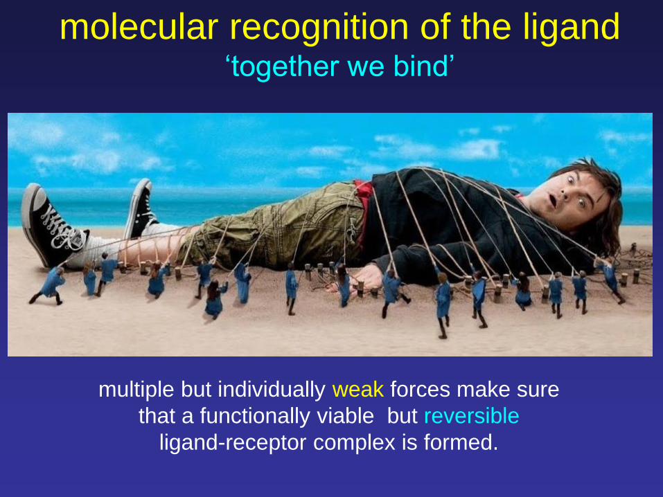
molecular recognition of the ligand‘together we bind’
multiple but individually weak forces make sure
that a functionally viable but reversible
ligand-receptor complex is formed.

An enzyme (COX-2) bound to a painkiller (Flurbiprofen)
recognition of the ligand at its
receptor site

Ligand binding assays address the first step of ligand-
receptor interaction – the physicochemical properties
and kinetics of ligand-receptor complex formation
Functional assays measure the actual biological
response (electrical or biochemical or physical)
evoked by the ligand via its receptor
techniques to measure ligand-receptor
interaction

% r
ece
pto
r b
ind
ing
[ligand]
100
50
Kd
quantification of ligand-receptor interaction
Kd affinity%
ma
x. re
sp
on
se
[ligand]
100
50
EC50
potencyEC50
quiz: why do the curves plateau?

quantification of ligand-receptor interaction
biological response receptor occupancy
receptor occupancy (%)
log [L]
[Rbound]
[RTotal] response
max. response (%)
A. J. Clark(1937)
Occupancy theory: EC50 = KD

quantification of ligand-receptor interactionmodification of the occupancy theory
Nickerson (1956); Nature
tissues tend to have ‘spare receptors’
EC50 values are typically <<Kd
occupancy 1% of histamine
receptors was adequate to
produce maximum effect

what allows EC50<<Kd: signal amplification

ligand conc. – biologic response relationship
log [ligand]
% m
ax. re
sp
on
se
[ligand]
100
50
EC50 EC50
% m
ax.
respon
se

quantification of ligand-receptor interactionmodification of the occupancy theory
= [bound receptors]
[total receptors] occupancy = magnitude of response
occupancy theory: EC50 = KD
But some ligands fail to produce maximum response even
at very high concentrations
magnitude of response = A . fractional occupancy
Ariens (1954) & Stephenson (1956)
efficacy or intrinsic activity

ligand-receptor interaction:
affinity vs efficacy
• affinity: ability to bind
• efficacy: ability to activate
• antagonists have same (or
higher) affinity but no efficacy
• partial agonists have
lower efficacy

certain contacts can be crucial for
activity
b-adrenergic receptor
(b-AR)
bAR blocker
what underlie the differences between
affinity and efficacy

various forms of receptor antagonism
receptor can be reversibly or irreversibly inhibited

various forms of receptor modulation

end of part II

Part III:
common receptor classes
terminating active receptors

major types of receptors

heterotrimeric G proteins act as
transducers for GPCRs
effector molecules
GPCR
stimuli
recognition &
reception
signal
transduction
enzymes
ion channelsexecution of
responses
cellular
effects

diversity of GPCRs

GPCR signalling shapes organism
response
‘fight or flight’ response
sympathetic nervous system
Adrenalin/noradrenalin mediated
via a-AR and b-AR
‘rest & digest’ response’
parasympathetic nervous system
acetylcholine mediated
via muscarinic and nicotinic AChR
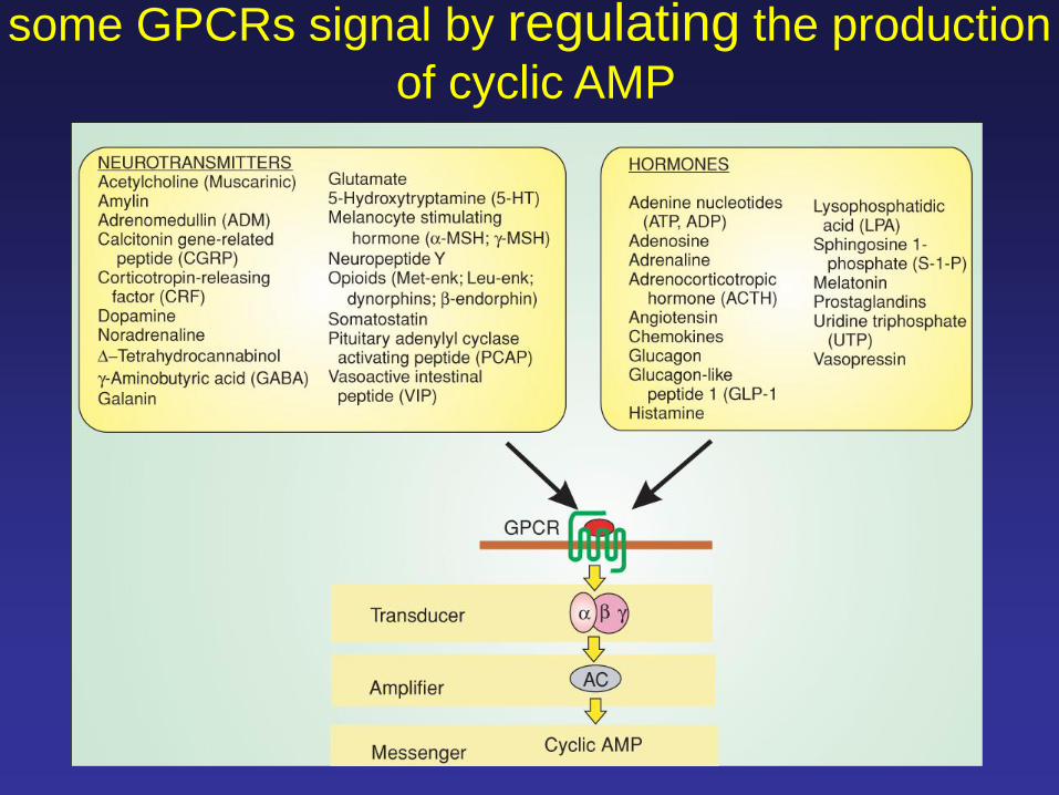
some GPCRs signal by regulating the production
of cyclic AMP

some GPCRs activate the inositol
phospholipid signalling pathway

termination of
GPCR signaling
(routes for
desensitization)

classical model of GPCR signalling
• signalling is mediated by Ga and bg only
• b-Arrestin is only involved for receptor
desensitization and internalisation
current view of GPCR signalling
• signalling is mediated by Ga and bg
as well as by b-Arrestin
• b-Arrestin also mediates receptor
desensitization and internalisation

original notionLigands that bind GPCR have unbiased
preference towards G protein and b-arrestin
mediated downstream signalling pathways
i.e. they signal equally through both
biased agonism or stimulus trafficking
current viewLigands that bind GPCR may selectively
promote either G protein or b-arrestin
mediated downstream signalling pathways

ion channels – receptors with holes
• integral membrane proteins
• contain ion conductive pores
• allow influx or efflux of ions across biological membranes
• can be activated
by changes in membrane potential
binding of particular ligands
changes in pH, temperature, cell volume
changes in intracellular Ca2+ concentration
mechanical deformation or stress

major types of ion channels
• voltage-gated
• ligand-gated
• gap junctions
• others e.g. mechanosensitive

the substrate protein can be an enzyme or an adaptor protein
receptors with enzyme activitygrowth factor receptors

intracellular receptors
ER/SR
nucleoplasm
lysosomeIP3R/RyR
IP3R
TPC
TRPML1
intracellular ionotropic
receptors (ion channels)
nuclear hormone receptors

A very good and up to date
resource on receptors & cell
signalling
http://www.cellsignallingbiology.org/csb/
If interested, may also consider visiting :
http://www.guidetopharmacology.org/

THANKS


