105
Introduction to the Skeletal System Structure, function, and classification of bones
| Date post: | 14-Dec-2015 |
| Category: |
Documents |
| Upload: | monica-stelling |
| View: | 225 times |
| Download: | 0 times |

Introduction to the Skeletal System
Structure, function, and classification of bones
Skeletal System
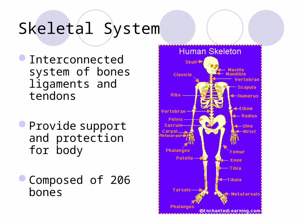
Interconnected system of bones ligaments and tendons
Provide support and protection for body
Composed of 206 bones
Functions of Skeletal System
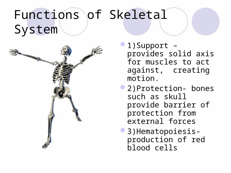
1)Support – provides solid axis for muscles to act against, creating motion.
2)Protection- bones such as skull provide barrier of protection from external forces
3)Hematopoiesis-production of red blood cells

Types of Bones
Bones are divisible into 5 class.LongShortFlat IrregularSesamoid

Long Bones
Found in the limbsEach bone is made of
a body (diaphysis) and two extremities (epiphyses)
Wall consists of dense tissue
Central canal called medullary canal is filled with marrow
Short Bones
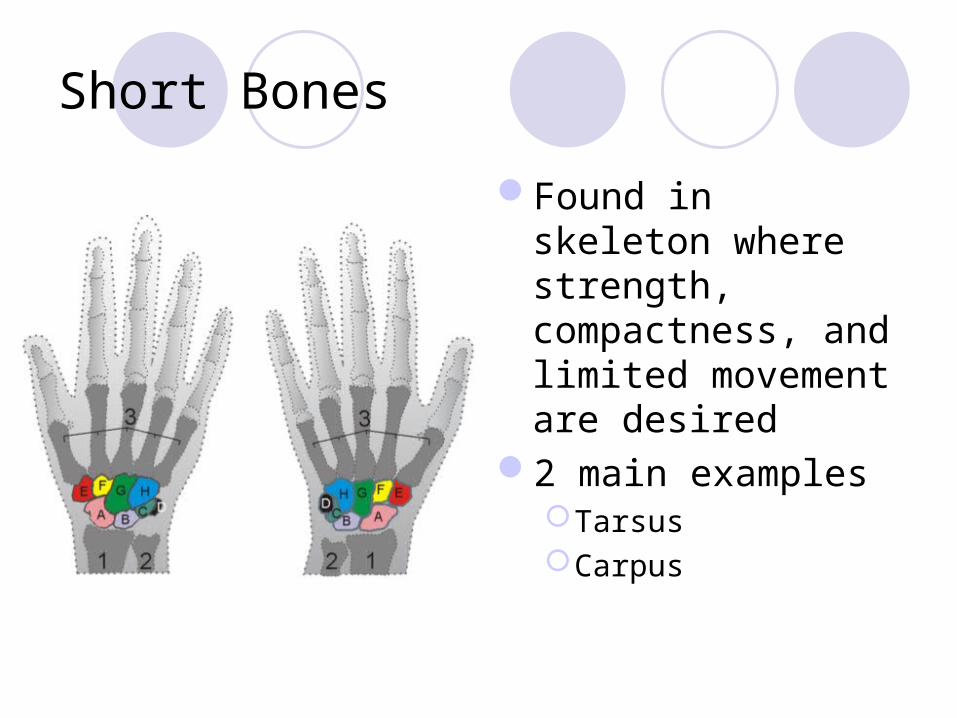
Found in skeleton where strength, compactness, and limited movement are desired
2 main examplesTarsusCarpus

Flat Bones
Used in spots where protection or muscular attachment is desired
Main locations are skull and scapula
Irregular Bones
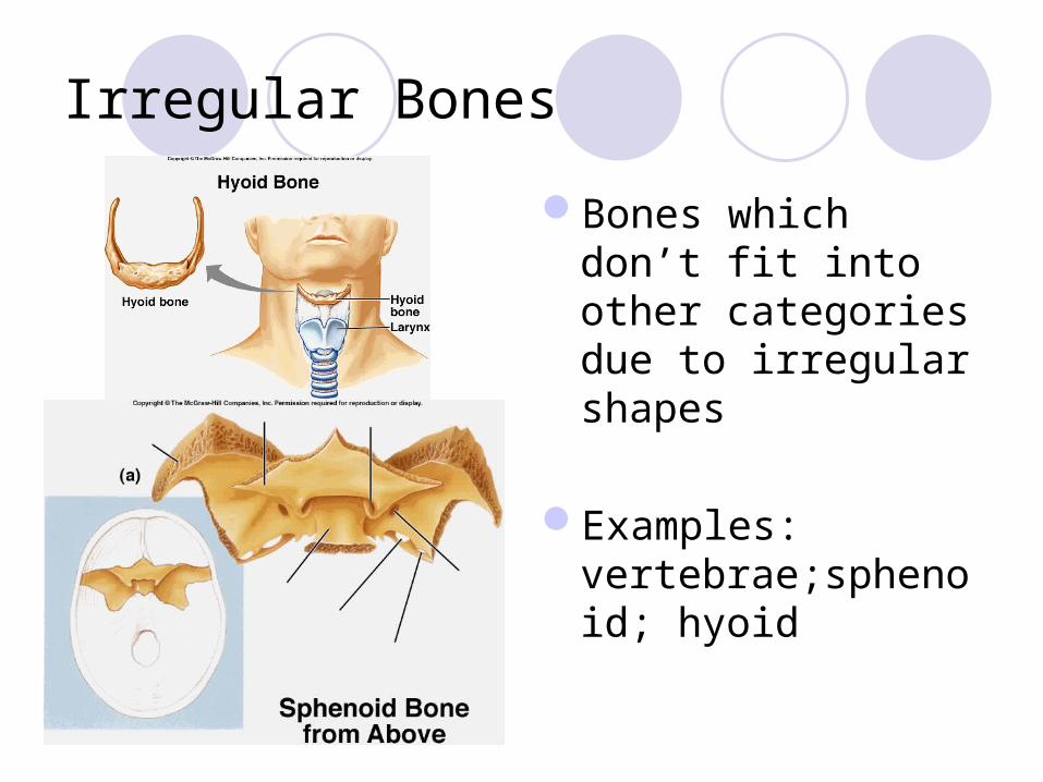
Bones which don’t fit into other categories due to irregular shapes
Examples: vertebrae;sphenoid; hyoid

Sesamoid (Round)BonesUsually small and
round.Embedded within
tendons adjacent to joints.
Example: patella (knee cap)
Bone Formation and Fractures

Fetal Skeleton
Begins as mainly cartilage
Calcifies in utero
At birth, fontanels remain
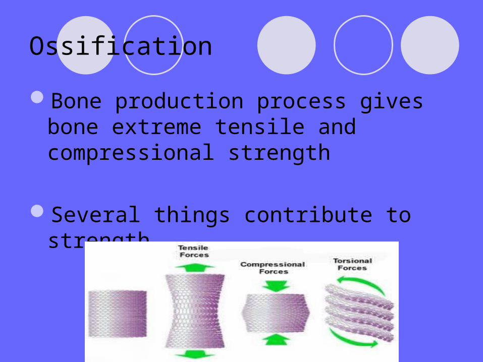
Ossification
Bone production process gives bone extreme tensile and compressional strength
Several things contribute to strength
Factors which contribute to bone growth
NutritionExposure to sunlightHormonal SecretionPhysical Exercise

Nutrition
Mainly calcium consumption
Increased blood calcium triggers release of calcitonin
Causes uptake of calcium by osteoblasts (bone builders)
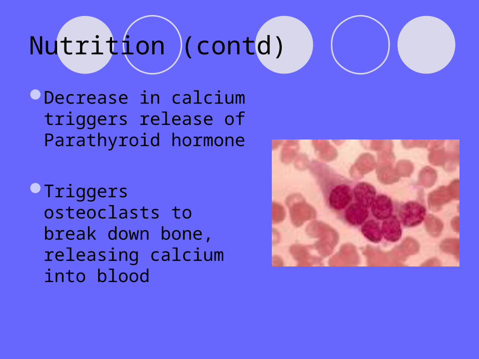
Nutrition (contd)
Decrease in calcium triggers release of Parathyroid hormone
Triggers osteoclasts to break down bone, releasing calcium into blood

Exposure to Sunlight
UV light on the skin causes Vitamin D production
Promotes proper absorption of calcium in the SI
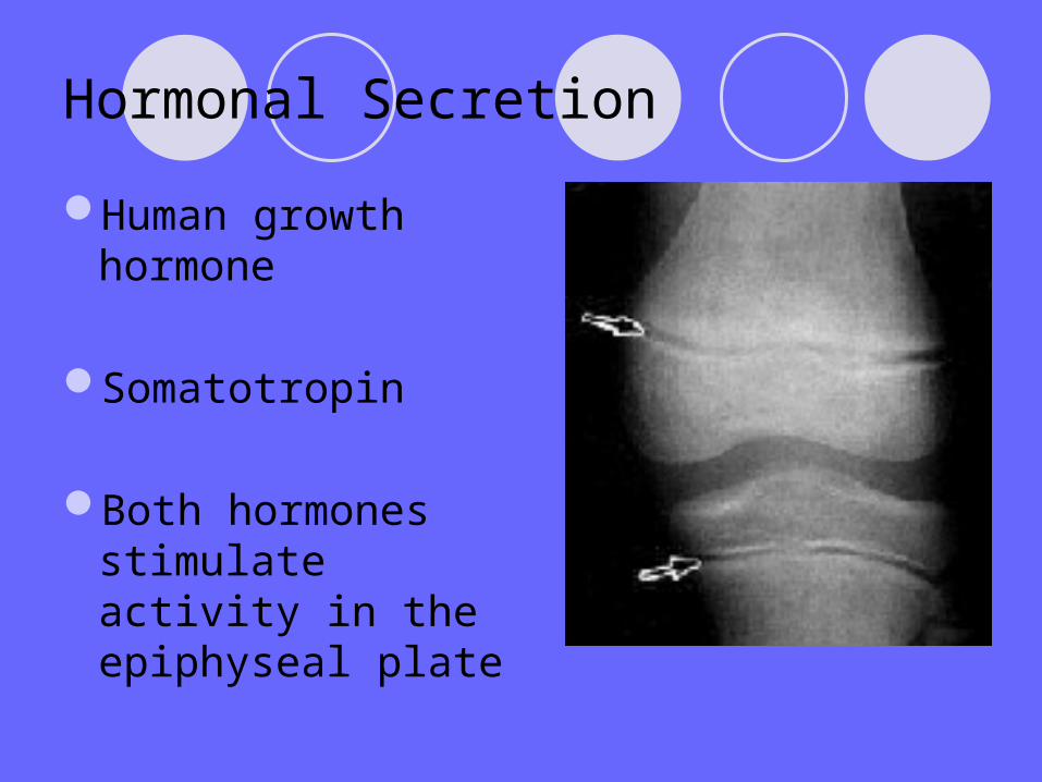
Hormonal Secretion
Human growth hormone
Somatotropin
Both hormones stimulate activity in the epiphyseal plate

Physical Activity
Increase in physical exertion on bone tissue actually increases bone density and strength
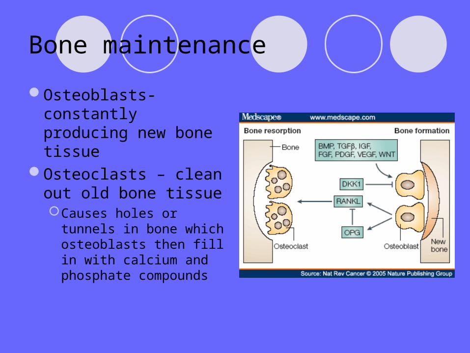
Bone maintenance
Osteoblasts-constantly producing new bone tissue
Osteoclasts – clean out old bone tissueCauses holes or
tunnels in bone which osteoblasts then fill in with calcium and phosphate compounds

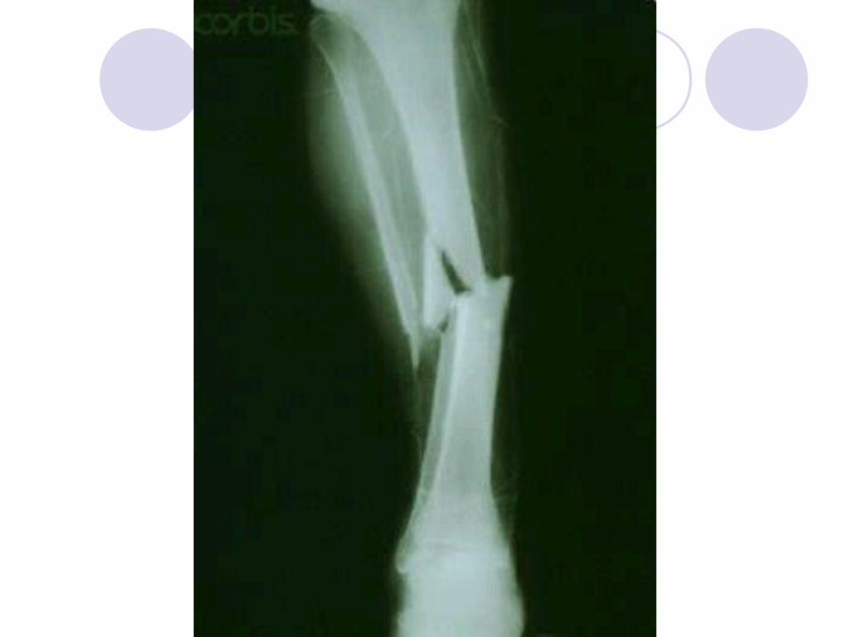
Fractures
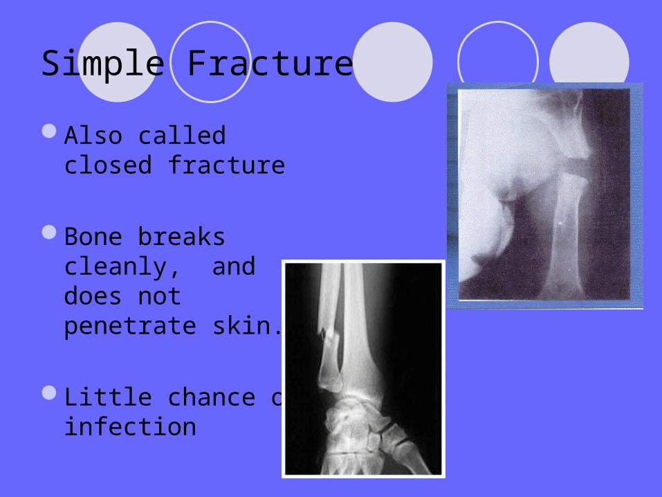
Simple Fracture
Also called closed fracture
Bone breaks cleanly, and does not penetrate skin.
Little chance of infection
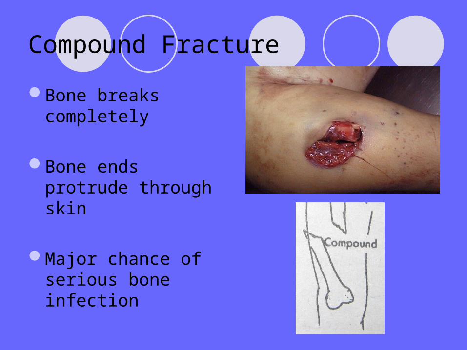
Compound Fracture
Bone breaks completely
Bone ends protrude through skin
Major chance of serious bone infection

Comminuted Fracture
Bone breaks into many fragments
Common in elderly

Compression Fracture
Bone is crushed
Common in porous bones
Especially common in vertebrae of osteoporosis patients
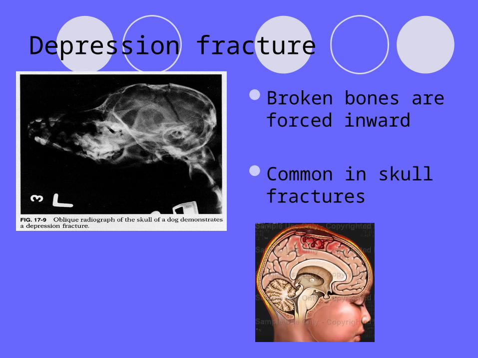
Depression fracture
Broken bones are forced inward
Common in skull fractures

Impacted Fracture
Broken bone ends are forced into each other
Common in falls (ie. From ladder) where person attempts to break their fall

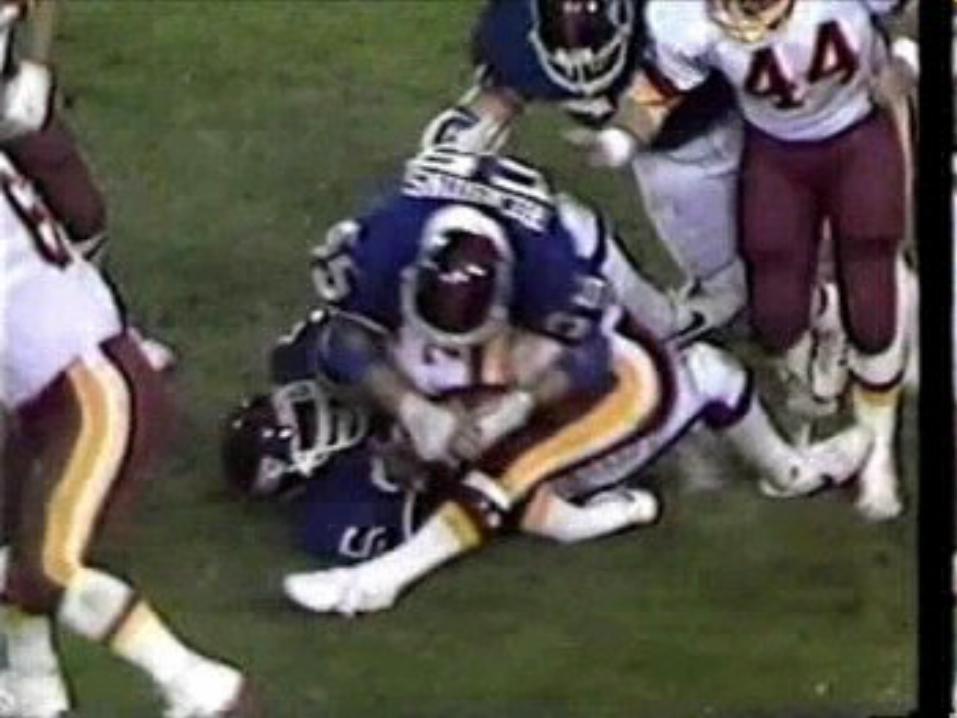
Spiral Fracture
Occurs from excessive twisting force on bone
Common in sports injuries
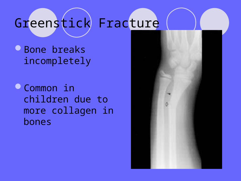
Greenstick Fracture
Bone breaks incompletely
Common in children due to more collagen in bones
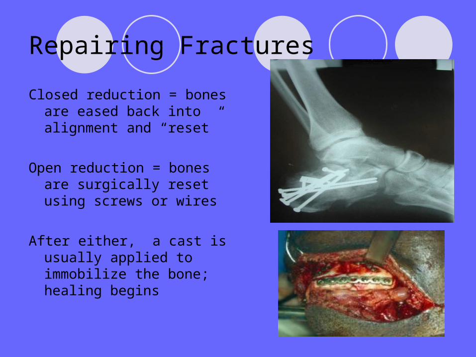
Repairing Fractures
Closed reduction = bones are eased back into alignment and “reset”
Open reduction = bones are surgically reset using screws or wires
After either, a cast is usually applied to immobilize the bone; healing begins

Internal Bone Repair
1)Hematoma forms from ruptured blood vessels.
2)After new capillaries form, fibrocartillage callus “splints” broken bone using cartilage and bony matrix.
3)Osteoblasts migrate to area, forming bone “patch” over break. Fibrocartilage is replaced by bony callus.
The Axial SkeletonThe Axial Skeleton

Divisions of the Skeletal SystemDivisions of the Skeletal System
Skeletal system is Skeletal system is divided into two main divided into two main divisiondivisionAxial – central skeleton Axial – central skeleton
that protects and that protects and supports vital organssupports vital organs
Appendicular – Appendicular – skeleton of the skeleton of the extremities extremities
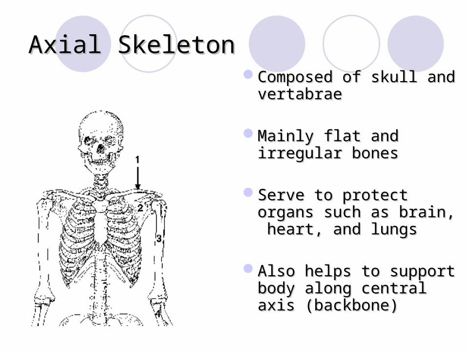
Axial SkeletonAxial SkeletonComposed of skull and Composed of skull and
vertabraevertabrae
Mainly flat and irregular Mainly flat and irregular bonesbones
Serve to protect organs Serve to protect organs such as brain, heart, and such as brain, heart, and lungslungs
Also helps to support Also helps to support body along central axis body along central axis (backbone)(backbone)
Parts of the axial skeletonParts of the axial skeleton
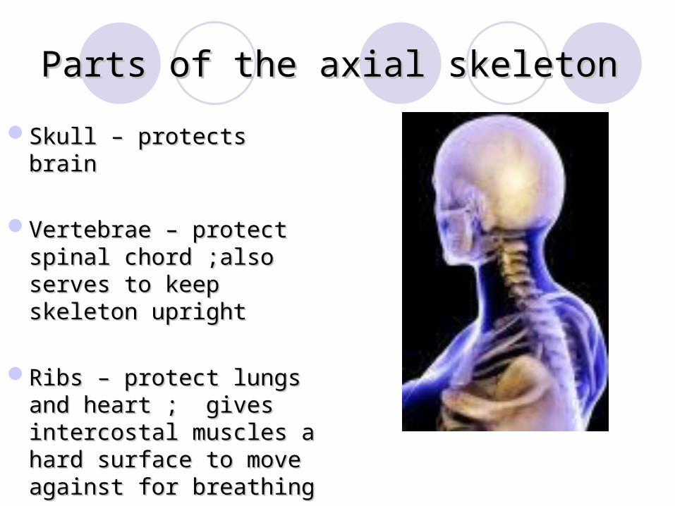
Skull – protects brainSkull – protects brain
Vertebrae – protect Vertebrae – protect spinal chord ;also serves spinal chord ;also serves to keep skeleton uprightto keep skeleton upright
Ribs – protect lungs and Ribs – protect lungs and heart ; gives intercostal heart ; gives intercostal muscles a hard surface muscles a hard surface to move against for to move against for breathingbreathing
Divisions of the skullDivisions of the skull
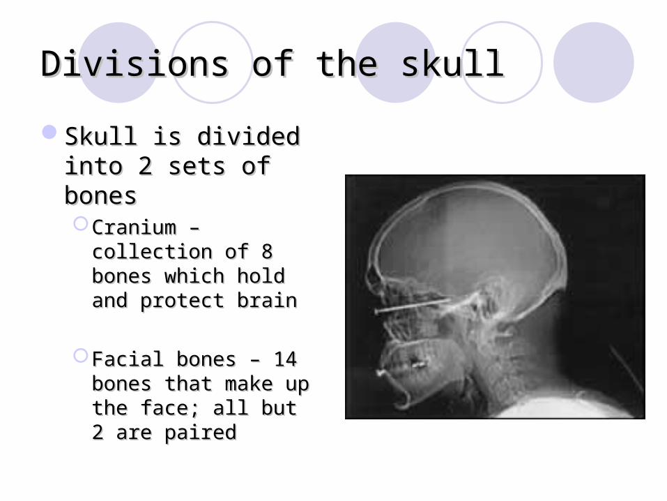
Skull is divided into 2 Skull is divided into 2 sets of bonessets of bonesCranium – collection of Cranium – collection of
8 bones which hold and 8 bones which hold and protect brainprotect brain
Facial bones – 14 Facial bones – 14 bones that make up the bones that make up the face; all but 2 are face; all but 2 are pairedpaired
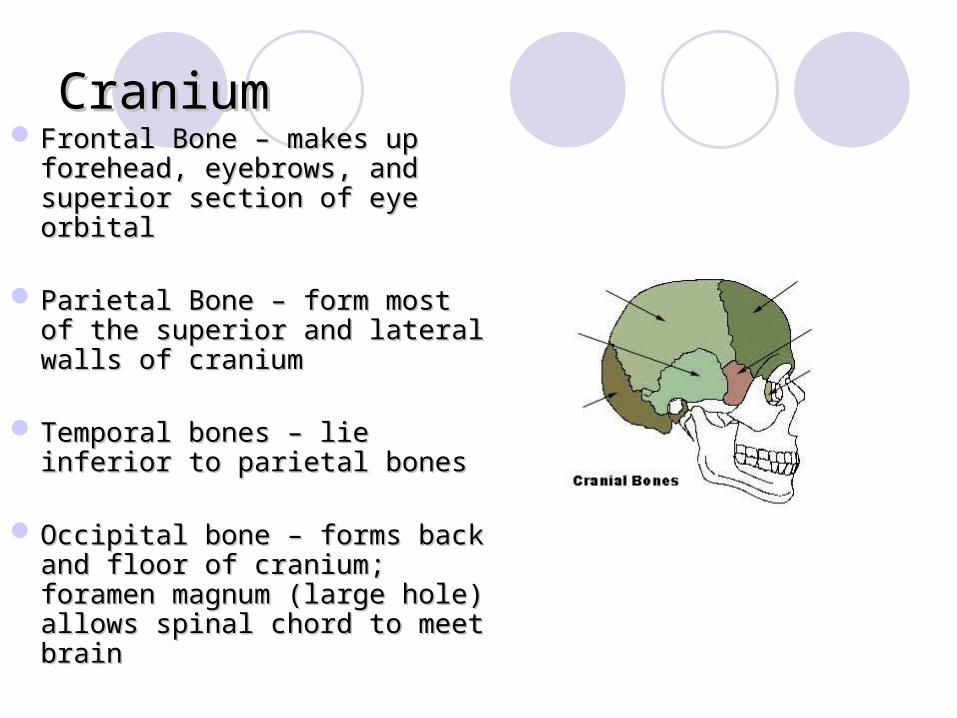
CraniumCranium Frontal Bone – makes up Frontal Bone – makes up
forehead, eyebrows, and forehead, eyebrows, and superior section of eye orbitalsuperior section of eye orbital
Parietal Bone – form most of Parietal Bone – form most of the superior and lateral walls the superior and lateral walls of craniumof cranium
Temporal bones – lie inferior Temporal bones – lie inferior to parietal bonesto parietal bones
Occipital bone – forms back Occipital bone – forms back and floor of cranium; foramen and floor of cranium; foramen magnum (large hole) allows magnum (large hole) allows spinal chord to meet brainspinal chord to meet brain

Facial BonesFacial Bones
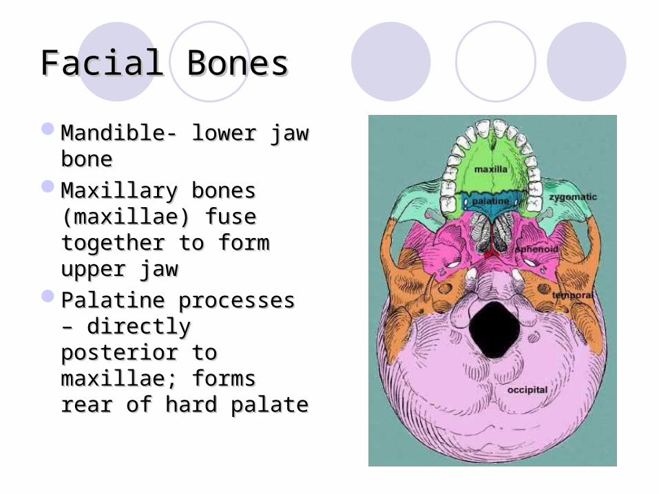
Mandible- lower jaw Mandible- lower jaw bonebone
Maxillary bones Maxillary bones (maxillae) fuse (maxillae) fuse together to form together to form upper jawupper jaw
Palatine processes – Palatine processes – directly posterior to directly posterior to maxillae; forms rear maxillae; forms rear of hard palateof hard palate
Facial Bones Contd.Facial Bones Contd.
Zygomatic bones – cheekbonesZygomatic bones – cheekbones
Lacrimal bones – inferior section of orbital Lacrimal bones – inferior section of orbital bones; provides passageway for tearsbones; provides passageway for tears
Ethmoid bone- forms roof of nasal cavityEthmoid bone- forms roof of nasal cavity

More Facial BonesMore Facial Bones
Nasal bones- form Nasal bones- form bridge of nosebridge of nose
Vomer – divides Vomer – divides nasal cavity in halfnasal cavity in half
Inferior conchae- thin Inferior conchae- thin curved bones which curved bones which project from interior of project from interior of nasal cavitynasal cavity
Axial SkeletonAxial Skeleton
Intervertebral DiscsIntervertebral Discs
Spinal curvaturesSpinal curvatures
Bony Thorax Bony Thorax
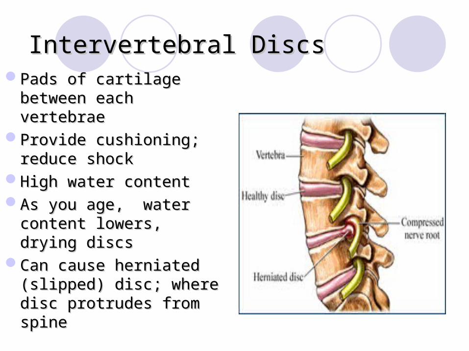
Intervertebral DiscsIntervertebral DiscsPads of cartilage Pads of cartilage
between each vertebraebetween each vertebraeProvide cushioning; Provide cushioning;
reduce shockreduce shockHigh water contentHigh water contentAs you age, water As you age, water
content lowers, drying content lowers, drying discsdiscs
Can cause herniated Can cause herniated (slipped) disc; where disc (slipped) disc; where disc protrudes from spineprotrudes from spine

Bony ThoraxBony Thorax
Made of bones which Made of bones which connect and protect connect and protect heart and lungsheart and lungs
Ribs, Costal Ribs, Costal Cartilage, and Cartilage, and SternumSternum
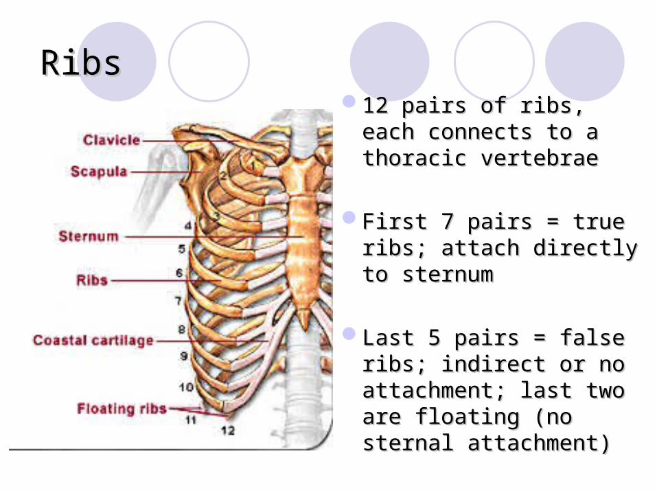
RibsRibs12 pairs of ribs, each 12 pairs of ribs, each
connects to a thoracic connects to a thoracic vertebraevertebrae
First 7 pairs = true ribs; First 7 pairs = true ribs; attach directly to sternumattach directly to sternum
Last 5 pairs = false ribs; Last 5 pairs = false ribs; indirect or no attachment; indirect or no attachment; last two are floating (no last two are floating (no sternal attachment)sternal attachment)

SternumSternum
Fusion of three bonesFusion of three bones1) Manubrium (top)1) Manubrium (top)2) Body (middle)2) Body (middle)3) Xiphoid Process 3) Xiphoid Process
(bottom)(bottom)Location for rib Location for rib
attachmentattachmentSurrounded by costal Surrounded by costal
cartilagecartilage

Sternal PunctureSternal Puncture
Process by which Process by which marrow is removed marrow is removed from sternumfrom sternum
Good location Good location because of proximity because of proximity to body surfaceto body surface

The Spinal ColumnThe Spinal Column
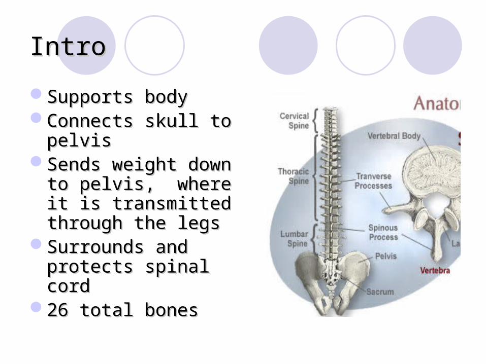
Intro Intro
Supports bodySupports bodyConnects skull to Connects skull to
pelvispelvisSends weight down to Sends weight down to
pelvis, where it is pelvis, where it is transmitted through transmitted through the legsthe legs
Surrounds and Surrounds and protects spinal cordprotects spinal cord
26 total bones26 total bones
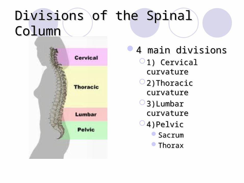
Divisions of the Spinal ColumnDivisions of the Spinal Column
4 main divisions4 main divisions1) Cervical curvature1) Cervical curvature2)Thoracic curvature2)Thoracic curvature3)Lumbar curvature3)Lumbar curvature4)Pelvic4)Pelvic
SacrumSacrumThoraxThorax
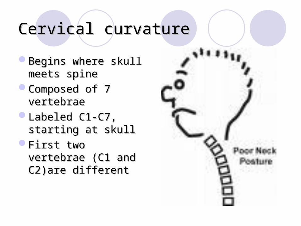
Cervical curvatureCervical curvature
Begins where skull Begins where skull meets spinemeets spine
Composed of 7 Composed of 7 vertebraevertebrae
Labeled C1-C7, Labeled C1-C7, starting at skullstarting at skull
First two vertebrae First two vertebrae (C1 and C2)are (C1 and C2)are differentdifferent
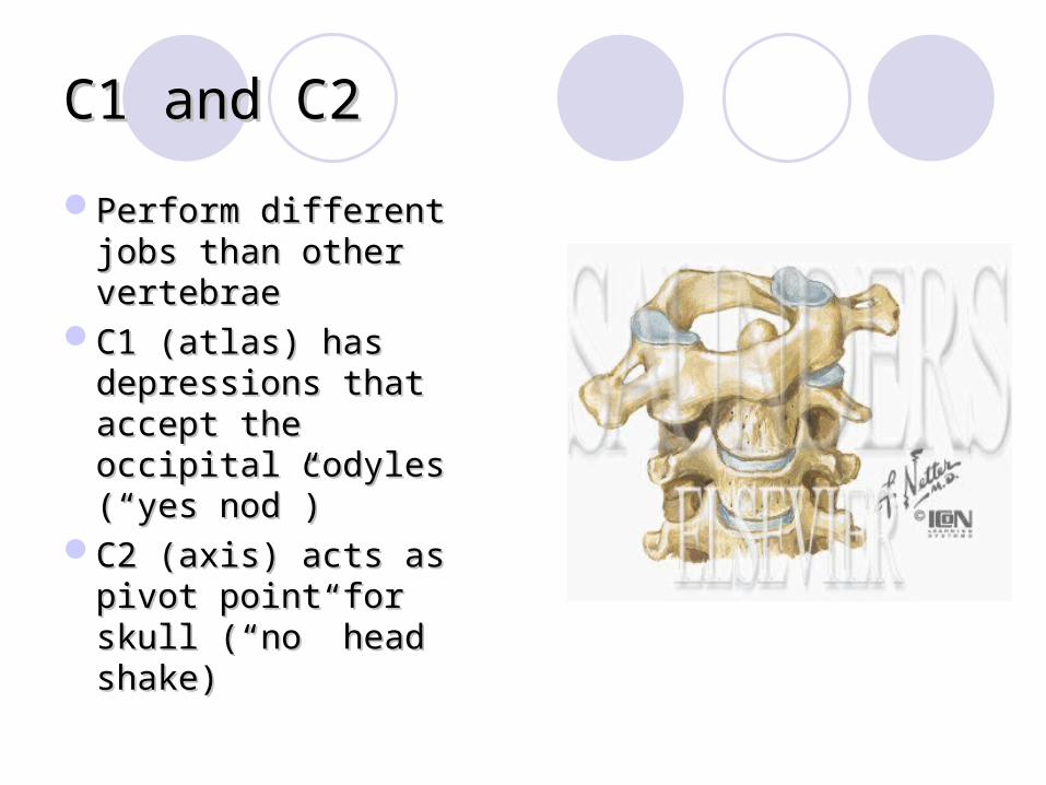
C1 and C2C1 and C2
Perform different jobs Perform different jobs than other vertebraethan other vertebrae
C1 (atlas) has C1 (atlas) has depressions that depressions that accept the occipital accept the occipital codyles (codyles (““yes nodyes nod””))
C2 (axis) acts as C2 (axis) acts as pivot point for skull pivot point for skull ((““nono”” head shake) head shake)
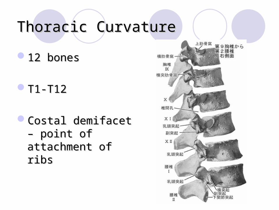
Thoracic CurvatureThoracic Curvature
12 bones12 bones
T1-T12T1-T12
Costal demifacet – Costal demifacet – point of attachment of point of attachment of ribsribs
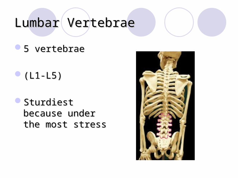
Lumbar VertebraeLumbar Vertebrae
5 vertebrae5 vertebrae
(L1-L5)(L1-L5)
Sturdiest because Sturdiest because under the most stressunder the most stress

SacrumSacrum
1 bone composed of 1 bone composed of 5 fused vertebrae5 fused vertebrae
““wing-likewing-like”” alae alae connect laterally with connect laterally with hip bones (forms hip bones (forms sacroiliac joints)sacroiliac joints)
Makes up posterior Makes up posterior wall of pelviswall of pelvis
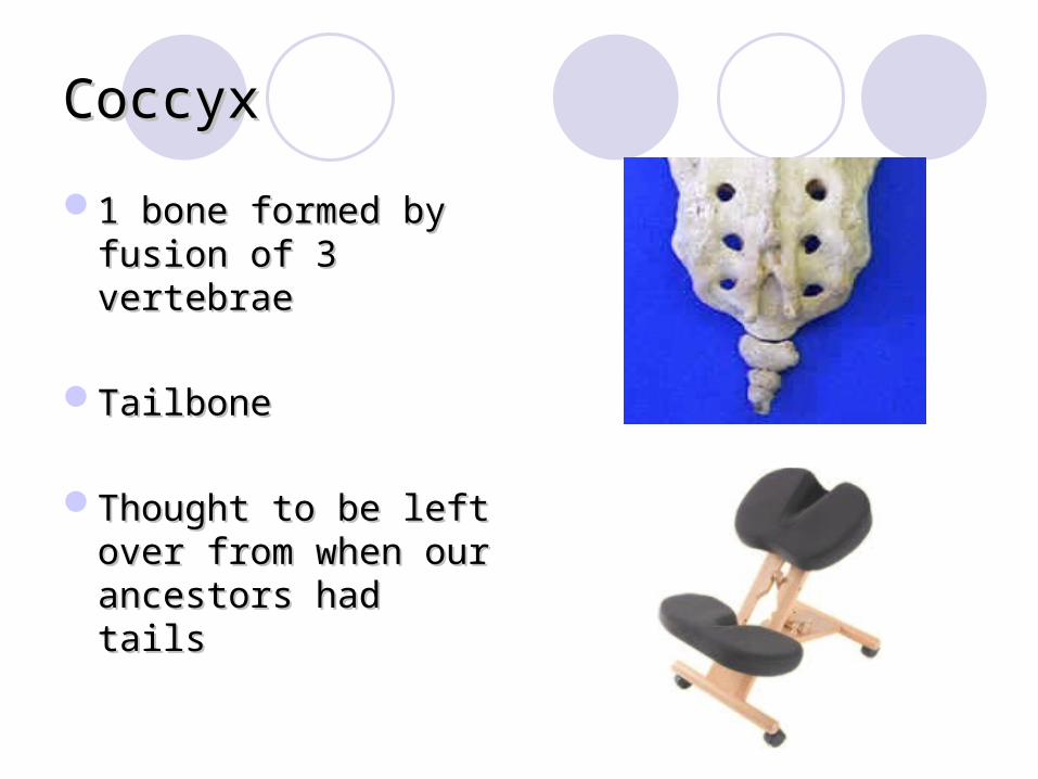
CoccyxCoccyx
1 bone formed by 1 bone formed by fusion of 3 vertebraefusion of 3 vertebrae
TailboneTailbone
Thought to be left Thought to be left over from when our over from when our ancestors had tailsancestors had tails
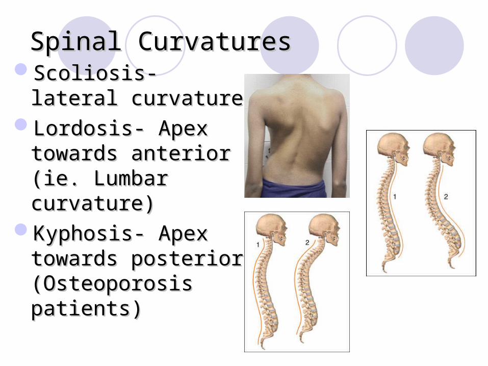
Spinal CurvaturesSpinal CurvaturesScoliosis- lateral Scoliosis- lateral
curvature curvature Lordosis- Apex Lordosis- Apex
towards anterior (ie. towards anterior (ie. Lumbar curvature)Lumbar curvature)
Kyphosis- Apex Kyphosis- Apex towards posterior towards posterior (Osteoporosis (Osteoporosis patients)patients)

Appendicular Skeleton
Pelvic Girdle

PelvisJuncture point for
axial skeleton and lower body
Holds internal organsDistributes weight
down legs3 fused bonesObturator foramen-
large hole through which nerves and muscles pass
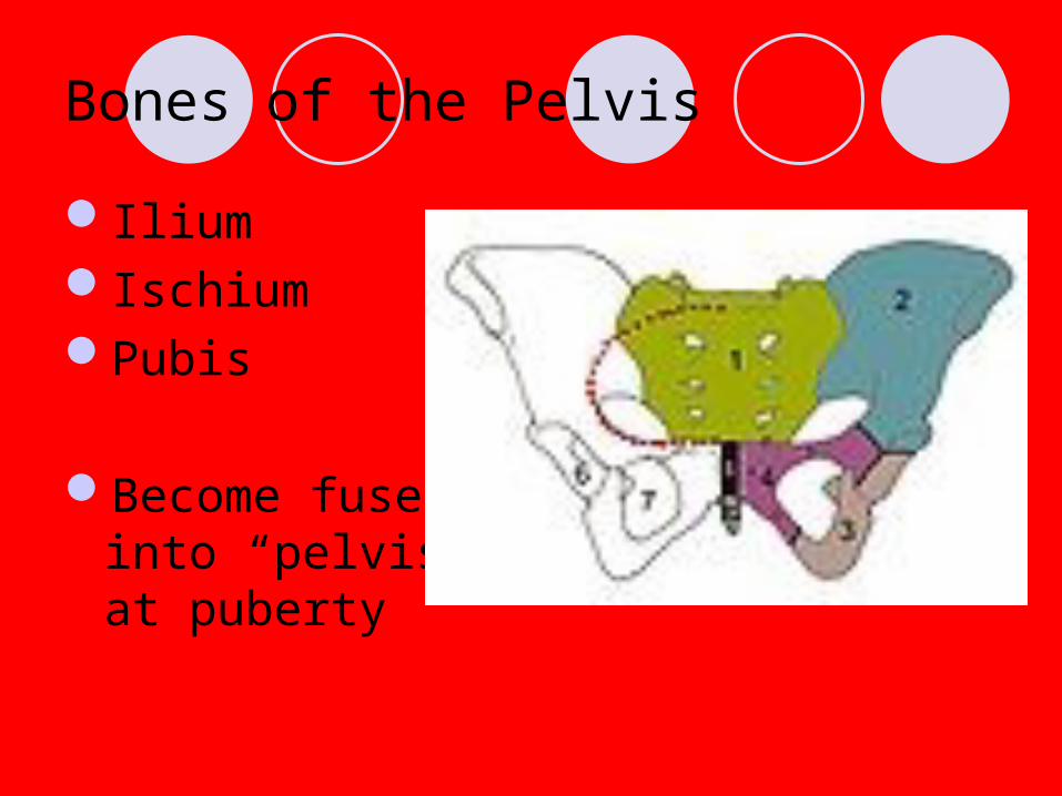
Bones of the Pelvis
IliumIschiumPubis
Become fused into “pelvis” at puberty
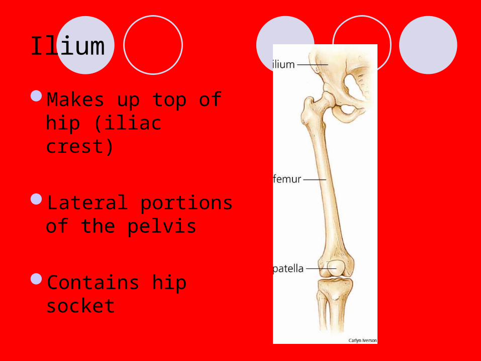
Ilium
Makes up top of hip (iliac crest)
Lateral portions of the pelvis
Contains hip socket

Features of the Ilium
Iliac crest – rounded projection on superior surface; makes up “hip”
Acetabulum- joint between femure and pelvis
Width from crest to crest = false pelvis
Width of actual inlet = true pelvis

Ischium
Inferior portion of pelvis
Ischial Tuberosity – point of muscle attachment; “sit bones”
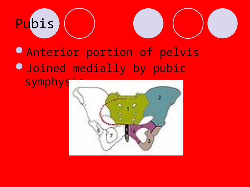
Pubis
Anterior portion of pelvisJoined medially by pubic symphysis
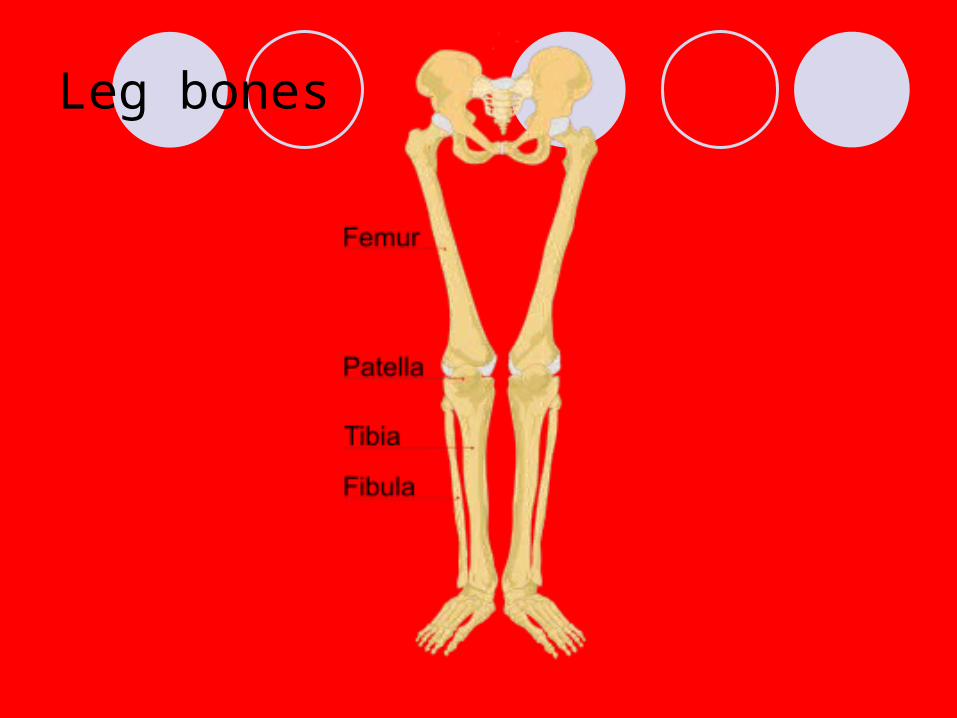
Leg bones
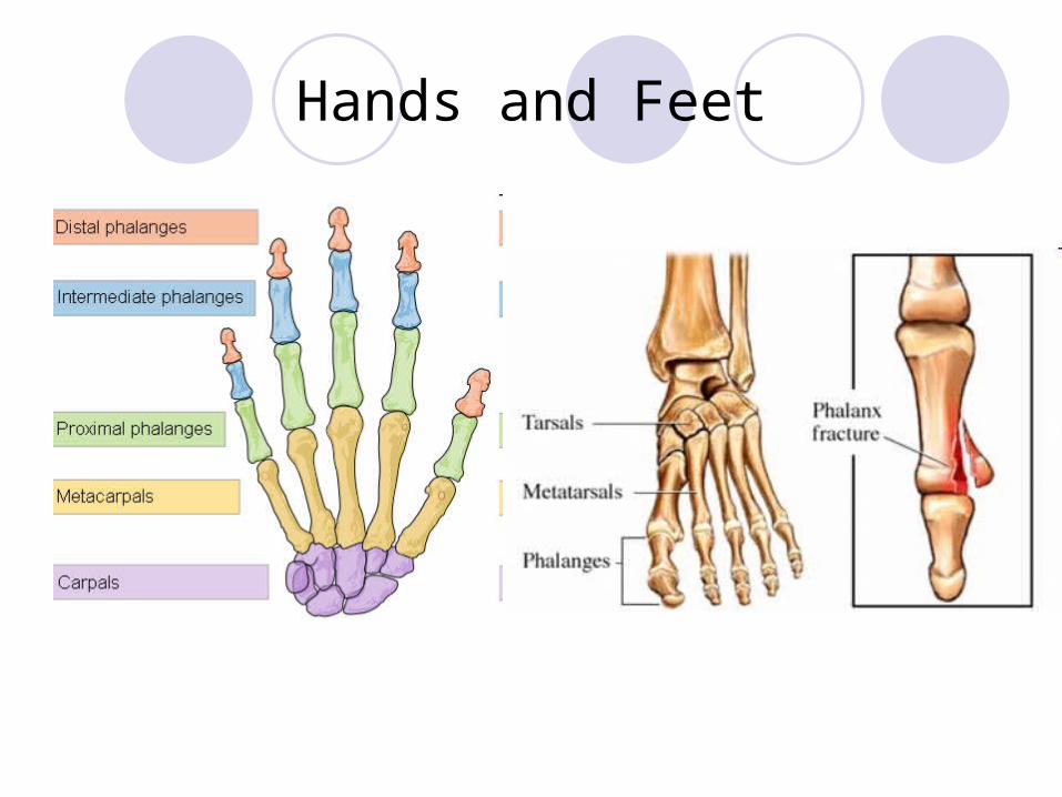
Hands and Feet
Appendicular SkeletonAppendicular SkeletonSuperior Extremities Superior Extremities
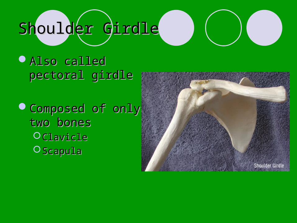
Shoulder GirdleShoulder Girdle
Also called pectoral Also called pectoral girdlegirdle
Composed of only Composed of only two bonestwo bonesClavicleClavicleScapulaScapula
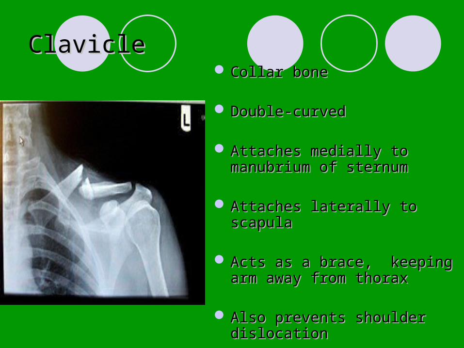
ClavicleClavicle Collar boneCollar bone
Double-curvedDouble-curved
Attaches medially to manubrium Attaches medially to manubrium of sternumof sternum
Attaches laterally to scapulaAttaches laterally to scapula
Acts as a brace, keeping arm Acts as a brace, keeping arm away from thoraxaway from thorax
Also prevents shoulder Also prevents shoulder dislocationdislocation

ScapulaScapulaShoulder BladeShoulder Blade
Main function is Main function is attachment of shoulderattachment of shoulder
Major point of muscle Major point of muscle attachment for movement attachment for movement of armsof arms
Weakly attached to Weakly attached to thorax, so moves easilythorax, so moves easily
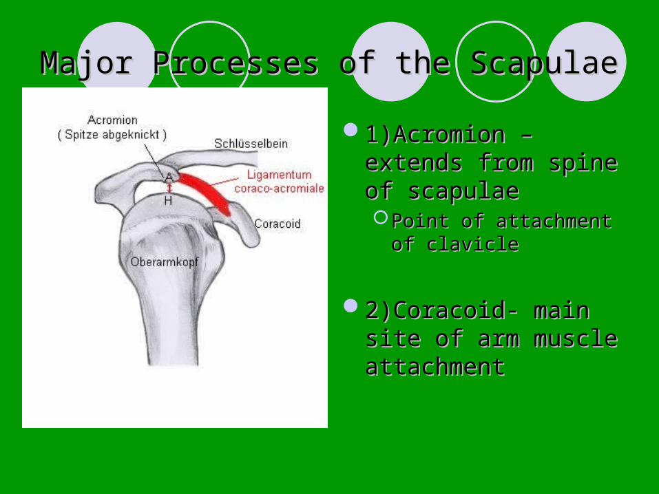
Major Processes of the ScapulaeMajor Processes of the Scapulae
1)Acromion – extends 1)Acromion – extends from spine of from spine of scapulaescapulaePoint of attachment of Point of attachment of
clavicleclavicle
2)Coracoid- main site 2)Coracoid- main site of arm muscle of arm muscle attachmentattachment
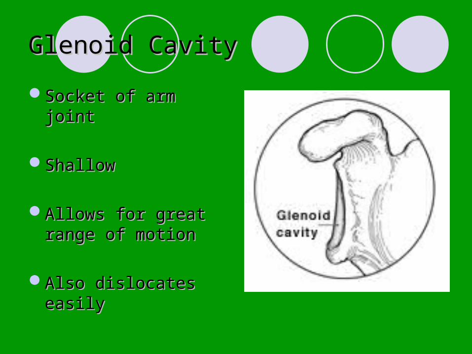
Glenoid CavityGlenoid Cavity
Socket of arm jointSocket of arm joint
ShallowShallow
Allows for great range Allows for great range of motionof motion
Also dislocates easilyAlso dislocates easily

Movement in the Shoulder GirdleMovement in the Shoulder Girdle
Very free moving becauseVery free moving because1)Only attaches at one point to axial 1)Only attaches at one point to axial
skeletonskeleton2)Loose attachment of scapula allows it to 2)Loose attachment of scapula allows it to
slideslide3)Glenoid cavity very shallow3)Glenoid cavity very shallow
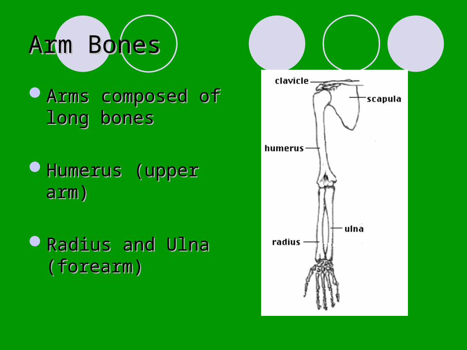
Arm BonesArm Bones
Arms composed of Arms composed of long boneslong bones
Humerus (upper arm)Humerus (upper arm)
Radius and Ulna Radius and Ulna (forearm)(forearm)
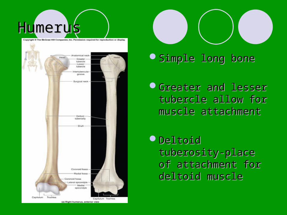
HumerusHumerus
Simple long boneSimple long bone
Greater and lesser Greater and lesser tubercle allow for tubercle allow for muscle attachmentmuscle attachment
Deltoid tuberosity-Deltoid tuberosity-place of attachment place of attachment for deltoid musclefor deltoid muscle
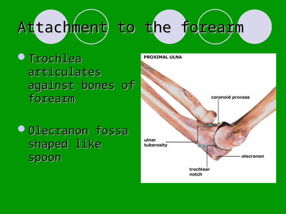
Attachment to the forearmAttachment to the forearm
Trochlea articulates Trochlea articulates against bones of against bones of forearmforearm
Olecranon fossa Olecranon fossa shaped like spoonshaped like spoon
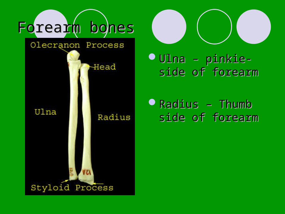
Forearm bonesForearm bones
Ulna – pinkie-side of Ulna – pinkie-side of forearmforearm
Radius – Thumb side Radius – Thumb side of forearmof forearm
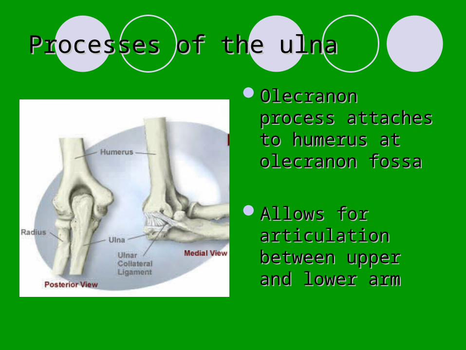
Processes of the ulnaProcesses of the ulna
Olecranon process Olecranon process attaches to humerus attaches to humerus at olecranon fossaat olecranon fossa
Allows for articulation Allows for articulation between upper and between upper and lower armlower arm

Hands and Feet
Joints

Intro
Any point where bones meet
Also called articulations
Every bone (except hyoid) articulates with at least 1 other bone

Classifications of Joints
Can be classified by mobility, or by the type of tissue which connects the bones
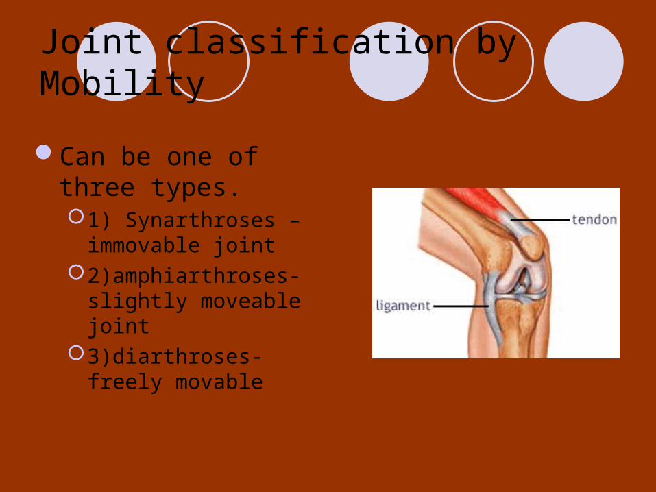
Joint classification by Mobility
Can be one of three types.1) Synarthroses –
immovable joint2)amphiarthroses- slightly
moveable joint3)diarthroses- freely
movable

Classification by connective tissue type
Joints are connected by either fibrous, cartilage, or synovial connective tissue.
Fibrous is usually synarthroses,
Synovial – diarthroses

Fibrous Joints
Fibrous tissueExample= sutures of
the skullTight fibrous tissue
allows for essentially no movement
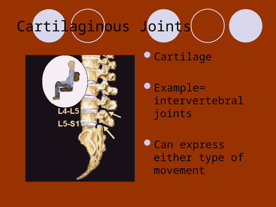
Cartilaginous Joints
Cartilage
Example= intervertebral joints
Can express either type of movement
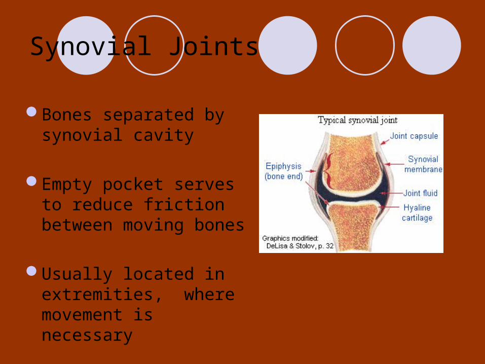
Synovial Joints
Bones separated by synovial cavity
Empty pocket serves to reduce friction between moving bones
Usually located in extremities, where movement is necessary
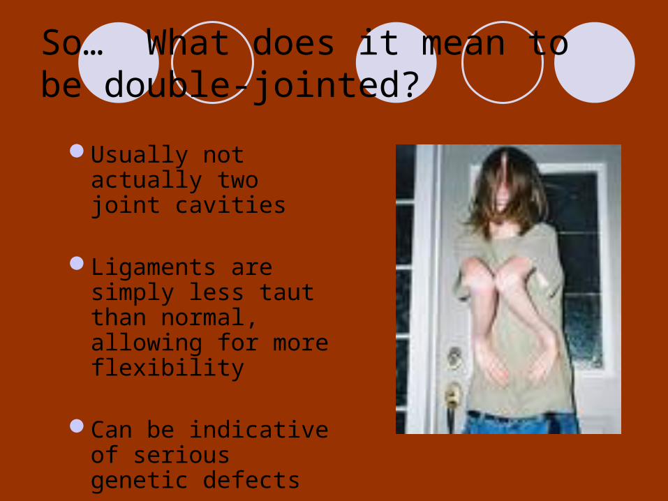
So… What does it mean to be double-jointed?
Usually not actually two joint cavities
Ligaments are simply less taut than normal, allowing for more flexibility
Can be indicative of serious genetic defects
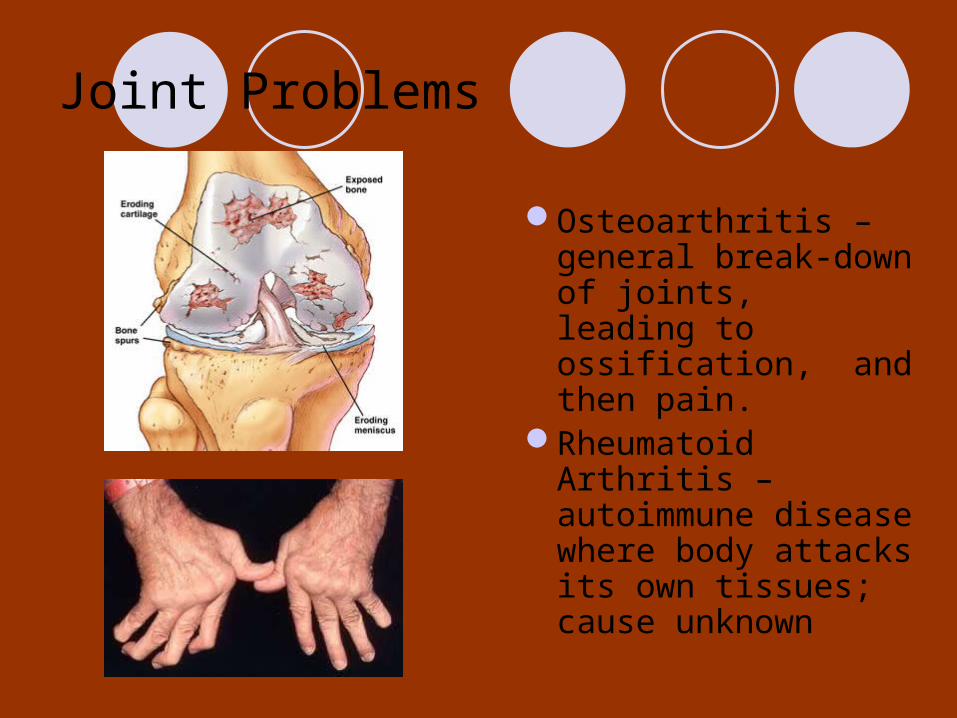
Joint Problems
Osteoarthritis – general break-down of joints, leading to ossification, and then pain.
Rheumatoid Arthritis – autoimmune disease where body attacks its own tissues; cause unknown
Features of the Skull
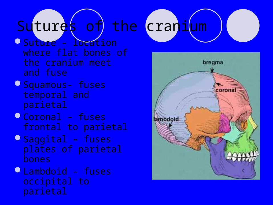
Sutures of the craniumSuture – location
where flat bones of the cranium meet and fuse
Squamous- fuses temporal and parietal
Coronal – fuses frontal to parietal
Saggital – fuses plates of parietal bones
Lambdoid – fuses occipital to parietal
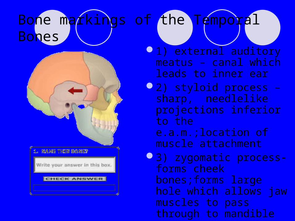
Bone markings of the Temporal Bones
1) external auditory meatus – canal which leads to inner ear
2) styloid process – sharp, needlelike projections inferior to the e.a.m.;location of muscle attachment
3) zygomatic process- forms cheek bones;forms large hole which allows jaw muscles to pass through to mandible

Temporal bone markings (contd.)4) mastoid process –
posterior and inferior to e.a.m.;location of muscle attachment for muscles of the neck
5) jugular foramen- at junction of occipital and temporal bones; allows jugular vein to pass through from brain
6) carotid canal – anterior to j.f. Allows carotid artery to pass to brain

Occipital Condyles
Lie lateral to the foramen magnum
Rest upon the spinal column
Provides point of attachment for skull to spinal column
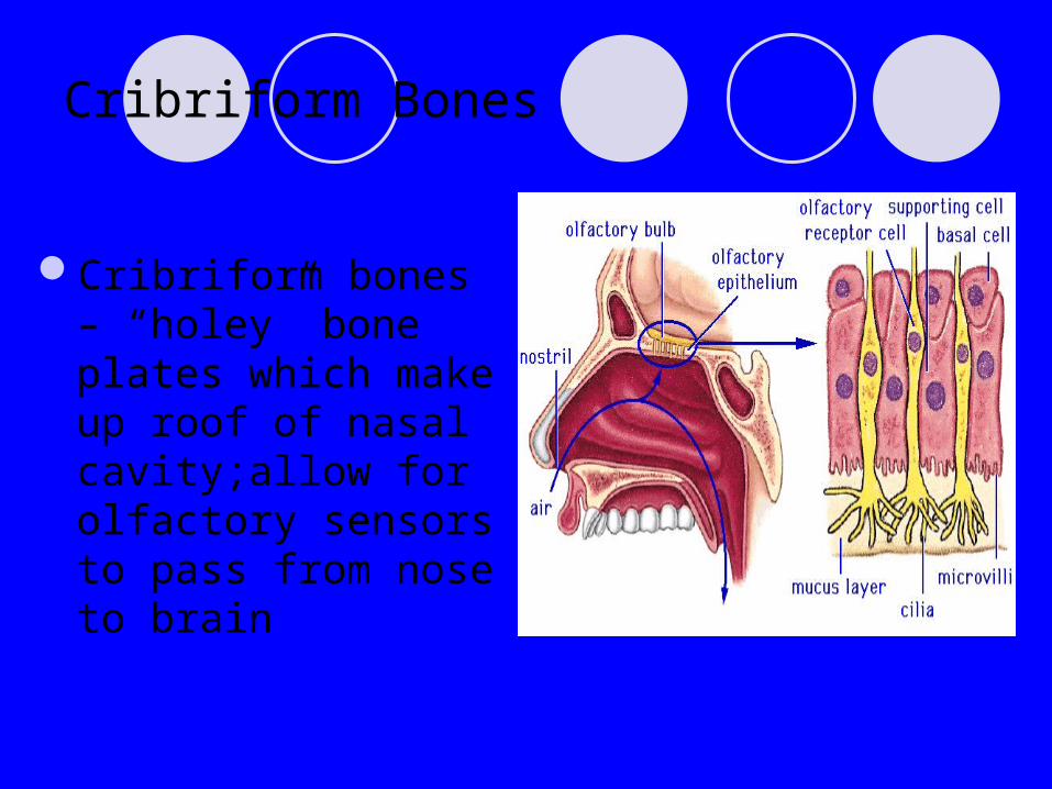
Cribriform Bones
Cribriform bones – “holey” bone plates which make up roof of nasal cavity;allow for olfactory sensors to pass from nose to brain

Sinuses
Empty pocket inside bones which are lines with mucous membranes
Paranasal sinus- surrounds nasal cavity
Lighten skull, and thought to amplify sounds when speaking
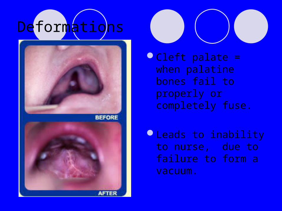
Deformations
Cleft palate = when palatine bones fail to properly or completely fuse.
Leads to inability to nurse, due to failure to form a vacuum.
Male vs. Female Skeleton
In general
Male skeleton is larger, with thicker bones
Female bones maintain many characteristics of prepubescent skeleton
Male features change at puberty (usually at points of muscular attachment)

Skull
Male mastoid process more pronounced
Superior portion of female orbital (brow ridge) less pronounced
Female mandible is pointed, while male is squared
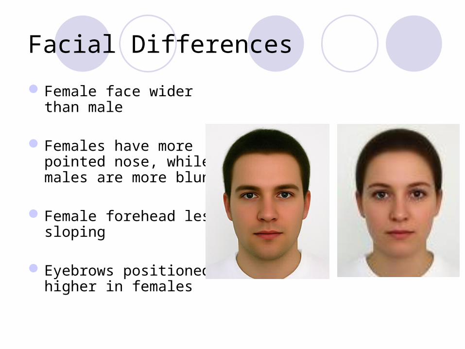
Facial Differences
Female face wider than male
Females have more pointed nose, while males are more blunt
Female forehead less sloping
Eyebrows positioned higher in females
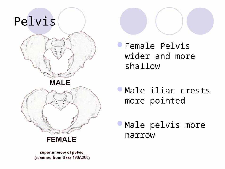
Pelvis
Female Pelvis wider and more shallow
Male iliac crests more pointed
Male pelvis more narrow
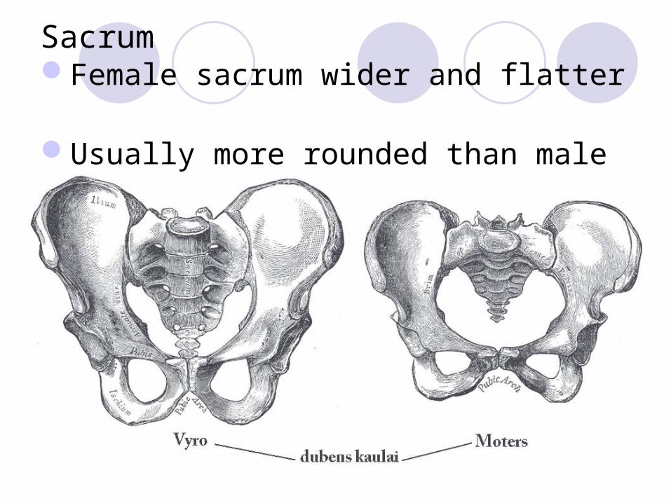
SacrumFemale sacrum wider and flatter
Usually more rounded than male
Forensic anthropology
Most will say that there is no exact way to determine sex from skeleton (not exact science)
Pelvis is probably most reliable feature to analyze, followed by mandible


