28
Introduction to XAFS Experiments IUCr XAFS Tutorial Montreal, August 5 2014 Bruce Bunker Physics Department University of Notre Dame Director, Materials Research CAT, APS
Introduction to XAFS Experiments
IUCr XAFS TutorialMontreal, August 5 2014
Bruce BunkerPhysics Department
University of Notre DameDirector, Materials Research CAT, APS
In this talk …
• Reminder about x-ray techniques
• Experiment “modalities”
• X-ray sources
• X-ray beamlines
• “In-hutch” instrumentation
• Sample considerations
3
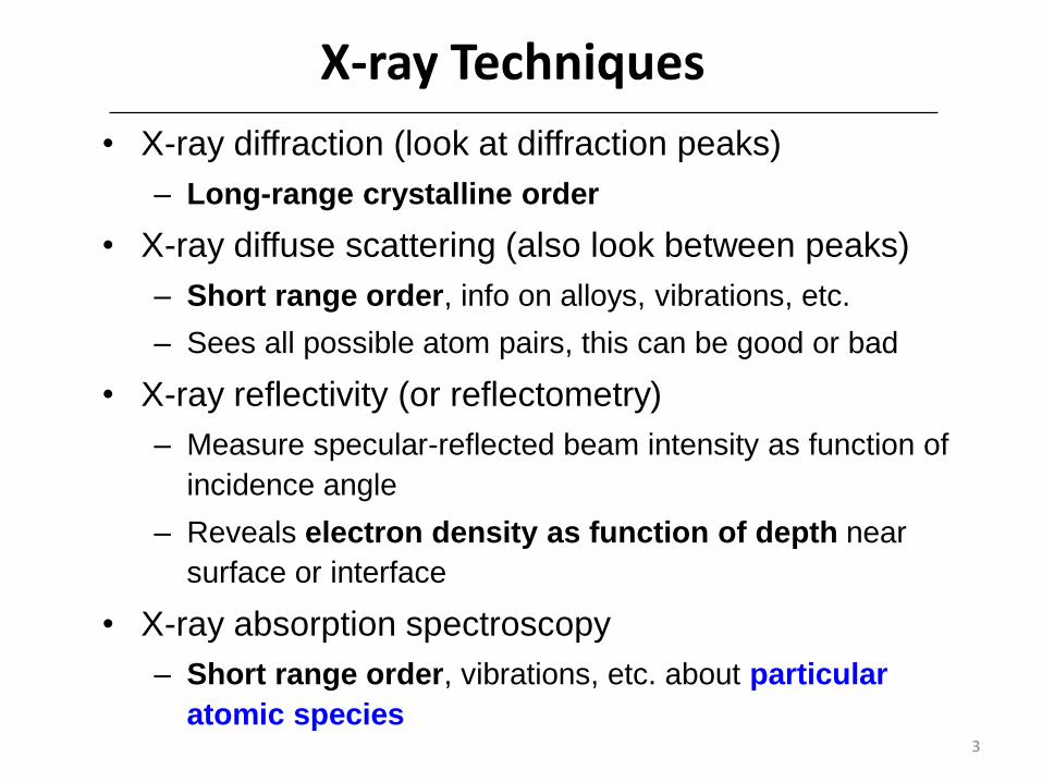
X-ray Techniques
• X-ray diffraction (look at diffraction peaks)
– Long-range crystalline order
• X-ray diffuse scattering (also look between peaks)
– Short range order, info on alloys, vibrations, etc.
– Sees all possible atom pairs, this can be good or bad
• X-ray reflectivity (or reflectometry)
– Measure specular-reflected beam intensity as function of
incidence angle
– Reveals electron density as function of depth near
surface or interface
• X-ray absorption spectroscopy
– Short range order, vibrations, etc. about particular
atomic species

4
X-ray Absorption
Many contributions to
absorption, but largest in
x-ray region is the
photoelectric effect:
Ionization of inner-shell
atomic electrons
Absorption Edge:
High enough energy for
excitation of atomic core
electrons to unoccupied
states (either bound or
continuum)
Figures courtesy Matt Newville, University of Chicago / CARS
5
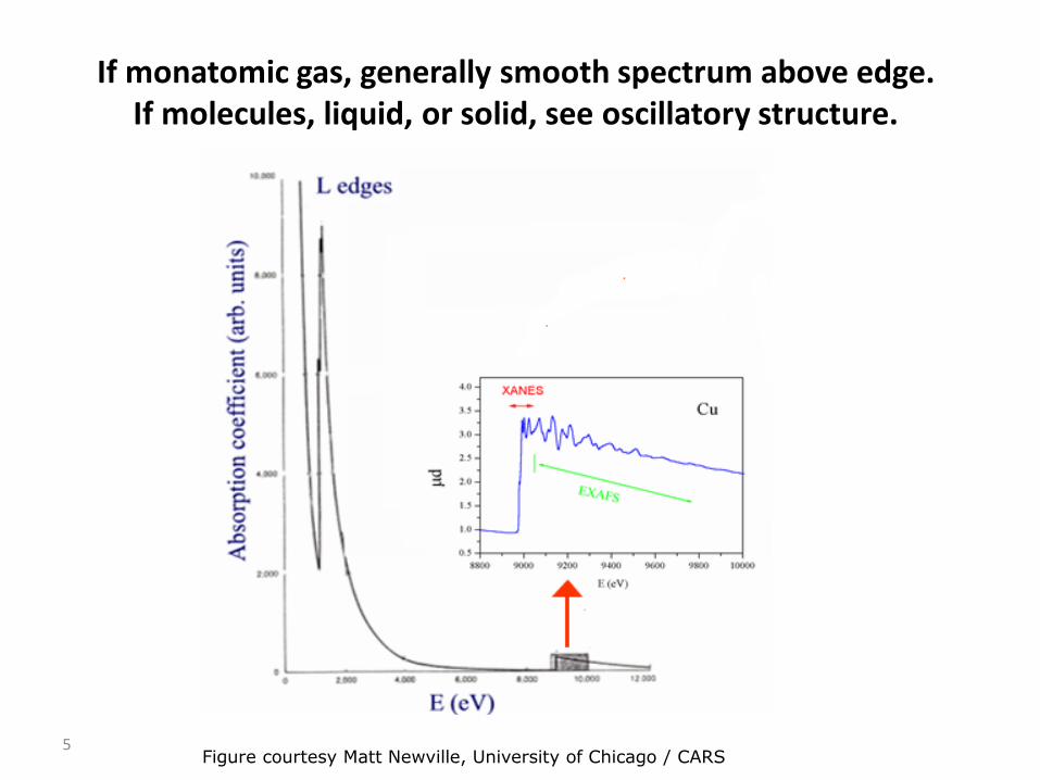
If monatomic gas, generally smooth spectrum above edge.If molecules, liquid, or solid, see oscillatory structure.
Figure courtesy Matt Newville, University of Chicago / CARS

The x-ray spectroscopy acronym game
• X-ray Absorption Spectroscopy (XAS)
– X-ray Absorption Fine-structure Spectroscopy (XAFS)
– Extended X-ray Absorption Fine-structure Spectroscopy (EXAFS)
– X-ray Absorption Near-Edge Spectroscopy (XANES) or
– Near-Edge X-ray Absorption Fine Structure (NEXAFS)
– And many more variations of techniques
• In all cases, variations in x-ray absorption coefficient as function of energy related to structural or electronic properties of sample

Back to extraction of structural information in a minute, first…
• These experiments require x-ray beam that is
– extremely intense
– well-collimated (for some experiments)
– broad-spectrum so that we can tune x-ray energy
• By far best source for most experiments is synchrotron radiation
7

Synchrotron Radiation and Storage Rings
• Accelerated charged particles (e.g. electrons) radiate electromagnetic radiation
• If highly relativistic electrons, radiation in x-ray region, strongly focused in forward direction
Courtesy David Atwood, UC Berkeley
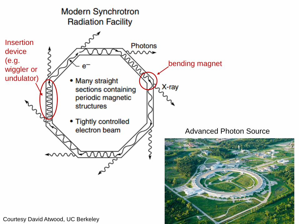
Advanced Photon Source
Courtesy David Atwood, UC Berkeley
bending magnet
Insertion
device
(e.g.
wiggler or
undulator)

Three Common Sources of Synchrotron Radiation
Courtesy David Atwood, UC Berkeley

11
Undulator Magnets
Extremely “bright” beam (small
source size, small divergence)
Great for spatial resolution,
angular resolution, etc.
Undulators have sharp peaks in
spectrum that can be tuned by
changing gap
Wigglers have stronger field, broad
spectrum (and lots of heat!)
Image courtesy SPRing8
Image courtesy DESY

12
Schematic of beamline Instrumentation
X-ray beamline components include…• “Front-end” components (cooled slits, etc.)• Bragg crystal monochromator (two Si crystals, first cooled
with liquid nitrogen)• Harmonic-rejection mirror (monochromator lets through
not only energy of interest, but harmonics, e.g. 3𝐸0)– Alternative: detune monochromator
• Detectors (several different types)• Goniometers, etc., for sample positioning

Now, have monoenergetic x-ray beam
• What do we do with it?
• Obviously want to measure x-ray absorption as a function of energy, but can do it as simple transmission experiment or indirectly
• Start out with the simplest …
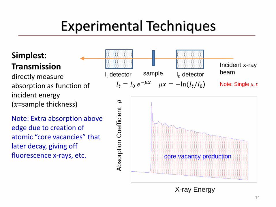
14
Experimental Techniques
Simplest:Transmissiondirectly measure absorption as function of incident energy (𝑥=sample thickness)
X-ray Energy
Ab
sorp
tio
n C
oe
ffic
ien
t
core vacancy production
Note: Extra absorption above edge due to creation of atomic “core vacancies” that later decay, giving off fluorescence x-rays, etc.
It detector
Incident x-ray
beamsample I0 detector
𝐼𝑡 = 𝐼0 𝑒−𝜇𝑥 𝜇𝑥 = −ln(𝐼𝑡/𝐼0)
𝜇
Note: Single 𝜇, 𝑡

15
“Indirect” XAFS Detection Methods:(proportional to absorption)
• x-ray fluorescence, or
• emitted electrons (total or partial “electron yield”)
Measure emitted flux as function of incident beam energy
If detector
Incident x-ray
beam
sample
I0 detector
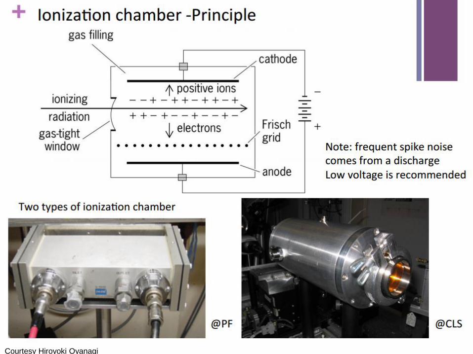
Courtesy Hiroyoki Oyanagi

17
More on Fluorescence Measurements
• X-rays from sample include not only fluorescence signal, but also background:– Elastic and Compton scattered x-rays– Fluorescence from other atomic species
Figure courtesy Matt Newville, University of Chicago / CARS
For many
systems,
background can
be 10-100 times
larger than
desired
fluorescence
18
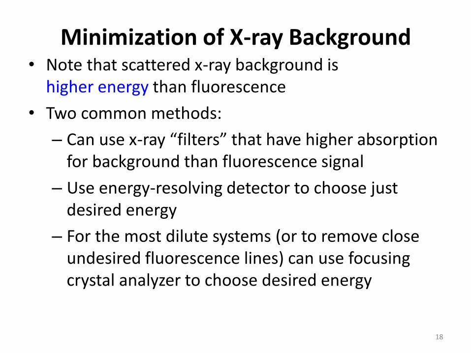
Minimization of X-ray Background• Note that scattered x-ray background is
higher energy than fluorescence
• Two common methods:
– Can use x-ray “filters” that have higher absorption for background than fluorescence signal
– Use energy-resolving detector to choose just desired energy
– For the most dilute systems (or to remove close undesired fluorescence lines) can use focusing crystal analyzer to choose desired energy
19
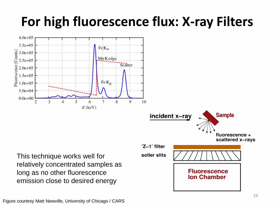
For high fluorescence flux: X-ray Filters
This technique works well for
relatively concentrated samples as
long as no other fluorescence
emission close to desired energy
Figure courtesy Matt Newville, University of Chicago / CARS

20
Energy-discriminating solid-state detectors(usually either Ge or Si)
• Select just energy region of interest
• Problems: Relatively low count rate, possible non-linearity from “dead time”
• Almost always use multi-element detectorsFigure courtesy Matt Newville, University of Chicago / CARS
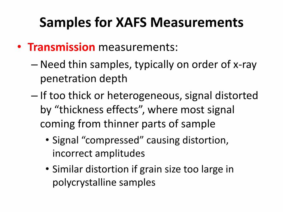
Samples for XAFS Measurements
• Transmission measurements:
– Need thin samples, typically on order of x-ray penetration depth
– If too thick or heterogeneous, signal distorted by “thickness effects”, where most signal coming from thinner parts of sample
• Signal “compressed” causing distortion, incorrect amplitudes
• Similar distortion if grain size too large in polycrystalline samples
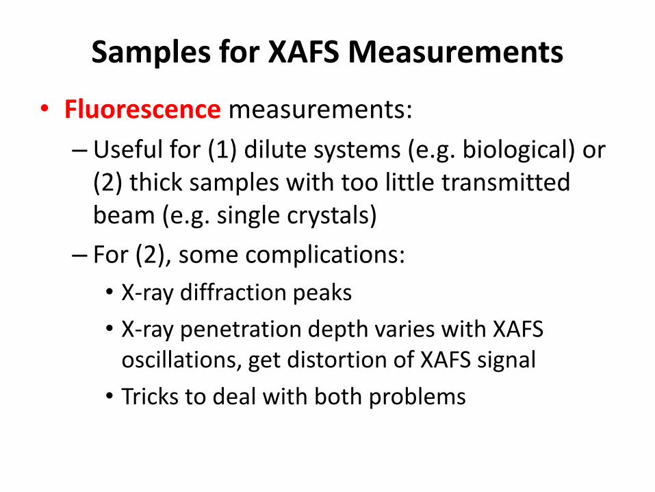
Samples for XAFS Measurements
• Fluorescence measurements:
– Useful for (1) dilute systems (e.g. biological) or (2) thick samples with too little transmitted beam (e.g. single crystals)
– For (2), some complications:
• X-ray diffraction peaks
• X-ray penetration depth varies with XAFS oscillations, get distortion of XAFS signal
• Tricks to deal with both problems
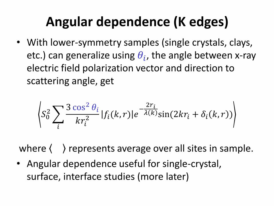
Angular dependence (K edges)
• With lower-symmetry samples (single crystals, clays, etc.) can generalize using 𝜃𝑖, the angle between x-ray electric field polarization vector and direction to scattering angle, get
𝑆02
𝑖
3 cos2 𝜃𝑖
𝑘𝑟𝑖2 𝑓𝑖(𝑘, 𝑟) 𝑒
−2𝑟𝑖𝜆 𝑘 sin(2𝑘𝑟𝑖 + 𝛿𝑖 𝑘, 𝑟 )
where represents average over all sites in sample.
• Angular dependence useful for single-crystal, surface, interface studies (more later)
24
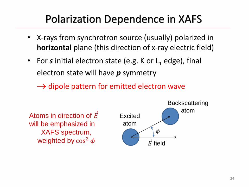
Polarization Dependence in XAFS
• X-rays from synchrotron source (usually) polarized in horizontal plane (this direction of x-ray electric field)
• For s initial electron state (e.g. K or L1 edge), final
electron state will have p symmetry
dipole pattern for emitted electron wave
Atoms in direction of 𝐸will be emphasized in
XAFS spectrum,
weighted by cos2 𝜙 𝐸 field
Excited
atom
Backscattering
atom
𝜙
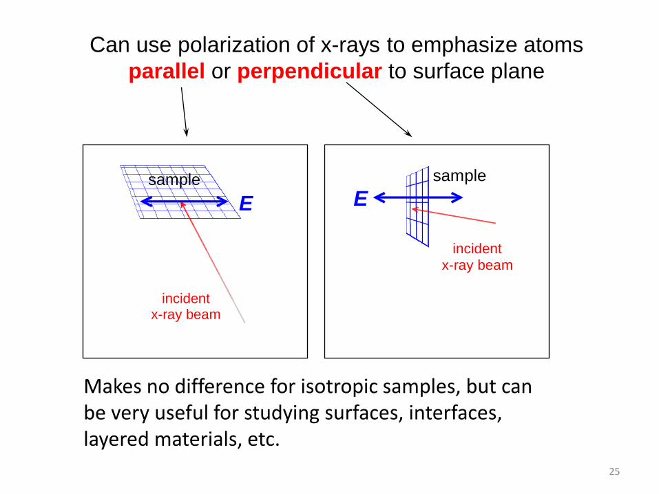
25
E
sample
incidentx-ray beam
Esample
incidentx-ray beam
Makes no difference for isotropic samples, but can be very useful for studying surfaces, interfaces, layered materials, etc.
Can use polarization of x-rays to emphasize atoms
parallel or perpendicular to surface plane

26
When performing experiments, need to keep track of (depending on beamline)
• If undulator line, set gap to optimize for your energy region: choose taper and/or scanning parameters
• Monochromator (optimize scanning, feedback stabilization, glitches)
• Harmonic rejection mirror (optimize for your energy range) (or detune monochromator)
• Detectors (I0, It, fluorescence; optimize gases if ion chambers, set regions of interest if counting detectors)
• Samples (design for optimum thickness, uniformity, etc. During measurements need to monitor temperature, radiation damage, etc.)
• Sample environment (temperature, gas-handling, high pressure, etc.

Not covered here (but wish I could)• Techniques for extreme environments, e.g. diamond
pressure cells
• Micro- and nano-focusing using mirrors, Fresnel zone plates, capillaries, etc.
• Time-resolved techniques
– Quick/slew scanning
– Energy dispersive techniques
– Pump/probe measurements using laser excitation
• Spin-resolved measurements: Magnetic circular dichroism
• X-ray emission spectroscopy
Next…
• Specifics for different types of applications (e.g. materials science, biological systems, etc.)
• Different approaches to data analysis and interpretation
• Stick around!


