+ All Categories
Home > Documents > INTRODUCTION - sharedocs.ca file · Web viewA 2-year-old girl presented with left leukocorea (white...

INTRODUCTION - sharedocs.ca file · Web viewA 2-year-old girl presented with left leukocorea (white...

Date post: 23-Aug-2019
Category:
Upload: truongngoc
View: 213 times
Download: 0 times
Share this document with a friend
34
Format of the review article: - A word limit of 5,000 words; - Less than 80 references; - No strict limit to the number of tables and figures (8-10 recommended); - An unstructured abstract of ≤ 250 words; - The maximum number of authors: 6 Genetics and Molecular Diagnostics in Retinoblastoma - An Update Authors: Sameh E. Soliman, MD, 1-2 Hilary Racher, PhD, 3 Chengyue Zhang, MD , 4 Hilary Racher, PhD Heather MacDonald, 1 Brenda L. Gallie. 1,5 Affiliations: 1 Department of Ophthalmology and Vision Sciences, University of Toronto, Ontario, Canada 2 Department of Ophthalmology, Faculty of Medicine, University of Alexandria, Alexandria, Egypt. 3 Impact Genetics, Bowmanville, Ontario.
Transcript

Format of the review article:

- A word limit of 5,000 words;

- Less than 80 references;

- No strict limit to the number of tables and figures (8-10 recommended);

- An unstructured abstract of ≤ 250 words;

- The maximum number of authors: 6

Genetics and Molecular Diagnostics in

Retinoblastoma - An Update

Authors:

Sameh E. Soliman, MD,1-2 Hilary Racher, PhD,3 Chengyue Zhang, MD,4 Hilary Racher, PhDHeather

MacDonald,1 Brenda L. Gallie.1,5

Affiliations:

1Department of Ophthalmology and Vision Sciences, University of Toronto, Ontario, Canada

2Department of Ophthalmology, Faculty of Medicine, University of Alexandria, Alexandria, Egypt.

3Impact Genetics, Bowmanville, Ontario.

4Department of Ophthalmology, Beijing Children’s Hospital, Capital Medical University, Beijing, China.

5Departments of Ophthalmology, Molecular Genetics, and Medical Biophysics, University of Toronto,

Toronto, Canada.

Corresponding author:

Brenda L. Gallie: Hospital for Sick Children, 555 University Ave, Toronto, Ontario, Canada M5G 1X8.

Telephone: +1-294-9729

Gallie Brenda, 01/04/17,
Organizing Text: Number the pages of the manuscript consecutively, beginning with the introduction as page 1. The text of an original article should not exceed 4,000 words with up to 8 images and tables and 50 references while that of a review article should not exceed 6,000 words with up to 8 images and tables and 100 references. The text of an annual review should not exceed 15,000 words with up to 200 references.
Gallie Brenda, 01/04/17,
Title page: Include on the title page (a) complete manuscript title; (b) authors’ full names, highest academic degrees, and affiliations; (c) name and address for correspondence, including fax number, telephone number and email address; (d) address for reprints if different from that of corresponding author; and (e) sources of support that require acknowledgement.

Disclosures:

Both SS and HR contributed equally to this review as first co-first authors.

We confirm that this manuscript has not been and will not be submitted elsewhere for publication, and all

co-authors have read the final manuscript within their respective areas of expertise and participated

sufficiently in the review to take responsibility for it and accept its conclusions.

HR is a paid employee and BG is an unpaid medical advisor at Impact Genetics. No other authors have

any financial/conflicting interests to disclose.

This paper received no specific grant from any funding agency in the public, commercial or not-for-profit

sectors.

Word Count: (/5000)

Key Words: retinoblastoma, RB1 gene, bilateral, unilateral, DNA sequencing, genetic counselling,

prenatal screening.

3

Unstructured abstract

Abstract: (120/250)

Retinoblastoma is an intraocular malignancy that affects one or both eyes of young children, that is

initiated by biallelic mutation of the retinoblastoma gene (RB1) in a developing retinal cell. A good

understanding of retinoblastoma genetics supports optimal care for retinoblastoma children and their

families. In this scenario the genetics trait description was conducted by the conversation between a

family with a retinoblastoma child and their attending who is mostly the ophthalmologist but can be any

member of the retinoblastoma multidisciplinary team of physicians, nurses and genetic counselors. All the

questions are true and high frequently asked by the parents. This scenario aims to try to simplify the

information around genetics for ophthalmologists to help them improve their patient and family care.

bilateral, unilateral, DNA sequencing, genetic counseling prenatal screening

4

Gallie Brenda, 01/04/17,
Review articles should emphasize new developments and areas of controversy in clinical or laboratory ophthalmology. An unstructured abstract of no more than 250 words should be submitted on a separate page.

5321/5000 words

INTRODUCTION

Retinoblastoma is the most common childhood intraocular malignancy that affects one or both eyes.

{Dimaras, 2015 #10881} Because of the strong links between clinical care and genetic causation,

{Knudson, 1971 #11106} retinoblastoma is considered the prototype of heritable cancers.{Theriault, 2014

#8591} Worldwide, about 8,000 children are newly diagnosed with retinoblastoma every year (1/16,000

live births).{Seregard, 2004 #10380;Dimaras, 2015 #10881} Genetics underlies many aspects of

retinoblastoma: clinical presentation, choice of treatment modalities and follow-up for both child and

family. We now highlight the genetic etiology of retinoblastoma in the context of individual children and

families.

CASE SCENARIO

A 2-year-old girl presented with left leukocorea (white pupil), noticed by her family in a photograph 5

days earlier. They sought medical advise from their family physician, who suspected retinoblastoma and

referred them urgently to the pediatric ophthalmologist. The family had never before heard of

retinoblastoma, and the mother was 33 weeks pregnant. The child was very uncooperative but the

ophthalmologist was able to visualize a white retinal mass in the left eye. He could see the inferior retina,

intact optic nerve and fovea in the right eye and diagnosed retinoblastoma in the left eye. The following

discussion took place between the pediatric ophthalmologist and the family.

Q1: Father: What is retinoblastoma?

A: (Pediatratric Ophthalmologist) “Retinoblastoma is a cancer that arises from a developing retinal cell

in babies and young children. Retinoblastoma can affect one (unilateral) or both eyes (bilateral) and in 5%

of children is associated with a midline brain tumor (trilateral).{de Jong, 2014 #10885} Without timely

and effective treatment, retinoblastoma may spread through optic nerve to the brain, or via blood

5

Gallie Brenda, 01/04/17,
I have DD in mind I was thinking of having images of both her eyes but I found the imaging not done by cynthia or Leslie. I think very poor quality for publication. BG Not sure who you mean…….

particularly to bone marrow, which will result in death. To be sure of the diagnosis and the best treatment

for this rare disease, I will refer your daughter to the Retinoblastoma Centre, where experts treat many of

these children. I will phone now!”

Q2: Father: why this is presenting at such a young age?

A: (Retinoblastoma Ophthalmic Specialist) “The cell of origin of retinoblastoma is most likely a

developing cone photoreceptor precursor cell that has lost both copies of the RB1 tumor suppressor gene,

and remains in the inner nuclear layer of the retina, unable to migrate to the outer retina and function

normally.{Dimaras, 2015 #10881;Rootman, 2013 #11096;Xu, 2014 #9924} The susceptible cell that

becomes cancer is only present in the retinas of young children, from before birth, up to around 7 years of

age. Rarely, retinoblastoma is first diagnosed in older persons, but likely there was previously an

undetected small tumor (retinoma) present from childhood, that later became active.{Gallie, 1982

#10343;Dimaras, 2008 #13250} The mean age at presentation is 1 year in bilateral disease and 2 years in

unilateral disease.

Despite the fact that we can see tumor in only one eye by clinical examination of your daughter, we

cannot be sure the other eye is normal until we examine it under anesthetic (EUA).”

Q3: Mother: What caused retinoblastoma? How can a gene cause cancer in a

baby?

A: (Retinoblastoma Ophthalmic Specialist) “No one knows what really causes the damage to the RB1

gene. Maybe a random cosmic ray passes through Planet Earth and hits that large, important gene.

In nearly 50% of patients the first RB1 gene is damaged in most, or all, normal cells, resulting in

predisposition to retinoblastoma. A retinal tumor develops when the second RB1 gene is also damaged in

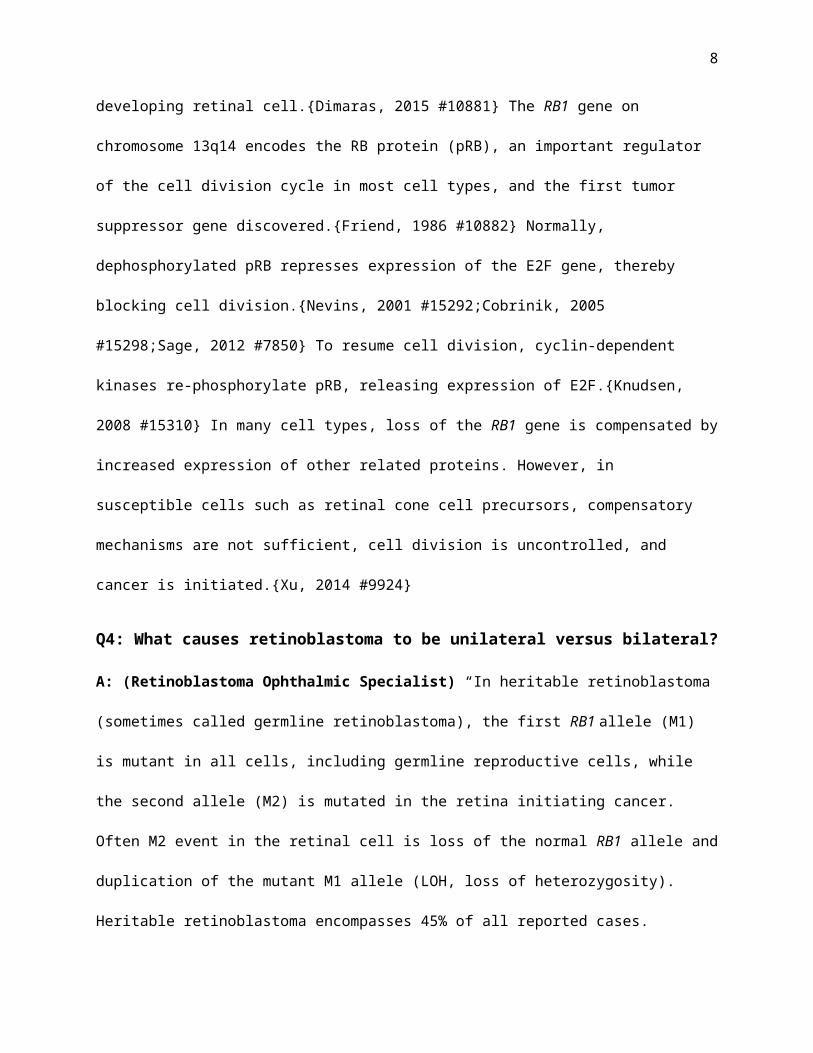
a developing retinal cell.{Dimaras, 2015 #10881} The RB1 gene on chromosome 13q14 encodes the RB

protein (pRB), an important regulator of the cell division cycle in most cell types, and the first tumor

suppressor gene discovered.{Friend, 1986 #10882} Normally, dephosphorylated pRB represses

6

expression of the E2F gene, thereby blocking cell division.{Nevins, 2001 #15292;Cobrinik, 2005

#15298;Sage, 2012 #7850} To resume cell division, cyclin-dependent kinases re-phosphorylate pRB,

releasing expression of E2F.{Knudsen, 2008 #15310} In many cell types, loss of the RB1 gene is

compensated by increased expression of other related proteins. However, in susceptible cells such as

retinal cone cell precursors, compensatory mechanisms are not sufficient, cell division is uncontrolled,

and cancer is initiated.{Xu, 2014 #9924}

Q4: What causes retinoblastoma to be unilateral versus bilateral?

A: (Retinoblastoma Ophthalmic Specialist) “In heritable retinoblastoma (sometimes called germline

retinoblastoma), the first RB1 allele (M1) is mutant in all cells, including germline reproductive cells,

while the second allele (M2) is mutated in the retina initiating cancer. Often M2 event in the retinal cell is

loss of the normal RB1 allele and duplication of the mutant M1 allele (LOH, loss of heterozygosity).

Heritable retinoblastoma encompasses 45% of all reported cases.{MacCarthy, 2009 #8367;Moreno, 2014

#9935;Wong, 2014 #15170} with either bilateral (80%), unilateral (15%) or trilateral (5%) tumors.

{Dimaras, 2015 #10881} Germline retinoblastoma carries risk of second primary cancers higher than

normal, most commonly osteosarcoma, fibrosarcoma and melanoma. These persons can benefit from

regular surveillance for such cancers for their lifetime.

Of non-heritable retinoblastoma, 98% have RB1 M1 and M2 arise in a retinal cell. The remaining 2%

the retinoblastoma is induced by somatic amplification of the MYCN oncogene, in the presence of normal

RB1 genes.{Rushlow, 2013 #11102}” Germline retinoblastoma carries the risk of development of second

primary cancers, most commonly osteosarcoma and fibrosarcoma due to loss of RB1 gene. This is why

these children should be kept under surveillance for the rest of their lives.

Q5: Mother: What caused these mutations? Did I cause them?

A: (Retinoblastoma Expert) “No one is to blame for the mutations causing retinoblastoma. Many

environmental forces induce DNA damage, including cosmic rays, X-rays, DNA viruses, UV irradiation

7

and smoking. The DNA damage may be point mutations, small and large deletions, promotor methylation

shutting down RB1 expression and rarely, chromothripsis.{Lohmann, 1999 #9272;McEvoy, 2014 #8499}

The majority of RB1 mutations arise de novo, unique to a specific patient or family. However, some

recurrent mutations are found in unrelated individuals, such as those that affect 11 sites CpG DNA

sequence sites, which are hyper-mutable and make up 22% of all RB1 mutations.{Rushlow, 2009

#10337;Richter, 2003 #11998}

When there is no family history of retinoblastoma, a de novo RB1 germline mutation may arise either

pre- or post-conception. Pre-conception mutagenesis of RB1 usually occurs during spermatogenesis,

perhaps because cell division (and opportunity for mutation) is very active during spermatogenesis, but

not during oogenesis.{Zhu, 1989 #6514;Dryja, 1997 #15586;Munier, 1998 #10955} Advanced paternal

age increases risk for retinoblastoma,{Toriello, 2008 #15506} suggesting that base substitution errors

may increase in aging men. The affected child carries the de novo RB1 mutation in every cell, typically

presenting with 4-5 tumors and bilateral retinoblastoma. In contrast, if mutagenesis occurs post-

conception, during embryogenesis, only a portion (1-50%) of cells carrying the RB1 mutation and the

person will be mosaic for the RB1 mutation. If the mutation arises during retinal development, the child

will have unilateral retinoblastoma.{Dimaras, 2015 #10881}

Q6: Father: So, only RB1 mutation causes retinoblastoma?

A: (Retinoblastoma Expert) “There are two answers to this question: RB1 mutation only causes a

benign precursor to retinoblastoma, retinoma, and other genes are modified to cause progression to

cancer;{Dimaras, 2008 #13250} and 2% of retinoblastoma have normal RB1 and are caused by a different

gene.

In addition to loss of RB1, specific alterations in copy number of other genes are common in RB1-/-

retinoblastoma. There are gains (4-10 copies) in oncogenes MDM4, KIF14 (1q32), MYCN (2p24), DEK

and E2F3 (6p22), and loss of the tumor suppressor gene CDH11 (16q22-24).{Corson, 2007

#9909;Theriault, 2014 #8591} Other less common genomic alterations in retinoblastoma tumors include

8

differential expression of specific microRNAs{Huang, 2007 #8613} and recurrent single nucleotide

variants/insertion-deletions in the genes BCOR and CREBBP.{Kooi, 2016 #14338} In comparison to the

genomic landscape of other cancers, retinoblastoma is one of the least mutated.{Kooi, 2016 #14338}”

There is a newly recognized form of retinoblastoma with normal RB1 genes. Two percent of unilateral

patients have RB1+/+MYCNA tumors, with the MYCN oncogene is amplified (28-121 instead of the normal

2 DNA copies).{Rushlow, 2013 #11102} These children are diagnosed at median age 4.5 months

compared to 24 months for non-heritable unilateral RB-/- patients, and the tumors are distinct

histologically, with advanced features at diagnosis.

Retinoma is a premalignant precursor to retinoblastoma with characteristic clinical features: translucent

white mass, reactive retinal pigment epithelial proliferation and calcific foci.{Gallie, 1982 #10343}

Pathology of retinoma reveals fleurettes{Tso, 1970 #3456} that are not proliferative.{Dimaras, 2008

#13250} Comparison of adjacent normal retina, retinoma and retinoblastoma shows in retinoma loss of

both RB1 alleles and early genomic copy number changes, that are amplified further in the adjacent

retinoblastoma.{Dimaras, 2008 #13250} Many retinoblastoma have underlying elements of retinoma.

Retinoma can transform to retinoblastoma even after many years of stability.{Theodossiadis, 2005

#5578}

Q6: Father: Could we have discovered retinoblastoma earlier?

The only way to find retinoblastoma tumor early is to look with specific expertise, which we can not

for every child. If we know to look because a relative had retinoblastoma, the smallest visible tumors are

round, white retinal lesions that obscure the underlying choroidal pattern. Centrifugal growth results in

small tumors being round; more extensive growth produces lobular growth, likely related to genomic

changes in single (clonal) cells, that provide a proliferative advantage.{Murphree, 2005 #11984;Balmer,

2006 #8323} Next, tumor seeds spread out of the main tumor a result of poor cohesive forces between

tumor cells into the subretinal space, or the vitreous cavity as appearing as dust, spheres or tumor clouds.

{Munier, 2014 #11111} Advanced vitreous tumor seeds can migrate to the anterior chamber producing a

9

pseudo-hypopyon. Enlarging tumor can push the iris lens diaphragm forward causing angle closure

glaucoma. Advanced tumors may induce iris neovascularization. Rapid necrosis of tumor can cause an

aseptic orbital inflammatory reaction resembling orbital cellulitis, sometimes showing central retinal

artery occlusion.{Balmer, 2007 #8320;Balmer, 2006 #8323;Murphree, 2005 #11984} Untreated,

retinoblastoma spreads into the optic nerve and brain, or hematogenous spread occurs through choroid,

particularly to grow in bone marrow. Direct tumor growth through the sclera can present as orbital

extension and proptosis.

The earliest signs of retinoblastoma detectable by parents are leukocorea (white pupil), either

directly or in photographs (photo-leukocorea) and strabismus when the macula is involvement by tumor.

{Balmer, 2007 #8320} In developing countries, buphthalmos and proptosis due to advanced and

extraocular disease respectively is common.{Canturk, 2010 #13461} Less common presentations include;

heterochromia irides, neovascular glaucoma, vitreous hemorrhage, hypopyon or aseptic orbital cellulitis.

{Balmer, 2007 #8320} Retinoblastoma (unilateral or bilateral) might be associated with a brain tumor in

the pineal, suprasellar or parasellar regions (Trilateral retinoblastoma){Popovic, 2007 #9156;Antoneli,

2007 #10877} with the median age of diagnsosis 17 months after retinoblastoma and before the age of 5

years. Retinoblastoma might present as 13q deletion syndrome, with facial features and various degrees

of hypotony and mental retardation.{Baud, 1999 #8118;Bojinova, 2001 #13205;Skrypnyk, 2004 #5276}

The main differential diagnosis includes Coats’ disease, persistent hyperplastic primary vitreous and

ocular toxicariasis.{Balmer, 2007 #8320}

Q7: Do all affected individuals with RB1 mutations develop retinoblastoma?

Each offspring of a person carrying an RB1 mutant gene has 50% risk to inherit the RB1 mutant gene

[Figure # Pedigree – full penetrance]. Nonsense and frame-shift germline mutations, which lead to absent

or truncated dysfunctional pRB, result in 90% bilateral retinoblastoma (nearly complete penetrance).

Often the second mutational event in the retinal cell is loss of the second RB1 allele (LOH, loss of

heterozygosity). For partially functional RB1 mutant alleles, reduced penetrance and expressivity is

10

observed, with later onset and fewer tumors{Soliman, 2016 #15159}, and some carriers never develop

retinoblastoma. Some reduced penetrance mutations reduce RB1 protein expression: (i) mutations in

exons 1 and 2,{Sanchez-Sanchez, 2007 #6108} (ii) mutations near the 3’ end of the gene in exons 24 to

27,{Bremner, 1997 #12040;Mitter, 2009 #7216} (iii) splice and intronic mutations{Zhang, 2003

#8986;Schubert, 1997 #4830;Lefevre, 2002 #4903} and (iv) missense mutations.{Scheffer, 2000

#15178;Cowell, 1998 #10958} Strangely, large deletions encompassing RB1 gene and MED1 gene also

cause reduced expressivity/penetrance, because RB1-/- cells cannot survive in the absence of MED4.

{Dehainault, 2014 #12140;Bunin, 1989 #4280} In comparison, patients with large deletions with one

breakpoint in the RB1 gene typically present with bilateral disease.{Mitter, 2011 #7339;Matsunaga, 1980

#357;Albrecht, 2005 #10898} A measure of expressivity of a mutant retinoblastoma allele is the disease-

eye-ratio (DER) (number of eyes affected with tumor divided by the total number of eyes in carriers of

the mutation).{Lohmann, 1994 #10954}

There are two specific RB1 mutations showing a parent-of-origin effect: intron 6 c.607+1G>T

substitution{Klutz, 2002 #8593;Schuler, 2005 #5551} [Figure # Pedigree – reduced penetrance family

with parent of origin….] and c.1981C>T (p.Arg661Trp).{Eloy, 2016 #12079} Both may be explained by

at differential methylation of intron 2 CpG85, which skews RB1 expression in favor of the maternal allele.

{Buiting, 2010 #7661;Kanber, 2009 #16381} When the allele is maternally inherited there is sufficient

tumor suppressor activity to prevent retinoblastoma development and 90% of carriers remain unaffected.

However, when the p.Arg661Trp allele is paternally transmitted, very little RB1 is expressed, leading

retinoblastoma in 68% of carriers.

Q10: What are the treatments and what govern the choice?

A: “Treatment and prognosis depend on the stage of disease at initial presentation. Factors predictive of

outcomes include size, location of tumor origin, extent of subretinal fluid, presence of tumor seeds and

the presence of high risk features on pathology.{Mallipatna, 2017 #14252} Multiple staging systems have

predicted likelihood to salvage an eye without using radiation therapy, but published evidence is

11

confusing because significantly different versions have emerged.{Dimaras, 2015 #10881;Mallipatna,

2017 #14252} The 2017 TNMH classification is based on international consensus and evidence from an

international survey of 1728 eyes, and separates more clearly initial clinical and pathological features

relevant to outcomes, in retrospective comparison to 5 previous eye staging systems.{Mallipatna, 2017

#14252} (Table X)

Retinoblastoma is the first cancer in which staging recognizes the impact of genetic status on

outcomes: presence of a positive family history, bilateral or trilateral disease or high sensitivity positive

RB1mutation testing, is stage H1; without these features bfore testing blood, HX; and H0 for those

relatives shown to not carry the proband’s specific RB1 mutation.{Mallipatna, 2017 #14252} We propose

H0* for patients with M1 and M2 RB1 mutant alleles of the tumor not detectable in blood, but with

remaining low risk (<1%) of mosaicism.

Choice of treatment depends on the laterality of disease, tumor stage and genetic status. Focal therapy

only can control cT1a eyes, but visually threatening or large cT1b tumors and cT2 eyes need

chemotherapy (systemic or intra-arterial chemotherapy) to reduce the size of the tumor followed by

consolidation focal therapies (laser therapy or cryotherapy) as initial treatment. Enucleation of eyes with

advanced tumors in unilateral disease where the other eye is normal is a definitive cure.{Dimaras, 2015

#10881} Ancillary therapies for specific indications include plaque radiotherapy and periocular

chemotherapy. Intravitreal chemotherapy for vitreous disease has recently dramatically improved safe eye

salvage.{Munier, 2012 #8588;Munier, 2012 #8587} For persons carrying RB1 mutations, external beam

radiation therapy is rarely indicated due to the high risk of inducing later second cancers.{Dimaras, 2015

#10881}

Saving life is the priority of retinoblastoma treatment, followed by vision salvage; the least important

is eye salvage. The child’s job is to play and develop in a healthy life; the many procedures and their

complications that may span years for at best a 50% chance to save a blind eye with risk of tumor spread,

are not justified, especially when the other eye is normal.{Soliman, 2015 #10948;Soliman, 2016 #14269}

12

Q11: Is retinoblastoma lethal?

A: “If untreated, retinoblastoma is lethal. If treated before metastasis occurs, cure is nearly 100%. If

metastasis occurs, the treatment becomes challenging and there is around 40% chance of mortality.

Delayed diagnosis and treatment due to lack of knowledge by ophthalmologists and parents,

socioeconomic{Soliman, 2015 #10948} and cultural factors are major causes of mortality. Asia and

Africa have the highest mortality, with >70% of affected children dying of retinoblastoma, compared

with <5% in developed countries.{Chantada, 2011 #13420;Canturk, 2010 #13461} ibute to reducing

mortality from retinoblastoma.

Germline retinoblastoma carries the risk of development of second primary cancers, most commonly

leiomyosarcoma, osteosarcoma, and melanoma.{MacCarthy, 2013 #11093} Occasionally metastatic

retinoblastoma may confused with a second cancer; blue round cell tumors on cytopathology may not

differentiate from retinoblastoma, but molecular demonstration of the same RB1 mutations as the

intraocular retinoblastoma will confirm metastases.{Racher, 2016 #13990}

Q12: How can we test for retinoblastoma mutations?

A: “If the patient is bilaterally affected, the probability of finding a germline mutation in the RB1 gene in

DNA extracted from blood is high (97% in a comprehensive RB1 laboratory). In 3% of bilateral

retinoblastoma patients, the predisposing RB1 mutation may be mosaic at a low level. Identification of

M1 and M2 RB1 mutations in DNA from tumor lead to identification of a germline mutation.{Astudillo,

2014 #10893;Rushlow, 2009 #10337;Canadian Retinoblastoma, 2009 #14251}

Similarly, to detect the 15% of unilateral patients carrying a germline mutation, optimal strategy is to

first tumor DNA, then check for those mutations in blood. If the blood is not found to carry one of the

tumor RB1 mutations, risk of germline status is reduced to <1% (Table) for parents, siblings and cousins.

{Canadian Retinoblastoma, 2009 #14251}

13

Quality of genetic results depends on quality of DNA. Fresh or frozen tumor samples are fine, but

formalin fixed paraffin embedded tumors generally produce highly degraded DNA. For blood genomic

whole blood in EDTA or ACD, provide high quality DNA.{Banfi, 2007 #15789}

The RB1 gene can be mutated in many ways, best identified by a series of techniques. Single

nucleotide variants (SNVs) and small insertions/deletions can be identified by DNA sequencing (Sanger

dideoxy-sequencing or next-generation sequencing (NGS) methods.{Singh, 2016 #19381;Li, 2016

#19404;Chen, 2014 #19419} The most appropriate technology depends on the clinical question being

asked. NGS may be the most effective screening strategy to investigate for an unknown de novo mutation

in an affected proband, and may have a lower limit of detection (analytic sensitivity) for mosaic

mutations.{Chen, 2014 #14457} To screen family members for a known sequencing-detectable RB1

mutation, targeted Sanger sequencing is cost and time effective.

Large RB1 deletions or duplications that span whole exons or multiple exons typically cannot be

detected by DNA sequencing. Multiplex ligation-dependent probe amplification (MLPA), quantitative

multiplex PCR (QM-PCR) or array comparative genomic hybridization (aCGH) are used identify RB1

deletions and duplications, and other genomic copy number alterations, such as MYCN amplification.

New developments in bioinformatics analysis suggest that NGS data can be interrogated for copy number

variants,{Devarajan, 2015 #15675;Li, 2016 #19404} but sensitivity is not yet optimized.

Somatic mosaicism can arise in either the presenting patient or their parent. Allele-specific PCR (AS-

PCR) has excellent sensitivity when the RB1 mutation is known,{Rushlow, 2009 #10337} and with

primers specific to the mutation can detect as low as 1% mosaicism.

The second mutational event in 70% of retinoblastoma tumors is loss of heterozygosity (LOH), a

common event associated with loss of the normal allele in tumor from individuals with an inherited

cancer predisposition syndrome.{Cavenee, 1983 #9210} Microsatellite marker analysis is also important

in identity testing and in maternal cell contamination in prenatal diagnostic tests.

14

Epigenetic changes can also initiate retinoblastoma development.{Ohtani-Fujita, 1993 #2258}

Hypermethylation of the RB1 promoter CpG island results in inhibition of RB1 gene transcription in 10-

12% of retinoblastoma tumors, commonly involving both alleles.{Richter, 2003 #11998;Zeschnigk, 1999

#15496} This epigenetic gene silencing event primarily occurs in somatic cells, but heritable RB1

promoter mutations and translocations disrupting RB1 regulatory sites may cause RB1 promoter

hypermethylation.{Quinonez-Silva, 2016 #12111} Occasionally hypermethylation of the RB1 promotor

is found in blood, strongly suggestion the presence of a translocation, for example to the X Chromosome.

In rare instance, no RB1 mutation is identified in the coding, promoter or flanking intronic sequence

in blood from a bilateral patient. Deep intronic sequencing alterations that disrupt RB1 transcription by

interfering with correct splicing in patients with retinoblastomaare best detected by analysis of the RB1

transcript by reverse-transcriptase PCR (RT-PCR).{Zhang, 2008 #7502;Dehainault, 2007 #5872} RNA

studies also clarify pathogenicity of intronic sequence alterations.{Zhang, 2008 #7502;Dehainault, 2007

#5872} As NGS costs continue to decrease, whole genome sequencing (WGS) may uncover deep intronic

changes.

Karyotype, fluorescent in situ hybridization (FISH) or array comparative genomic hybridization

(aCGH) of peripheral blood lymphocytes can be used to identify large deletions and rearrangements in

patient’s suspected of 13q14 deletion syndrome.{Caselli, 2007 #15862;Mitter, 2011 #7339} In parents of

13q14 deletion patients, karyotype analysis discover balanced translocations in carriers, informing about

risk of retinoblastoma in subsequent generations.{Baud, 1999 #8118}

Q13: Are these tests available worldwide?

A: “High sensitivity RB1 mutation testing is established in core labs mainly in high-income countries.{,

2015 #10824} In low- and middle-income countries, genetic counseling as a specialty does not exist, so

families may not understand the benefits of genetic testing and counseling in retinoblastoma treatment

and follow-up.{Dimaras, 2015 #10881} In many places, existing health insurance does cover genetic

testing testing. The new Chinese government policy allowing one more child per family will make

15

Sameh Gaballah, 01/01/17,
References Jeffrey.
Gallie Brenda, 01/04/17,
Hilary, I know we have one case at least and there was one in the literature which I can not find!Can you look?

genetic testing and counseling more important. A novel collaboration between Impact Genetics and

Geneseeq in China is optimizing expertise to achieve high quality RB1 testing for families.

In Egypt,{Soliman, 2016 #14713} Genetic testing for retinoblastoma is not available and genetic

counseling is the only way to address the issues. Ophthalmologists with insufficient training in

retinoblastoma genetics and counseling are burdened with the task. Genetic counseling was found to

increase knowledge in families retinoblastoma genetics, but gaps remained in translation of knowledge

into action.{Soliman, 2016 #14713}

Q14: What is done after finding the RB1 mutation?

A: “Targeted familial testing{Canadian Retinoblastoma, 2009 #14251;Dimaras, 2015 #10881} is used to

determine if a predisposing RB1 mutation has occurred de novo, through investigation of parental DNA

from PBL. Even if neither parent is identified to be a carrier, recurrence risk in siblings is still increased

due to the risk of germline mosaicism. DNA from PBL for all siblings of affected patients should be

tested for the proband’s mutation. As well, DNA from PBL for children of all affected patients should

also be tested for the predisposing mutation. Table Y shows the risk of having retinoblastoma in different

family relatives.

If the proband’s mutation was identified to be mosaic (ie postzygotic in origin) in DNA from PBL,

parents and siblings of the proband are not at risk to carry the predisposing mutation. However, the

children of mosaic proband should be tested, as their risk of inheriting the predisposing RB1 mutation can

be as high as 50% depending on the mutation burden in the probands germline.

When a RB1 mutation has been identified in a family, the known RB1 mutation of the proband can be

tested in his offspring. Couples may consider multiple options with respect to planning a pregnancy.

16

Hilary Racher, 01/01/17,
Should I use the new Skalet table instead?
Sameh Soliman, 01/01/17,
Can you write this down Hilary. I think this already available on Impacts website.
Gallie Brenda, 01/04/17,
BG to here

Q15: Can we use the known mutation to test my upcoming child? I am 33 weeks

pregnant

Prenatal genetic testing is usually performed early in the course of the pregnancy and is available in

many countries worldwide. Two early procedures are available: 1) chorionic villus sampling (CVS) and

2) amniocentesis. CVS is a test typically performed between 11-14 weeks gestation during which as

sample of the placenta is obtained either by transvaginal or transabdominal approach. Amniocentesis is a

test performed after 16 weeks of gestation whereby as sample of the amniotic fluid is gathered with a

transabdominal approach. CVS has a procedure-associated risk of miscarriage of ~1%. Amniocentesis

has a procedure-associated risk of miscarriage between 0.1-0.5%. Though uncommon, there is a risk for

maternal cell contamination that occurs more frequently with CVS.{Akolekar, 2015 #9479}

Genetic testing results can be used by the family and health care team to manage the pregnancy. If a

mutation is not identified, the pregnancy can proceed with no further intervention, as there is no increased

risk for retinoblastoma beyond the general population risk. If the mutation is identified, some couples

may decide to stop the pregnancy, while other couples may decide to continue with the pregnancy and

apply appropriate interventions, such as early delivery.{Soliman, 2016 #15159}

Some couples know that they wish to continue their pregnancy regardless of the genetic testing results

and are concerned by the risk of miscarriage associated with early invasive prenatal testing. Where

available, couples can also consider the option of late amniocentesis, performed between 30-34 weeks

gestation. When amniocentesis is performed late into the pregnancy, the key complication becomes early

delivery rather than miscarriage.{Akolekar, 2015 #9479} The risk for procedure-associated significant

preterm delivery is low (<3%). Results of genetic testing will be available with enough time to plan for

early delivery when a mutation has been inherited.

In many countries around the world, the option for prenatal genetic testing is not available. Even

where available, some couples may elect to not do invasive testing during the course of the pregnancy.

For these conceptions, if the pregnancy is at 50% risk for inheriting a RB1 mutation, it is crucial that the

17

pregnancy does not go post-dates. Induction of labour should be seriously considered if natural delivery

has not occurred by the due date.{Soliman, 2016 #15159;Canadian Retinoblastoma, 2009 #14251}

Q16: What is the benefit of prenatal mutation detection versus postnatal

screening?

A: “RB1 mutation detection can be performed either prenatal, as discussed previously, or it can be

performed at birth via umbilical cord blood (postnatal screening). This will help either eliminate the 50%

theoretical risk of the proband’s RB1 mutation heritability or confirm it to be 100% risk. Both screening

methods are effective in improving visual outcome and eye salvage compared to non-screened children.

However, prenatal screening allows for planning for earlier delivery in positive children (late

preterm/early term); this was shown to have less number of tumors at birth (20% versus 50%) with only

15% visual threatening tumors in prenatal screening. Prenatal screening with early delivery showed less

tumor and treatment burden with higher treatment success, eye preservation and visual outcome.

{Soliman, 2016 #15159}

Q17: Can we plan our next pregnancy to avoid having this RB1 mutation?

A: “In some countries around the world, there is an in vitro fertilization option available to couples called

preimplantation genetic diagnosis (PGD).{Dhanjal, 2007 #9216;Dommering, 2004 #10248;Xu, 2004

#9246;Girardet, 2003 #9219} For PGD, a couple undergoes in vitro fertilization. Conceptions are tested at

an early stage of development (typically 8-cell) for the presence of the familial mutation. Only those

conceptions that do not carry the mutation will be used for fertilization. The procedure is costly, ranging

from $10,000-$15,000 per cycle. In some countries, there may be full or partial coverage of the costs

associated with procedure. In addition to cost, couples must consider the medical and time impact of

undergoing in vitro fertilization. Couples also need to be aware that the full medical implications of PGD

are not yet understood; there is emerging evidence that there may be a low risk for epigenetic changes in

the conception as a result of the procedure. For couples that undergo PGD, it is recommended that typical

18

Hilary Racher, 01/01/17,
CDN or US $?

prenatal testing be pursued during the course of the pregnancy to confirm the results.{Dhanjal, 2007

#9216;Dommering, 2004 #10248;Girardet, 2003 #9219;Xu, 2004 #9246}

Q18: what is genetic counseling?

A: “Genetic counseling is both a psychosocial and educational process for patients and their families with

the aim of helping families better adapt to the genetic risk, the genetic condition, and the process of

informed decision-making.{Uhlmann, 2009 #15690;Shugar, 2016 #15715;Shugar, 2016 #15725}. Genetic

testing is an integral component of genetic counseling that results in more informed and precise genetic

counseling. Concrete knowledge of the genetic test outcomes results in specificity, reducing the need for

other possible scenarios to be discussed with the family. This enhances the educational component of

genetic counseling and also provides further time for psychosocial support to be provided to the family.

Q19: Can genetic counseling suffice alone? If yes, what are the benefits of

genetic testing?

A: “In countries where genetic testing is not available or unaffordable, genetic counseling is the option. It

was found that genetic testing is more cost effective than examining all the at-risk family members.

Patients with bilateral retinoblastoma at presentation are presumed to have heritable retinoblastoma and a

RB1 mutation (H1 in the TNMH classification). Genetic testing provides (1) more accurate information

about the type of heritable retinoblastoma and allows for straightforward testing to determine if additional

family members are at risk. (2) Through genetic testing, a patient may be found to have a large deletion

extending beyond the RB1 gene as part of the 13q deletion spectrum. Individuals with 13q deletion

syndrome are at risk for additional health concerns requiring appropriate medical management and

intervention. (3) Results may reveal a mosaic mutation which indicates that the mutation is definitively de

novo; only the individual’s own children are at risk and no further surveillance or genetic testing is needed

for other family members. (4) The results may find a low-penetrance mutation which indicates the patient

is at reduced risk to develop future tumours. As genetic testing for retinoblastoma becomes more common

19

Sameh Gaballah, 01/01/17,
Is it presumed or sure?

and data accumulate, surveillance of the proband may one day be matched more precisely to the level of

risk for new tumours for individuals with low penetrance mutations.

Patients with unilateral retinoblastoma greatly benefit from genetic testing and counselling.

Approximately 15% of patients with unilateral retinoblastoma will be found to have heritable

retinoblastoma. Correctly identifying these patients can be lifesaving, for both the patients and their

families. Genetic testing laboratories focused on enhanced detection of RB1 mutations are able to identify

nearly 97% of all retinoblastoma mutations. Genetic testing of the patient’s blood is sensitive enough

when thorough methods are used that not finding a mutation results in a residual risk of heritable

retinoblastoma low enough to remove the need for examinations under anesthesia. This reduces the health

risk for the patient and the cost to the health care system. Testing is even more accurate when a tumour

sample is collected and tested when available. When mutations are identified in the tumour and are

negative in blood, the results can eliminate the need for screening of family members and provide

accurate testing for the patient’s future children. Whether or not a tumour sample is available, finding a

RB1 mutation in a patient’s blood confirms that this patient has heritable retinoblastoma. This patient now

benefits from increased surveillance designed to detect tumours at the earliest stages and awareness of an

increased lifelong risk for second primary cancers. Members of the patient’s family can have appropriate

genetic testing to accurately determine who is at risk. As with patients with bilateral retinoblastoma,

knowing the specific type of mutation provides the most detailed provision of medical management and

counselling.

Q20: When is the appropriate timing for collecting samples for genetic testing?

For blood samples, they can be collected at any time but preferably when the child is under EUA where

there is no fear from the needle prick. For tumor samples, they would be collected from the enucleated

eye just after enucleation. Tumor cells will be preserved in a specific transport medium that allows the

cells to grow. We can also freeze some tumor cells (cryopreservation) for future necessity or for research

purposes.

20

Q21: If we know the mutation prenatally, is there any treatment to prevent

retinoblastoma from occurring?

A: “

Retinoblastoma genetics is challenging to understand, but once understood It largely affect the level

of care presented to retinoblastoma patients and their families. It helps alleviate the psychological burden

of the families regarding moving forward with their life choices regarding the affected child and future

siblings. It also helps the family to understand the risks of different family members giving them the

chance of the level of disclosure they wish.

21

Sameh Gaballah, 01/01/17,
References Jeffrey.
Sameh Gaballah, 01/01/17,
Is it presumed or sure?
Sameh Gaballah, 01/01/17,
I need a hint to references from here.
Sameh Gaballah, 01/01/17,
Reference?
Sameh Gaballah, 01/01/17,
I am finding difficulty citing here Hilary. Can you please give me any hints about the papers?
Sameh Gaballah, 01/01/17,
Hilary, please organize this part
Sameh Gaballah, 01/01/17,
-?A and B pockets-Also describe the role in genomic instability (Demaris. Rushlow)
Sameh Gaballah, 01/01/17,
I think this comes after Knudson hyposthesis and before the penetrance.
Hilary Racher, 01/01/17,
Moved to RB1 mutation section
Sameh Gaballah, 01/01/17,
Hilary, Can you please write a small paragraph explaining this with citations?
Gallie Brenda, 01/01/17,
BELONGS UP IN CLINICAL, not in genetics???
Sameh Gaballah, 01/01/17,
I preferred putting this here. Open for discussion.
Sameh Gaballah, 01/01/17,
Can we delete unilateral?
Sameh Gaballah, 01/01/17,
Please Hilary, can we rephrase to a simpler sentence?
Hilary Racher, 01/01/17,
Sameh – add section on retinoma
Hilary Racher, 01/01/17,
Sameh – define true H0 (*) vs most likely H0
Sameh Soliman, 01/01/17,
Table attached
Gallie Brenda, 01/01/17,
Sameh – integrate into the clinical section
Sameh Soliman, 01/01/17,
I would recommend a figure to show clinical features. BG: NO, this will be elsewhere in the revie issue. We chould stick to genetics.BG: not sure, this is one article in a review issue where we are assigned the genetics…..
Hilary Racher, 01/01/17,
Jeffry - Build more details from the Dimaris Nature Primer paper into this paragraph
Sameh Gaballah, 01/01/17,
I would remove eye salvage because in developing countries the concept of eye salvage before mortality should be changed. I prefer to speak on mortality only as the paragraph started.
Sameh Gaballah, 01/01/17,
I still think that this is an over-statement. I don’t think this number is true? What is your reference here Jeffrey??
Sameh Soliman, 01/01/17,
I find this a common question asked. We can answer Not yet, but there is research going on to target the epigenetic changes and MYCN. If approve this question, Hilary can add a line here ?She will add the epigenetic changes previously and can add what possible mechanisms here.

REFERENCES

Uhlmann, WR; Schuette, JL; Yashar, B. (2009) A Guide to Genetic Counseling. 2nd Ed. Wiley-

Blackwell.

Shugar, A. (2016) Teaching Genetic Counseling Skills: Incorporating a Genetic Counseling

Adaptation Continuum Model to Address Psychosocial complexity. J Genet Counsel. Epub ahead of print.

PMID: 27891554 DOI: 10.1007/s10897-016-0042-y

22

Gallie Brenda, 01/01/17,
Journal article 1. Boisjoly HM, Bernard PM, Dube I, et al. Effects of factors unrelated to tissue matching on corneal transplant endothelial rejection. Am J Ophthalmol 1989; 107: 647­54. References double-spaced in AMA style

Table X:

Subretinal Fluid (RD)

No≤ 5 mm

>5 mm - ≤ 1 quadrant

> 1quadrant

Tum

or

Tumors ≤ 3 mm and further than 1.5 mm from the disc and fovea cT1a/A cT1a/B cT2a/C cT2a/D

Tumors > 3 mm or closer than 1.5 mm to the disc and fovea cT1b/B cT1b/B cT2a/C cT2a/D

Se

edin

g Localized vitreous/ subretinal seeding cT2b/C cT2b/C cT2b/C cT2b/Ddiffuse vitreous/subretinal seeding cT2b/D

High

risk

feat

ures

Phthisis or pre-phthisis bulbi cT3a/ETumor invasion of the pars plana, ciliary body, lens, zonules, iris or anterior chamber cT3b/ERaised intraocular pressure with neovascularization and/or buphthalmos cT3c/EHyphema and/or massive vitreous hemorrhage cT3d/EAseptic orbital cellulitis cT3e/EDiffuse infiltrating retinoblastoma ??/E

Extraocular retinoblastoma cT4/??

clinical T (cT) versus International Intraocular retinoblastoma Classification (IIRC) (cT/IIRC); ?? Not

applicable ; RD Retinal detachment

23


Recommended