Spyttkjertler Inflammasjon og benigne neoplasier Spyttkjertelpatologi Store spyttkjertler: Parotis - normalt med 20-25 små lkn i Parotis Submandibularis Sublingualis Små spyttkjertler Ca 500- 1000 stk Ikke kapselavgrensing, små lobuli 1-5 mm SCHEMATIC OF BASIC SALIVARY GLAND UNIT Cytomorphologic features of various portions of the salivary system from the secretory end piece, left, to the oral cavity, right. AFIP: Tumors of Salivary Glands Introduksjon Mucocele. Obstruksjon av utførselsgang liten spyttkjertel med epiteltap i dilatert utførselsgang – typisk underleppe Ranula. Obstruksjon av utførselsgang glandula Sublingualis, lokalisert til munngulv. Sjelden Underliggende liten spyttkjertel med del av utførselsgang Normal, liten spyttkjertel x 40
Transcript
Spyttkjertler
Inflammasjon og benigne neoplasier
Spyttkjertelpatologi
Store spyttkjertler:
Parotis - normalt med 20-25 små lkn i Parotis
Submandibularis
Sublingualis
Små spyttkjertler
Ca 500- 1000 stk
Ikke kapselavgrensing, små lobuli 1-5 mm
SCHEMATIC OF BASIC SALIVARY GLAND UNIT
Cytomorphologic features of various portions of the salivary
system from the secretory end piece, left, to the oral cavity, right.
AFIP: Tumors of Salivary Glands
Introduksjon Mucocele. Obstruksjon av utførselsgang liten spyttkjertel
med epiteltap i dilatert utførselsgang – typisk underleppe
Ranula. Obstruksjon av utførselsgang glandula
Sublingualis, lokalisert til munngulv. Sjelden
Underliggende liten spyttkjertel
med del av utførselsgang Normal, liten spyttkjertel x 40
Atrofi. 40x som
forrige bilde
Relativ økning av
utførselsganger
Årsaker ??
AmEurConsGr Classification ACR Classification
Inclusion criteria Inclusion criteria
1. Ocular symptoms None
Xerophthalmia, sensations of sand or
gravel, use of tear substitutes
2. Oral symptoms None
Xerostomia, swollen salivary glands,
liquid aid needed to swallow
3. Ocular signs 1. Ocular signs
Schirmer test, Rose Bengal ocular stain Objective evidence of xerophthalmia
(Rose Bengal ocular stain)
4. Histopathology 2. Histopathology
MinSalivGlandBiopsy MSGB
5. Salivary gland involvement None
Whole salivary flow, sialography,
salivary scintigraphy
6. Autoantibodies 3. Autoantibodies
Anti-SSA/Ro without or without anti-
SSB/
Sicca Syndrom: 4 av 6 kriterier, inkl kriterium 4 el 6.
Wicheta et al J Oral maxillofac Surg 2017.
Journal of Oral and Maxillofacial Surgery
Volume 75, Issue 12, December 2017, Pages 2573-
2578
Pathology
Minor Salivary Gland Biopsy—An Important
Contributor to the Diagnosis of Sjögren Syndrome
87 pas med spyttkjertel biopsier
15 hadde Sjøgrens sykdom
12 fikk diagnose pga spyttkjertelfunn
Vanlig problemstilling (ukentlig)
Helst (minst) 5 små spyttkjertler, 3 opplegg på glass
Måle areal av spyttkjertelvev tilgjengelig - ved
inflammasjon, ikke nødvendig hvor inflammasjon er
fraværende
FOKUSSKÅR 1: Funn av aggregert minst 50
mononukleære celler pr 4 kvadrat mm
Positivt funn fokusskår >= 1
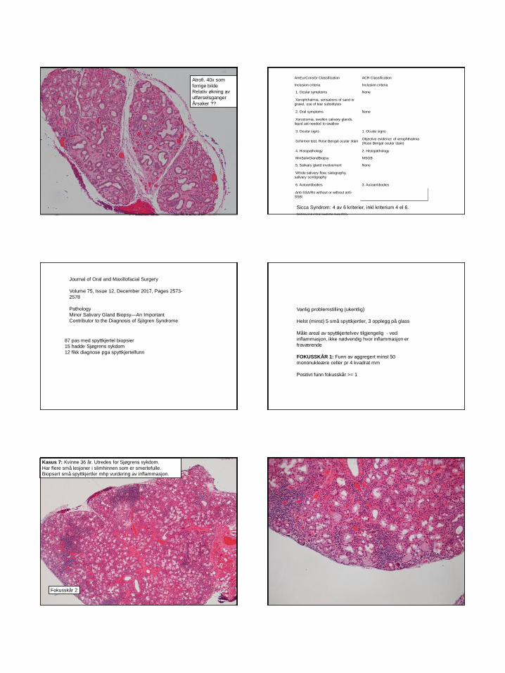
Kasus 7: Kvinne 36 år. Utredes for Sjøgrens sykdom.
Har flere små lesjoner i slimhinnen som er smertefulle.
Biopsert små spyttkjertler mhp vurdering av inflammasjon.
Fokusskår 2
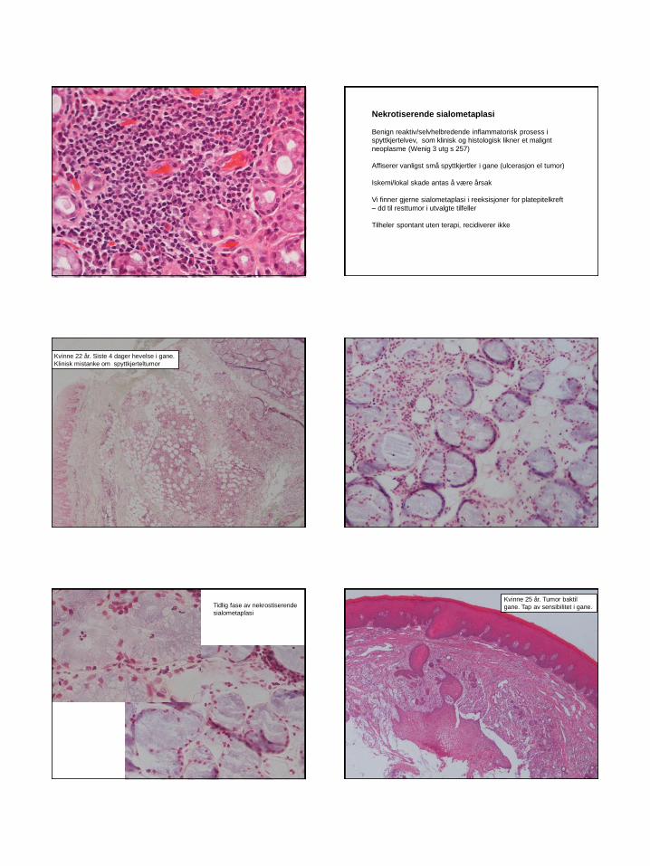
Nekrotiserende sialometaplasi
Benign reaktiv/selvhelbredende inflammatorisk prosess i
spyttkjertelvev, som klinisk og histologisk likner et malignt
neoplasme (Wenig 3 utg s 257)
Affiserer vanligst små spyttkjertler i gane (ulcerasjon el tumor)
Iskemi/lokal skade antas å være årsak
Vi finner gjerne sialometaplasi i reeksisjoner for platepitelkreft
– dd til resttumor i utvalgte tilfeller
Tilheler spontant uten terapi, recidiverer ikke
Kvinne 22 år. Siste 4 dager hevelse i gane.
Klinisk mistanke om spyttkjerteltumor
Tidlig fase av nekrostiserende
sialometaplasi
Kvinne 25 år. Tumor baktil
gane. Tap av sensibilitet i gane.
WHO histological classification of salivary gland tumors 2017
Benign epithelial tumors
Pleomorhic adenoma
Myoepithelioma
Basal cell adenoma
Warthins tumor
Canalicular adenoma
Lymphadenoma, sebaceous/non-sebaceous
Ductal papilloma – inverted papilloma
- intraductal papilloma
- sialadenoma papilliferum
Cystadenoma
FNAC av spyttkjerteltumores
-Gir vanligvis et adekvat cellemateriale
-Anbefaler lufttørkede MGG fargede preparat – fordi «kliniker fikser
dette best»
- MGG-farging viser best ekstracellulære komponenter
-Unngå tørkeartefakter i fikserte PAP-fargede utstryk
-Fiksering/PAP-farging – gir økt celletap i fargeprosess
- viser bedre kjernedetaljer/kromatinstruktur og
cytoplasmatisk keratinisering
MGG-farging erfaringsmessig sterkt varierende fargekvalitet fra de
forskjellige norske labber, til dels substandard kvalitet
For adekvat farging se
1: Piaton E m fl 2016: Guidelines for MGG staining in…
2: UK-NEQAS Staining Criteria Handbook – non gyn..
Pleomorphic adenoma
• Def WHO 2017
• Pleomorphic adenoma is a benign tumor with variable
cytomorphological and architectural manifestations. The identification
of epithelial and myoepithelial/stromal components is essential for the
diagnosis of PA.
• Insidens 2-3.5 tilfelle pr 100.000 pr år
Cytology
-fibrillary chondromyxoid ground substance
- spindle shaped ”mesenchymal”cells seen mainly in stromal matrix
- epithelial cells single and in poorly cohesive clusters and sheets
- regular ovoid nuclei with bland nuclear chromatin and well defined
cytoplasm
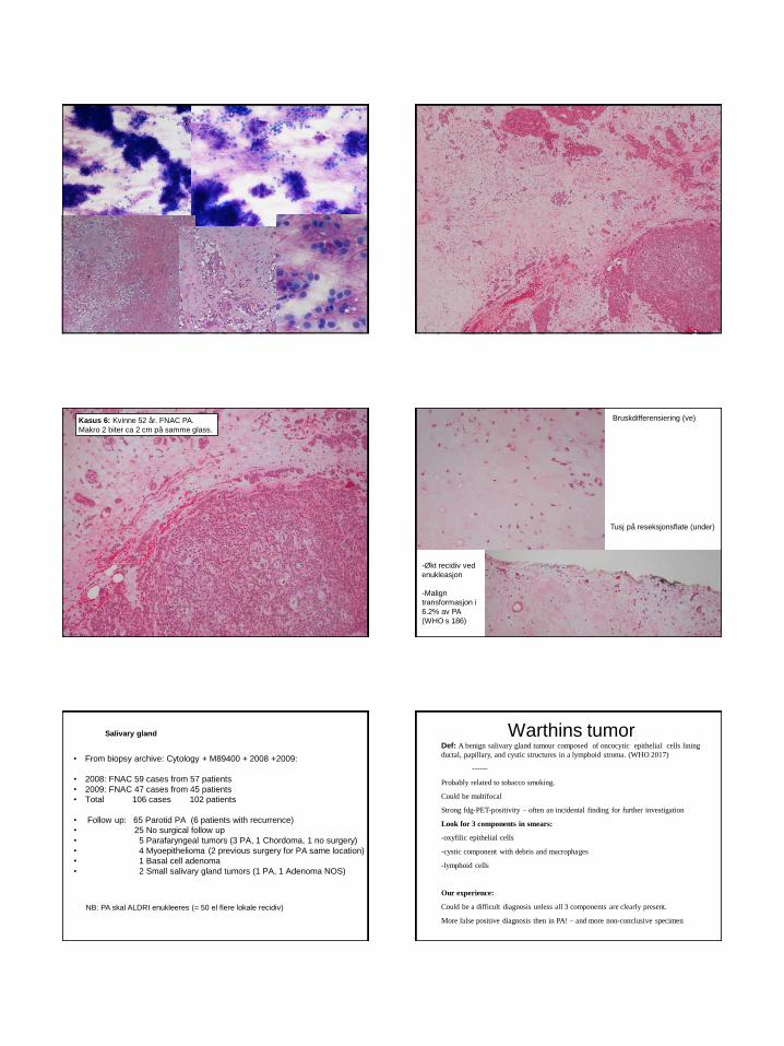
Kasus 6: Kvinne 52 år. FNAC PA.
Makro 2 biter ca 2 cm på samme glass.
Bruskdifferensiering (ve)
Tusj på reseksjonsflate (under)
-Økt recidiv ved
enukleasjon
-Malign
transformasjon i
6.2% av PA
(WHO s 186)
• From biopsy archive: Cytology + M89400 + 2008 +2009:
• 2008: FNAC 59 cases from 57 patients
• 2009: FNAC 47 cases from 45 patients
• Total 106 cases 102 patients
• Follow up: 65 Parotid PA (6 patients with recurrence)
compared to cells from neck – and neck tumor was removed.
Other constituents of cellmaterial with onkocytic differentiation – clue to Warthins
tumor?
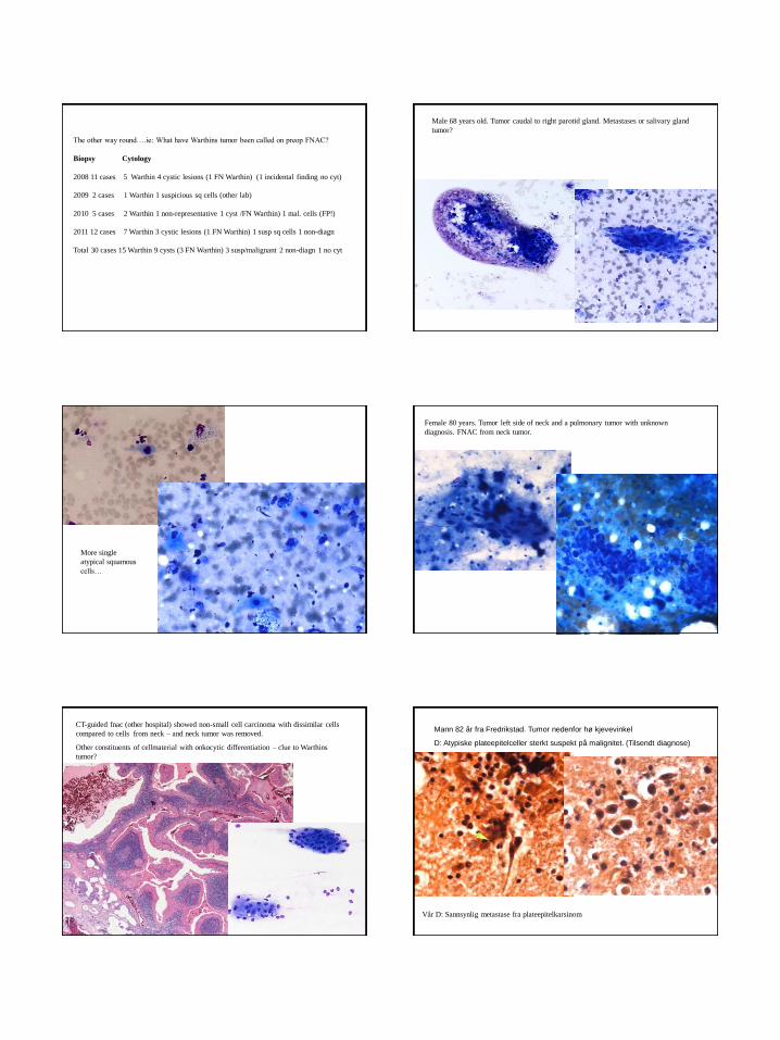
Mann 82 år fra Fredrikstad. Tumor nedenfor hø kjevevinkel
D: Atypiske plateepitelceller sterkt suspekt på malignitet. (Tilsendt diagnose)
Vår D: Sannsynlig metastase fra plateepitelkarsinom
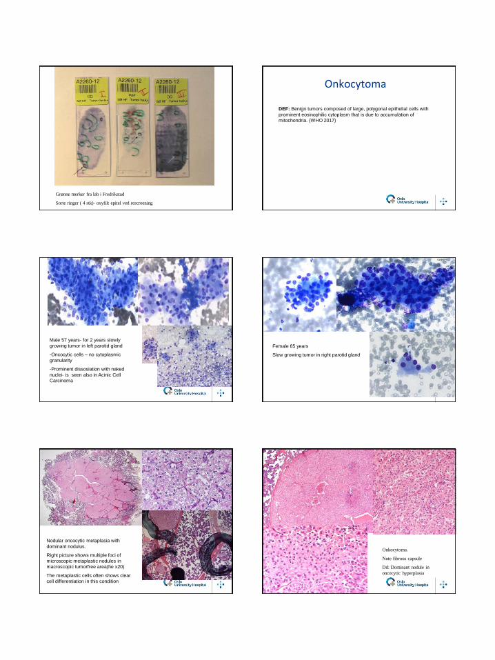
Grønne merker fra lab i Fredrikstad
Sorte ringer ( 4 stk)- oxyfilt epitel ved rescreening
Onkocytoma
DEF: Benign tumors composed of large, polygonal epithelial cells with
prominent eosinophilic cytoplasm that is due to accumulation of
mitochondria. (WHO 2017)
Male 57 years- for 2 years slowly
growing tumor in left parotid gland
-Oncocytic cells – no cytoplasmic
granularity
-Prominent dissosiation with naked
nuclei- is seen also in Acinic Cell
Carcinoma
Female 65 years
Slow growing tumor in right parotid gland
Cg09 904
Bg 10 3085
Nodular oncocytic metaplasia with
dominant nodulus.
Right picture shows multiple foci of
microscopic metaplastic nodules in
macroscopic tumorfree area(he x20)
The metaplastic cells often shows clear
cell differentiation in this condition
Onkocytoma.
Note fibrous capsule
Dd: Dominant nodule in
oncocytic hyperplasia
Basal cell adenoma WHO 2017 Basal cell adenoma is a benign salivary gland neoplasm composed of small basaloid cells, with occasional inner ductal epithelial cells forming nests and cords. Syn: Monomorft adenom 1-3.5% av spyttkjerteltumores. Fortrinnsvis i parotis. Membranøs subtype kan være multifokal og residivere.
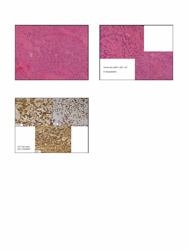
Kasus 10: Kvinne 60 år. Tumor ve parotis. Usikker cyt. Superfisiell parotidektomi
p63
ck7
Bifasisk immunprofil
Myoepithelioma
WHO 2017.
Def: Myoepithelioma is a benign salivary gland tumor composed
almost exclusively of cells with myoepithelial differentiation.
Min tilføyelse: - that may exhibit spindle, plasmacytoid, epitheloid or
clear cytoplasmic features
- har unntaksvis glandulære strukturer
- vanligvis pos ck5-6/p63/p40 (m.actin,s100)
- myoepitel angis også å kunne være pos ck7 (WHO s 187)
• Myoepithelial cells - Can look like other cells
- Plasmacytoid
- Spindle celle
- Epitheloid
- Clear cell
- Myoepithelial immun markers:
- P63,(P40) ck5/6 (s-100, SMA, SOX10)
- CK7 pos iflg WHO 2017 p 187, også i duktalt epitel)
Myoepithelial tumor cells
Spindled myoepithelial cells – WHO 2005
Epitheloid myoepithelial cells
Plasmacytoid myoepithelial cells – preferred
manifestation in small salivary gland tumors Clear cell variant of myoepithelial cells


![[PPT]TUMOR TRAKTUS UROGENITAL - FK UWKS 2012 C | … · Web viewTUMOR TRAKTUS UROGENITAL I. Tumor Ginjal A. Tumor Grawitz B. Tumor Wilms II. Tumor Urotel III. Tumor Testis IV. Karsinoma](https://static.documents.pub/doc/80x56/5ade93b87f8b9ad66b8bb718/ppttumor-traktus-urogenital-fk-uwks-2012-c-viewtumor-traktus-urogenital.jpg)


![BRAIN TUMOR-1.ppt [Read-Only] - ocw.usu.ac.idocw.usu.ac.id/.../bms166_slide_brain_tumor.pdf · introduction •• brain tumor intra kranial, med spinalis and meninges •• two](https://static.documents.pub/doc/80x56/5cd0246288c99375718d4765/brain-tumor-1ppt-read-only-ocwusuacidocwusuacidbms166slidebraintumorpdf.jpg)

![Salivary gland nodules [Schreibgeschützt] - ESHNR · salivary gland nodules C. Czerny ... • Tumor benign – malignant • Posttherapie Introduction ... Normale Parotis Perfusion](https://static.documents.pub/doc/80x56/5ca9d62488c993c9218d4289/salivary-gland-nodules-schreibgeschuetzt-salivary-gland-nodules-c-czerny.jpg)


