21
INTUSSUSCEPTION Author Nina Le, MS4 Editor Ilse Castro Aragon MD X-Ray Ultrasound

INTUSSUSCEPTIONAuthor Nina Le, MS4
Editor Ilse Castro Aragon MD
X-Ray Ultrasound

CASE HISTORY
10 month old male otherwise healthy presents with vomiting and bloody stool for past 10 hours. Had NBNB emesis with mucus. Noticed to have had 3 bouts of mucoid bloody stool. No fever, no sick contacts. Patient has been refusing his bottle since morning with decreased activity.
Labs show fecal occult blood (+).

ABDOMINAL X-RAY of INTUSSUSCEPTION
X-ray of abdomen of a 10 month old male shows a soft tissue density in the mid abdomen (yellow arrows) and lateralization of small bowel with mild air distention (red arrow) concerning for intussusception.

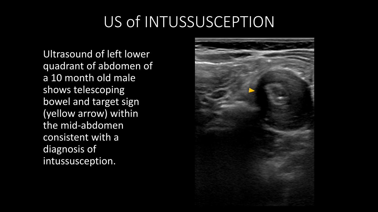
US of INTUSSUSCEPTION
Ultrasound of left lower quadrant of abdomen of a 10 month old male shows telescoping bowel and target sign (yellow arrow) within the mid-abdomen with entrapped fluid between loops of bowel (red arrow) consistent with a diagnosis of intussusception.
US of INTUSSUSCEPTION
Ultrasound of left lower quadrant of abdomen of a 10 month old male shows telescoping bowel and target sign (yellow arrow) within the mid-abdomen consistent with a diagnosis of intussusception.
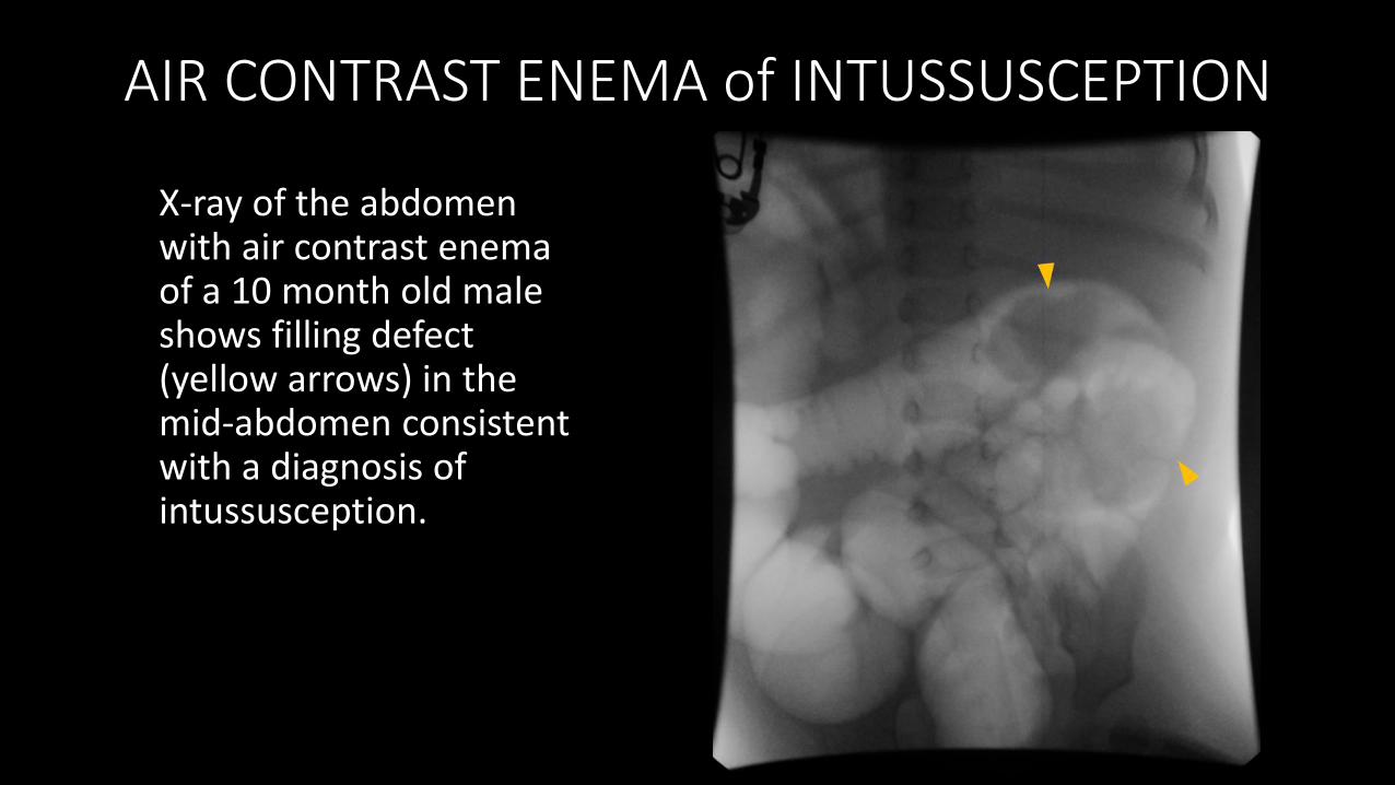
AIR CONTRAST ENEMA of INTUSSUSCEPTION
X-ray of the abdomen with air contrast enema of a 10 month old male shows filling defect (yellow arrows) in the mid-abdomen consistent with a diagnosis of intussusception.
CLINICAL FOLLOW UP
Following air contrast enema the intussusception could not be reduced, so the patient required laparoscopic reduction and was found to have ileocolic intussusception secondary to enlarged lymph nodes in the terminal ileum mesentery.

IN A NUTSHELL Most important findings include;
target sign – concentric rings
telescoping bowel
Other relevant findings include;
bowel dilation
Potential complications include;
bowel ischemia leading to perforation
peritonitis
Remember
Evaluate for perforation for which a contrast enema is contraindicated
VOICE RAD ULTRASOUND AND INTUSSUSCEPTION
by Dr. Castro-Aragon (2020)
OLA
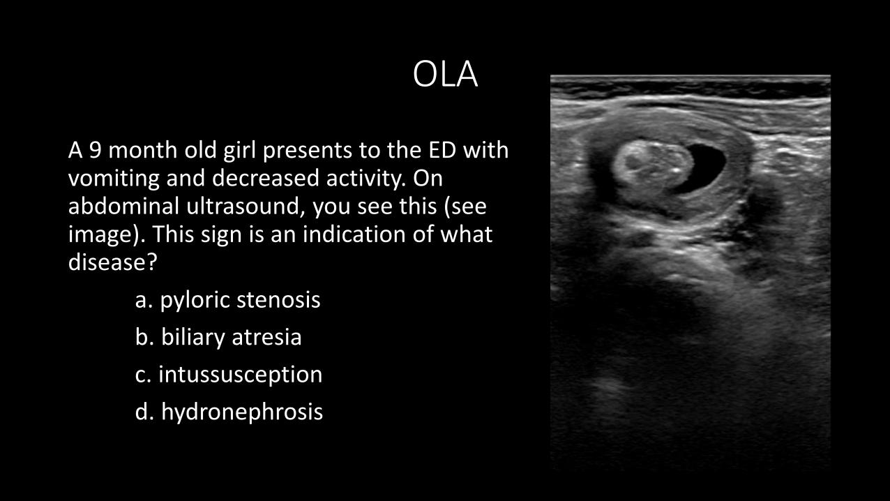
A 9 month old girl presents to the ED with vomiting and decreased activity. On abdominal ultrasound, you see this (see image). This sign is an indication of what disease?
a. pyloric stenosis
b. biliary atresia
c. intussusception
d. hydronephrosis

OLA
The abdominal ultrasound of a 7 month old boy show signs concerning for intussusception. Follow up abdominal x-ray reveals no signs of perforation. He is hemodynamically stable. What is the best treatment option?
a. monitor closely
b. laparoscopic reduction
c. colonoscopy
d. air enema

IMAGING SPECTRUM of INTUSSUSCEPTION
Ultrasound of an 8 year old male shows multiple loops of intussuscepted small bowel (yellow arrows) in the mid abdomen, consistent with ileoileal intussusceptions. Differential diagnosis includes appendicitis.
IMAGING SPECTRUM of INTUSSUSCEPTION
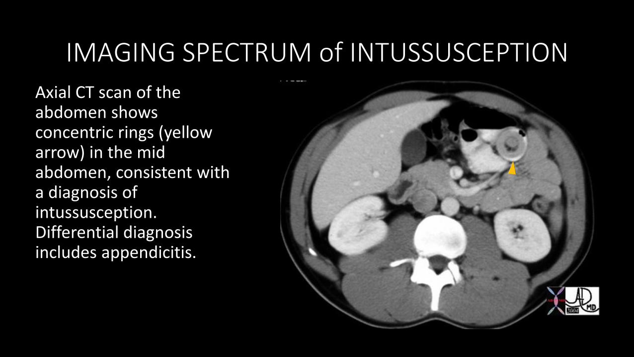
Axial CT scan of the abdomen shows concentric rings (yellow arrow) in the mid abdomen, consistent with a diagnosis of intussusception. Differential diagnosis includes appendicitis.
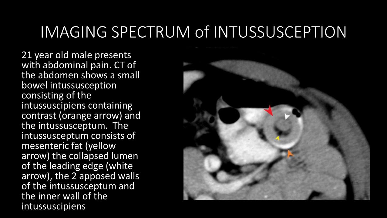
IMAGING SPECTRUM of INTUSSUSCEPTION21 year old male presents with abdominal pain. CT of the abdomen shows a small bowel intussusception consisting of the intussuscipiens containing contrast (orange arrow) and the intussusceptum. The intussusceptum consists of mesenteric fat (yellow arrow) the collapsed lumen of the leading edge (white arrow), the 2 apposed walls of the intussusceptum and the inner wall of the intussuscipiens

Acknowledgements: European Society of RadiologyReferences: Dr S Kuzmich
IMAGING SPECTRUM of INTUSSUSCEPTION
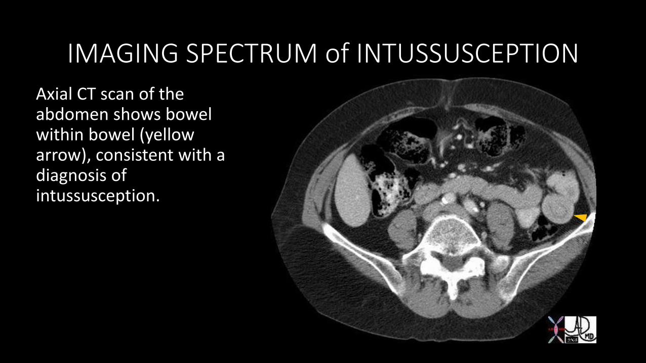
Axial CT scan of the abdomen shows bowel within bowel (yellow arrow), consistent with a diagnosis of intussusception.
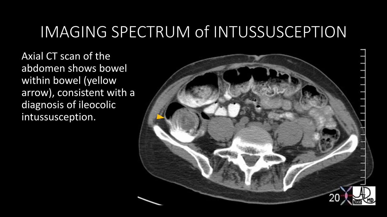
IMAGING SPECTRUM of INTUSSUSCEPTION
Axial CT scan of the abdomen shows bowel within bowel (yellow arrow), consistent with a diagnosis of ileocolic intussusception.

IMAGING SPECTRUM of APPENDICITIS
Ultrasound of the abdomen of a 5 year old female shows a distended, hypervascular and non-compressible appendix (yellow arrow) consistent with a diagnosis of appendicitis. Differential diagnosis includes intussusception.
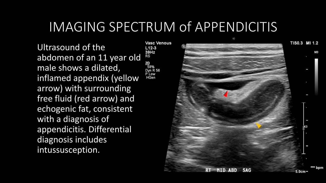
IMAGING SPECTRUM of APPENDICITIS
Ultrasound of the abdomen of an 11 year old male shows a dilated, inflamed appendix (yellow arrow) with surrounding free fluid (red arrow) and echogenic fat, consistent with a diagnosis of appendicitis. Differential diagnosis includes intussusception.

DISCUSSION
Intussusception occurs mostly in younger children around the ages of 3 months to 3 years old and are most common in the ileocolic region. It is characterized by sudden abdominal pain associated with vomiting and bloody stool and may cause bowel obstruction. It is most often seen on abdominal ultrasound as a pathognomonic “target sign,” representing telescoping bowel. Interestingly, there is an association of rotavirus vaccine and increased risk of intussusception.
In most cases, it is treated with an air contrast enema guided by fluoroscopy. Surgery is indicated if the intussusception is irreducible or if there is a bowel perforation.
LINKS AND REFERENCES
National GuidelinesACR Guidlines
Consistent References: RadiopediaRadioGraphics
Other Journals and TextsImaging Intussusception in Children's Hospitals in the United States: Trends, Outcomes, and Costs
VideosRadiology Capsules
OLAS


