Invasion of the Fallopian Tube by Enterobius vermicularis
SWEI H. TSUNG, M.D. and W EI-PING LOH, M.D., Ph .D.
Department o f Pathology, Methodist Hospital o f Gary, Inc.,
Gary, IN 46402
ABSTRACT
A solitary granuloma containing a gravid female pinworm was incidentally found in the fallopian tube of a 27 year old woman. On rare occasions, pinworms have been found outside the gastrointestinal tract, most frequently in the peritoneal cavity. The reported cases have been found at all levels of the female reproductive tract from the introitus to the fallopian tube. Thus, the hypothesis is supported that the worms reach the pelvic cavity via the genital tract. In addition, pinworm granulomas have also been found in the liver, lung, prostate and the renal pelvis. Awareness that such lesions may occur is important since the lesions may be in terpreted as being malignant with subsequent unnecessary surgical intervention.
The m ature pinworm (Enterobius vermicularis) inhabits the lumen ofthe intestine in the region of the lower most part of the ileum, cecum, the proximal part o f the ascending colon and the appendix. The gravid females migrate down the colon to the anogenital region where they lay eggs.
P inw orm eggs have b een found in routine vaginal smears,6' 9,17 endocervical aspiration,13 uterus,7,12,15 ovary16 and the peritoneal cavity in wom en.1,4,5,8,14,16,19 These reports leave little doubt that the adult gravid females can find their way into the genital tract during their nocturnal w andering over the perineum and gain access to the peritoneal cavity by crawling up the vagina, uterus and fallo-
Address reprint requests to: Swei H. Tsung, M.D., Department of Pathology, Methodist Hospital o f Gary, Inc., 600 Grant Street, Gary, IN 46402.
pian tubes. A survey of the literature reveals few instances of pinworm infestation of the fallopian tube.11,16,19 It is the purpose of this report to describe an additional patient and to review the topic of “ectopic Enterobius vermicularis.”
Case History
A 27-year-old Mexican-American woman, gravida3, para 2, was admitted to the Methodist Hospital of Gary on June 23, 1978, for voluntary sterilization. Past history revealed that the patient’s health had been good. She was asymptomatic. The values ofthe laboratory data measurements were within normal limits. The patient underwent bilateral salpingectomy. The resected specimen consisted of an essentially normal segment of the left fallopian tube and a segment of the right fallopian tube that contained a nodular area measuring 1.0 x 0.8 x 0.5 cm. Section through this area revealed com plete obliteration of the lumen with a thickened wall. Microscopic sections of this nodular area revealed
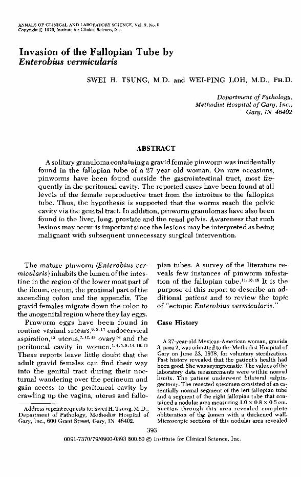
F igure 1. Photomicrograph o f section through gravid fem ale pinworm , show ing num erous eggs in worm which possessed the thornlike crest projecting on each side from the c u t ic le (H em a to x y lin eosin stain x 100).
one gravid female worm which possessed the characteristics o f the pinworm, with the thornlike crest projecting on each side from the cuticle (figure 1). The ova inside the worm also had the morphologic features resembling those of pinworm egg. Immediately around the worm was a layer of necrotic tissue which was surrounded by a thin band of loose connective tissue infiltrated by lymphocytes, occasional eosinophils and occasional giant cells.
A stool examination on a concentrated smear was negative for the parasites and their ova. Scotch tape touch preparation from the perineum also was negative. The patient stated that her two children were under treatment for pinworm infection.
Discussion
The pinworm or the egg rem aining after d isin teg ra tion and resorption of o ther parts of the worm has been identified in several extraintestinal locations. These w ere c o m p re h en s iv e ly rev ie w e d by Sym m ers19 in 1950 and by Sjovall and Ackerman16 in 1968. Since then, few case s tud ies have been p u b lish e d . In the majority of cases, the Enterobius ver- micularis was found in the peritoneal cavity . Som e cases o f E n te ro b iu s ver- micularis were recorded as being found in the female genital tract. It is interesting to note that all recorded cases of peritoneal in v o lv em en t have b e e n in w om en. Brandt3 recorded one case of pinworm
granuloma in the lung of a 50-year-old woman.
Ectopic lesions in male have been only reported five times. Symmers reported finding a granuloma owing to pinworm in the renal pelvis of a boy19 and in the prostate gland of a man.20 Slais18 found a p inworm granuloma in the liver of a 57-year- old man and Beaver et al2 described the case of granuloma owing to pinworm in the lung of a 23-year-old man.
Although inflamm ation of the reproductive system was found to be associated with the presence of the pinworm, it is difficult to decide w hether pinworm infestation of that system can give rise to inflammation or w hether the worms have settled in a previously inflam m ed area. T here are two possible routes for the spread of pinw orm to the abdom inal cavity,—tissue penetration of the in testinal wall or migration through the female genital tract. That all cases o f peritoneal involvement are in women lends support to the migration theory. The transport of pinworm s through the uterus and the tubes can be explained by its own activity which, however, may be enhanced by retrograde movements of the uterine tubal muscular tissue.16 Such movements do oc
INVASION OF THE FALLOPIAN TUBE BY ENTEROBIUS VERMICULARIS 395
cur. W ell known to any pelvic surgeon is the phenom enon of retrograde bleeding from fim briated ends of the tubes into the abdomial cavity during menstruation.
In a few cases, the presence of parasites or their ova in the peritoneal cavity appear to have caused sufficient reaction to produce symptoms and lead to surgical intervention, but in the great majority the lesions have been incidental findings which cannot be correlated with any event in the patient’s history.
Grossly, the granulomatous lesions can s im u la te leiom yom a, fibrom a, endo- m etriom a or tuberculous lesion. M oreover, awareness that such lesions may occur is im portant since the lesions may be interpreted as being malignant with subsequent unnecessary surgical intervention.
References1. Ar t h u r , H. R. and To m lison , P. E.: Oxyuris
granulomata of fallopian tube and peritoneal surface of ovarian cyst. J. Obst. Gynec. Brit. Emp. 65:996-997, 1958.
2. Be a v e r , I. C., Kr iz , J. J., and La u , T. J.: Pulm onary nodule caused by E n te ro b iu s verm icu la r is . Amer. J. Trop. M ed. Hyg. 22:711-713, 1973.
4. Brook , T. J., Go e t z , C. C., Bl a u c h e , W. C., e t AL: P elv ic granuloma due to E n te ro b iu s vermicularis. ]. Amer. Med. Assoc, 179:116- 118, 1962.
5. Ca m pb e l l , C. G. and Bow m an , J.: Enterobius verm icularis granuloma of pelvis. Amer. J. Obst. Gynec. 8 1 :256-258, 1961.
6. C r is t o b a l , A. S. and M u n d i , A.: Enterobius verm icularis larvae in vaginal smears. Acta C ytol. 20:190-192, 1976.
7. F a t h e r e e , J. P., C a r r e r a , G. M., and Be a v e r , P. C .: E n te ro b iu s verm icu la ris in human uterus. Mississippi Doctor 29:159-161, 1951.
8. L a n s m a n , H. H., L a p in , A., andB l a u s t e in , A.: P elv ic granuloma associated w ith en dometriosis. Amer. J. Obst. Gynec. 79:1178-1180, 1960.
9. L a n g l in a is , P. C.: Enterobius verm icularis in a vaginal smear. Acta Cytol. 13:40-41, 1969.
10. L it t l e , M. D ., C u e l l o , C . J., and D ’A l e s s a n - DRO, A.: Granuloma of the liver due to Enterobius vermicularis. Amer. J. Trop. Med. Hyg. 22:567-569, 1973.
11. M y e r , W. R.: Uher reizlos verkalkte im oileiter. Zbl. Allg. Path. 87:385-387, 1951.
12. N a ir n , R. C. and D u g u id , H. L . D .: Oxyuris granuloma of endometrium. J. C lin. Path. 7:228-230, 1954.
13. R a d , M. J . and J e a n n o t , A.: E n tero b iu s vermicularis in a cervical smear. Acta Cytol. 14:466-467, 1970.
14. Sc h e n k e n , J. R. and T a m isie a , J.: Peritoneal granulomas due to Enterobius vermicularis. Amer. J. Obst. Gynec. 72:309-311, 1956.
15. Sc h e n k e n , J. R. and T a m is ie a , J.: Enterobius vermicularis infection of endometrium. Amer. J. Obst. Gynec. 72:913-914, 1956.
16. Sjö v a l l , A. and A c k e r m a n , M.: Peritoneal granulomas in women due to the presence of Enterobius vermicularis. Acta Obst. Gynec. Scand. 47:361-372, 1968.
17. S h ip t o n , E. A ., M c In e r n e y , R. J. F ., and H u b e r t , L.: Parasitic ova in vag inal sm ears. M ed. J. Australia 1 :1014, 1973.
18. S l a i s , J .: A threadworm granuloma in the human liver. Helminthologia. 4:479-483,1963.
19. Sy m m e r s , W. S. C.: Pathology of Oxyuriasis : With special reference to granulomas due to the presence of Oxyuris vermicularis and its ova in the tissues. Arch. Path. 50:475-516, 1950.
20. SYMMERS, W. S. C.: Two cases of eosinophillic prostatitis due to metazoan infection (with Oxyuris vermicularis and with a larvae of Lin- guatula serrata). J. Path. Bact. 73:549-555,1957.