Page 1
INVESTIGATING THE OCCURRENCE OF
Vibrio parahaemolyticus
IN VARIOUS SEAFOOD CONSUMED IN THE
TURKISH REPUBLIC OF NORTHERN CYPRUS
A THESIS SUBMITTED TO THE GRADUATE
SCHOOL OF APPLIED SCIENCES
OF
NEAR EAST UNIVERSITY
By
HAFIZU IBRAHIM KADEMI
In Partial Fulfillment of the Requirements for
the Degree of Master of Science
in
Food Engineering
NICOSIA, 2016
INV
ES
TIG
AT
ING
TH
E O
CC
UR
RE
NC
E O
F V
ibrio
para
haem
olyticu
s IN V
AR
IOU
S S
EA
FO
OD
CO
NS
UM
ED
IN T
UR
KIS
H R
EP
UB
LIC
OF
NO
RT
HE
RN
CY
PR
US
HA
FIZ
U IB
RA
HIM
KA
DE
MI N
EU
2016
Page 2
INVESTIGATING THE OCCURRENCE OF
Vibrio parahaemolyticus
IN VARIOUS SEAFOOD CONSUMED IN THE
TURKISH REPUBLIC OF NORTHERN CYPRUS
A THESIS SUBMITTED TO THE GRADUATE
SCHOOL OF APPLIED SCIENCES
OF
NEAR EAST UNIVERSITY
By
HAFIZU IBRAHIM KADEMI
In Partial Fulfillment of the Requirements for
the Degree of Master of Science
in
Food Engineering
NICOSIA, 2016
Page 3
I hereby declare that, all the information in this document has been obtained and presented in
accordance with academic rules and ethical conduct. I also declare that, as required by these
rules and conduct, I have fully cited and referenced all material and results that are not
original to this work.
Name, Last Name:
Signature:
Date:
Page 4
i
ACKNOWLEDGEMENTS
My gratitude is endless to the One and only One that makes impossible to become possible.
Distinctively, I would like to express my appreciation to all people who contributed in one
way or the other in my educational pursuit.
At first, I would like to express my deepest gratitude to my supervisor Dr. Perihan Aysal
ADUN, for her generosity and unwavering support as well as open-minded approach, without
whom this study would not have been completed. No amount of inks and papers are enough
to transcribe my appreciation.
I would like to thanks all the chairs in my jury for their scholarly recommendations, and my
esteemed regards to Assist. Prof. Meryem Güvenir for her help in the laboratory studies.
I would like to also use this opportunity to credit Mr. Buğra Demircioğlu, the coordinator of
Food Engineering Department for his tremendous counselling and mentorship. I am also
grateful to all the lecturers of Food Engineering Department of Near East University for their
support and encouragement.
It is a great pleasure to acknowledge my lecturers at Kust Wudil, particularly Malam Munir
Abba Dandago (my academic father and a role model); your teachings, guidance and support
are indelible in my mind.
I am highly grateful to Kano state government under the leadership of Engr. (Dr) Rabiu Musa
Kwankwaso for sponsoring my master’s program, may Allah (S.W.T) rewards him
abundantly. Accordingly, I wish to acknowledge Center of Excellence of Near East
University for sponsoring this research.
Above ground, I am indebted to my parents Malam Ibrahim Muhammad and Malama Halima
Ibrahim for giving me their all to live an examplanary life, I am indeed grateful.
Finally, I am thankful to all my colleagues, friends and relatives whose names are numerous
to mention.
Page 5
ii
To the entire ummah
Page 6
iii
ABSTRACT
This study investigates the presence of pathogenic Vibrio parahaemolyticus in seafood
consumed in the Turkish Republic of Northern Cyprus (TRNC). Sixty samples of fish were
obtained from major seafood outlets and sea costs of Famagusta, Kyrenia, Nicosia and
Morphou. Conventional culture technique was employed for the bacterial identification. After
having been enriched, isolation of this pathogen (V. Parahaemolyticus) from different
seafood was performed on Thiosulfate Citrate Bile Sucrose-Salts Agar (TCBS) medium. The
identity of the bacteria were confirmed by using BD Phoenix Instrument.
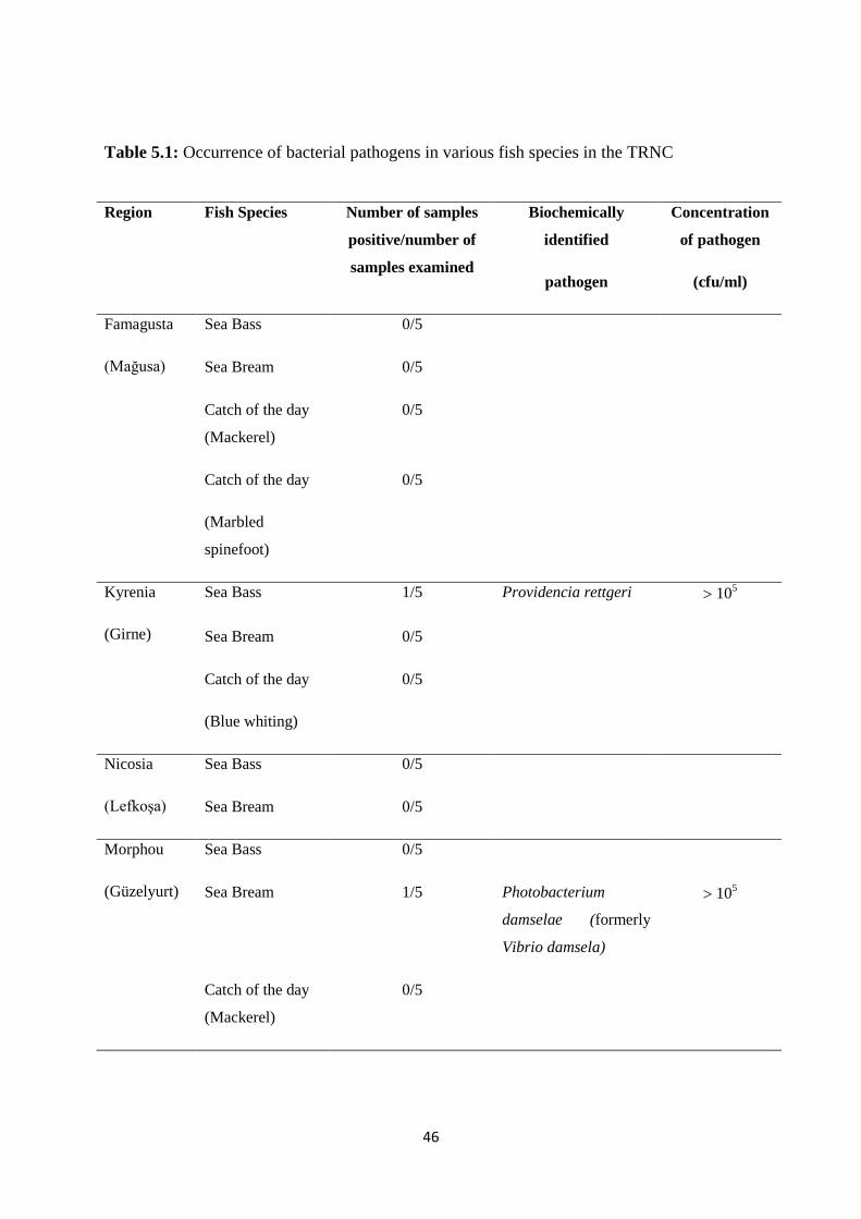
We could not find Vibrio parahaemolyticus in fish samples taken from different regions of
TRNC which is one of the most important seafoodborne pathogens. However seafood
consumed in TRNC might be a source of other bacterial pathogens like Photobacterium
damselae (formerly Vibrio damsela) and Providencia rettgeri species, since the
concentrations of these bacteria were found to be greater than 105
cfu/ml (minimum infective
dose) in sea bass and sea bream fishes from Kyrenia and from Morphou regions respectively.
Keywords: Isolation; V. Parahaemolyticus; TCBS; culture method; Seafood; investigating;
food safety; TRNC
Page 7
iv
ÖZET
Bu çalışmada Kuzey Kıbrıs Türk Cumhuriyeti’nde (KKTC) tüketilen deniz ürünlerindeki
patojen bir bakteri olan Vibrio parahaemolyticus’un olası varlığı araştırılmıştır. KKTC’nin
Mağusa, Girne, Lefkoşa ve Güzelyurt bölgelerindeki deniz ürünleri satan marketlerden ve
balıkçılardan 60 balık örneği toplanmıştır. Balıkların solungaç ve iç organları ayrıldıktan
sonra alkali peptonlu suda ayrı ayrı homojenize edilip zenginleştirilmiş ve Thiosulphate
Citrate Bile Salt Sucrose (TCBS) Agarda izole edilmiştir. TCBS agarda üreyen şüpheli
koloniler BD Phoenix cihazı kullanılarak tanımlanmışlardır.
Kültüre alınan örneklerin hiçbirinde Vibrio parahaemolyticus’a rastlanmamıştır. Girne’den
alınan levrek örneklerinden bir balığın iç organlarında patojen Providencia rettgeri ve
Güzelyurt’tan alınan çipura örneklerinden bir balığın yine iç organlarında patojen
Photobacterium damsalae (önceki adıyla Vibrio damsela) bulunmuştur.
Balık örneklerinde Vibrio parahaemolyticus bulunmaması halk sağlığı açısından sevindirici
bir sonuç olmakla beraber KKTC’de yaygın şekilde tüketilen balık örneklerinden bazılarında
105 cfu/ml (minimum infektif doz) düzeyinde rastlanılan Providencia rettgeri ve
Photobacterium damsalae patojen bakterilerinin varlığının araştırılması önerilmektedir.
Anahtar Kelimeler: İzolasyon ve identifikasyon; verifikasyon; Vibrio parahaemolyticus;
TCBS; deniz ürünleri; balık, gıda güvenliği; KKTC
Page 8
v
TABLE OF CONTENTS
ACKNOWLEDGEMENTS.............................................................................................. i
ABSTRACT....................................................................................................................... iii
ÖZET.................................................................................................................................... iv
TABLE OF CONTENTS................................................................................................... viii
LIST OF TABLES............................................................................................................... v
LIST OF FIGURES............................................................................................................. ix
LIST OF ABBREVIATIONS............................................................................................. x
CHAPTER1: INTRODUCTION........................................................................................ 1
1.1 Background Information.................................................................................................. 1
1.2 Overview on Seafood....................................................................................................... 4
1.2.1 Proximate composition and nutrition of seafood..................................................... 5
1.2.2 Seafood and foodborne pathogens........................................................................... 6
1.2.3 Prevalence, occurrence and distribution of V. parahaemolyticus in seafood........... 7
1.2.4 Microbiological criteria of seafood.......................................................................... 8
1.3 Fish................................................................................................................................... 9
1.3.1 Sea bream (Sparus aurata L.)................................................................................. 9
1.3.2 European Sea bass (Dicentrarchus labrax)........................................................... 10
1.4 Historical Background and Classification of Vibrios....................................................... 11
1.4.1 Factors affecting growth and biogenesis of Vibrios................................................ 13
1.5 Control of Vibrios in Seafood.......................................................................................... 14
CHAPTER 2: THEORETICAL FRAMEWORK............................................................. 17
2.1 Significance of Microbiological Investigations............................................................... 17
2.2 Vibrio parahaemolyticus.................................................................................................. 18
2.2.1 Classification of V. parahaemolyticus strains......................................................... 19
2.2.2 Pathogenicity of V. parahaemolyticus..................................................................... 20
2.2.3 Maximum infective dose....................................................................................... 22
2.3 Seafood sampling and sample processing........................................................................ 23
Page 9
vi
2.3.1 Sample size.............................................................................................................. 24
2.3.2 Primary sample........................................................................................................ 25
2.3.3 Composite sample................................................................................................... 25
2.3.4 Laboratory sample preparation.............................................................................. 25
2.3.5 Final sample............................................................................................................ 25
2.3.6 Sampling equipment................................................................................................ 25
2.3.7 Handling of the sample............................................................................................ 26
2.3.8 Sample storage........................................................................................................ 26
2.4 Conventional Culture Method......................................................................................... 27
2.4.1 Confirmation........................................................................................................... 30
CHAPTER 3: RELATED RESEARCH............................................................................ 31
CHAPTER 4: MATERIALS AND METHOD................................................................. 38
4.1 Study Area....................................................................................................................... 38
4.2 Sampling.......................................................................................................................... 38
4.3 Media, Test Kits and Equipment Used............................................................................ 40
4.3.1 Preparation of enrichment media........................................................................... 40
4.3.2 TCBS agar.............................................................................................................. 41
4.4 Bacteriological Analysis................................................................................................... 42
4.4.1 Analytical sample preparation................................................................................. 42
4.4.2 Reculture of control Vibrio parahaemolyticus ATCC 17802................................ 43
4.4.3 Isolation and identification of Vibrio parahaemolyticus........................................ 43
4.5 Confirmation.................................................................................................................... 43
4.5.1 Preparation of colony suspensions in Phoenix Inoculum Broth............................. 43
CHAPTER 5: RESULTS AND DISCUSSION.................................................................. 45
5.1 Results.............................................................................................................................. 45
5.2 Discussion........................................................................................................................ 47
CHAPTER 6: CONCLUSION AND RECOMMENDATIONS..................................... 49
Page 10
vii
REFERENCES.................................................................................................................... 50
APPENDICES..................................................................................................................... 73
Appendix 1: Vibrio species and their infections................................................................... 74
Appendix 2: Survival requirements of Vibrio parahaemolyticus........................................ 75
Appendix 3: Microbiological limits for Vibrio parahaemolyticus....................................... 76
Appendix 4: Advancements in culture methods................................................................... 77
Page 11
viii
LIST OF TABLES
Table 4.1: Sampling regions in TRNC and number of primary samples taken............... 39
Table 4.2: TCBS selective isolation media composition................................................... 41
Table 5.1: Occurrence of bacterial pathogens in various fish species in the TRNC........ 46
Page 12
ix
LIST OF FIGURES
Figure 1.1: Occurrence, prevalence and distribution of Vibrio parahaemolyticus in
various seafood...................................................................................................................................... 8
Figure 1.2: Gilthead Sea bream (Sparus aurata).................................................................................. 9
Figure 1.3: European Sea bass (Dicentrarchus labrax)...................................................................... 10
Figure 1.4: Main producer countries of Dicentrarchus labrax........................................................... 11
Figure 2.1: Images of Vibrio parahaemolyticus..................................................................... 19
Figure: 2.2 Sampling and preparation of analytical samples for the Vibrio parahaemolyticus
investigation in fish................................................................................................................ 27
Figure 2.3: Automated BD Phoenix Instrument..................................................................... 30
Figure 4.1: Map of Cyprus showing the study area in TRNC (KKTC)............................... 40
Figure 4.2: Prepared APW enrichment media and homogenization of fish samples.......... 42
Figure 4.3: steps for cultural identification of Vibrio parahaemolyticus in seafood............ 44
Figure 5.1: The suspected TCBS agar plates........................................................................ 45
Page 13
x
LIST OF ABBREVIATIONS AND SYMBOLS
API: Analytical profile index
APS: Alternative protein source
APW: Alkaline peptone water
aw: Water activity
BAM: Bacteriological analyses manual
BD: Becton Dickinson
CAC: Codex Alimentarius Commission
CDC: Centers for Disease Control and Prevention
CFU: Colony forming unit
D-value: Decimal reduction time/dose
DHA: Docosahexaenoic acid
EC: European Commission
EPA: Eicosapentaenoic acid
FAO: Food and Agriculture Organization of the United Nations
FDA: Food and Drug Administration
G: Gram
GAP: Good Aquaculture Practice
GHP: Good Hygiene Practice
GMP: Good Manufacturing Practice
GST: Glucose salt teepol
HACCP: Hazard Analysis Critical Control Points
Hr: Hour
IAEA: International Atomic Energy Agency
ICMFS: International Commission on Microbiological Specifications for Foods
Kg: Kilogram
KGy: Kilogray
KP: Kanagawa phenomenon
LAMP: Loop-mediated amplification assay
Page 14
xi
LOD: Limit of detection
M: Meter
MC: Microbiological criteria
MID: Minimum infective dose
ml: Milliliter
Min: Minute
MPN: Most probable number
NaCl: Sodium Chloride
Na+: Sodium ion
NGO: Non-Governmental Organization
pH: Hydrogen ion concentration
PCR: Polymerase chain reaction
SPB: Salt polymyxin broth
SCB: Salt colistin broth
ST: Sodium taurocholate
STS: Salt tripticase soy broth
TCBS Agar: Thiosulphate citrate bile salts sucrose agar
TCI: Thiosulphate chloride-iodide
TDH: Thermostable direct hemolysin
Tlh: Thermolabile hemolysin
TRH: TDH- related hemolysin
TRNC: Turkish Republic of Northern Cyprus
TSA: Tryptone soy broth
T3SS: Type three secretion systems
WHO: World Health Organization
°C: Degree Celsius
%: Percent
Page 15
1
CHAPTER 1
INTRODUCTION
1.1 Background Information
Foodborne infections caused by microorganisms are the most persistent non-communicable
infections all over the world and are the most frequent, costly and yet preventable public
health problems. Foodborne gastrointestinal infections cause significant morbidity and
mortality globally, and despite the huge resources spent for the control programs, these
infections continue to implicate public health and economy (Helms et al., 2006). Seafood is
implicated in a number of these infections throughout the world; with United States having
10-19%, Australia 20%, European Union 42.5%, Canada 62% and Japan 87% (Butt et al.,
2004; FAO, 2016a).
Seafood is consumed globally because of its significant contributions in nutrition and well-
being of the consumers. However, despite its significance, seafood contain a number of
deleterious microbial loads such as bacteria, viruses such as norovirus and microparasites
such as flukes.
The relevance of microorganisms associated with seafood after harvest depends on two major
factors: environmental conditions and microbial state of the harvesting water; water
temperature, degree of saltiness, proximity of harvesting ground to polluted areas, feeding
mechanism of seafood, method of harvest and preservation techniques employed (Feldhusen,
2000).
The bacterial biota of seawater is mostly Gram-negative; although, Gram-positive bacteria
exist there basically as ephemerals (Jay, 2000). Pathogenic bacteria associated with seafood
could be divided into three major groups: the indigenous bacteria (Vibrionaceae spp.,
Listeria monocytogenes, and Clostridium botulinum), enteric bacteria which occur due to
faecal contamination (Salmonella spp., Yersinia enterocolitica, Escherichia coli, amongst
others) and those encountered in the course of processing (Bacillus spp., Clostridium
perfringens and Staphylococcus aureus) (Feldhusen, 2000). Vibrionaceae is a family of
Proteobacteria inhabiting aquatic systems and seafood harvested from such systems. This
Page 16
2
includes the genus Photobacterium, Vibrios, Aeromonas and Plesiomonas (Colakoğlu et al.,
2006).
Occurrence of Vibrio species have been reported in seafood harvested from contaminated
waters, or which have been mishandled improperly after harvesting (Baffone et al., 2000).
They play significant role in seafood associated infections (Huss, 1997).
Nevertheless, not all vibrios pose dangers to humans. In all the 65 species of the genus, only
12 are known as human pathogens (Nair et al., 2006), and 8 species regarded as agents of
food poisoning in humans (Baffone et al., 2001). Most importantly, three species including
V. cholerae, V. parahaemolyticus and V. vulnificus are responsible for the pathogenicity in
food by food contamination (DePaola et al., 2010).
Vibrios associated with seafood gained more attention as they are an important cause of food
poisoning in humans (Quintoil et al., 2007 and DePaola et al., 2010). V. parahaemolyticus is
the leading causative agent of acute gastroenteritis in human after ingestion of contaminated
raw, undercooked, or mishandled marine food products (Letchumanan et al., 2014).
V. parahaemolyticus are enteropathogenic bacteria responsible for many seafoodborne
illnesses as a result of ingestion of contaminated seafood such as raw fish or shellfish. The
organism manifests through nausea and vomiting, abdominal cramps, fever and subsequent
watery to bloody diarrhea after a short period of time following ingestion of the food.
Although the mechanism of illness is not clear yet; fecal leukocytes are usually observed. The
disease occurs throughout the world with highest prevalence in areas where uncooked
seafood is used (Jawetz et al., 2013).
V. parahaemolyticus are the classical agents of seafood-associated gastroenteritis in the U.S
and many Asian countries (Mead et al., 1999), although rare cases have been reported in
European countries (Robert-Pillot et al., 2004). V. parahaemolyticus is frequently isolated in
seafood everywhere in the world (Martinez-Urtaza et al., 2005; Colakoğlu et al., 2006;
Fuenzalida et al., 2007; Iwamoto et al., 2010; Adebayo-Tayo et al., 2011; Francis et al.,
2012).
The growing consumption of seafood, the increase prevalence, and the elevated levels of
cross contamination caused by aquatic pathogenic microbes motivated us to investigate the
occurrence of Vibrio parahaemolyticus in seafood in the TRNC. Infections due to ingestion
of seafood contaminated with V. parahaemolyticus result in frequent hospitalizations with
Page 17
3
morbidity and mortality. V. parahaemolyticus has a greater seasonal and geographic range
than other Vibrios and it is generally more abundant year round. Because of its association
with seafood, this agent is a significant concern to the seafood industry and public health
agencies. V. parahaemolyticus can readily be detected and enumerated with available
facilities in the Near East University Laboratories. Seafood took significant portion in the diet
of people in the Turkish Republic of Northern Cyprus (TRNC) and that there are no or less
adequate information regarding the safety of seafood.
It is very unfortunate that nearly almost all marine environments have been polluted with
biological and chemical pollutants as a result of human activities. It is, therefore, obvious
seafood harvested from marine or aquatic environments contain some pathogenic
microorganisms.
Most of fish species consumed in the TRNC are imported from different countries around the
world, however, due to its significance, many attempts have been made to grow commercial
seafood in the TRNC. In addition to two established farms, another project aimed at
producing Sea bass and Sea bream has been planned to provide 29 tons in 2003 with hope of
increasing in the subsequent years. In TRNC, the estimated demand for finfish, in particular
Sea bass and Sea bream is above 1100 tons per year and is increasing continously
(Anonymous, 2012).
The aim of this study is to investigate the presence of Vibrio parahaemolyticus in various
types of seafood consumed in the TRNC. Objectives include:
I. To assess the safety of some seafood varieties in TRNC in terms of potentially
pathogenic Vibrio parahaemolyticus.
II. To acquire epidemiological and analytical data for risk assessment of V.
parahaemolyticus for seafood of the TRNC.
III. To evaluate the frequency of occurrence of this pathogen among various types of
seafood.
In terms of area, our research is limited to Turkish Republic of Northern Cyprus (TRNC).
Sampling area includes major seafood outlets of Nicosia (Lefkoşa), Famagusta (Mağusa),
Kyrenia (Girne) and Morphou (Güzelyurt). In the context of our research, seafood is limited
to fınfish specıes. Even though seafood may contain a lot of pathogenic microorganisms, this
Page 18
4
study is aimed to determine the presence of medically important V. parahaemolyticus in
various finfish varieties consumed in the TRNC.
1.2 Overview on Seafood
Potter and Hotchkiss (2007) defined seafood as a food originated from salt water only,
while foods originated from all aquatic environments either fresh or salt water are referred to
as marine foods. This shows that seafood are subclasses of marine foods or that marine foods
are the general nomenclature of all foods originated from aquatic environments.
Accordingly, Venugopal (2006) and Ronholm et al (2016) defined seafood as a vast group of
biologically diverse animals and their products; comprising of fish, whether of marine,
freshwater, or estuarine habitat, and shellfish, consisting crustacean and mollusks. The
crustacean consist of crab, lobster, crayfish and shrimp, while the mollusks comprises
subgroups of bivalves such as oyster, mussel, and scallop, univalve creatures which include
snail, conch and abalone, and cephalopods comprising cuttlefish, octopus and squid. By
extension, seafood refers to all edible forms of aquatic life either from marine or fresh water
habitat. Seafood comprises all flora and fauna found in aquatic habitat, the prominent one
being fish and shellfish.
Seafood comprises of other animals and plants such as seaweed and sea cucumber. Seafood
can also be in form of manufactured or processed foods usually frozen or canned. They
include precooked, battered, breaded, and frozen fillets, shrimps, fish sticks, canned tuna,
sardines and salmon. Moreover, fish are often pickled, salted, smoked or dried (Potter and
Hotchkiss, 2007).
Seafood is an excellent substrate for the survival of microorganisms in aquatic environments.
This is because of the soft texture of their flesh and similar living habits with these microbes
in the same ecological habitat, obviously these bacteria become part of microflora of seafood.
Consequently, inappropriate packaging, shipment and preservation of the seafood harvested
from contaminated aquatic environments give room for these pathogens to multiply rapidly
and cause life threatening foodborne illnesses to people who consume this contaminated
seafood (Colakoğlu et al., 2006). Seafood harvested from tropical and subtropical or from
temperate regions usually accommodates significant doses of V. parahaemolyticus. Routine
Page 19
5
analysis for V. parahaemolyticus indicates the presence of both pathogenic and
enteropathogenic strains.
1.2.1 Proximate composition and nutrition of seafood
Seafood serves as an important source of proteins and other nutrients in the diets of many
people and it is adding to food security of the growing world population. Proper attention in
post–harvest handling, processing and transportation of seafood are the cornerstone of
ensuring better quality and safety. Maintaining the nutritional value of the seafood,
preserving the benefits of its rich composition and avoiding costly and debilitating effect of
seafood-borne illnesses could not be overemphasized (FAO, 2015). Significant number of
people throughout the globe depend on seafood as a primary source of valuable nutrients
particularly protein, poly unsaturated fatty acids (PUFAs), vitamins and minerals (Francis et
al., 2012). Virtually, the nutritional value of seafood, fish in particular, led to its worldwide
acceptance and excessive consumption. The low fat nature of some seafood and the
availability of essential fatty acids in some fishes which are vital in tackling the risks of
coronary heart problems, have increased the public awareness of dietary and health
significance of seafood consumption (Amusan et al., 2010).
The chemical composition and nutritional attributes of a healthy fish of a given species vary
considerably with respect to the season of the year and maturity index (Potter and Hotchkiss,
2007), and artificial diet of aquacultured fish (Onwuka, 2014). For instance, the fat content in
muscle of herring may vary from about 8% to 20% depending on the period of the year and
availability of food. The average compositions of most fish are: 18-35% total solids, 14-20%
protein, 0.2-20% fat, meanwhile 1.0-1.8% is ash (Potter and Hotchkiss, 2007).
Nutritionally, finfish provide high quality protein compared to some categories of shellfish
especially mollusks, partly due to their high water content (Onwuka, 2014).
Proteins of finfish are highly digestible and are as good as red meat proteins in terms of
essential amino acids. Accordingly, the most essential role of finfish in the diet is the
provision of high quality proteins (Potter and Hotchkiss, 2007). In another statement,
Onwuka (2014) highlighted that fish proteins are basically similar to other animals’ proteins,
meaning they contain sarcoplasmic proteins (containing enzymes and myoglobin),
Page 20
6
myofibrillar or contractile proteins (such as chitin and myosin) and the connective tissue
proteins (i.e. collagen).
The fats present in fish are easily digestible and mostly liquid at room temperature because
they contain fewer amounts of saturated fatty acids. Seafood oil contains the omega-3-
polyunsaturated fatty acids, eicosapentaenoic acid (EPA) and docosahexaenoic acid (DHA)
which have been reportedly vital in preventing many diseases including coronary disease in
humans (Onwuka, 2014).
Seafood is a good source of important micronutrients (required in small amounts) like
vitamins and minerals. The fat of fish is an excellent source of the fat-soluble vitamins; A, D,
E and K and B-vitamins (thiamine, riboflavin and niacin). This is the rationale behind giving
cod liver oil to small children (Potter and Hotchkiss, 2007; Onwuka, 2014).
Seafood is an excellent source of essential mineral elements particularly Iodine (Potter and
Hotchkiss, 2007). Other minerals include Iron, Magnesium, Calcium and Phosphorus
(Onwuka, 2014).
1.2.2 Seafood and foodborne pathogens
The Food and Agriculture Organization of the United Nations (1994) declared that fish
provides about 60% of the world’s supply of protein and that 60% of the developing world
gains more than 30% of their protein from fish annually (Amusan et al., 2010).
Seafood is one of the most rapid growing sources of food. Since ancient times, seafood
played a significant role in the diet and served as main supply of animal protein worldwide
(Amusan et al., 2010). Significant number of people throughout the globe depend on seafood
as a primary source of valuable nutrients particularly protein, poly unsaturated fatty acids
(PUFAs), vitamins and minerals (Francis et al., 2012). Virtually, the nutritional value of
seafood, fish in particular, led to its worldwide acceptance and excessive consumption. The
low fat nature of some seafood and the availability of essential fatty acids in some fishes
which are vital in tackling the risks of coronary heart problems, have increased the public
awareness of dietary and health significance of seafood consumption (Amusan et al., 2010).
With increased seafood consumption; foodborne illnesses associated with seafood is also
increasing. Seafood is being responsible for significant figures of foodborne diseases
throughout the globe (Francis et al., 2012).
Page 21
7
According to Donnenberg (2005) raw fish has become the most vulnerable of all food to
microbial spoilage as microbes such as bacteria, fungi and viruses are commonly associated
with fresh fish as such may pose dangers to public health. Raw clams and oysters are known
to cause infectious diseases such as hepatitis and gastroenteritis (Potter and Hotchkiss, 2007).
It is very unfortunate that nearly almost all marine environments have been polluted with
biological and chemical pollutants as a result of human activities. Therefore, it is obvious that
seafood harvested from marine or aquatic environments contain some pathogenic
microorganism. Consumption of seafood that has been infected with microbes can result in
respiratory irritation in man (Potter and Hotchkiss, 2007).
More widely, the World Health Organization (WHO) stated that raw or undercooked seafood
provides good medium for several prevalence of food-borne diseases (WHO, 2002).
The possibility of contamination of raw foods by dangerous microorganisms is equally
applicable to seafood when compared to any other food possibly due to their soft texture.
Effects of processing, preservation factors and storage conditions affect the frequency or
level of contamination (Huss, 2003).
Vibrios and other pathogenic microorganisms may accumulate in molluscan bivalves through
filter feeding in the aquatic environments. Moreover, molluscan bivalves are usually
developed and harvested in shallow and near-shore estuarine habitat, so, they are susceptible
to contain large number of pathogens including Vibrios. They create a substantial health risk
to the consumers (Gram and Huss, 2000).
1.2.3 Prevalence, occurrence and distribution of V. parahaemolyticus in seafood
Naturally, V. parahaemolyticus occurs in aquatic environments and seafood harvested from
such environments. However, the occurrence of V. parahaemolyticus in seafood depends on
several factors including; the type of aquatic environment, seasonal temperature, degree of
contamination of the surrounding water and type or species of seafood. A number of studies
from various regions around the world justified the variations in occurrence, prevalence and
distribution of the total and pathogenic V. parahaemolyticus in seafood.
Generally, shellfish (fig. 1.2) contain high number of V. parahaemolyticus than finfish (Jones
et al., 2014; Odeyemi, 2016). Moreover, even among shellfish, oysters have the highest
number of occurrence of V. parahaemolyticus (Odeyemi, 2016).
Page 22
8
Figure 1.1: Occurrence, prevalence and distribution of V. parahaemolyticus in seafood
1.2.4 Microbiological criteria of seafood
“A microbiological criterion (MC) has been define by the Codex Alimentarius Commission
as a risk management metric which indicates the acceptability of a food, or the performance
of either a process, or a food safety control system following the outcome of sampling and
testing for microorganisms, their toxins/metabolites or markers associated with pathogenicity
or other traits at a specified point of the food chain” (CAC, 1997).
Seafood must comply with microbiological criteria (MC) that are relevance to seafood in
order to meet public health interest. MC are prepared to determine the effectiveness of Good
Hygiene Practices and Hazard Analysis Critical Control Point (HACCP).
MC are usually established based on international agreed principles as in Codex
Alimentarius. MC are established standards used in assessing the safety and quality of foods.
The Commission Regulation (EC) No. 2073/2005 on microbiological criteria for foodstuffs
maintained that developing reliable methods for detecting potentially pathogenic V.
parahaemolyticus is prerequisite for establishing effective microbiological criteria of seafood
which will subsequently help to implement good sanitary plan.
Additionally, because of its widespread distribution in marine environments, short generation
and fast replication times and low infectious doses of the pathogenic strains of V.
parahaemolyticus in humans (Kaysner & DePaola, 2000), intensive and continuous
0 10 20 30 40 50 60 70
Mussel, scallop, and periwinkle
Shrimp, prawn and crab
Fish, squid and cephalopod
Clam and cockle
Oyster
Page 23
9
monitoring and evaluation are highly needed in order to assess the potential health risk
arising from seafood consumption.
1.3 Fish
Fish or finfish have been described as aquatic vertebrates, ectothermic in nature (having
streamlined body), covered with scales, with two sets of paired fins and several unpaired fins
(Onwuka, 2014). More generally, the term “fish” is used to described any non-tetrapod
chordate (animal with backbone), with respiratory gills and limbs in form of fins (Onwuka,
2014).
In TRNC, like other Mediterranean countries, the most important finfish consumed are Sea
bream (Sparus aurata L.) and European Sea bass (Dicentrarchus labrax). According to a
report released by the Food and Agriculture Organization of the United Nations,
Mediterranean seafood production has been increased in the previous decades as a result of
large production of Sea bream and Sea bass (FAO, 2011).
1.3.1 Sea bream (Sparus aurata L.)
Sea bream (Sparus aurata L.) also known as gilthead sea bream (Turkish name ‘Çipura’) is a
protandrous fish species, hermaphrodite in nature which is commonly found in the
Mediterranean Sea, the coasts of Atlantic Sea and rarely in the Black Sea (Figure 1.2).
Figure 1.2: Gilthead Sea bream (Sparus aurata L.)
(http://ec.europa.eu/fisheries/marine_species/farmed_fish_)
Due to euryhaline and eurythermal nature of this species, it is usually farmed in an extensive
system in coastal lagoons and ponds, until 1980s when intensive farming systems were
developed. Around 1981-82, genetic modification was successfully carried out leading to
Page 24
10
massive production. This fish species added largely to aquaculture production in the
Mediterranean region due its high adaptability to intensive farming conditions which is
capable of attaining high market value in just 18-24 months after hatching.
The production capacity of Sea bream farming industry is increasing in the last few decades
like that of salmon farming industry. In 2014, the world aquaculture production of gilthead
Sea bream is about 158,389 tonnes and in the EU, it is one of the three main farmed fish
species after rainbow trout (Onchorynchus mykiss) and Atlantic salmon (Salmon salar)
(FAO, 2014b).
Mediterranean countries are the major producers, Greece being the largest producer, with
production capacity of (51.50%), seconded by Turkey (15.00%) and Spain (14.60%).
Additionally, considerable production occurs in Cyprus, and other neighboring countries
along the coast of Mediterranean Sea (FAO, 2014b).
However, infections caused by pathogenic bacteria associated with seafood result in huge
economic loss to the aquaculture industries (Balebona et al., 1998), and V. parahaemolyticus
is among the pathogenic bacteria of public health interest that is frequently isolated from Sea
bream (Kusuda et al., 1979; Li et al., 1999; Li et al., 2013).
It is therefore imperative to investigate this fish species for the occurrence of V.
parahaemolyticus in order to meet local and international trade requirements.
1.3.2 European Sea bass (Dicentrarchus labrax)
European Sea bass (Dicentrarchus labrax) ( Turkish name ‘Levrek’) is a marine fish species
from Moronidae family. It is found mostly in and around Mediterranean regions up to
Northeastern Atlantic Ocean (through Norway to Senegal), and also in the Black Sea coasts.
European Sea bass is abundantly distributed in coastal waters, lagoons, estuaries and rivers.
Figure 1.3: European Sea bass (Dicentrarchus labrax)
Page 25
11
European Sea bass was named Dicentrarchus because of the presence of two dorsal fins
(Figure 1.3). Morphologically, it possesses silver sides and white belly, sterrated and spinned
operculum, can be as long as 1m in length and 15kg in weight ( Froese et al., 2006).
The European Sea bass were traditionally farmed in coastal lagoons and tidal reservoirs
before the need to develop mass-production of juveniles started in the 1960s. It was during
this time, France and Italy developed reliable mass-production techniques for this fish species
and by the late 1970s, these techniques reached most of the Mediterranean countries. The
European Sea bass became the first cultured non-salmonid species in Europe and it is widely
cultured in most Mediterranean regions, with Greece, Turkey, Italy, and Spain as major
producers’ followed by Croatia and Egypt, and considerable productions in other
Mediterranean countries (FAO, 2016b).
Figure 1.4: Main producer countries of Dicentrarchus labrax
(FAO Fishery Statistics 2006)
1.4 Historical Background and Classification of Vibrios
The microorganisms of genus Vibrio derived their names from Italian scientist Filippo Pacini
(1854) who first isolated them in clinical specimens from cholera patients in Florence, Italy.
However, his findings were not widely considered due to the prevalence of non-pathogenic
Vibrios in the environment (Adams and Moss, 2000). Eventually, Robert Koch (1843-1910)
Page 26
12
established the cause and effect relationship between V. cholerae and outbreak of cholera
(Adams and Moss, 2000).
Another historic backup for the occurrence of vibrios is the isolation and identification of V.
cholerae biotypes by Gotschlich in 1906 at the El Tor quarantine station for pilgrims in the
city of Sinai, Egypt. This is responsible for the seventh pandemic of V. cholerae throughout
the world (Adams and Moss, 2000).
Vibrios and other members of the same family (Vibrionaceae) Aeromonas, Campylobacter,
Helicobacter, and Plesiomonas species are gram-negative rods that are widely found in
nature. The vibrios are dominantly found in marine and surface waters (Jawetz et al., 1995).
Their cellular arrangements may be linked end to end producing S shapes and spirals. They
used single polar-flagellum for movements, classified as oxidase-positive, non-spore-formers
and withstand both aerobic and anaerobic conditions (Nafees et al., 2010). They are also
known to metabolize through fermentation (Michael and John, 2006).
Mckane and Kandel (1996) described Vibrios as comma-shaped bacilli that are responsible
for the frequent and deadly epidemics of gastrointestinal diseases all over the world
especially in developing countries.
Different species of vibrio (Table 1.1) (see Appendix 1) have been named as agent of
diseases, causing different health irregularities such as cholera, gastrointestinal problems,
wound and ear infections and septicemia. In Japan, about 50-70% of the first foodborne
gastroenteritis outbreak has been linked to enteropathogenic V. parahaemolyticus. V. fluvialis
has been randomly isolated from various cases of diarrhea especially in warm countries. V.
vulnificus causes severe extra-intestinal infections such as septicemia often without diarrhoea.
This normally occurs on disease-suffering individuals who ate seafood, particularly shellfish
(Adams and Moss, 2000).
All vibrios species, with exception of V. cholerae and V. mimicus require sodium chloride
(NaCl) media for their growth (Drake et al., 2007). The optimal growth of enteropathogenic
Vibrios is around 37°C and the general temperature range is between 5-43°C. Despite,
approximately 10°C is considered minimum in natural habitats. In favorable conditions
Vibrios can multiply rapidly in generation times of as little as 11min and 9min for Vibrio
parahaemolyticus and other non-pathogenic marine Vibrios such as V. natringens
Page 27
13
respectively (Adams and Moss, 2000). The minimum aw, for growth of V. parahaemolyticus
varies between 0.937 and 0.986 depending on the solute used.
There are about sixty five (65) species in the genus vibrio; fortunately, twelve (12) are
regarded as disease-causing to humans (Nair et al., 2006). These include V. cholerae, V.
mimicus, V. parahaemolyticus, V. alginolyticus, V. cincinnatiensis, V. hollisae, V. vulnificus,
V. furnissii, V. fluvialis, V. damsela, V. metshnikovii, and V. carchariae (Drake et al., 2007).
However, eight (8) species are usually observed in food (Baffone et al., 2001). Some Vibrio
species and their associated infections are given in Table 1.1 (see Appendix 1).
Nonetheless, among all the extant species of the genus Vibrio, only three species including
V. cholerae, and other two non-cholera Vibrios (V. parahaemolyticus, and V. vulnificus) are
the most significant and responsible for epidemic associated with food (DePaola et al., 2010).
1.4.1 Factors affecting growth and biogenesis of Vibrios
Many factors influence the growth and biogenesis of Vibrios either singly or in combination.
Among these factors include:
i. Temperature: Water temperature can greatly influence the availability of Vibrios in
seafood. Vibrios can grow rapidly between 20 and 40°C. Optimum temperature
(37°C) can increase the rate of growth and generation times of 9 to 10 minutes have
been found (ICMFS, 1996a). The minimum and maximum growth temperatures of
these organisms range from 5°C to 43°C respectively (Adams and Moss, 2000). All
Vibrios are heat-sensitive. In seafood especially shellfish, heating to internal
temperature of at least 60°C for some minutes is sufficient to destroy the pathogenic
vibrios (Adams and Moss, 2000). Lower temperatures can critically control or prevent
the growth of Vibrios. It is well documented that V. parahaemolyticus is positively
correlated with increased in temperature (Mudoh et al., 2014). Accordingly, one study
indicated that V. parahaemolyticus can survive at higher temperatures of between 15
to 44°C and died at -20 to 10°C (Boonyawantang et al., 2012).
ii. Effect of pH and other factors: All Vibrios can survive in acidic condition, yet grow
best at pH values slightly above neutrality, i.e. 7.5 to 8.5. They can also survive in
drying condition. More strongly, V. parahaemolyticus has an absolute Na+ ion
Page 28
14
requirement and grows optimally at about 2 to 4% NaCl. Freshwater incapacitates this
organism (Adams and Moss, 2000).
1.5 Control of Vibrios in Seafood
As already been discussed in the literature, seafood support the economies of various
countries besides its role in nutrition. Despite, seafood may contain a number of pathogenic
microorganisms either from aquatic environment such as Vibrios, Aeromonas or from the
general environment after catch such as C. botulinum and L. monocytogenes.
The environments where seafood lived also determined the type of pathogenic bacteria they
contain and the hazards encountered. The pathogenic bacteria can be found on both live and
raw fish material. Some of the common pathogenic bacteria associated with seafood include
Vibrio spp., Aeromonas, and Clostridium botulinum type E (naturally found in aquatic
environment) and Salmonella spp., Listeria monocytogenes, C. perfringens and C. botulinum
type A and B (present in the general environment). Although, the occurrence of later
organisms does not draw much attention since they occur in numbers insignificant to cause
disease, but accumulation of large numbers of Vibrio spp. in filter-feeding mollusks poses
public concern especially when they are consumed in raw form (Huss, et al 2000). The
Minimum Infective Dose (MID) of these pathogenic bacteria is almost (>105-10
6 cells)
(Twedt, 1989).
Vibrios are among the inherent pathogens in seafood causing many outbreaks, a lot of control
measures should be put in place to eliminate or reduce these pathogens from seafood. (Huss,
et al 2000) suggested that monitoring seafood raw material on-board fishing containers
should be included in seafood safety preventive control programs.
In general, control of pathogenic microorganisms in seafood varied across the types of
seafood, shellfish accommodate more pathogens than finfish. Among the shellfish molluscan
bivalve are the major concern, for example the European Union Regulations have established
guidelines with respect to control of live bivalve mollusks. This is based on classifying
growing waters and examining the faecal contamination, test for Salmonella and toxic algae
in the final product. Nonetheless, there is still doubt on the effectiveness of controlling
indigenous pathogenic bacteria in raw or lightly steamed seafood (EU Regulation, 1991 as
cited in Huss, 1997).
Page 29
15
Nowadays, various emerging technologies can be used to reduce, suppress, or destroy
pathogenic vibrios in seafood without changing the organoleptic and sensory properties of the
product. Technologies like high pressure preservation, preservation with natural compounds
of plant origin, phage lysis and irradiation were found effective in controlling pathogenic
vibrios in seafood (Ronholm et al., 2016).
It is well documented that Vibrios spp. are sensitive to irradiation. Many irradiation processes
can destroy Vibrios and prevent decontamination of seafood. Because of their sensitivity to
radiation, 1 kGy dose may destroy them in raw seafood (IAEA, 2001).
A number of studies reported that ionizing radiation can effectively decontaminate fish and
seafood from life-threatening pathogens. Doses of 1.0-2.0 kGy can completely eliminate V.
parahaemolyticus from seafood without damaging the products (Matches and Liston, 1971;
Molins et al., 2001).
The response of V. parahaemolyticus to ionizing radiation was examined in alkaline
phosphate saline and frozen shrimp homogenate. The D10 values were found to be 0.03 to
0.05 kGy and 0.04 to 0.06 kGy respectively. The study indicated that 0.90 kGy would be
enough to decontaminate the frozen shrimp from all pathogenic bacteria without changing the
nutritional quality and sensory attributes (Bandekar et al., 1987). The D10 value of V.
parahaemolyticus was further reaffirmed by Ito and others (1989) to be 0.03 kGy in NaCl+
0.067 M phosphate buffer, while the equivalent value in raw and cooked shrimp was 0.38
kGy.
Other studies conducted by Rashid et al. (1992) and Ito et al. (1993) reported that 3.0 kGy
and 3.50 kGy doses can reduce the numbers of Vibrionaceae and Listeria
monocytogenes/Salmonella spp. respectively from frozen shrimp. V. cholerae and V.
vulnificus can be completely eliminated from crabmeat at doses of 1.0 kGy and 0.35 kGy
respectively (Grodner and Hinton, 1986 and Grodner and Watson, 1990).
Additionally, from farm to fork, the control of Vibrios and other pathogenic bacteria
associated with seafood can be achieved by effective and efficient adoption of Good
Aquaculture Practices (GAPs), Good Manufacturing Practices (GMPs) and Hazard Analysis
and Critical Control Points (HACCP) food safety programmes.
Recently, food industry, organization of producers, governments and Non-governmental
organizations (NGOs) have collectively developed GAP codes, standards and regulations
Page 30
16
aimed at codify agricultural practices at farm level. The objectives include realization of trade
and regulatory requirements (food safety and quality), capturing new market demands,
improving natural resources utilization and many more (FAO, 2008).
In Turkey, Fisheries Regulation No 22223 is concerned with legislation pertaining food
safety issues in fisheries and aquaculture. It entails procurement of operating licenses by the
firm, sanitary requirements of facilities, technical requirements for the processing of fresh
seafood, frozen fishery products and processed seafood products and characteristics of fresh
seafood intended for human consumption (FAO/Turkey, 2016).
While HACCP-based safety programmes are routinely implemented in the manufacture of
seafood products, the practice of such programmes at farm levels is at an early stage.
Although, not only seafood sector and few animal husbandry sectors were lag behind in terms
of efficient implementation of HACCP-based food safety programmes at farm levels,
judiciously attributed to inadequate scientific data pertaining the quality of on-farm control of
pathogenic microorganisms (FAO, 1998). The introduction of HACCP-based food safety
programmes from farm levels to point of consumption might reduce the risk of pathogenic
Vibrios.
Moreover, indigenous bacteria can be controlled by the application of probiotic technology
particularly in aquaculture production system. Selected bacterial species can be introduced to
change the microbial composition of the growing waters. Probiotic strains of Bacillus species
could be added into water bodies to displace pathogenic Vibrios (David, 1999).
Eradicating these bacteria from seafood is somehow not possible, though strategies could be
developed in favor of the growth of some and inhibits others through optimizing the presence
of probiotics and other potential vectors. Additionally, tools that may reduce the number of
Vibrios at any stages of seafood production could be useful in reducing the occurrence of
these pathogens in seafood.
Page 31
17
CHAPTER 2
THEORETICAL FRAMEWORK
2.1 Significance of Microbiological Investigations
Investigation of microbial pathogens in food is recognised as one of the most important
control measures in the prevention of foodborne diseases (Velusamy et al., 2010). Estimation
of bacterial populations in foods is vital in assessing the presumptive microbial safety of
foods. This involves sampling, microbial examinations and evaluation of results.
Microbiological analysis constitutes essential part of food safety programme. It is
irreplaceable during compliance testing for defined microbiological criteria and in assessing
management commitments for overall quality. Microbiological analyses have various roles to
play including monitoring of food production processes, verification and validation of
HACCP systems and establishing guidelines and policies for domestic and international trade
(FAO, 2005; FSSAI, 2012), and also in settling dispute among food production firms,
regulatory bodies and consumers (Jarvis et al., 2007).
The quantities and species of microorganisms present in foods signify adherence to good
hygiene and safety practices (Jarvis et al., 2007). This depends on the commitments of the
authorities concern along the food chain (Jasson et al., 2010). Qualitative analysis is usually
performed for the detection of pathogenic Vibrios (Denovan and Netten, 1995). Although,
quantitative analysis can also be performed rarely (Kaysner et al., 1989; Cook et al., 2002; Su
and Liu, 2007; Blanco-Abad et al., 2009). Moreover, European Commission Regulation
acknowledged that epidemiological studies should be performed based on standard culture
techniques for isolating pathogens in foods (EC 2073/2005).
Seafood (fish and shellfish), like other animals accommodate various types and number of
pathogenic microorganisms, and the quantities differ in various parts of the body. In fish,
gills and intestines are the resting place of pathogenic Vibrios (Cahill, 1990). Fish used gills
for the movement of water in and out of their bodies, as a result; gills accommodate large
quantities of foreign matters including bacteria. When the conditions are favorable for these
bacteria, they grow and inhabit gills (Horsley, 1973).
Page 32
18
The inner parts of live fish do not support bacterial growth due to the role of body immune
system. However, when the fish die, the bodies remain inactive in which the pathogenic and
spoilage bacteria gain entry and multiply easily (Huss et al., 2003). When the fish die, the
bacteria that inhabit the gills and surface of the skin can penetrate into the inner parts such as
intestine and contaminate them. All seafood contain certain doses of pathogenic bacteria and
the prevalence of these pathogens is influenced by a number of extrinsic factors such as
geographical zone, time of storage, and temperature fluctuations in the course of handling
(Huss et al., 2003).
Shellfish employed filter feeding mechanism to obtain food and water necessary for their
survival, and in this mechanism they accumulate pathogenic bacteria like V.
parahaemolyticus to doses even higher than those obtained from the surrounding water
(Yeung and Boor, 2004).
2.2 Vibrio parahaemolyticus
V. parahaemolyticus (Figure 2.2) is a human enteropathogenic, sucrose non-fermenting,
facultative and halophilic bacterium that is widely distributed in both marine and estuarine
habitats, and in seafood harvested from aquatic environments worldwide (Odeyemi, 2016).
This marine-based enteropathogenic bacterium is responsible for the majority of seafood-
borne bacterial illnesses leading to gastrointestinal problems (Su and Liu, 2007). The
bacterium can be characterized by its high genetic diversity which, sometimes made the strain
relatedness and epidemiological isolation complicated (Lüdeke et al., 2015). This is solely
due to high rate of genetic transformation (Gonzalez-Escalona et al., 2008). Pertaining
research and epidemiological studies, V. parahaemolyticus are the most widely observed
among cholera and non-cholera Vibrios in the United States (Levine and Patricia, 1993), and
isolates are often characterized for their unique virulence genes, ribotypes, serotypes and
response to Pulsed-Field Gel Electrophoresis (Broberg et al., 2011; Jones et al., 2012;
Banerjee et al., 2014 and Xu et al., 2015).
V. parahaemolyticus is generally less withstanding at higher temperatures, so also its
numbers decline slowly at chill temperatures below its growth minimum and under frozen
conditions a 2-log reduction has been observed after 8 days at – 18 °C (Adams and Moss,
2000).
Page 33
19
V. parahaemolyticus is largely found in coastal inshore waters rather than the open sea. It is
infrequently isolated from water with temperatures below 15°C (Adams and Moss, 2000 and
ICMSF, 1996b).
Figure 2.1: Images of V. parahaemolyticus
(https://kswfoodworld.wordpress.com)
Various studies revealed different D-values for V. parahaemolyticus, for example in a study
with clam slurry, the D49 of V. parahaemolyticus is 0.7 min whilst it is 5 min in peptone
water (3%NaCl) at 60°C with 4-5 log reductions. Pre-growth of V. parahaemolyticus in salt
media enables the organism to increase heat resistance (Adams and Moss, 2000).
In terms of pH conditions, V. parahaemolyticus grows best at pH range slightly above neutral
point (7.5-8.5). This unique property of V. parahaemolyticus is used as the basis for their
isolation, although some growth has been detected at 4.5-5.0 (Adams and Moss, 2000). Table
2.1 contains the characteristics for the growth/survival of Vibrio parahaemolyticus (Appendix
2).
2.2.1 Classification of V. parahaemolyticus strains
Iniatially, V. parahaemolyticus starains has been classified based on antigens present in their
cells (serotype) (Drake et al., 2007). Presently, more than 20 serovariants were available,
these include; O3:K6, O4:K68, O1:K25 and O1:KUT (Nair et al., 2007). However, the
Page 34
20
present-day classifications focused on the presence of specific genes, and such particular
genes determined the pathogenicity of V. parahaemolyticus.
Thus, for general species characterization, thermolabile hemolysin (tlh) can be applied. The
presence of thermostable direct hemolysin (tdh) and/or TDH-related hemolysin (trh) genes in
V. parahaemolyticus strains signifies that particular strain is pathogenic (Drake et al., 2007).
These genes (tdh and/or trh) and their relationship to pathogenicity are summarised in
subsection below.
2.2.2 Pathogenicity of V. parahaemolyticus
Pathogenicity of V. parahaemolyticus depends on their hemolytic reaction on Wagatsuma
agar, usually referred to as Kanagawa Phenomenon (KP). As a result, Kanagawa
Phenomenon is used as a scientific frame for measuring the pathogenicity of V.
parahaemolyticus (Honda and Iida, 1993). In fact majority of the virulence factors are seen to
take part in the pathogenicity of V. parahaemolyticus. Among the virulence factors that are
susceptible to cause disease include those associated with beta-hemolysis, various enzymes
and the product of the tdh, trh and ure genes (Drake et al., 2007).
Nonetheless, some strains of V. parahaemolyticus are not pathogenic. Most often the clinical
isolates are KP-positive (produce either TDH or TRH genes) meanwhile very little (1% to
2%) of the environmental isolates are KP-positive (Sakazaki et al., 1968; Miyamoto et al.,
1969; Nashibuchi and Kaper, 1995).
Eventually, it was discovered that the thermostable direct hemolysin (TDH) protein is related
to Kanagawa Phenomenon (KP) (Nashibuchi and Kaper, 1995), and it was named TDH
because it withstand high temperature (100°C for 10 min) and because addition of lecithin
does not affect its activity on erythrocytes (Sakurai et al., 1973; Nashibuchi and Kaper,
1995).
The first cloning of the TDH protein encoded gene from V. parahaemolyticus WP1, was
conducted by Kaper and colleguages (1984) which was designated as tdh1. They
subsequently applied the probes derived from this gene to detect tdh genes in other V.
parahaemolyticus strains.
The following years Hida and Yamamota (1990) observed that V. parahaemolyticus strain
WP1 contained another different tdh gene, so named tdh2. This was suppoted by a survey
Page 35
21
conducted by Nashibuchi and Kaper (1990) suggesting that all KP-positive (the clinical
isolates) of V. parahaemolyticus possess 2 tdh genes while others (clinical and environmental
isolates) that show weak response on wagatsuma agar (KP-intermediate) have only 1 tdh
gene. By looking at the KP-negetive strains (mostly environmental isolates), it was
discovered that only 16% contained 1 copy of the tdh gene, others are believed to have no tdh
gene implying that TDH protein cannot be produce by KP-negetive strains (Nashibuchi et al
1985; Nashibuchi and Kaper, 1995).
Oftenly, some strains of other Vibrios including V. cholerae non-O1, V. hollisae and V.
mimicus are said to contained the tdh gene (Nashibuchi and Kaper, 1995).
Irrespective of the role play by Kanagawa factor and TDH protein in V. parahaemolyticus
infections, some outbreaks of gastroenteritis have been linked to KP-negetive strains of V.
parahaemolyticus. For instance, Honda and colleagues (1987, 1988) showed that KP-
negetive produced similar but somehow different type of TDH protein so-called TDH-related
hemolysin (TRH) which was initially observed in O3:K6.
Additionally, TRH which is usually associated with environmental isolates was found to have
adverse effects in the tested mouse (Sarkar et al., 1987). There is almost 69% similarity
which shows that trh genes resemble the tdh genes in the nucleotide sequence indicating that
they are from the same ancestor (Honda et al., 1987; Nashibuchi et al., 1989).
Furthermore, there is strong evidence indicating various forms of trh gene among some
vibrios that vary in their nucleotide sequence and hemolytic activity and they equally share
common ancestor (Kishishita et al., 1992).
It is well documented that both the tdh and trh genes are present in some clinical isolates,
meanwhile most of the environmental isolates do not have the tdh and trh genes (Xu et al.,
1994).
More recently, the CDC noted that many cases of V. parahaemolyticus infection are due to V.
parahaemolyticus strains lacking any of the tdh and/or trh genes (Yu et al., 2006).
Studies indicated that adhesiveness plays a significant role in V. parahaemolyticus
pathogenicity. For example, Hackney and colleagues (1980) revealed that all the tested
clinical and environmental strains of V. parahaemolyticus were capable of adhering to HFI
(human fetal intestinal) cells, although there is variability in the degree of adherence.
Page 36
22
Regardless of their Kanagawa reaction, V. parahaemolyticus strains isolated from patients
were found to have high adherence capacity compared to Kanagawa-negetive strains isolated
from seafood which exhibited weak adherence. Accordingly, it was noted that the ability of
V. parahaemolyticus clinical isolates to adhere to human intestinal mucosa is a function of
hemagglutinin levels in human or erythrocytes in guinea pig (Yamamoto and Yakota, 1989).
Several enzymes were found to contribute to pathogenicity of V. parahaemolyticus. For
instance, Baffone and colleagues (2001) tested various enzymatic (gelatinase, lipase and
hemolysin), biological (cytotoxicity, enterotoxicity and adhesiveness) and enteropathogenic
activities of V. parahaemolyticus isolated from seawater. They concluded that all the strains
had gelatinase and lipase activity. They also revealed that 80% and 90% had adhesive and
cytotoxicity activities respectively.
For the previous few decades, urea hydrolysis has been used as a basis to measure the
pathogenicity of V. parahaemolyticus strains. Findings from Abbot and others (1989) was the
basis of this phenomenon. Briefly, it was found that urease-positive phenotype is linked to V.
parahaemolyticus of O4:K12 serotype. Accordingly, Kaysner and others (1994) noted that
tdh-positive isolates (clinical and environmental) were also urease-positive, correspondingly,
Osawa and coworkers (1996) found that all clinical and environmental strains with trh gene
were urease-positive.
Similarly, Iida and coworkers (1997) reported that urease production in V. parahaemolyticus
was due to the presence of ure gene and as such ure and trh genes are related genetically as
shown by restriction endonuclease digestion. Subsequent research by Lida and colleagues
(1998) highlighted that there is close proximity among tdh, trh and ure genes on the
chromosome of potentially pathogenic V. parahaemolyticus.
It was reported that consumption of raw or undercooked seafood that has been contaminated
(at 107-10
8 CFU) of this organism may cause acute gastroenteritis with subsequent clinical
manifestations such headaches, diarrhoea, vomiting, nausea, abdominal cramps and
sometimes low fever (Yeung and Boor, 2004).
2.2.3 Maximum infective dose
V. parahaemolyticus is among most widely known non-cholera Vibrios implicated in food
poisoning in the world. FAO recommended that organism of V. parahaemolyticus should be
Page 37
23
more 106 CFU/g to cause disease (FAO, 2002b). Hence, seafood containing 10
7-10
8 CFU/g
can cause severe gastroenteritis with diarrhoea, abdominal cramps, nausea, vomiting,
headaches and sometimes fever. Accordingly, the number of virulence factors and dose of V.
parahaemolyticus determined the possibility of occurrence and intensity of gastroenteritis
(Zhang and Austin, 2005).
Additionally, V. parahaemolyticus can cause wound infection to individuals exposed to
polluted waters. Although, the number of this organism which can cause disease is high
enough (107-10
8 CFU), its short generation time (less than 20min) enables it to increase
rapidly at ambient temperatures thereby forming maximum infective dose within short
intervals (FAO, 2002a).
2.3 Seafood Sampling and Sample Processing
Sampling is the cornerstone of any analysis. In microbiological investigations, the adequacy
and condition of the sample are of paramount importance. Accordingly, the laboratory results
will be valueless if samples are not systematically collected or could not represent the
sampled lot.
Establishing sampling procedures must be uniformly applied to allow general interpretations
on a large group of foods based on relatively small sample from the lot. Sampling procedures
should be designed in a logical and coherent manner to provide the basis for valid results for
the sample lot and/or the consignment (FDA/BAM, 2003). Samples should be taken
independently and randomly. A number of factors should be considered in designing a good
sampling plan; these include nature of the food, production processes, storage conditions,
associated risks, targeted consumers and practical limitations (CFS, 2014). A comprehensive
sampling plan should consider the following subjects:
1. The microbe or group of microbes in question.
2. Number of samples to be taken (n).
3. Method(s) of investigation.
4. Microbiological limit(s), c, m and M. Refer to Table 3.1 for more information (see
Appendix 3).
Acceptable (≤ m).
Marginally acceptable (> m and ≤ M).
Page 38
24
Unacceptable (> M).
5. Number of samples which fall into each category of microbiological limit (i.e
acceptable, marginally acceptable or unacceptable) (CFS, 2014).
To allow or ensure transparency and confidence in the sample collection, the food business
operator should be involved. Information and rights of all the parties concern in ensuring
food safety (food analysist, food business operator and food standards Authority) should be
included in the final report (FSSI, 2012). Sampling can be done for many purposes; these
include monitoring, surveying and checking the compliance with legislation (Reg. EC No
2073/2005). Several obligations were set down by regional and international regulatory
authorities for food business operators to ensure that microbiological criteria are met. This
will help establish efficient and effective traceability systems (EC 2073/2005; CAC/GL,
2008) and in ensuring the natural habitat and individuals involved in the food chain are
protected (Denovan and van Netten, 1995).
Proper sampling, weighing and measurements of reagents and diluents should be correctly
performed. Inefficient sample homogenization, unnecessary delay during analysis, and
variations in media preparation and formulation, incubation temperature, atmosphere should
be taken care of in order to minimize errors (Jarvis et al., 2007).
2.3.1 Sample size
Sample size is of paramount important as it determine the number of representative samples
to be taken from the lot. ICMSF has recommended five (5) units per lot of fresh and frozen
and cold-smoked finfish for V. parahaemolyticus investigations (ICMSF, 1986). A “lot” of
seafood is a shipment or part of shipment of fresh fish produced and processed by the same
producer in a period of one day. Representative sample is the one in which the units selected
for analysis exhibit all the properties of the lot in an appropriate manner. Five (5) sample
units of finfish (approximately 250g per unit) can be drawn in one lot size (CFIA, 2013). The
procured sample should be carefully divided into three parts (representative portions), then
labelled and sealed as quickly as possible to ensure clear and easy sample identification.
However, if it is not possible to uniformly mix the samples from the three representative
sample containers, then it is advisable to take one for analysis (FSSI, 2012).
Page 39
25
2.3.2 Primary sample
Primary sample refers to the first portion of seafood generated from a lot in the initial stage of
sampling. The primary sample should be drawn from the entire parts of the lot; any deviation
should be taken care of. The samples should be sufficient enough to conduct laboratory
analysis. Relevant procedures and precautions must be followed to keep the homogeneity and
integrity of the samples such that laboratory samples fully represent the primary sample taken
from the lot.
2.3.3 Composite sample preparation
This could be obtained by mixing the primary samples from the lot.
2.3.4 Laboratory sample preparation
All containers and equipment will be sterilized thoroughly before they can be used for sample
preparation. The sample should be comminuted homogenously to obtain true representative
analytical portion for liquids or semi-solid, if the sample is solid the analytical unit can be
obtained from different parts within the representative unit (Kiiyukia, 2003). The sample
should be measured separately in triplicate (25g each), dissolved, blended and homogenized
in alkaline peptone water.
2.3.5 Final sample
The bulk or bulked sample should appropriately form the final sample ready for analysis.
However, when it is not possible to analyze the bulk or bulked sample, the final sample may
be extracted from it through appropriate ‘Reduction Method’ (FSSI, 2012).
Sample reduction can be done by dividing the sample into four equal parts (quartering) such
that each part may represent the initial sample and can therefore be used for microbial
analysis (FAO, 2012).
2.3.6 Sampling equipment
Equipment, materials and containers suitable for keeping the sample condition must be used
when obtaining samples. Cleaning and sterilizing methods that may result in accumulation of
Page 40
26
residues on the equipment should be avoided, as it may affect the results. The sample meant
for analysis must be taken in clean, portable and inert container capable of preventing
subsequent damage, leakage or contamination during transportation. The container should be
appropriately sealed, sampling document must be attached and the sample transported to the
laboratory as quickly as possible. In addition, the container should have temper resistant
closures and seals (FSSI, 2012). Some of the approved materials and apparatus for sampling
include plastic bags, clean, hard-sided cooler and Ice packs, utility knife, hand towels, and
hand coverings (CFIA, 2013).
2.3.7 Handling of the sample
Since all seafood samples must accurately meet the bacteriological conditions during
sampling, it is imperative that analysis of samples is carried out in a short time following
samples arrival; otherwise the samples must be stored in suitable temperatures that can
maintain the original flora without decreasing or increasing the number due to death or new
population generation. This can be done in one of two ways:
i. Chilling: Samples intended for use in short periods of time may be stored at 0°C
(32°F) by placing the sample containers in melting Ice.
ii. Freezing: This method can be employed for some reasons (example long distance
from sampling area to laboratory) which may prevent the samples from being
analysed within the possible time frame (say 8hrs). Care has to be taken because
freezing can diminish the original bacterial flora or reduce the viability of the bacteria
in the samples if the samples are stored under protracted conditions (Bonnell, 1994).
2.3.8 Sample storage
Clean, dry, leak-proof, wide-mouthed, sterile and portable containers can be used for sample
storage. The containers must be clearly labelled with a marked strip of masking tape or
etiquette to avoid confusion. Initial storage conditions of the samples should be maintained as
appropriately as possible to nurture the microbial flora during the course of transportation.
Rapid cooling destroys Vibrios and may results into false negative outcomes, but ambient
temperatures favor the growth of Vibrios in seafood. Vibrios do not thrive or withstand
extreme temperatures (heat and cold); storage of seafood under mild refrigeration is a best
Page 41
27
practice that enhances their survival. The procured samples should be aseptically collected,
cooled (7-10°C) and analysed as quickly as possible and also storage under high temperatures
is not encouraged, since Vibrios can grow significantly at ambient temperatures which may
eventually change the initial content of microbial flora of the food (FDA/BAM, 2003).
Figure 2.2: Sampling and preparation of analytical samples for the Vibrio parahaemolyticus
investigation in fish
2.4 Conventional Culture Method
Microbiological methods that involved cultivation, isolation and serotyping of microbes
gained more importance and are routinely used due to their effectiveness, sensitivity,
reproducibility, ease of use and low cost (Gracias and McKillip, 2004). Culture method has
been traditionally utilized for detection and enumeration of pathogenic bacteria for the past
decades. The method is based on the growth and isolation of bacteria on selective culture
Sampling from selected sites
Taking primary fish samples (n=5)
Transfering of laboratory samples to the laboratory @
cold temperatures
Separation of gills and intestines
Homogenisation of separated organs in APW
Analytical sample
Page 42
28
media followed by standard biochemical tests for serological differentiation of the individual
species (Kong et al., 2002; Amin and Salem, 2012).
Basically, culture method involved growing, isolating, and enumerating microorganisms of
interest, while preventing the growth of unintended microorganisms by using appropriate
culture media (López-Campos et al., 2012). Cultural detection methods are largely used for
qualitative analyses of pathogenic microbes, usually in 25g of food sample extracted out of
small or large samples of food (Jasson et al., 2010).
Most of control laboratories are established under standardized methods, usually developed
by the ISO protocols. These standardized methods are nothing but culture methods that
involve growing, isolating and identifying target pathogens on appropriate culture media
while inhibiting the growth of other indigenous microbial flora in the food. More interesting
characteristics of these methods are the culture media and materials are abundantly found
from a number of suppliers and the techniques are sufficiently described (Jasson et al., 2010).
Although molecular detection methods are faster, they are often hampered by limitations such
as similarities in inter species 16Sr RNA sequence and occurrence of multiple copies of 16Sr
RNA gene (Shikongo-Nambatorbi et al., 2012). Culture method is the oldest, standard and
remained the most accurate and reliable technique for isolation, detection and identification
of pathogenic microorganisms in foods including Vibrios (Lazcka et al., 2007; Velusamy
et al., 2010).
Vibrios and many other Gram-negative bacteria can be grown under relatively high levels of
bile salts condition. They are among facultative anaerobic microbes that grow best in high pH
conditions. This phenomenon provides clue for their isolation in food samples, and the
culture media used are prepared with compounds that have alkaline properties like NaCl.
Various sort of enrichment media for the cultivation of Vibrios are available. Among these
media, APW perform better and is widely accepted. Chemically, APW is made up of 10g
peptone and 10g NaCl mixed in 1000ml distilled water. The pH level of approximately 8.5-9
and 3% NaCl (w/v) included in this medium favor the growth of Vibrio spp. while inhibiting
other microbial flora present in the sample (Depaola and Kaysner, 2004; Letchumanan et al.,
2014).
Alkaline peptone water is an enrichment medium chiefly used for the isolation of many
species of Vibrios from food, water and clinical samples. Alkaline peptone water (APW) is a
Page 43
29
well nourished and enriched, cultured broth officially approved by the U.S. Food and Drug
Administration (FDA), for the growth of all Vibrios including V. parahaemolyticus (Farmer
et al., 2003; Depaola and Kaysner, 2004).
Thiosulphate citrate bile-salts sucrose (TCBS) agar has been strongly recommended for the
enumeration of Vibrio parahaemolyticus (FDA/BAM 2001). It is a selective medium
generally used for plating, propagating and isolating Vibrios from seafood (Elliot et al., 1995
as cited in FDA/BAM, 2003; Blanco-Abad et al., 2009). Since its acceptance as a selective
medium for the isolation of Vibrios, TCBS agar has been widely applied on food, water and
clinical samples (Blanco-Abad et al., 2009). Typical composition of TCBS agar is presented
in table 2.2 (FDA/BAM, 2003) (see Appendix 5). The medium acts by supporting the growth
of Vibrios while inhibiting other non-vibrio species (Kobayashi, et al 1963 as cited in
FDA/BAM 2003). It is known for its high selectivity for pathogenic Vibrios associated with
seafood and environmental samples (Lotz et al., 1983). TCBS agar medium is superior to the
newly developed culture media, thiosulphate chloride-iodide (TCI) for the isolation of
Vibrios and it has, therefore been considered as the most effective selective medium for
Vibrio spp. (Morris et al., 1979; Pfeffer and Oliver, 2003).
Although, conventional culture method is often associated with intensive labor and time
consumption, scientists and researchers are continuously improving these methods for more
accurate and reliable recoveries (Hara-kudo et al., 2001; Bisha, et al., 2012).
Additionally, a lot of improvements and automatization have been incorporated into
conventional culture methods (Appendix 4) for the investigation of bacterial pathogens in
foods. These include modifications and automation in sample preparation, plating techniques
and test kits for enumeration and identification (De Boer and Beumer, 1999).
In the past decades, microbial colonies are counted manually without employing mechanical
devices. Automatic colony-counters are now available and frequently used with the help of
spiral platers, eliminating difficulty, slow and tedious nature of manual counting (Corry et al.,
2007). There has been a progress in reducing inaccuracies from automatic counters as a result
of coalescence of colonies and differences in colony size (Marotz et al., 2001), because of
high percentage of counting errors in replicate manual counting (Fowler et al., 1978).
The time to detection could be reduced or at least the performance of the method could be
improved by introducing little modifications to the classical methods. Nowadays, researchers
Page 44
30
developed interests towards reducing the time required for the preparation of enrichment
broths by introducing one-step enrichment broths capable of preserving injured target cells
and enabling rapid growth of isolatable numbers (Jasson et al, 2010). Various one-step
enrichment broths are available in the market. Salt polymyxin broth (HyServe, Germany) is a
typical example of one-step enrichment broths for isolation of Vibrios (Hara-kudo et al.,
2001). In order to do away with cumbersome classical confirmation steps, numerous
confirmation and identification kits were available. They are equipped with miniaturized test
tubes and dehydrated reagents which can be inoculated with presumptive cell colony. Various
identification kits are available and can be in different formats, examples include BD
Phoenix™, API®
test kits, MicroLog™, and Vermicon Identification Technology kits (VIT®)
(Jasson et al., 2010; Velusamy et al., 2010).
2.4.1 Confirmation
Automated microbiology system like BD Phoenix™ uses smart software to detect microbes
without addition of reagents (Figure 2.3). BD Phoenix allows simultaneous identification,
flexible data entry, reduced waste disposal, single or batch inoculation, and gives rapid and
accurate results.
Thus, any modification or improvement in conventional culture method that may reduce
labor and time of analysis can be regarded as rapid method (Mandal et al., 2011).
Figure 2.3: Automated BD Phoenix Instrument
Page 45
31
CHAPTER 3
RELATED RESEARCH
This chapter provides information on similar studies carried out in different parts of the world
by exploring the issues surrounding the prevalence and methods of detection and molecular
characterization of pathogenic Vibrios particularly, V. parahaemolyticus in various seafood.
Various studies carried out on seafood in different parts of the world demonstrated the
presence and contamination of these foods by pathogenic bacteria, predominantly Vibrio
species (Colakoğlu et al., 2006; Adebayo-Tayo et al., 2011 and Francis et al., 2012).
V. parahaemolyticus is marine based bacterium and a leading seafoodborne pathogen causing
severe clinical conditions mostly gastroenteritis coupled with mild to moderate diarrhea,
nausea, vomiting, abdominal cramp, headache, chill and sometimes low fever (Chatterjee et
al., 1970; Joseph et al., 1982). Although, few V. parahaemolyticus strains in seafood can
cause disease (Drake et al., 2007). It is a leading cause of gastroenteritis associated with
seafood consumption in the world (Letchumanan et al., 2014).
V. parahaemolyticus was originally discovered in 1950s by Tsunesaburo F. following shirasu
(whitebit) outbreaks in Japan (Fujino et al., 1953). Presently, the illness occurs throughout the
world (Daniels et al., 2000; Ansaruzzaman et al., 2005; Martinez-Urtaza et al., 2005;
Fluenzalida et al., 2007; Iwamoto et al., 2010). Since then, V. parahaemolyticus is repeatedly
isolated in seafood from different regions around the world (Martinez-Urtaza et al., 2005;
Francis et al., 2012 and AbdElghany and Sallam, 2013).
With the emergence of highly virulent pandemic O3: K6 clone in 1996, the incidence of V.
parahaemolyticus-associated infections has been increased (Okuda et al., 1997; Wong et al.,
2000). This emerging serotype and its serovariants disseminated the aquatic environments
and seafood in four different continents; Africa (Ansaruzzaman, et al 2005), Asia (Han et al
2016), America (DePaola et al., 2000) and Europe (Martinez-Urtaza et al., 2005) were
resulting in several outbreaks. The outbreaks were mostly due to consumption of raw,
undercooked or re-contaminated ready-to-eat seafood (Alam et al., 2003; DePaola et al.,
2003; Tuyet et al., 2002; Wong et al., 2000; Martinez-Urtaza et al., 2005; Fluenzalida et al.,
2007).
Page 46
32
The continuing incidences of V. parahaemolyticus following the first outbreaks in 1950s is
correlated with the frequent isolation of V. parahaemolyticus in foods resulting from 61 to
71% of the total outbreaks between 1996 and 1999 in Taiwan (Chiou et al., 2000). The
incidence of V. parahaemolyticus in aquatic habitats is a function of many ecological factors.
Seawater temperature and organic matter play a significant role for the survival of this
organism in aquatic habitats. In japan, for example it accounts for about 20-30% of all food
poisoning outbreaks (Alam et al., 2003).
V. parahaemolyticus strains were isolated following investigation of seafood-associated
outbreaks in Chile during the summer of 2006. The outbreaks is the second of its kind that is
connected with consumption of seafood carrying the pathogenic V. parahaemolyticus serovar
O3:K6 pandemic clone. The outbreaks occurred unevenly between two geographical regions
of the country. The outbreaks was reported in the summer of 1998 in Antofagasta (northern
Chile), leaving some rare cases. The second and most frequent outbreaks was reported in
2004 at Puerto Montt (southern Chile). There was uneven occurrence of the outbreaks, large
in Puerto Montt and rare in Antofagasta due to the differences in seawater temperatures
between the two regions. For better understanding of the importance of seafood in the latter
outbreaks, V. parahaemolyticus strains in clinical and shellfish samples from Puerto Montt
were analysed following diarrheal outbreaks in 2006 and shellfish harvested from
Antofagasta where there were no reported cases. V. parahaemolyticus was detected in 80% of
the samples from Puerto Montt whereas no growth was detected in the samples from
Antofagasta (Fuenzalida et al., 2007).
The increase in seafood-associated infections instigate the investigation of various seafood to
ascertain the prevalence and levels of this organism and, when necessary, establish guidelines
and control measures to protect seafood-consuming populace (Feldhusen, 2000; Adedeji et
al., 2012).
The occurrence of V. parahaemolyticus in seafood has been documented in the southern part
of Cyprus (Eleftheriadou et al., 2002). The investigated types of seafood carrying this
medically important pathogen were frozen and raw shrimps and prawns. Standard culture
method and ISO 891:1990 method were employed in this analysis, the presumptive isolates
were subsequently taken for biochemical test and further confirmed by Analytical Profile
Index (API) 20 E.
Page 47
33
Jaksic et al. (2002) used conventional culture technique in the investigation of pathogenic
Vibrios associated with seafood. V. parahaemolyticus was the most prevalent among all
human pathogenic Vibrios in the tested samples as revealed by the most probable number
(MPN) quantitative method.
Microbiological quality of seafood has been assessed in the Adriatic Sea region of Croatia.
Different varieties of seafood were investigated for the presence of pathogenic microbes
including Vibrios. V. parahaemolyticus has been detected from the studied samples using
standard culture method coupled with rapid detection test kit (API 20 NE) to facilitate the
identification of the Vibrio isolates (Popovic et al., 2010).
Colakoğlu et al. (2006) investigated the presence of some indigenous pathogenic bacteria of
aquatic environs in various shellfish consumed in the Dardanelles coast of Turkey. Following
traditional analysis by culture technique, a number of indigenous aquatic bacterial pathogens
were isolated. Although, the occurrence of V. parahaemolyticus in this study was meager,
other pathogenic Vibrios such as V. alginolyticus and V. vulnificus were significantly isolated.
Di pinto et al. (2008) carried out research on the occurrence of pathogenic V.
parahaemolyticus in shellfish harvested from the coastal regions of southern Italy. V.
parahaemolyticus strains was isolated by conventional culture technique with confirmatory
biochemical tests, followed by molecular (PCR) analysis indicating the presence of
pathogenic TDH genes of V. parahaemolyticus. Eventhough the TRH gene was not detected
in their study, the presence of tdh gene which is the major virulence factor, indicates safety
concerns. Based on this study V. parahaemolyticus is regarded as dangerous organism
seeking proper inspection to safeguard the health of shellfish consumers.
The distribution of V. parahaemolyticus in finfish and shellfish varieties has been reported.
The bacterium occurs most commonly in shellfish than finfish. Effect of environmental
condition on the distribution of this bacterium was also noted. Further, occurrences of V.
parahaemolyticus in finfishes from different aquatic environs was analysed to provide
insights on the unequal distribution of this bacterium from different sources (Das et al.,
2009).
Additionally, incidences of pathogenic Vibrios were reported in freshly harvested seafood in
Nigeria. Different species of finfish and shellfish were examined using standard
microbiological techniques. The isolated Vibrios include V. cholerae, V. mimicus, V. fluvialis
Page 48
34
and V .vulnificus. The occurrence of these pathogens in seafood should not be neglected,
raising the need for proper inspection at the very beginning of seafood harvest before
allowance for human consumption (Adebayo-Tayo et al., 2011).
Salmonella and Vibrios species have been described as the most important microorganisms in
terms of seafood-associated infections. Bakr and colleagues 2011 investigated the occurrence
of Salmonella and some Vibrios in seafood marketed in the historic city of Alexandria, Egypt.
TCBS was employed in the isolation of pathogenic Vibrios. V. parahaemolyticus was largely
distributed and detected in the analysed seafood samples.
In another study, Francis et al (2012) investigated the prevalence and distribution of
pathogenic V. parahaemolyticus in various finfish sold in Cochin, India. V. parahaemolyticus
were identified from various organs of pelagic and demersal fish species by the standard
conventional culture technique. The species specific thermolabile hemolysin (tlh) was
confirmed by the polymerase chain reaction (PCR) method. This study revealed that there is
high occurrence of V. parahaemolyticus on the skin and gills of pelagic fish, accordingly high
strains of V. parahaemolyticus was isolated in the intestine of demersal fish. The authors
mentioned that the occurrence of V. parahaemolyticus in finfish required control measures to
avoid recontamination of seafood after harvest.
In their study, AbdElghany and Sallam (2013) examined the occurrence of potentially
pathogenic V. parahaemolyticus in different shellfish sold in Mansoura, Egypt. The study is
based on the detection of thermostable direct haemolysin (tdh) and tdh related (trh) virulent
genes. Both conventional and molecular methods confirmed the presence of the target strains
of V. parahaemolyticus although there was major variations between the methods. It was
found that the tested samples of seafood (crab, cockle and shrimp ) contain an adequate
number of the target species with shrimp having the highest number of the verified
pathogenic strains of V. parahaemolyticus. This study demonstrated that the analysed
samples may be a threat to public health as they contained potentially pathogenic V.
parahaemolyticus strains. The use of reliable molecular detection methods in the subsequent
investigation of V. parahaemolyticus was also encouraged.
Alkaline peptone water (APW) broth was mostly used in the isolation of V.
parahaemolyticus in seafood, but, advances in research showed that there are many other
Page 49
35
forms of enrichment broths that could be used to detect the pathogenic V. parahaemolyticus
in seafood.
Sodium taurocholate (ST) broth, salt polymyxin broth (SPB), salt colistin broth (SCB),
Alternative protein source (APS) broth and glucose salt teepol (GST) broth can replace APW
broth (Bisha, et al 2012). In this regard, an alternative method for determining V.
parahaemolyticus in seafood has been developed (Hara-kuda, et al 2000). The method is
based on enriching and plating of the homogenate in chromogenic agar. The study employed
and tested two distinct media for V. parahaemolyticus isolation; the salt polymyxin broth
(SPB) and salt tripticase soy (STS) broth, which are selective and non-selective media
respectively. In essence, the method consists of two distinct steps; initial culturing of sample
in STS broth followed by pouring the cultured STS onto SPB broth. The combine media
yielded more growth of V. parahaemolyticus compared to SPB alone.
Raghunatath et al (2009) compared the modified enrichment broth containing bile salt and
sodium taurocholate with traditional APW broth. The enrichment broth were compared
following conventional isolation, colony hybridization and PCR. The authors revealed that
the newly formed enrichment broth has higher efficiency over popularly used APW broth in
terms of detection and isolation for the strains of pathogenic V. parahaemolyticus in seafood.
Hassan et al. (2012) determined the frequency of occurrence of V. parahaemolyticus in
various seafood marketed in some seafood outlets in The Netherlands by cultural and
phenotypic detection techniques. The study is aimed at comparing the percent recoveries of
two distinct selective culture media (TCBS agar and CHROMagr Vibrio (CV)) of the
examined seafood samples. The study concluded that the two media showed no significant
difference in terms of growth of V. parahaemolyticus in seafood samples.
Some studies emphasized on the importance of V. parahaemolyticus characterization
following isolation and confirmation. Characterization is based on the presence or absence of
pathogenic strains. The pathogenicity of V. parahaemolyticus is accessed by its hemolytic
ability on wagatsuma agar (kanagawa phenomenon), urea hydrolysis, adherence factors
(Drake et al 2007), and cytotoxicity on host body (Broberg et al., 2011; Zhen et al., 2014).
The thermostable direct hemolysin (tdh) and TDH related hemolysin (trh) genes were
basically recognised as indicators of pathogenicity in V. parahaemolyticus, until recently two
Page 50
36
type III secretion systems designated as T3SS1 and T3SS2 were also discovered (Makino et
al., 2003).
V. parahaemolyticus has been a major cause of seafood-borne diseases and understanding of
its characteristic features could reduced an outbreak of seafood related diseases
(Boonyawantang et al., 2012).
Vongxay et al. (2008) investigated 216 V. parahaemolyticus isolates of seafood and clinical
sources. The isolates were analysed for the presence of hemolytic and urea producing
phenotypes, and prominent virulence genes of tdh and trh. Pathogenetic strains of V.
parahaemolyticus was tested for hemolytic activity, urease activity, adherence to Caco-2 cells
and entheropathogenicity and cytotoxicity against test animals and cell lines.
Avşar and others (2016) investigated the frequency of Vibrio spp. of two fish varieties
(anchovy and garfish) in the Sinop region of Turkey. Many Vibrio spp. including V.
parahaemolyticus have been detected by conventional culture method and characterized by a
series of morphological, physiological as well as biochemical examinations of the suspect
isolates.
Several methods for detection of total and pathogenic strains of V. parahaemolyticus were
available in the literature.
Raghunath et al. (2008) used colony hybridization method to detect the total and tdh+ strains
of V. parahaemolyticus in seafood. In this study, special set of seafood samples was used to
evaluate the prevalence of V. parahaemolyticus in the selected seafood. The study was aimed
at detecting the pandemic clones in different seafood samples.
Rosec et al. (2009) compared the standard culture method with PCR protocols using pR72H
and toxR and determined the total and pathogenic V. parahaemolyticus in various molluscan
bivalve. The resultant primer pairs were compared based on V. parahaemolyticus-specific
toxR gene, pR72H fragment as well as tdh and trh genes. Both methods were critically
observed for their limits of detection (LOD). In V. parahaemolyticus-toxR, the LOD ranged
from 7-24 pg of the purified DNA per reaction tube whereas pR72H depends on the V.
parahaemolyticus strains used. The PCR protocols effectively identified thirty three (33) V.
parahaemolyticus isolates in the samples , more than 3 V. parahaemolyticus isolates found
with conventional culture method. PCR sequencing was applied to authenticate the presence
of V. parahaemolyticus in the PCR prototocols (toxR) samples.
Page 51
37
Khamesipour and others (2014) used culture method and PCR assay in the investigation of
indigenous and non-indigenous bacterial pathogens in seafood. Samples of crayfish were
analysed in this study. Vibrio species of concern in this research include: V. vulnificus, V.
alginolyticus, V. mimicus and V. harveyi.
Malcolm and others (2015) investigated the quantity of potentially pathogenic V.
parahaemolyticus in various shellfish using multiplex PCR and loop-mediated isothermal
amplification assay (LAMP). Briefly, the multiplex PCR was used to detect the pathogenic
genes (tox R+), (tdh+) and (trh+) in detected strains of V. parahaemolyticus while the
LAMP was used to detect pathogenic strains only. Significant number of the tested shellfish
were positive for tdh+ whereas positive samples of trh+ were significantly low in terms of
most probable number per gram (MPN/g). Although the key objective of this study was to
compared the differences between these methods, detection of tdh+ across the sample shows
no variation between the methods, however, in terms of trh+ singnificant difference was
observed between the tested methods. In view of their findings, the authors suggested that V.
parahaemolyticus present in shellfish could pose potential risk when preventive or safety
measures are not properly applied.
In a study involving mussel, seawater and fish samples, Terzi Gulel and Martinez-Urtaza
(2016) investigated the occurrence of pathogenic V. parahaemolyticus in the Black Sea
region of Turkey using Pulsed-field gel electrophoresis (PFGE) and Real-time PCR following
conventional culture method. The virulent strain markers (tdh and trh genes), serotype
(O4:KUT, O2:KUT as well as O3:KUT serovars) and genetic profiles of V. parahaemolyticus
were analysed.
Page 52
38
CHAPTER 4
MATERIALS AND METHOD
4.1 Study Area
Cyprus, a third largest island in the historic Mediterranean Sea, is located at approximately 75
km to the South of Turkey, 200 km to the North West of Israel and Palestine territory, 800
km to the South East of Greece, and 380 km to the North of Egypt (The Early Prehistory of
Cyprus: From Colonization to Exploitation, 2001). Demographically, Cyprus is broadly
divided into two distinct ehtnic groups, one is Greek Cypriot and the other is Turkish Cypriot
(2011 census, 2006 North Cyprus data, entire Island) (statistical service republic of Cyprus,
2011). The geographical location of Cyprus was found to be essential to the previous
civilisations. Sequentially, the Island has been ruled by the Persians, Assyrians, Hellenistic,
Arabs, Romans, Byzantine and Ottoman Empires.
In the TRNC, seafood are consumed in significant quantities and can be a source of vibriosis.
Accordingly, ensuring the safety of seafood by investigating the pathogenic V.
parahaemolyticus is crucial.
4.2 Sampling
Fish samples were taken from major seafood outlets of Nicosia (Lefkoşa), Famagusta
(Mağusa), Kyrenia (Girne) and Morphou (Güzelyurt), and also directly from the coasts and/or
bays of the Meditterranean Sea of the above named districts. A total of 60 seafood samples,
(20 from seacoasts) and (40 from seafood outlets) of the four regions were asceptically taken
during the summer period of 2016 as can be seen in Table 4.1. These representative samples
were drawn in accordance with standardised procedures for fresh fish sampling (ICMFS,
1986; CFIA, 2013). See Appendix 3.
Seafood samples in this study include European Sea bass (Dicentrarchus labrax-Levrek),
Gilt-head bream (Sparus aurata L.- Çipura), Blue whiting (Micromesistius poutassou-
Voppa), Marbled spinefoot (Siganus rivulatus-Sokan) and Mackerel (Scomber scombrus-
Palamut).
Page 53
39
Table 4.1: Sampling regions in TRNC and number of primary samples taken
Region Fish species Number of
primary samples
Famagusta (Mağusa) Sea bass 5
Sea bream 5
Catch of the day: Mackerel 5
Catch of the day: Marbled spinefoot 5
Kyrenia (Girne) Sea bass 5
Sea bream 5
Catch of the day: Blue whiting 5
Nicosia (Lefkoşa) Sea bass 5
Sea bream 5
Morphou (Güzelyurt) Sea bass 5
Sea bream 5
Catch of the day: Mackerel 5
These fish varieties were selected because they are widely consumed and are available in all
seasons of the year.
Seafood samples were packed into a clean polyethylene bag then labelled and transferred into
icebox and then taken directly to the laboratory of Microbiology Department, Faculty of
Medicine, Near East University for bacteriological analyses.
Direct contact of the seafood samples with storage medium was avoided to ensure maximal
survival and recovery of Vibrio parahaemolyticus.
Page 54
40
All samples were processed immediately upon their arrivals to the laboratory. Additionally,
aseptic procedures were strictly adhered to during collection, transportation, and analysis of
the seafood samples.
Figure 4.1: Map of Cyprus showing the study area in TRNC (KKTC)
4.3 Media, Test Kits and Equipment used
Media and test kits: Alkaline Peptone Water (APW), TCBS Agar plates (20), TCBS Agar
(100), Vibrio Parahaemolyticus ATTC (17802), Sodium Chloride (NaCl), Phoenix
NMIC/ID, Phoenix ID Broth, Phoenix AST Broth and Phoenix AST Indicator.
Equipment: BD Phoenix 100×100, NMIC (1 KUT), Incubator (36±20C), autoclave, pH meter,
sterile forceps, pipettes, bottles, jars and glass wares.
4.3.1 Preparation of enrichment media
Alkaline peptone water was prepared by weighing 20 g of APW powder (Liofichem srl, Italy)
on a digital balance (Shimadzu, Japan) and transferred into 1000 ml distilled water
volumetric container and dissolved in distilled water by shaking (according to manufacturer’s
instructions). The solutions were autoclaved at 121°C for 15 minutes.
Page 55
41
4.3.2 TCBS agar
Thiosulphate citrate bile-salts sucrose (TCBS) agar is a selective medium generally used for
plating, propagating and isolating Vibrios from seafood (Table 4.2). In the study, ready-to use
plates were used (Liofichem srl, Italy).
Table 4.2: TCBS agar selective isolation media composition (FDA/BAM, 2003)
Components gram/liter
Yeast extract 5
Peptone 10
Sucrose 20
Sodium thiosulfate 5H2O 10
Sodium citrate 2H2O 10
Sodium cholate 3
Oxgall 5
NaCl 10
Ferric citrate 1
Bromthymol blue 0.04
Thymol blue 0.04
Agar 15
Distilled water 1 liter
Plates were stored in the dark at 2.8°C avoiding freezing and overheating and minimizing
exposure to light prior to use. The medium is allowed to warm to room temperature before
inoculation.
Page 56
42
4.4 Bacteriological Analysis
The isolation and identification of Vibrio parahaemolyticus by conventional culture
technique has been done in accordance with Food and Drug Administration/Bacteriological
Analyses Manual (FDA/BAM, 2001).
4.4.1 Analytical sample preparation
Seemingly, apathogenic and pathogenic bacteria live on the skin, the gills and in the
intestines of fish (Feldhusen, 1999). Therefore the gills and intestines from each fish sample
were separated and then homogenised in 225ml of alkaline peptone water (APW) with 3%
NaCl for 1min. The homogenates were tranferred into sterile polythene stomacher bags and
stored in an incubator (Thermo Scientific) at 37 0C for 18 – 24 hr.
After incubation period (24 hr), one ml of each homogenate were taken aseptically using a
sterile wooden cotton applicator stick and streaked onto sterile surface dried TCBS agar
plates. The plates were then incubated at 37 0C for 18 – 24 hr. Figure 4.3 summarizes the
distinctive steps involved in cultural detection of Vibrio parahaemolyticus from seafood
samples.
Figure 4.2: Prepared APW enrichment media and homogenization of fish samples
Page 57
43
4.4.2 Reculture of control Vibrio parahaemolyticus ATCC 17802
Vibrio parahaemolyticus ATCC 17802 was used as control. It was lyophilized pellet. One
pellet was dissolved in 500 µl brain hearth infusion broth and then inoculate onto TCBS agar
at 37 oC for 24 hours. These control colonies were used for the detection of presumptive
Vibrio parahaemolyticus colonies in the fish samples.
4.4.3 Isolation and identification of Vibrio parahaemolyticus
Following plate incubation, TCBS plates were checked for suspect colonies which are
sucrose non-fermenting with green or bluish green color and dark blue or green center about
3-5 mm in length indicating the presence of Vibrio parahaemolyticus and carefully selected.
The suspect colonies were purified and further characterized by carrying out catalase and
Gram staining tests. Suspect isolates which are positive for catalase and Gram staining tests
were selected for biochemical identification and confirmation.
4.5 Confirmation
After counting, suspect isolates with varying morphological features were screened by
Phoenix automated and rapid identification biochemical test kits (Becton Dickinson, USA).
4.5.1 Preparation of colony suspensions in Phoenix Inoculum Broth
Phoenix Inoculum Broth was used to make the initial McFarland suspension of
microorganisms when utilizing Phoenix MIC panels (PMIC, NMIC, SMIC).
The Inoculum Density window enables us to see the default McFarland concentration for
inoculum. The default density is 0.5 for Gram Negative and Gram Positive panel types. In our
study, 0.5 was also selected (acceptable density is 0.50-0.60) for Gram Positive and Gram
Negative panels (Strep panels use only 0.5). The Phoenix Inoculum Broth was used to reach
the correct inoculum density.
For NMIC panels used in our study, one drop of the Phoneix AST Indicator was added to
each AST-S broth tube. And then 25 µl of the Phonenix Inoculum broth suspension was
transfered to the Phonenix AST tubes, capped and gently inverted. Then the prepared panels
were placed into BD Phoneix Instrument for 24 hours for bacterial identification.
Page 58
44
Figure 4.3: Steps for cultural detection of Vibrio parahaemolyticus in fish samples
Separation of gills and intestines from fish samples
Homogenization of individual fish gill/intestine separately in 225 ml
APW for 1 min
Incubation of the homogenates at 37°C for 18-24 h
Inoculation onto TCBS agar plates and incubation at 37°C for 18-24h
Isolation of V. parahaemolyticus colonies
Identification and confirmation of V. parahaemolyticus isolates by using
BD Phoenix automated system
Page 59
45
CHAPTER 5
RESULTS AND DISCUSSION
Results obtained in this study were presented in this chapter. Findings related to our study
were explored and compared, taking into consideration the various studies that investigate
Vibrio parahaemolyticus from different finfish varieties.
5.1 Results
Apathogenic and pathogenic microorganisms are widely distributed in aquatic environment
and of course in seafood harvested from such environment. Specifically, the present study
investigated the occurrence of Vibrio parahaemolyticus in finfish marketed in four major
cities and other species caught off from the Mediterranean coasts of the TRNC. Whereas
Vibrio parahaemolyticus could not be found in any of the examined fish samples, other
Gram- negative bacteria were detected in the intestines of sea bass from Kyrenia and sea
bream from Morphou. Three bacterial species including Photobacterium damselae (formerly
Vibrio damsela), Providencia rettgeri and Pseudomonas fluorescens were confirmed
following biochemical test with BD Phoenix Identification Instrument. Two of these bacteria,
namely Photobacterium damsalae (formerly Vibrio damsela) and Providencia rettgeri are
pathogenic in humans and in animals. Results for fish species, locations and pathogens are
presented in Table 5.1 and suspected bacterial colonies on TCBS agar in Figure 5.1.
Figure 5.1: The suspected TCBS agar plates
Page 60
46
Table 5.1: Occurrence of bacterial pathogens in various fish species in the TRNC
Region Fish Species Number of samples
positive/number of
samples examined
Biochemically
identified
pathogen
Concentration
of pathogen
(cfu/ml)
Famagusta
(Mağusa)
Sea Bass 0/5
Sea Bream 0/5
Catch of the day
(Mackerel)
0/5
Catch of the day
(Marbled
spinefoot)
0/5
Kyrenia
(Girne)
Sea Bass 1/5 Providencia rettgeri 105
Sea Bream 0/5
Catch of the day
(Blue whiting)
0/5
Nicosia
(Lefkoşa)
Sea Bass 0/5
Sea Bream 0/5
Morphou
(Güzelyurt)
Sea Bass 0/5
Sea Bream 1/5 Photobacterium
damselae (formerly
Vibrio damsela)
105
Catch of the day
(Mackerel)
0/5
Page 61
47
5.2 Discussion
Recent epidemiological studies identified the importance of minimizing or preventing
pathogens contamination and control of temperature as a key to increasing shelf life and
minimizing the occurrence of seafoodborne illnesses (Ronholm et al., 2016). Surveying,
monitoring and detection of pathogens in foods are the most important approaches for
reducing, controlling or preventing foodborne bacterial infections (Zhao et al., 2014).
Bacterial infections mostly due to consumption of fish and shellfish have been attributed to
pathogenic Vibrios (Ronholm et al., 2016). V. cholerae, V. parahaemolyticus and V.
vulnificus are the most debilitating of all Vibrios in humans (DePaola et al., 2010).
Vibrio parahaemolyticus, a potential aquatic and seafood pathogen, happens to be the
principal cause of seafoodborne diseases throughout the planet Earth. The debilitating effects
of V. parahaemolyticus is due to the presence of virulence genes (tdh and trh), type III
secretion systems (T3SS1 and T3SS2), clonal serotypes (O3:K6 and its serovariants) and
extracellular proteases (Okuda et al 1997; Makino et al., 2003; Drake et al., 2007; Mahoney
et al., 2010; Letchumanan et al., 2014; Caburlotto et al., 2016).
To our knowledge, occurrence of Vibrio parahaemolyticus in the seafood consumed in
TRNC has never been investigated. The present study examined the occurrence of the total
Vibrio parahaemolyticus in fresh fish species marketed in four major cities of the TRNC.
Fortunately, Vibrio parahaemolyticus was not found in any fresh fish species sampled in our
study, although some studies from the United States reported the elevations in the number of
Vibrio infections associated with seafood (Iwamato et al., 2010), meanwhile in Europe,
Vibrio parahaemolyticus has been considered as emerging foodborne pathogen responsible
for most of the recent sporadic and epidemic seafoodborne infections (Powell et al., 2013).
However, our results are in agreement with previous study conducted in some European
countries where fish samples sourced from France and Great Britain contain no V.
parahaemolyticus (Davis et al., 2001).
Nonetheless, other aquatic bacterial pathogens like Photobacterium damselae (formerly
Vibrio damsela) and Providencia rettgeri were found in our fish samples from Kyrenia and
Morphou. Photobacterium damselae is a pathogen for several species of fish and shellfish. In
humans, this bacterium can cause wide range of infections which may result into necrotizing
fasciitis usually with severe clinical consequences.
Page 62
48
Providencia rettgeri is one of the major causes of diarrhea in humans. It is also a major
source of Tetrodotoxin, predominantly in some Asian countries and recently in Europe (Tu et
al., 2014)
These bacteria could be isolated because they are also sugar-fermenting, Gram negative like
V. parahaemolyticus and share similar growing conditions in sea.
Page 63
49
CHAPTER 6
CONCLUSION AND RECOMMENDATIONS
Based on the results of our study, we could not find Vibrio parahaemolyticus in fish samples
taken from different regions of the TRNC which is one of the most important seafoodborne
pathogens. However seafood consumed in the TRNC might be a source of other bacterial
pathogens like Photobacterium damselae (formerly Vibrio damsela) and Providencia rettgeri
species, since the concentrations of these bacteria were found to be greater than 105
cfu/ml
(minimum infective dose) in sea bass and sea bream fishes from Kyrenia and Morphou
regions respectively.
It is highly recommended to investigate the occurrence of Photobacterium damselae
(formerly Vibrio damsela) and Providencia rettgeri in various seafood products consumed in
the TRNC.
Page 64
50
REFERENCES
Adams, R.M., and Moss, O.M. (2000). Food Microbiology, Second Edition, The Royal
Society of Chemistry, Thomas Graham House, Science Park, Milton Road, Cambridge
CB4 OWF, UK.
Adams, R. M., and Moss, O. M. (2008). Food Microbiology, Third Edition, The Royal
Society of Chemistry, Thomas Graham House, Science Park, Milton Road, Cambridge
CB4 OWF, UK.
Adebayo-Tayo, B.C., Okonko, I.O., Esen, C.U., Odu, N.N., Onoh, C.C., and Igwiloh, N. J.
P. (2011). Incidence of Potentially Pathogenic Vibrio Spp. In Fresh Seafood from Itu
Creek in Uyo, Akwa Ibom State, Nigeria. World Applied Sciences Journal, 15(7), 985-
991.
Adedeji O.B., Okerentugba P.O., Innocent-Adiele, H.C., and Okonko I.O. (2012). Benefits,
Public Health Hazards and Risks Associated with Fish Consumption. Journal of New
York Science, 5(9), 33-61.
Alam, M.J., Miyoshi, S.I., and Shinoda, S. (2003). Studies on pathogenic Vibrio
parahaemolyticus during a warm weather season in the Seto Inland Sea, Japan. Journal
of Environmental Microbiology, 5(8), 706-710.
Alasalvar, C., Shahidi, F., Miyashita, K., and Wanasundara, U. (2011). Handbook of Seafood
Quality, Safety and Health Applications, Blackwell Publishing Ltd, Chichester, West
Sussex, UK.
Amin, R.A., and Salem, A.M. (2012). Specific Detection of Pathogenic Vibrio species in
Shellfish by Using Multiplex Polymerase Chain Reaction. Journal of Global
Veterinaria, 8(5), 525-531.
Amusan, E., Oramadike C.E., Abraham-Olukayode A.O., and Adejonwo O.A. (2010).
Bacteriological Quality of Street Vended Smoked Blue Whiting (Micromesistus
poutasou). Journal of Food Safety, 12, 122-126.
Page 65
51
Anonymous, (2011). Basin Bildirisi KKTC Nüfus ve Konut Sayimi. Retrieved March 28,
2016 from http://www.devplan.org/Nufus-2011/nufus%20son_.pdf.
Anonymous, (2012). Agriculture Sector Overview, Retrieved March 20, 2016 from
http://idbgbf.org/assets/2012/5/2/pdf/ca41bf40-25a5-4df8-8bd7-748db6689401.pdf.
Ansaruzzaman, M., Lucas, M., Deen, J.L., Bhuiyan, N.A., Wang, X.Y., Safa, A., Sultana, M.,
Chowdhury, A., Nair, G.B., Sack, D.A., Von Seidlein, L., Puri, M.K., Ali, M.,
Chainagt, C., Clemens, J.D. and Baretto, A. (2005). Pandemic serovars (O3:K6 and
O4:K68) of Vibrio parahaemolyticus associated with diarrhea in Mozambique: spread
of the pandemic into the African continent. Journal of Clinical Microbiology, 43, 2559–
2562.
Archer, D.L. (1984). Diarrhoeal episodes with diarrhoeal disease: acute disease with chronic
implications. Journal of Food Protection, 47, 322-328.
Archer, D.L., and Kvenberg, V.E. (1985). Incidence and cost of foodborne diarrhoeal disease
in the United States. Journal of Food Protection, 48, 887-894.
Abbott, S.L., Powers, C., Kaysner, C.A., Takeda, Y., Ishibashi, M., Joseph, S.W. and Janda,
J. M. (1989). Emergence of a restricted bioserovar of Vibrio parahaemolyticus as the
predominant cause of Vibrio-associated gastroenteritis on the west coast of the United
States and Mexico. Journal of Clinical Microbiology, 27, 2891–3.
Avşar, C., Berber, I., and Yıldırım, A.K. (2016). Isolation and characterization of Vibrio spp.
from anchovy and garfish in the Sinop province. Turkish Bulletin of Hygiene and
Experimental Biology, 73(2), 121-130.
Baffone, W., Pianetti, A., Bruscolini, F., Barbieri, D. and Citterio, B. (2000). Occurrence and
expression of virulence related properties of Vibrio species isolated from widely
consumed seafood products. International Journal of Food Microbiology, 54, 9-18.
Baffone W., Citterio B., Vittoria, E., Casaroli, A., Pianetti A., Campana, R., Bruscolini, F.,
(2001). Determination of several potential virulence factors in Vibrio spp. isolated from
sea water. Journal of Food Microbiology, 18, 479–88.
Page 66
52
Baird-Parker, A.C. (1994). Foods and Microbiological Risks. Journal of Microbiology, 140,
687-695.
Baker-Austin, C., Stockley, L., Rangdale, R., and Martinez-Urtaza, J. (2010). Environmental
occurrence and clinical impact of Vibrio vulnificus and Vibrio parahaemolyticus: a
European perspective, Mini review. Environmental Microbiology Reports, 2(1), 7–18.
Bakr, W.M.K., Hazzah, W.A. and Abaza, A.F. (2011). Detection of Salmonella and Vibrio
species in some seafood in Alexandria. Journal of American Science, 7(9), 663-668.
Balebona, M.C., Zorrilla, I., Miguel A. Morinigo, M.A., Juan, J., and Borrego, J.J. (1998).
Survey of bacterial pathologies affecting farmed gilt-head sea bream (Sparus aurata L.)
in southwestern Spain from 1990 to 1996. Journal of Aquaculture, 166 (1-2), 19–35.
Bandekar, J.R., Chander, R., and Nerkar D.P. (1987). Radiation Control of Vibrio
parahaemolyticus in Shrimp. Journal of Food Safety, 8, 83-88.
Banerjee, S.K., Kearney, A.K., Nadon, C.A., Peterson, C L., Tyler, K., Bakouche, L., Clark,
C.G., Hoang, L., Gilmour, M.W., and Farber, J.M. (2014). Phenotypic and genotypic
characterization of Canadian clinical isolates of Vibrio parahaemolyticus collected from
2000-2009. Journal of Clinical Microbiology, 52(4), 1081-1088.
Bisha, B., Simonson, J., Janes, M., Bauman, K., and Goodridge, L.D. (2012). A review of the
current status of cultural and rapid detection of Vibrio parahaemolyticus. International
Journal of Food Science & Technology, 47, 855–899.
Bonnell, A.D. (1994). Quality Assurance in Seafood Processing: A Practical Guide,
Chapman & Hall, Inc., Dordrecht, P. 97. ISBN 978-1-4615-2642-1.
Boonyawantang, A., Mahakarnchanakul, W., Rachtanapun, C. and Boonsupthip, W. (2012).
Behavior of pathogenic Vibrio parahaemolyticus in prawn in response to temperature in
laboratory and factory. Journal of Food Control, 26, 479- 485.
Page 67
53
Blanco-Abad, J.V., Ansede-Bermejo, Rodriguez-Castro, A., and Martinez-Urtaza, J. (2009).
Evaluation of different procedures for the optimized detection of Vibrio
parahaemolyticus in mussels and environmental samples. International Journal of Food
Microbiology, 129, 229–236.
Broberg, C.A., Calder, T.J., and Orth, K. (2011). Vibrio parahaemolyticus cell biology and
pathogenicity determinants. Journal of Microbes Infections. 13(12-13), 992–1001.
Butt, A.A., Aldridge, K E. and Sanders C.V. (2004). Infections related to the ingestion of
seafood Part I: viral and bacterial infections; Reviews. Journal of Lancet Infectious
Diseases, 4, 201–212.
Canigral, I., Moreno, Y., Alonso, J.L., Gonzalez, A., and Ferrus, M.A. (2010). Detection of
Vibrio vulnificus in seafood, seawater and wastewater samples from a Mediterranean
coastal area. Journal of Microbiology, 165, 657- 664.
Canadian Food Inspection Agency (CFIA) (2013). Fish Inspection Program Sampling Policy.
Standards and Methods Manual sampling documents, Canada. Retrieved May 20, 2016
from http://www.inspection.gc.ca/food/fish-and-seafood/manuals/standards-and-
methods
Caburlotto, G., Suffredini, E., Toson, M., Fasolato, L., Antonetti, P., Michela Zambon, M.
and Manfrin, A. (2016). Occurrence and molecular characterisation of Vibrio
parahaemolyticus in crustaceans commercialised in Venice area, Italy. International
Journal of Food Microbiology, 2, 39-49.
Cahill, M.M. (1990). Bacterial flora of fishes: a review. Journal of Microbial Ecology, 19,
21- 41.
Center for Disease Control and Prevention, CDC (2013). Increase in Vibrio parahaemolyticus
Illnesses Associated with Consumption of Shellfish from Several Atlantic Coast Harvest
Areas, United States. Retrieved 15th April, 2016. from http://www.cdc.gov /vibrio /
investiga tions/index.html
Page 68
54
Center for Disease Control and Prevention, CDC (2015). Foodborne Germs and Illnesses.
Retrieved 27th May, 2016. From http://www.cdc.gov/foodsafety/foodborne-germs.html
Centre for Food Safety, CFS (2014). Microbiological Guidelines for Food (For ready-to-eat
food in general and specific food items), Food and Environmental Hygiene Department,
66 Queensway, Hong Kong.
Chiou, C.S., Hsu, S.Y., Chiu, S.I., Wang, T.K., and Chao, C.S. (2000). Vibrio
parahaemolyticus serovar O3:K6 as cause of unusually high incidence of food-borne
disease outbreaks in Taiwan from 1996 to 1999. Journal of Clinical Microbiology 38,
4621- 4625.
Codex Alimentarius Commission, CAC. (1997). Principles and Guidelines for the
Establishment and Application of Microbiological Criteria Related to Foods. CAC/GL-
21 (Pp. 1-6).
Codex Alimentarius Commission, CAC (2010). Guidelines on the application of general
principles of food hygiene to the control of pathogenic Vibrio species in seafood.
CAC/GL 69-2008 (Pp. 1-14).
Codex Alimentarius Commission, CAC (2008). Guidelines for the validation of food safety
control measures. CAC/GL 73-2010 (Pp. 1-10).
Colakoğlu, F.A., Sarmasik, A. and Koseoğlu, B. (2006). Occurrence of Vibrio spp. and
Aeromonas spp. in shellfish harvested off Dardanelles cost of Turkey. Journal of Food
Control, 17, 648–652.
Commission Regulation (EC) No. 2073/2005 on microbiological criteria for foodstuffs.
Official Journal of the European Union, 22, 338, 1-26.
Page 69
55
Cook, D.W., O'Leary, P., Hunsucker, J.C., Sloan, E.M., Bowers, J.C., Blodgett, R.J. and
Depaola, A. (2002). Vibrio vulnificus and Vibrio parahaemolyticus in U.S. Retail Shell
Oysters: A National Survey from June 1998 to July 1999. Journal of Food Protection,
1, 5-237, 79-87.
Corry, J.E.L., Jarvis, B., Passmore, S. and Hedges, A. (2007). A critical review of
measurement uncertainty in the enumeration of food micro-organisms. Journal of Food
Microbiology 24, 230–253.
Daniels, N.A., Mackinnon, L., Bishop, R., Altekruse, S., Ray, B., Hammond, R.M.,
Thompson, S., Wilson, S., Bean, N.H., Griffin, P M. and Slutsker, L. (2000). Vibrio
parahaemolyticus infections in the United States, 1973–1998. Journal of Infectious
Diseases 181, 1661–1666.
Davies, A.R., Capell, C., Jehanno, D., Nychas, G.J.E and Kirby, R.M. (2001). Incidence of
foodborne pathogens on European fish. Food Control, 12, 67-71.
De Boer, E. and Beumer, R.R. (1999). Methodology for detection and typing of foodborne
microorganisms. International Journal of Food Microbiology, 50, 119–130.
Donovan, T.J. and Netten, P.V. (1995). Culture media for the isolation and enumeration of
pathogenic Vibrio species in foods and environmental samples. International Journal
of Food Microbiology, 26(1), 77-91.
DePaola, A., Kaysner, C.A., Bowers, J., and Cook, D.W. (2000). Environmental
Investigations of Vibrio parahaemolyticus in Oysters after Outbreaks in Washington,
Texas, and New York (1997 and 1998). Journal of Applied and Environmental
Microbiology, 66(11), 4649–4654.
DePaola, A., Ulaszek, J., Kaysner, C.A., Tenge, B.J., Nordstrom, J.L., Wells, J., Puhr, N. and
Gendel, S.M. (2003). Molecular, serological, and virulence characteristics of Vibrio
parahaemolyticus isolated from environmental, food, and clinical sources in North
America and Asia. Journal of Applied and Environmental Microbiology, 69(7), 3999-
4005.
Page 70
56
DePaola, A., and Kaysner, C.A. (2004). Vibrio. Bacteriological Analytical Manual Online.
Washington, DC: U.S. Food and Drug Administration.
DePaola, A., Jones, J. L.,Woods, J., Burkhardt,W., Calci, K.R., Krantz, J.A., Bowers, J.C.,
Kaskuri, K., Byars, R.H. Jacobs, E., Williams-Hill, D. and Nabe, K. (2010). Bacterial
and viral pathogens in live oysters: 2007 united states market survey. Journal of
Applied Environmental Microbiology, 76, 2754–2768.
Di Pinto, A., Ciccarese, G., De Corato, R., Novello, L., and Terio, V. (2008). Detection of
pathogenic Vibrio parahaemolyticus in southern Italian shellfish. Journal of Food
Control, 19, 1037–1041.
Drake, S.L., Depaola, A. and Jaykus, L. (2007). Comprehensive Reviews in Food Science
and Food Safety, “An Overview of Vibrio vulnificus and Vibrio parahaemolyticus”.
Journal of Institute of Food Technologists, 6, 120-144.
Donnenberg, M.S., Mandel, G.I., Bennet, J.E., John, R., and Mandel, D. (2005).
Enterobacteriaceae, principles and practice of infectious diseases, Sixth Edition.
Elhadi N., Radu S., Chen C.H. & Nishibuch M. (2004). Prevalence of potentially pathogenic
Vibrio species in the seafood marketed in Malaysia. Journal of Food Protection, 67,
1469-1475.
Eleftheriadou. M., Varnava-Tello, A., Metta-Loizidou, M., Nikolaou, A. and Akkelidou D.
(2002). The microbiological profile of foods in the Republic of Cyprus. Journal of Food
Microbiology, 19, 463-471.
Elliot, E.L., Kaysner, L., Jackson, and M.L. Tamplin. (1995). Vibrio cholerae, V.
parahaemolyticus, V. vulnificus and other Vibrio spp, p. 9.019.27. In FDA Bacteriological
Analytical Manual, Eighth Edition, AOAC International, Gaithersburg, MD, USA.
Farmer, J.J., Janda, J.M., and Birkhead, K. (2003). “Vibrio,” in Manual of Clinical
Microbiology, Editor, P.R. Murray, Washington, DC: ASM Press.
Feldhusen, F. (1999). Seafood related diseases. Journal of Dtsch. Tierärztl. Wschr, 106, 319–
326.
Page 71
57
Feldhusen, F. (2000). The role of seafood in bacterial foodborne diseases. Journal of Microbes
and Infection, 2, 1651−1660.
Food and Agriculture Organization of the United Nations, FAO (1994). Review of the State of
the World Fishery Resources; Marine Fisheries. FAO Fishery circular No 920. Rome.
Retrieved on 16th
April, 2016. from http://www.fao.org/3/a-y5852e.pdf
Food and Agriculture Organization, FAO (2002). Discussion paper on Risk Management
Strategies for Vibrio spp. in seafood. FAO, Rome, CX/FH 03/5, Add.3, 1-23. Retrieved
on 20th April 2015 from http://ftp.fao.org/codex/ccfh35/fh0305ce.pdf
Food and Agriculture Organization of the United Nations, FAO (2008). Good Agricultural
Practices benefits and challenges, Rome. Retrieved on 28th April, 2016 from
http://www.fao.org/prods/gap/
Food and Agriculture Organization of the United Nations, FAO (2005). Perspectives and
guidelines on food legislation, with a new model food law. Rome. Retrieved on 17th
April, 2016 from http://www.fao.org/3/a-a0274e.pdf
Food and Agriculture Organization of the United Nations, FAO (2011). The State of World
Fisheries and Aquaculture (SOFIA), 2010. Rome. Retrieved on 17th May, 2016 from
http://www.fao.org/docrep/013/i1820e/i1820e00.htm
Food and Agriculture Organization of the United Nations, FAO (2012). Methods of sampling
and analysis, Manual of Good Practices for the Feed Industry. Rome.
Food and Agriculture Organization of the United Nations, FAO (2014). The State of World
Fisheries and Aquaculture (SOFIA). Rome. Retrieved on 17th May, 2016 from
http://www.fao.org/3/a-i3720epdf
Food and Agriculture Organization, FAO (2015). Fishery utilization and trade, Fisheries and
Aquaculture Department, Rome, http://www.fao.org/fishery/utilization_trade/en.
(Retrieved on 15th May, 2016).
Food and Agriculture Organization of the United Nations, FAO (2016). Seafood Safety-
Economics of Hazard Analysis Critical Control Point “Seafoodborne Disease and
Page 72
58
Illnesses”, Fisheries and Aquaculture Department, Rome. Retrieved on 27th May, 16
from http://www.fao.org/docrep/003/x0465e/x0465e04.htm
Food and Agriculture Organization of the United Nations, FAO (2016). Fisheries and
Aquaculture Department, National Aquaculture Legislation Overview, Turkey. from
http://www.fao.org/fishery/legalframework/nalo_turkey/en (Retrieved on May 2, 16)
Food and Agriculture Organization of the United Nations, FAO (2016). Fisheries and
Aquaculture Department, Rome. Retrieved on February, 19th, 2016 from
http://www.fao.org/fishe ry/culturedspecies/Dicentrarchus_labrax/en
Food and Drug Administration/Bacteriological Analysis Manual, FDA/BAM (2003). “Food
Sampling and Preparation of Sample Homogenate”, Silver Spring, Md, USA, 2001.
Retrieved on 25th
May, 2016 from
http://www.fda.gov/Food/FoodScienceResearch/LaboratoryMethods/ucm063335.htm
Food Safety and Standards Authority of India (FSSAI), (2012). Manual on Guidelines on
Sampling. Ministry of Health and Family Welfare Government of India, New Delhi.
Retrieved on 20th
June, 2016 from
http://www.fssai.gov.in/Portals/0/Pdf/15Manuals/SAMPLING.pdf
Fowler, J.L., Clark, W.S., Foster, J.F., Hopkins, A., (1978). Analyst variation in doing the
standard plate count as described in Standard Methods for the Examination of Dairy
Products. Journal of Food Protection, 41, 4–7.
Francis, B., Santha S., Puthenkandathil S.D., and Ammanamveetil A.M. Hatha. (2012)
Prevalence and distribution of Vibrio parahaemolyticus in finfish from Cochin (south
India). Journal of Veterinaria Italiana, 48(3), 269‐281.
Froese, Rainer and Pauly, Daniel, eds. (2006). "Dicentrarchus labrax" in FishBase.
Fuenzalida, L., Armijo, L., Zabala, B., Hernández, C., Rioseco, M.L., Riquelme, C. and
Espejo, R. T. (2007). Vibrio parahaemolyticus strains isolated during investigation of
the summer 2006 seafood related diarrhea outbreaks in two regions of Chile.
International Journal of Food Microbiology, 117, 270–275.
Page 73
59
Fujino, T., Okuno, Y., Nakada, D., Aoyama, A., Mukai, T. and Ueho, T. (1953). On the
bacteriological examination of shirasu-food poisoning. Medical Journal of Osaka
University, 4, 299-304.
Gracias, K.S. and McKillip, J.L. (2004). A review of conventional detection and enumeration
methods for pathogenic bacteria in food. Canadian Journal of Microbiology, 50(11),
883-890.
Gram, L. and Huss, H. H. (2000). Fresh and Processed Fish and Shellfish. In: Lund BM,
Baird-Parker TC & Gould GW, editors. The Microbiological Safety and Quality of
Food Volume I. Maryland, Aspen Publishers.
Gonzalez-Escalona, N., Martinez-Urtaza, J., Romero, J., Espejo, R.T., Jaykus, L.A., and
DePaola, A. (2008). Determination of molecular phylogenetics of Vibrio
parahaemolyticus strains by multi locus sequence typing. Journal of Bacteriology, 190,
2831–2840.
Gronder, R.M. and Hinton A.H (1986). Low dose gamma irradiation of Vibrio cholerae in
crabmeat (Callinectes sapidus). Proceedings of the Eleventh Annual Tropical and
Subtropical Fisheries Conference of the Americas, Texas Agricultural Extension
Service.
Gronder, R.M. and Watson, M.A. (1990). Low dose gamma irradiation of Vibrio vulnificus
in crabmeat (Callinectes sapidus). Proceedings of the Eleventh Annual Tropical and
Subtropical Fisheries Conference of the Americas, Orlando, FL.
Hackney, C.R., Kleeman, E.G., Ray, B., and Speck, M.L. (1980). Adherence as a method for
differentiating virulent and avirulent strains of Vibrio parahaemolyticus. Journal of
Applied and Environmental Microbiology, 40, 652–8.
Han, F., and Ge, B. (2010). Quantitative detection of Vibrio vulnificus in raw oysters by real
time loop-mediated isothermal amplification. International Journal of Food
Microbiology, 142, 60-66.
Page 74
60
Han, C., Tang, H., Ren, C., Zhu, X., and Han, D. (2016). Sero-Prevalence and Genetic
Diversity of Pandemic V.parahaemolyticus Strains Occurring at a Global Scale. Frontiers
in Microbiology Journal, 7, 567.
Hara-Kudo, Y., Nishina, T., Nakagawa, H., Konuma, H., Hasegawa, J. and Kuma-gai, S.
(2001). Improved method for detection of Vibrio parahaemolyticus in seafood. Journal
of Applied Environmental Microbiology, 67, 5819–5823
Hassan, Z.K., Zwartkruis-Nahuis, J.T.M., and De Boer, E. (2012). Occurrence of Vibrio
parahaemolyticus in retailed seafood in the Netherlands. International Food Research
Journal, 19(1), 39-43.
Helms, M., Simonsen, J., and Mølbak, K. (2006). Foodborne Bacterial Infection and
Hospitalization: A Registry-Based Study. Journal of Clinical Infectious Disease, 42(4),
498-506.
Hida, T. and Yamamoto, K. (1990). Cloning and expression of two genes encoding highly
homologous hemolysins from a Kanagawa-phenomenon-positive Vibrio
parahaemolyticus T4750 strain. Journal of Gene, 93, 9–15.
Honda, S., Goto, I., Minematsu, I., Ikeda, N., Asano, N., Ishibashi, M., Kinoshita, Y.,
Nishibuchi, M., Honda, T., and Miwatani, T. (1987). Gastroenteritis due to Kanagawa
negative Vibrio parahaemolyticus. Journal of Lancet, 1, 331–2.
Honda, T., Ni, Y., and Miwatani, T. (1988). Purification and characterization of a hemolysin
produced by a clinical isolated of Kanagawa phenomenon-negative Vibrio
parahaemolyticus and related to the thermostable direct hemolysin. Journal of Infect
Immun., 56, 961–5.
Honda, T. and Iida, T. (1993). The pathogenicity of Vibrio parahaemolyticus and the role of
thermostable direct hemolysin and related hemolysins, Rev. Journal of Med.
Microbiology, 43, 106–113.
Page 75
61
Hooper, W.L., Baurow, G.I., and Mcnab, D.J.N. (1974). Vibrio parahaemolyticus food
poisoning in Britain. Lancet Journal of Infectious Disease, 1(7866), 1100-2.
Horsley, R.W. (1973). The bacterial flora of Atlantic salmon (Salmon salar L.) in relation to
its environment. Journal of Applied Bacteriology, 36, 377-386.
Huss, H.H. (1997). Control of indigenous pathogenic bacteria in seafood. Journal of Food
Control, 8(2), 91-98.
Huss, H.H., Reilly, A., and Ben Embarek, P.K. (2000). Prevention and control of hazards in
seafood. Journal of Food Control, 11, 149-156.
Huss, H.H. (2003): Assessment and Management of Seafood Safety and Quality. FAO
Fisheries Technical Paper, No 444. Food and Agriculture Organization of the United
Nations, Rome. Accessed on 13 February, 16 from
http://www.fao.org/docrep/006/y4743e/y4743e00.htm
Hutt, P.B. and Hutt, P.B. (1984). A history of government regulation of adulteration and
misbranding of food. Food Drug Cosmetics Law Journal, 39, 2-73.
Iida, T., Suthienkul, O., Park, K.S., Tang, G.Q., Yamamoto, R.K., Ishibashi, M., Yamamoto,
K. and Honda, T. (1997). Evidence for genetic linkage between the ure and trh genes in
Vibrio parahaemolyticus. Journal of Medical Microbiology, 46, 639–45.
International Atomic Energy Agency, IAEA (2001). Irradiation to Control Vibrio Infection
from Consumption of Raw Seafood and Fresh Produce. Results of a coordinated research
project organized by the Pan American Health Organization and the Joint FAO/IAEA
Division of Nuclear Techniques in Food and Agriculture, Vienna, Austria. Vol. 12/13 Pp.
4-22.
International Commission on Microbiological Specifications for Foods, ICMSF (1986).
Microorganisms in Foods 2. Sampling for microbiological analysis: Principles and
specific applications. Second Edition, London: University of Toronto Press, Toronto; P.
192.
Page 76
62
International Commission on Microbiological Specifications for Foods, ICMSF (1996).
Vibrio cholerae. In: Microorganisms in Foods 5. Characteristics of Microbial
Pathogens. London: Blackie Academic & Professional; P. 414-425.
International Commission on Microbiological Specifications for Foods, ICMFS (1996).
Vibrio parahaemolyticus. In: Microorganisms in Foods 5. Characteristics of Microbial
Pathogens. London: Blackie Academic & Professional; P. 426-435.
International Commission on Microbiological Specifications for Foods, ICMFS (1998). Fish
and Fish Products. In: Microorganisms in Foods 6. Microbial Ecology of Food
Commodities. London: Blackie Academic & Professional; P. 130-189.
Ito, H., Adulyatham, P., Sangthon, N., and Ishigaki I. (1989). Effects of gamma-radiation on
frozen shrimps to reduce microbial contamination. Journal of Radiation. Physics and
Chemistry, 3, 1009-1011.
Ito, H., Rashid, H., Sangthon, N., Adulyatham, P., Rattagool, P. and Ishigaki I. (1993).
Effects of gamma-radiation on frozen shrimps for decontamination of pathogenic bacteria.
Journal of Radiation. Physics and Chemistry, 42(1-3), 279-286.
Iwamoto, M., Ayers, T., Mahon, B. E. and Swerdlow, D. L. (2010). Epidemiology of seafood
associated infections in the United States. Journal of Clinical Microbiol. Review, 23,
399-411.
Jaksic, S., Uhitil, S., Petrak T., Bazulic, D. and Karolyi L. G. (2002). Occurrence of Vibrio
spp. in sea fish, shrimps and bivalve molluscs harvested from Adriatic Sea. Journal of
Food Control, 13, 491–493.
Jarvis, B. Hedges, A. J. and Corry, J. E. L. (2007). Assessment of measurement uncertainty
for quantitative methods of analysis: Comparative assessment of the precision
(uncertainty) of bacterial colony counts. International Journal of Food Microbiology,
116, 44–51.
Page 77
63
Jasson, V., Jacxsens L., Luning, P., Rajkovic, A. and Uyttendaele, M. (2010) Review.
Alternative microbial methods: An overview and selection criteria. Journal of Food
Microbiology, 27, 710–730.
Jay, J.M. (2000). Modern Food Microbiology, Sixth Edition, Aspen Food Science Series,
Gaithersburg, (Maryland), USA.
Jawetz, Melnick and Adelberg (2013). Medical Microbiology, Twenty Sixth Editions, The
McGraw Hill Companies, New York City, USA.
Jones, M.K. and Oliver, J.D. (2009). Vibrio vulnificus: disease and pathogenesis. Journal of
Infect. Immun., 77, 1723-1733.
Kaferstein, F.K. and Sims, J. (1987) Food safety: a worldwide public health issue. World
Health Organiztion (3), 308-328.
Kaysner, C.A., Abeyta, C.J., Trost, P.A., Wetherington, J.H., Jinneman, K.C., Hill, W.E. and
Wekell, M.M. (1994). Urea hydrolysis can predict the potential pathogenicity of Vibrio
parahaemolyticus strains isolated in the Pacific Northwest. Journal of Applied
Environmental Microbiology, 60, 3020–2.
Kaysner, C.A., & De Paola, A. (2000). Outbreaks of Vibrio parahaemolyticus gastroenteritis
from raw oyster consumption: Assessing the risk of consumption and genetic methods
for detection of pathogenic strains. Journal of Shellfish Research, 19, 657–660.
Kishishita, M., Matsuoka, N., Kumagal, K., Yamasaki, S., Takeda, Y. and Nishibuchi, M.
(1992). Sequence variation in the thermostable direct hemolysin related hemolysin (trh)
gene of Vibrio parahaemolyticus. Journal of Applied Environmental Microbiology, 58,
2449–57.
Kiiyukia, C. (2003). Laboratory Manual of Food Microbiology for Ethiopian Health and
Nutrition Research Institute. UNIDO Project (YA/ETH/03/436/11-52) (Vol. 1) P. 99.
Page 78
64
Kobayashi, T., Enomoto, S. and R. Sakazaki. (1963). A new selective isolation medium for
the vibrio group on modified Nakanishi's medium (TCBS agar). Japanese Journal of
Bacteriology, 18, 387-392.
Kong, R.Y.C., Lee, S.K.Y., Law, T.W.F., Law, S.H.W. and Wu, R.S.S. (2002). Rapid
detection of six types of bacterial pathogens in marine waters by multiplex PCR.
Journal of Water Resources, 36, 2802-2812.
Kubuabola, I. & Vally, H. (2006). Isolation of Vibrio cholerae O1 strains similar to pre-
seventh pandemic El Tor strains during an outbreak of gastrointestinal disease in an
island resort in Fiji. Journal of Medical Microbiology, 55, 1559-1562.
Kusuda, R., Sake, H. & Kawai, K., (1979). Classification of vibrios isolated from diseased
fishes. Part 1. On the morphological and biochemical properties. Journal of Fish
Pathology, 13, 123-138.
Lazcka, O. Del Campo, F.J. and Mu˜noz, F.X. (2007). Pathogen detection: A perspective of
traditional methods and biosensors, Review. Journal of Biosensors and Bioelectronics,
22: 1205–1217.
Letchumanan., Kok-GanChan, K. and Learn-HanLee, L. (2014). Vibrioparahaemolyticus: a
review on the pathogenesis, prevalence, and advance molecular identification
techniques. Journal of Frontiers in Microbiology/Food Microbiology, 5, 705-718.
Li, J., Yie, J., Foo, R.W.T., Ling, J.M.L., Xu, H. and Woo, N.Y.S. (1999). Antibiotic
Resistance and Plasmid Profiles of Vibrio Isolates from Cultured Silver Sea Bream, Sparus
sarba. Marine Pollution Bulletin, 39(1-12), 245-249.
Li, L., Lin, S.L., Deng, L. and Liu, Z.G. (2013). Potential use of chitosan nanoparticles for
oral delivery of DNA vaccine in black seabream Acanthopagrus schlegelii Bleeker to
protect from Vibrio parahaemolyticus. Journal of Fish Diseases, 36, 987–995.
Page 79
65
Levine, W.C. and Patricia, M.G. (1993). Vibrio Infections on the Gulf Coast: Results of First
Year of Regional Surveillance. The Journal of Infectious Diseases, 16(2), 479-483.
López-Campos, G., Martinez-Suarez, J.V., Aguado-Urda, M. and Victoria Lopez Alonso
V.L. (2012). Microarray Detection and Characterization of Bacterial Foodborne
Pathogens, Springer Briefs in Food, Health, and Nutrition, New York, USA.
Lotz, M.J., Tamplin, M.L. and Rodrick, G.E. (1983). Thiosulfate-citrate-bile salts-sucrose
agar and its selectivity for clinical and marine vibrio organisms. Journal of Ann Clin Lab
Sci. 13(1), 45-8.
Lüdeke, C.H.M., Gonzalez-Escalona, N., Fischer, M. and Jones, J.L. (2015). Examination of
clinical and environmental Vibrio parahaemolyticus isolates by multi-locus sequence
typing (MLST) and multiple-locus variable-number tandem-repeat analysis (MLVA),
frontiers in Microbiology Journal, 6, 564.
Ma, C., Deng, X., Ke, C., He, D., Liang, Z., Li W., Ke, B., Li, B., Zhang, Y., Ng L., and Cui,
Z. (2013). Epidemiology and Etiology Characteristics of Foodborne Outbreaks Caused
by Vibrio parahaemolyticus during 2008–2010 in Guangdong Province, China. Journal
of Foodborne Pathogens and Disease, 11(1), 21-29.
Mahoney, J.C., Gerding, M.J., Jones, S.H., and Whistler, C.A. (2010). Comparison of the
pathogenic potentials of environmental and clinical Vibrio parahaemolyticus strains
indicates a role for temperature regulation in virulence. Journal of Applied
Environmental Microbiology, 76, 7459–7465.
Makino, K., Oshima, K.,Kurokawa, K., Yokoyama, K., Uda, T., and Tagomori, K. (2003).
Genome sequence of Vibrio parahaemolyticus: a pathogenic mechanism distinct from
that of Vibrio cholerae. Lancet Journal, 361, 743–749.
Mandal, P. K., Biswas, A. K., Choi, K. and Pal, U.K. (2011). Methods for Rapid Detection of
Foodborne Pathogens: An Overview. American Journal of Food Technology, 6(2), 87-
102.
Page 80
66
Martinez-Urtaza, J., Simental, L., Velasco, D., DePaola, A., Ishibashi, M., Nakaguchi, Y.,
Nishibuchi, M., Carrera-Flores, D., Rey-Alvarez, C., and Pousa, A. (2005). Pandemic
Vibrio parahaemolyticus O3:K6, Europe. Journal of Emerging Infectious Diseases, 11,
1319-1320.
Marotz, J., Lubbert, C., Eisenbeiss, W., (2001). Effective light recognition for automated
counting of colonies in Petri dishes (automated colony counting). Comput. Methods
Prog. Biomed. Journal, 66, 183–198.
Matches, J. and Liston J. (1971). Radiation destruction of Vibrio parahaemolyticus. Journal
of Food Science, 36(2), 339–340.
Mead, P.S., Slutsker L., Dietz V., McCaig L.F., Bresee J.S., Shapiro, C., Griffin P.M &
Tauxe, R.V. (1999). Food-related illness and death in the United States. Journal of
Emerg Infect Dis, 5, 607-625.
Mckane, L. and Kandel, J. (1996). Microbiology Essentials and Applications, Second
Edition, York Graphic Services, New Aster, New York, USA.
Michael, T.M. and J., M.M. (2006). Block Biology of Microorganisms, Eleventh Editions,
Pearson Education Inc., New Jersey, USA.
Miyamoto, Y., Kato, T., Obara, Y., Akiyama, S., Takizawa, K. and Yamai, S. (1969). In vitro
hemolytic characteristic of Vibrio parahaemolyticus: its close correlation with human
pathogenicity. Journal of Bacteriology, 100, 1147–9.
Molins, R.A., Motarjemi, Y. and Kaferstein, F.K. (2001). Irradiation: a critical control point
in ensuring the microbiological safety of raw foods. Journal of Food Control, 12, 347-
356.
Page 81
67
Morris, G.K., Merson, M.H., Huq, I., Kibrya A.K. and Black, R. (1979). Comparison of four
plating media for isolating Vibrio cholerae. Journal of Clinical Microbiology, 9(1), 79-
83.
Motarjemi, Y. (1993). Contaminated weaning food: a major risk factor for diarrhoea and
associated malnutrition. Bulletin of World Health. Organization, 71, 79-92.
Mudoh, F.M., Parveen, S., Schwarz, J., Rippen, T. and Chaudhuri, A. (2014). The Effects of
Storage Temperature on the Growth of Vibrio parahaemolyticus and Organoleptic
Properties in Oysters. Journal of Frontiers in Public Health, 45(2), 1-7.
Nafees, A., Lawrence, W.D. and James J.P. (2010). Medical Microbiology, Fifth Edition,
Glyph International, New York, USA.
Nair, G.B., Safa, A., Bhuiyan, N.A., Nusrin, S., Murphy, D., Nicol, C., Valcanis, M., Iddings,
S. and Nelson, J.S. (2006). Fisheries of the World. John Wiley & Sons, Inc., USA.
Nair, G.B., Ramamurthy, T., Bhattacharya, S.K., Dutta, B., Takeda, Y., and Sack, D.A.
(2007). Global dissemination of Vibrio parahaemolyticus serotype O3:K6 and its
serovariants, Review. Journal of Clinical Microbiology, 20, 39–48.
Nishibuchi, M. and Kaper, J.B. (1985). Nucleotide sequence of the thermostable direct
hemolysin gene of Vibrio parahaemolyticus. Journal of Bacteriology, 162, 558–64.
Nishibuchi, M. and Kaper, J. B. (1990). Duplication and variation of the thermostable direct
hemolysin (tdh) gene in Vibrio parahaemolyticus. Journal of Molecular Microbiology,
4, 87–99.
Nishibuchi, M. and Kaper, J.B. (1995). Thermostable direct hemolysin gene of
Vibrioparahaemolyticus: a virulence gene acquired by a marine bacterium. Minireview.
Journal of Infections Immun., 63, 2093–9.
Page 82
68
Nishibuchi, M., Ishibashi, M., Takeda, Y. and Kaper, J.B. (1985). Detection of the
thermostable direct hemolysin gene and related DNA sequences in DNA colony
hybridization test. Journal of Infections Immun., 49, 481–6.
Okuda, J., Ishibashi, M., Hayakawa, E., Nishino, T., Takeda, Y., Mukhopadhyay, A. K.,
Garg, S., Bhattacharya, S. K., Nair, G. B. and Nishibuchi, M. (1997). Emergence of a
unique O3:K6 clone of Vibrio parahaemolyticus in Calcutta, India, and isolation of
strains from the same clonal group from Southeast Asian travelers arriving in Japan.
Journal of Clinical Microbiology, 35, 3150–3155.
Onwuka, G.I. (2014). Food Science and Technology, Naphtali Prints, Lagos, Nigeria.
Osawa, R., Okitsu T, Morozumi H, Yamai S. (1996). Occurrence of urease-positive Vibrio
parahaemolyticus in Kanagawa, Japan, with specific reference to presence of
thermostable direct hemolysin (TDH) and the TDH-related-hemolysin genes. Journal of
Applied Environmental Microbiology, 62, 725–7.
Popovic, N.T., Skukan, A.B., Dzidara, P.D., Coz-Rakovac, R., Strunjak-Perovic, I.S.,
Kozacinski, L., Jadan, M. and Brlek-Gorski, D. (2010). Microbiological quality of
marketed fresh and frozen seafood caught off the Adriatic coast of Croatia. Journal of
Veterinarni Medicina, 55(5), 233–241.
Potter, N.N. and Hotchkiss, J.H. (2007). Food Science, Fifth Edition, CBS Publishers, New
Delhi, India.
Powell, A., Baker-Austin, C., Wagley, S., Bayley, A. and Hatnell, R. (2013). Isolation of
Pandemic Vibrio parahaemolyticus from UK Water and Shellfish Produce. Journal of
Microbial Ecology, 65: 924- 927.
Page 83
69
Pfeffer, C. and Oliver, J.D. (2003). A comparison of thiosulphate-citrate-bile salts-sucrose
(TCBS) agar and thiosulphate-chloride-iodide (TCI) agar for the isolation
of Vibrio species from estuarine environments. Letters in Applied Microbiology Journal,
36(3), 150–151.
Quintoil M.N., Porteen, K. & Pramanik, A.K. (2007). Studies on occurrence of Vibrio
parahaemolyticus in finfishes and shellfishes from different ecosystem of West Bengal.
Journal of Livestock Resources Rural Development, 19, 215- 219.
Raghunath, P., Acharya, S., Bhanumathi, A., Karunasagar, I. and Karunasagar I. (2008).
Detection and molecular characterization of Vibrio parahaemolyticus isolated from
seafood harvested along the southwest coast of India. Journal of Food Microbiology,
25, 824– 830.
Raghunath, P., Karunasagar, I. and Karunasagar I. (2009). Improved isolation and detection
of pathogenic Vibrio parahaemolyticus from seafood using a new enrichment broth.
International Journal of Food Microbiology, 129, 200–203.
Rashid, H., Ito, H., and Ishigaki I. (1992). Distribution of pathogenic vibrios and other
bacteria in imported frozen shrimps and their decontamination by gamma-irradiation.
World Journal of Microbiology and Biotechnology, 8, 494-499.
Robert-Pillot, A., Guenole, A., Lesne, J., Delesmont, R., Fournier, J.M., & Quilici, M.L.
(2004). Occurrence of the tdh and trh in Vibrio parahaemolyticus isolates from waters
and raw shellfish collected in two French coastal areas and from seafood imported into
France. International Journal of Food Microbiology, 91, 319-325.
Ronholm, J., Lau, F. and Banerjee S.K. (2016). Emerging Seafood Preservation Techniques
to Extend Freshness and Minimize Vibrio Contamination. Frontiers in Microbiology Journal,
7, 350.
Rosec, J., Simon, M., Causse, V. and Boudjemaa, M. (2009). Detection of total and
pathogenic Vibrio parahaemolyticus in shellfish: Comparison of PCR protocols using
pR72H or toxR targets with a culture method. International Journal of Food
Microbiology, 129, 136–145.
Page 84
70
Sakazaki, R., Tamura, K., Kato, T., Obara, Y., Yamai, S. and Hobo, K. (1968). Studies of the
enteropathogenic, facultatively halophilic bacteria, Vibrio parahaemolyticus. III.
Enteropathogenicity. Japanese Journal of Medical Science Biology, 21, 325–31.
Sakurai, J., Matsuzaki, and A, Miwatani, T. (1973). Purification and characterization of
thermostable direct hemolysin of Vibrio parahaemolyticus. Journal of Infection Immum.,
8, 775–80.
Sarkar, B.L., Kumar, R., De, S.P. and Pal, SC. (1987). Hemolytic activity of and lethal toxin
production by environmental strains of Vibrio parahaemolyticus. Journal of Applied
Environmental Microbiology, 53, 2696–8.
Shikongo-Nambabi, M.N.N.N., Petrus, N.P. and Schneider, M.B. (2012). The role, isolation
and identification of Vibrio species on the quality and safety of seafood. Journal of
Biotechnology and Molecular Biology Review, 7(2), 16-30.
Statistical Service Republic of Cyprus, 2011. Preliminary Results of the Census of
Population. Retrieved May 25, 2016 from http://www.mof.gov.cy/mof/ cystat/statisti
cs.nsf/ All/732265957BAC953AC225798300406903?
Su, Y. and Liu, C. (2007). Vibrio parahaemolyticus: A concern of seafood safety, Review.
Journal of Food Microbiology, 24(6), 549–558.
Terzi Gulel, G. and Martinez-Urtaza, J. (2016). Molecular characterizations of Vibrio
parahaemolyticus in seafood from the Black Sea, Turkey. Letters in Applied
Microbiology Journal, 62(6), 494-500.
The Earliest Prehistory of Cyprus: From Colonization to Exploitation, (2001). In: Swinny, S.
(Ed.) Cyprus American Archaeological Research Institute Monograph Series vol. 2.
American Schools of Oriental Research Publications, Boston.
Twedt, R.M. (1989) Vibrio parahaemeolyticus. In Foodborne Bacterial Pathogens, ed. M. P.
Doyle, pp. 543-568. Marcel Dekker Inc, New York, USA.
Page 85
71
Tu, N., Tu, Q., Tung, H., Hieu, D. and Romeo-Jovel, S. (2014). Detection of tetrotoxin-
producing bacteria Providencia rettgeri T892 in Lagocephalus Pufferfish. World
Journal of Microbiology and Biotechnology, 30(6), 1829-1835.
Tuyet, D.T., Thiem, V.D., Seidlein, L.V., Chowdhury, A., Park, E., Canh, D.G., et al. (2002).
Clinical, epidemiological, and socioeconomic analysis of an outbreak of Vibrio
parahaemolyticus in Khanh Hoa Province, Vietnam. Journal of Infectious Diseases, 186,
1615-1620.
Velusamy, V.K., Arshak, O., Korastynska, K., Oliwa V. and Adley, C. (2010). An overview
of foodborne pathogen detection in the perspective of Biosensors, Research review
paper. Journal of Biotechnology Advances, 28: 232-254.
Venugopal, V. (2006). Seafood Processing “Adding Value through Quick Freezing,
Retortable Packaging, and Cook-Chilling”. CRC Press Taylor & Francis Group, United
States.
Vongxay, K., Wang, S., Zhang, X., Wu, B., Hu, H., Pan, Z., Suyun Chen, S. and Fang, W.
(2008). Pathogenetic characterization of Vibrio parahaemolyticus isolates from clinical
and seafood sources. International Journal of Food Microbiology, 126: 71–75.
World Health Organization, WHO (2002). Foodborne diseases, emerging. Fact sheet No. 124
Geneva. (http://www.who.int/mediacentre/factsheets/fs124/en/ (Retrieved on 3 May,
2016).
World Health Organization, WHO (2015): Estimates of the Global Burden of Foodborne
Diseases, Geneva. http://www.who.int/mediacentre/news/releases/2015/foodborne-
disease-estimates/en/ (Retrieved on 27 May, 2016).
Wong, H.C., Liu, S.H., Wang, T.K., Lee, C.L., Chiou, C.S., Liu, D.P., Nishibuchi, M. and
Lee, B.K. (2000). Characteristics of Vibrio parahaemolyticus O3:K6 from Asia.
Journal of Applied Environmental Microbiology, 66, 3981–3986.
Page 86
72
Wong, H.C. and Wang, P. (2004). Induction of viable but nonculturable state in Vibrio
parahaemolyticus and its susceptibility to environmental stresses. Journal of Applied
Microbiology, 96, 359-366.
Wu, Y., Wen, J., Ma, Y., Ma, X. and Chen, Y. (2014). Epidemiology of foodborne Diseases
Outbreaks by Vibrio parahaemolyticus, China, 2003-2008. Journal of Food Control, 46,
197-202.
Xu, M., Iida, T., Yamamoto, K., Takarada, Y., Miwatani T. and Honda T. (1994).
Demonstration and characterization of simultaneous production of a thermostable direct
hemolysin (TDH) and a TDH-related hemolysin (TRH) by a clinically isolated Vibrio
parahaemolyticus strain, TH3766. Journal of Infections Immun., 62:166–71.
Xu, F., Ilyas, S., Hall, J.A., Jones, S.H., Cooper, V.S., and Whistler, C.A. (2015). Genetic
characterization of clinical and environmental Vibrio parahaemolyticus from the Northeast
USA reveals emerging resident and non-indigenous pathogen lineages. Frontiers in
Microbiology Journal, 6: 272.
Yamamoto, T. and Yokota, T. (1989). Adherence targets of Vibrio parahaemolyticus in
human small intestines. Journal of Infections Immun., 57: 24, 10–9.
Yeung, P.S. and Boor, K.J. (2004). Epidemiology, pathogenesis, and prevention of foodborne
Vibrio parahaemolyticus infections. Journal of Foodborne Pathogen Diseases, 1, 74-
88.
Yu, P., Puhr, N., Bopp, C., Gerner-Smidt, P. and Painter, J. (2006). Vibrio parahaemolyticus
hemolysins associated with decreased hospitalization from shellfish-borne illness.
Presented at the 2006 Intl. Conference on Emerging Infectious Diseases, Atlanta, Ga.,
2006 March 19–22. Abstract 475.
Zhang, X.H. and Austin, B. (2005). Haemolysins in Vibrio species. Journal of Applied
Microbiology, 98(5), 1011-1019.
Zhao, X., Lin, C., Wang, J. and Oh, D.H. (2014). Advances in Rapid Detection Methods for
Foodborne Pathogens, Review. Journal of Microbiology Biotechnology, 24(3), 297–312.
Page 88
74
APPENDIX 1
VIBRIO SPECIES AND THEIR INFECTIONS
Table 1.1: Vibrio species and infections caused by each (Adams and Moss, 2008)
Species Infections
V. cholerae , O1 Cholera, wound infection
V. cholerae, non-O1 Diarrhoea, gastroenteritis, wound infection,
secondary septicaemia
V. mimicus Diarrhoea, gastroenteritis, wound infection
V. parahaemolyticus Gastroenteritis, wound infection, otitis media
V. fluvialis Diarrhoea
V. furnissii Diarrhoea
V. hollisae Diarrhoea
V. vulnificus Wound infection, primary septicaemia,
secondary septicaemia
V. alginolyticus Wound infection, otitis media
V. damsela Wound infection
Page 89
75
APPENDIX 2
SURVIVAL REQUIREMENTS OF VIBRIO PARAHAEMOLYTICUS
Table: 2.1: Characteristics for survival/growth of Vibrio parahaemolyticus
Parameters Optimum Extremes
Temperature (°C) 37 5-43
Ph 7.8-8.6 4.8-11
Water activity(aw) 0.981 0.940-0.996
NaCl (%) 1.5-3 0.5-10
Page 90
76
APPENDIX 3
MICROBIOLOGICAL LIMITS FOR V. PARAHAEMOLYTICUS
Table: 3.1 Recommended microbiological limits for V. parahaemolyticus in fish (ICMSF,
1986)
Product n1 c
2
Bacteria per gram
or per cm2
m3 M
4
Fresh and frozen fish and cold smoked fish 5 2
Frozen or raw crustaceans 5 1 102 10
3
Frozen cooked crustaceans 5 1 102 10
3
Cooked, chilled and frozen crabmeat 10 1 102 10
3
Fresh and frozen bivalve molluscs 10 1 102 10
3
1 n is the number of representative samples
2 c is the maximum number of acceptable samples with bacterial counts between m and M
3 m is the maximum recommended bacterial counts for good quality seafood
4 M is the maximum recommended bacterial counts for marginally acceptable seafood
Page 91
77
APPENDIX 4
ADVANCEMENTS IN CULTURE METHODS
Table 4.1: Some improvements and automatization in conventional culture methods (De
Boer and Beumer, 1999)
Method Application
Sample preparation
Gravimetric diluter Diluent addition
Stomacher™
(Colworth) Homogenization
PulsifierTM (Kalyx) Homogenization
Plating techniques
Spiral plater Enumeration
Dipslides Enumeration
Chromogenic/fluorogenic culture media Detection
Motility enrichment Detection
Petrifilm™
(3M) Enumeration and detection
HGMF Enumeration and detection
Counting automation Colony counting
Confirmation/identification test kits Confirmation, characterization