Contents lists available at SciVerse ScienceDirect
Journal of Controlled Release
j ourna l homepage: www.e lsev ie r .com/ locate / jconre l
Investigating the relationship between nanomaterial hazard and physicochemicalproperties: Informing the exploitation of nanomaterials within therapeutic anddiagnostic applications
Helinor Johnston ⁎, David Brown, Ali Kermanizadeh, Eva Gubbins, Vicki StoneNanosafety Research Group, School of Life Sciences, Heriot-Watt University, Edinburgh, EH14 4AS,UK
Article history:Received 7 June 2012Accepted 14 August 2012Available online 23 August 2012
Keywords:NanomaterialNanotoxicologyProteinCorona
Nanomaterials (NMs) have the potential to improve the treatment and diagnosis of disease as they are suit-able candidates for a number of diagnostic and therapeutic applications. On entering the body via a variety ofexposure routes, and during their translocation to secondary target sites it is inevitable that NMs interactwith biological molecules, such as proteins. These interactions may influence the behaviour and toxicity ofNMs following exposure. As the surface of NMs is what interacts with cells and tissues it is necessary to iden-tify the influence of NM surface properties on their toxicity, and determine how this is influenced by theroute of exposure, and physico-chemical characteristics of NMs. The term protein corona is used to describethe coating of the NM surface with protein. The protein corona is a dynamic and complex structure whosecomposition is dictated by the biological medium and the physico-chemical properties of NMs (such astheir size, composition, hydrophobicity and charge) as this influences protein binding specificity and affinity.Depending on the route of exposure (e.g. inhalation or injection) NMs will encounter different proteins. Wehave observed that i) the composition of protein corona of NMs is likely to be dictated by their route of entry, ii)the translocation of NMs to secondary target sites may influence the composition of the protein corona (i.e. theyencounter different proteins on their transport in the body) so that the composition of the protein corona evolvesover time, iii) the physico-chemical characteristics of NMs dictate the composition of the protein corona, and thetoxicity of NMs and iv) NMs can affect secondary target sites that vary according to delivery route and coronacomposition following exposure. These findings, and evidence from the wider literature has therefore ledus to hypothesise that NM toxicity is dictated by the exposure route due to the acquisition of a surface coat-ing (protein corona) that is determined by the route of entry and physico-chemical properties of the NM.This information can be exploited within the intelligent design of NMs in the future (e.g. to control proteinadsorption and the subsequent cellular response), and be used to improve the design of toxicology inves-tigations (e.g. to inform how NMs should be dispersed within in vitro experiments to more accurately re-flect in vivo conditions).
Nanotechnology involves the employment of engineerednanomaterials within products, or processes performed at the nano-scale (generally considered to be 1–100 nm). The development, pro-duction, and exploitation of engineered nanomaterials (NMs) withindiverse products are expanding rapidly. NMs have at least one dimen-sion in the nanoscale (b100 nm) and their behaviour has been demon-strated to be strikingly different to that of their larger ‘bulk’counterparts. Consequently, pharmaceutical, cosmetic, textile and elec-tronic industries are harnessing the size related properties of NMswith-in a variety of applications such as medicines, clothes, sunscreens andfood. The size of materials has been demonstrated to be instrumental
rights reserved.
to their toxicity, so that as particle size decreases, toxicity generally in-creases. This phenomenon has been consistently demonstrated formany different materials including carbon black, polystyrene, titaniumdioxide, and silver [1–4]. The importance of particle size to NM toxicitysuggests that their surface area drives their toxicity, as the surface areaofmaterials increaseswith a decrease in particle size. In fact, the toxicityof particles is well correlated to their surface area [5]. As the surface ofNMs interacts with cells and tissues following exposure, the surfaceproperties of NMs are likely to be fundamental to their behaviour. How-ever, althoughNMs share the commonality that they are ‘small’ they area diverse population of NMs that can vary with respect to their size,composition, charge, surface area, solubility, crystal structure, surfacechemistry and shape. It is therefore critical to identify the physical andchemical properties of NMs that confer toxicity.
Of particular interest to the 12th European Symposium on Con-trolled Drug Delivery is the potential for NM exploitation within
308 H. Johnston et al. / Journal of Controlled Release 164 (2012) 307–313
various medical applications. The area of nanomedicine is concernedwith the design and development of NMs with a medical application,and important uses of NMs in this area includes their exploitation asnovel drug delivery devices, diagnostic and therapeutic agents to im-prove the diagnosis and treatment of disease. Furthermore, individualNMs can have both therapeutic and imaging activities to allow for thesimultaneous diagnosis and treatment of disease, and these NMs aretermed theranostics [6].
NMs offer numerous advanceswithin thediagnosis and treatment ofdisease which has stimulated optimism surrounding the benefits asso-ciated with their exploitation. In fact, a number of NM based medicalapplications are in transition from the research to clinical phase [7].However, critical to the development of NMs is the rigorous assessmentof their safety. Therefore, in order to support innovation within theemerging field of nanomedicine, and realise the benefits to humanhealth aswell as themany financial gains promised it is necessary to ad-dress the potential adverse impacts of NMs to human health.
The inherent properties of NMs can be exploited for imaging (diag-nostic) or for therapeutic purposes. For example iron oxide NMs can beused as contrast agents for magnetic resonance imaging (MRI) to detecttumours [8], and the optical properties of gold NMs allows for their ex-ploitation as photothermal agents using hyperthermia to kill tumourcells [9]. Furthermore, the chemical and physical properties of NMscan also be tailored so thatNMs perform specific functions. For example,NMs are excellent carriers of therapeutic or diagnostic agents. Typically,the therapeutic load is conjugated to the surface of the NM (which is fa-cilitated by the high surface area of NMs), or encapsulated within theNM (hollow) core [10] and then released in a sustained or controlledmanner. Surfacemodification of NMs is also frequently used to improvecirculation time in blood (e.g. polyethylene glycol (PEG)), target NMs tospecific sites, and prevent NM agglomeration so that NMs are deliveredto the required location for a period that is sufficient to realise the de-sired therapeutic or diagnostic activity, but reduces adverse effects.The specific targeting of NMs to their required site of action is requiredto minimise adverse effects associated with systemic administration.This can be achieved through passive or active targeting processes. Forexample, the physical and chemical properties of NMs (e.g. size) canallow for the passive delivery to tumours due to the leaky nature ofblood vessels that supply tumours [9]. In addition, modification of NMsurface properties using carefully selected targetingmoieties can enablethe targeteddelivery of NMs to the required target site [9]. It is thereforecritical to understand how the surface properties of NMs influence theirinteraction with cells in the body, and their toxicity as this will dictatethe biological and toxicological response to NMs.
Following exposure NMs have the ability to interact with a numberof biological molecules (such as proteins) at the exposure site. Further-more, NMs have the capacity to accumulate within secondary target or-gans, such as the liver, following exposure [e.g. 11]. The movement ofNMs throughout the body therefore suggests that NMs will encounter,and interact with a variety of biological molecules which will be dictat-ed by their exposure site and their biodistirbution. ‘Protein corona’ isthe term that has been coined to describe the coating of the NM surfacewith proteins [12]. The formation of NM–protein complexes is able toinfluence the biological response to NMs [13]. The composition of thecorona is driven by the physico-chemical properties of NMs, as thesedictate protein binding specificity and affinity [12]. For example, thesize, hydrophobicity, surface area and charge ofNMs has been repeated-ly demonstrated to affect protein binding to the NM surface [13–17]. Ithas been suggested that a stable ‘hard’ core of biological macromole-cules interacts strongly with the NM surface (termed the hard corona),and a more loosely bound outer layer of biological macromoleculeswhich associatesmoreweakly to the particle surface and to the hard co-rona [12]. Importantly, binding of proteins to the NM surface is not sim-ply related to their relative abundance within biological fluids [13]which suggests that different proteins vary in their affinity for bindingwhich is likely to be related to the physico-chemical properties of the
NM. However, the most abundantly bound proteins may not be themost influential to the biological response induced by NMs [18]. Thecomposition of the corona may evolve over time, whereby proteins as-sociate and disassociate from the NM surface as proteins compete forbinding. The composition of the protein corona is therefore likely tochange with time; for example proteins in the greatest abundance orthose with the highest mobility may attach to the NM surface in thefirst instances and then are later replaced by less abundant or motileproteins that have a higher affinity for the surface, and this processmay take several hours [19]. Furthermore, themovement of NMswithinthe body following exposure (e.g. the translocation of NMs from thelung to the liver via blood) means that the composition of the proteincorona is likely to evolve throughout the NM's lifetime in the body[12]. The rates of protein association and dissociation from the NM sur-face are likely to vary considerably with protein and NM type. The pro-tein corona is therefore a complex and dynamic structure.
The formation of NM–protein complexes can have a variety of impli-cations on the biological and toxicological response followingNMexpo-sure; i) the attached proteins may block the NM surface, and therebyreduce NM reactivity and toxicity; ii) protein binding to the NM surfacemay result in the bolus delivery of active protein to the cell surface,iii) the attached proteins may undergo changes in their structural con-formation and be denatured preventing active protein activity; iv) thetoxicity of NMs may be underestimated due to their interference withthe assays used to assess their toxicity (e.g. cytokine production detec-tion [14]) and, v) the adsorption of proteins onto the NM surface canimprove the dispersion of NM suspensions (i.e. prevent against NM ag-glomeration and aggregation). It is therefore essential to identify the bi-ological molecules which ‘coat’ the surface of NMs, and how theformation of NM–protein complexes can impact on NM toxicity, andstudies are ongoing in this area. NMs are readily coated in vitro withproteins of various compositions in a size dependent manner [14].Pro-inflammatory cytokines (e.g. IL-8 and TNF-α) can lose some oftheir activity when adsorbed to 14 nm ultrafine carbon black (ufCB)NMs [14]. Also of importance is that the biological effect of ufCB is re-duced during this interaction [14]. The surface of NMs is routinelymod-ified with the intention of controlling NM–protein interactions. Forexample, the modification of the NM surface with polyethylene glycolis a commonly used strategy within the area of drug delivery which in-creases the circulation time of drugs by preventing recognition by thereticuloendothelial system through the introduction of ‘stealth’ proper-ties and reducingprotein absorption (i.e. opsonisation) [10]. NMswith aPEG coating have reduced uptake by macrophages in vitro [20] which istherefore likely to increase their circulation time in the body.
2. Investigating the relationship between the protein corona andNM toxicity
On entering the body, it is likely that NMs will interact with a va-riety of proteins and other biological molecules, which is influencedby the route of exposure. The importance of NM–protein interactionsto NM toxicity were investigated, and how this could be related to theroute of exposure and physico-chemical properties of NMs. We wereprimarily interested in the injection of NMs directly into the circula-tion (as this exposure route is likely to be used widely within thearea of nanomedicine), the pulmonary exposure of NMs (due to thelarge number of studies that have assessed the pulmonary toxicityof NMs), and subsequent accumulation and toxicity of NMs withinthe liver (due to the preferential accumulation of NMs in the liver fol-lowing exposure via a variety of routes [e.g. 11]). The findings of threeindependent research studies are outlined in order to identify the im-portance of NM–protein interactions with regard to NM toxicity.These studies were concerned with investigating; i) the interactionsthat occur between NMs and proteins contained in relevant biologicalmedia/dispersants, and how the physico-chemical characteristics ofNMs influence the affinity and degree of protein binding, ii) whether
309H. Johnston et al. / Journal of Controlled Release 164 (2012) 307–313
the inclusion of physiologically relevant dispersants are able to influ-ence the toxicity of NMs in vitro; and iii) whether NMs can manifesttoxicity at sites that are distal to the exposure site in vivo. Taken to-gether these studies can provide a greater understanding of howthe route of exposure, physico-chemical properties of NMs, and inter-actions with proteins are able to influence NM toxicity. The findingsof these studies have allowed us to develop a hypothesis that relatesNM toxicity to the formation of a surface coating (protein corona) onthe NM surface that is determined by the route of entry, biodistributionand physico-chemical properties of the NM.
3. Identification of hard corona composition
Following injection, NMs are likely to interact with proteinscontained in blood. Iron oxide NMs may be used as contrast agents formagnetic resonance imaging (MRI) within the detection of disease.The proteins contained in serum or plasma or lung lining fluid (LLF),thatwere bound to the surface of iron oxide (45 nmor 280 nm)was in-vestigated in order to determine the nature of the protein corona that isformed following the injection of NMs into blood (using plasma andserum to suspend theNMs), or exposure via the lungs (using lung liningfluid to suspend the NMs). The OECD has identified a panel of NMswhose safety should be assessed with highest priority (due to their an-ticipatedwidespread commercial use or due to concerns regarding theirsafety) within the OECD sponsorship programme for NM safety assess-ment. The iron oxide materials investigated were part of the OECD pri-ority group of materials. The NMs selected are being used at aninternational level to develop standardised approaches for testing NMsafety (for a variety of endpoints in eco and mammalian toxicity).Two sizes of iron oxide were selected in order to investigate the size de-pendency of any observed effects.
NMs were incubated in biological media; 10% serum, 10% plasma,or 10 μg/ml LLF lung lining fluid (LLF) at 37 °C for one hour in a shak-ing water bath. The 10% serum is of relevance as it reflects the in vitromedium used in a majority of cell culture models, the 10% plasma isrelevant as following injection NMs will enter blood and encounterserum proteins, likewise, following inhalation into the lung, NMsmay become coated with LLF. The proteins that were strongly associ-ated with the nanomaterials (i.e. those that remained attached after aseries of centrifugation/washing steps) were then identified using acombination of SDS-Page electrophoresis and mass spectroscopy. Acomparison was then made to the proteins that were bound to NMswhen they had been pre-coated with lung lining fluid, and then ex-posed to serum or plasma, and the composition of the ‘hard corona’investigated. This was included to encompass the possibility that in-haled NMs entering the lungs can then translocate and enter thecirculation.
The proteins containedwithin the ‘hard corona’ of NMswere dictatedby both the particle and the biological media used, insinuating that thecharacteristics of the particle surface and the composition of biologicalmedia determines the extent, specificity and affinity of protein bindingto the NM surface. For example, mass spectroscopy findings indicatedthat the most abundant protein found in the plasma corona (of 280 nmFe2O3 particles) was apolipoprotein B-100 and complement C3 proteins,and in the serum corona was serum albumin (Gubbins et al., manuscriptin preparation). Importantly, protein binding to the NM surface was notrelated to their abundance in the biological media, as observed previous-ly [13]. NMs that had been pre-coated in LLF and then suspended in plas-maor serumhad a greater amount of proteinwithin the corona (Gubbinset al.,manuscript in preparation). Pre-coating of NMs with LLF was there-fore able to influence the composition of the protein corona, whereby adifferent profile of proteins was bound to the NM surface. A largerpanel of NMs has now been tested, as well as different concentrationsof biologicalmedia (e.g. plasma), in order to includemore physiologicallyrelevant exposure conditions (Gubbins et al.,manuscript in preparation).However, the preliminary findings suggest that the route of entry into
the body is able to dictate the nature of the NM protein corona. In addi-tion, it is evident that the composition of the protein corona can evolvefollowing the transit of NMs within the body which has the potential toinfluence their behaviour and toxicity.
The finding that complement proteins bind to the NM surface is ofgreat interest due to the important role of the complement systemwithin the recognition, opsonisation and removal of foreign materialfrom the body. Following complement protein binding to the NM sur-face, complement activation may result in the generation of inflam-matory peptides that stimulate an inflammatory response, which ifpersistent could result in tissue damage. In addition, complement ac-tivation could promote the opsonisation of NMs to reduce their circu-lation time, which may be detrimental within the exploitation of NMswithin nanomedicines. Alternatively, complement protein sequestra-tion by NMs may diminish complement activation. It has been dem-onstrated that complement proteins can adsorb onto the surface ofNMs (carbon nanotubes) to result in complement activation [21,22].Furthermore Barlow et al. [23] observed that carbon black NPs acti-vate serum to induce macrophage migration in vitro via a mechanismthat involved reactive oxygen species and may involve complement.Further studies are required to investigate the surface properties ofNMs that determine whether complement activity is affected by com-plement protein binding to the NM surface. This information will pro-mote the safe design of NMs within the area of nanomedicine in thefuture to circumvent problems associated with complement activa-tion, such as reduced NM circulation time, or stimulation of an in-flammatory response which could result in tissue damage.
The isolation and identification of the proteins that are associatedwith the NM surface is a challenging task. It is essential that the meth-odologies employed do not disrupt the NM–protein complex or pro-mote additional protein binding [18]. In order to separate NM–proteincomplexes from unbound and weakly bound proteins, centrifugationhas been routinely used [18], as described for this study. This allowsfor the separation of NM–protein complexes but this process is associat-ed with a number of limitations. For example, centrifugation has theability to perturb the composition of the protein corona as high centrif-ugal forcemay encourage the association or dissociation from the parti-cle surface, and the number of washing steps (used to separate weaklybound proteins or unbound proteins from the protein corona) can influ-ence the composition proteins of the protein corona [24]. Alternativeapproaches are available to separate NM-protein complexes (e.g. sizeexclusion chromatography) and to characterise the protein corona(e.g. differential centrifugal sedimentation) but despite the limitationsassociated with centrifugation it is the most commonly employed ap-proach (for a review please see [24]Walkey and Chan, 2012). Followingseparation of the NM–protein complexes, the proteins are then isolatedfrom the NMs (using, for example, detergents, or high temperatures)and then SDS PAGE and mass spectroscopy are used to identify the at-tached proteins (for a review please refer to [24]). In addition to thecomplex analysis of the composition of the protein corona, complemen-tary approaches (such as differential centrifugal sedimentation (DCS),dynamic light scattering (DLS), and transmission electron microscopyhave been used to characterise the nature of the protein corona ofNMs following dispersion in biological media. Whilst these approachesmay not provide information about the composition of the corona itcan provide information on the structure of the corona e.g. thickness,size, and density.
It is also necessary to investigate how the protein corona evolveswith time [18,19]. Therefore, as well as obtaining information onbinding affinity (i.e. protein abundance within the protein corona) itis necessary to consider protein binding kinetics (rates of associationand disassociation), as this will inform whether the bound proteinsare attached for long enough to elicit a significant biological or toxico-logical response [18,19,25]. Furthermore, the exchange of proteins onthe NM surface will also be affected by the route of exposure and sub-sequent movement of NMs throughout the body, as each of these
0
10
20
30
40
50
60
70
80
90
100
0 7.8 15.6 31.2 62.5 125 250
LD
H (
% o
f T
ota
l)
Particle concentration (µg/ml)
Medium
BSA
LLF
Fig. 1. The cytotoxicity of gold NMs (20 nm) to primary rat hepatocytes when dis-persed in cell culture medium containing LLF (1 μg/ml), BSA (0.1%) or serum (0.1%)was investigated using the LDH assay (n=3). Gold NMs elicited a dose dependent in-crease in hepatocyte cytotoxicity when dispersed in cell culture medium 4 h post ex-posure. The toxicity of the gold NMs was enhanced by the inclusion of LLF within thecell culture medium used to disperse the NMs. The toxicity of gold NMs was reducedwith the inclusion of serum or BSA within the cell culture medium used to dispersethe NMs.
310 H. Johnston et al. / Journal of Controlled Release 164 (2012) 307–313
compartments will have a different composition of proteins and thisneeds to be investigated further in the future. Such detailed knowl-edge on the NM protein corona may allow for predictive modelingstudies to be performed (that predict the interaction between pro-teins and NMs and the subsequent biological response) and also in-form the intelligent design of NMs to control protein adsorption andsubsequent cellular response. In order to enable this, a more diversearray of NMs needs to be tested, and a more diverse array of biologicalmedia that are representative of different exposure sites (and sub-cellular compartments). Furthermore, such combinations then need tobe tested using in vitro cell models to see whether they better relateto in vivo responses than the simple serum dispersions that have beenused widely to date.
4. Influence of physiologically relevant dispersants on NM toxicityin vitro
The preparation of NMs within different biological media is able toinfluence their toxicity due to the nature of the protein corona that isformed. Gold NMs have many biomedical applications. Specifically,the optical properties of gold allow for imaging and exploitation asa diagnostic and therapeutic agent. The impact of gold NM dispersionwithin different biological media on their in vitro toxicity was investi-gated. Gold NMs (2, 20, and 100 nm) were suspended in cell culturemedium alone, or cell culture medium supplemented with LLF to rep-resent exposure via the lung, bovine serum albumin (BSA) or fetal calfserum to represent exposure via blood. Serum (in blood) and LLF (inthe lungs) are complex mixtures of proteins and other materials(such as lipids), which when bound to the surface of NMsmaymodifythe reactivity and toxicity of NMs. Gold NMs were suspended in cellculture medium that was supplemented with different physiologicaldispersants, and then exposed to primary rat hepatocytes for 4 h atconcentrations up to 250 μg/ml. Hepatocytes were included due toevidence of NM accumulation in the liver [e.g. 11]. This study aimedto evaluate how the use of physiologically relevant media which rep-resent different exposure routes can modify the toxicity of NMs.
It was demonstrated that the inclusion of serum, lung lining fluid orbovine serum albumin within cell culture mediumwas able to improvethe dispersion of the goldNMs,whichwas confirmed using electronmi-croscopy and dynamic light scattering (Brown et al.,manuscript submit-ted). As such, it is suggested that the proteins contained within thedifferent dispersants interact with, and coat the particle surface to pre-vent against NM agglomeration. It would now be of interest to investi-gate the composition of the protein corona, as this may help inunderstanding themechanism bywhich the stability of the NM suspen-sion is improved through the incorporation of protein within the bio-logical media. Importantly, this study is able to influence the meansby which NMs are dispersed within future toxicology investigations.This is important as the most appropriate means to disperse NMs is ofgreat topical interest and has been demonstrated to impact on the tox-icity of NMs as NMs have a tendency to agglomerate or aggregate toform larger structures within biological media. To improve the disper-sion of NMs a number of approaches have been attempted, includingthe incorporation of dispersants (such as proteins, detergents and sol-vents), and/or the utilisation of mechanical and physical processes(such as sonication). This study demonstrated that the effectiveness ofdifferent physiologically relevant dispersants to improve the dispersionof NMs within biological media is dictated by the dispersant andphysico-chemical properties of the NM under investigation.
The ability of the NMs to impact on the viability of hepatocyteswas investigated using the lactate dehydrogenase (LDH) assay. Therelease of pro-inflammatory cytokines (including interleukin (IL)-6,macrophage chemotactic protein (MCP)-1, tumour necrosis alpha(TNFα), IL-10 and IL-1β) following NM exposure was investigatedusing commercially available multiplex cytokine kits. LLF greatly en-hanced the cytotoxicity and pro-inflammatory cytokine production
following gold NM treatment when compared to serum as a disper-sant (Brown et al, manuscript in preparation, Figs. 1 and 2). In con-trast, preparation of NM suspensions using albumin and serumdecreased their toxicity to hepatocytes. This suggests that the inclu-sion of physiologically relevant dispersants influences the toxicity ofNMs, which may be mediated by the interactions that occur betweenproteins contained in the different biological media and the NMs. Thisis extremely important as hepatocytes in vivo are unlikely to encoun-ter ‘naked’ particles, and instead the NMs that they are exposed towill have a protein corona that is dictated by their route of entryinto the body, and the physico-chemical properties of the NM. Thisstudy therefore demonstrates the potential importance of NM–proteininteractions to NM toxicity. As such we hypothesise that the exposureroute can influence the subsequent toxicity of NMs due to the acquisitionof a surface coating that is determined by the route of entry. Further in-vestigations are required to investigate the importance of NM–proteininteractions to NM toxicity. For example, the findings demonstratedthat complex media that contain proteins or mixtures of proteins andlipids are able tomodify particle induced cytotoxicity, perhaps by coatingthe particle surface. It is now necessary tomodel ‘real-life’ exposure con-ditions more accurately within in vitro investigations. Specifically,pre-coating NMswith LLF and then serumproteins prior to the exposureof hepatocytes would more realistically represent the exposure of hepa-tocytes in vivo following pulmonary exposure. Exposure of animalmodels in vivo will also be required to validate the findings obtainedfrom the in vitro study, and these are ongoing.
5. Investigating the distal effects of NMs followingpulmonary exposure
The FP7 funded programme ENPRA is currently investigating the tox-icity of a panel of 10 NMs (including zinc oxide (ZnO), titanium dioxide(TiO2), silver, and multi walled carbon nanotubes (MWCNTs)) at differ-ent target sites using both in vitro and in vivomodels. The ranking of NMtoxicity was consistent across in vitro models of the lung epithelium,renal epithelium, fibroblasts, macrophages, hepatocytes and endothelialcells. TheNMs investigated could therefore be grouped into low and hightoxicity groups which suggest that the physico-chemical properties ofNMs were important to their toxic potency (e.g. [26]). Furthermore,the mechanism (e.g. pro-inflammatory, or oxidant driven response) bywhich NMs produce a toxic effect was also reliant on their physical andchemical properties [26]. This study also revealed that systemic ef-fects were observed in response to NM exposure. Specifically,
600
500
400
300
200
100
0Vehicle 1 4 8
NM 300 (Ag) dose (μg)
Glu
tath
ion
e n
mo
les/
mg
pro
tein
600
500
400
300
200
100
0Glu
tath
ion
e n
mo
les/
mg
pro
tein
16 32 64 128
Vehicle 1 4 8
NRCWE 001 (Ti2) dose (μg)16 32 64 128
∗ ∗ ∗∗∗
∗∗
Fig. 3. Effects of NM intratracheal instillation (24 h, 1–128 μg/mouse) on the total gluta-thione content of C57/BL6 mouse liver tissue. Values represent mean±SEM (n=3), sig-nificance indicated by *=pb0.05 and **=pb0.005, when NM treatments are comparedto the control.
0
50
100
150
200
250
300
350
IL-6 MCP-1 IL-10 TNF-a IL-1b
Cyt
oki
ne
rele
ase
(% o
f co
ntr
ol)
Medium
BSA
LLF
Fig. 2. The release of pro-inflammatory mediators from primary rat hepatocytes fol-lowing exposure to gold NMs (20 nm, 125 μg/ml, 4 h) when suspended in different bi-ological media/dispersants was investigated using Bioplex multiplex assays (n=3).NM-induced release of pro-inflammatory cytokines was enhanced by LLF inclusionwithin the cell culture medium.
311H. Johnston et al. / Journal of Controlled Release 164 (2012) 307–313
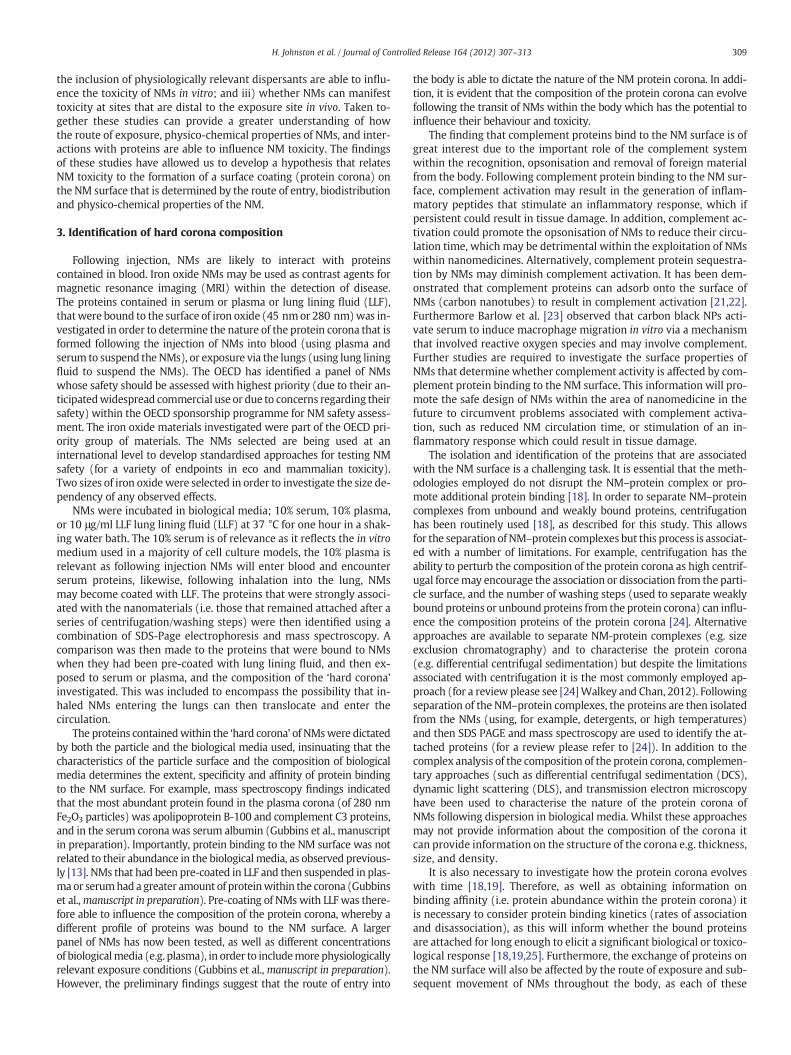
following intratracheal instillation (doses administered ranged from1 to 128 μg/mouse) it was observed that glutathione (GSH) levelswere depleted in the liver, but that this response was not evidentfor all NMs tested (Gosens et al., 2012, manuscript in preparation,Fig. 3). This correlated with the findings of in vitro investigations whereglutathione depletion was observed within hepatocytes (Kermanizadehet al.,manuscript submitted). Surprisingly, nanomaterials that did not in-duce a significant lung inflammatory response (e.g. TiO2) were still ableto induce glutathione depletion in the liver. The inflammatory andoxida-tive responses within the liver were further investigated using PCR. Itwas demonstrated that there was a significant decrease in the levels ofC3 gene expression while higher levels of both IL-6 and IL-10 weredetected and no change in the expression of TNFα and CXCL2 was ob-served (Gosens et al., 2012 manuscript in preparation, Fig. 4). It is nownecessary to elucidate whether these effects are manifested due to thetranslocation of NMs to the liver, or whether systemically acting factorsare produced at the exposure site.
6. Discussion
On entering the body, it is inevitable that NMs will interact withbiological molecules. The small size and large surface area of NMs al-lows for the adsorption of biological molecules such as protein. Thecomposition of the protein coating (or corona) is likely to be dictatedby the route of exposure. The translocation of NMs from their expo-sure site to secondary target sites is likely to influence NM behaviourand toxicity due to the acquisition of different surface coatings duringtheir movement in the body, indicating that the protein corona is acomplex and dynamic structure. The widespread use of NMs withindiverse applications means that exposure via inhalation, ingestion,dermal application and injection is expected within occupational,consumer and environmental settings. Within the described researcha focus has been placed on the fate and toxicity of NMs following in-halation and injection. However, future studies should assess the fateof NMs following ingestion and dermal exposure, as well as how thisinfluences the composition of the NM–protein complexes that areformed and how this relates to NM physico-chemical characteristicsto affect NM toxicity.
Taken together the findings of the three independent researchstudies described demonstrate that the surface properties of NMsare important to their toxicity, and that the formation of NM–proteincomplexes is influenced by the exposure route and physico-chemicalproperties of NMs. Specifically, the following important observationswere made; i) the composition of the protein corona of NMs is likely
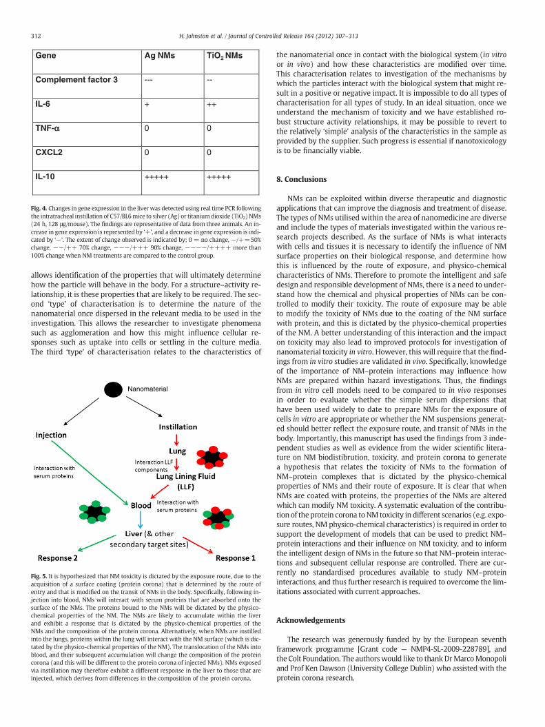
to be dictated by their route of entry into the body, ii) the transloca-tion of NMs to secondary target sites can influence the composition ofthe protein corona (i.e. they encounter different proteins on their move-ment in the body) so that the composition of the protein corona evolvesthroughout NM's lifetime in the body, iii) the physico-chemical charac-teristics of NMs dictate the composition of the protein corona, and thetoxicity of NMs, and iv) NMs can affect secondary target sites followingexposure. We therefore hypothesise that NM toxicity is dictated by theexposure route, due to the acquisition of a surface coating (protein coro-na) that is determined by the route of entry and that is modified on thetransit of NMs in the body (Fig. 5). This hypothesis therefore needs to betested further, in order to determine its applicability to more diverseforms of NMs, and exposure routes. In this respect, a number of lessonscan be learnt from the drug delivery field (e.g. opsonisation).
Binding of proteins to the NM surface (to form the protein corona)is able to influence the toxicity of NMs. Not only is the biological ac-tivity of the NM modified by the adsorption of proteins onto the NMsurface, but structural changes in adsorbed proteins have also beenobserved, which can have implications for normal protein function(e.g. [27,28]). Ongoing research in this area will help improve our un-derstanding of the composition of the protein corona, and the impli-cations of NM–protein complex formation.
7. Physico-chemical characterisation
There is increasing pressure frommultiple sources (e.g. reviewers,journals, and peers) to conduct physico-chemical characterisation ofnanomaterials used in toxicology studies. There are multiple pur-poses for such characterisation. The first involves characterisation ofthe pristine particles as provided by the supplier. This characterisa-tion allows the researcher to verify the properties of the substancethat they are about to investigate. In addition, this characterisation
Gene Ag NMs TiO2 NMs
Complement factor 3 --- --
IL-6 + ++
TNF-αα 0 0
CXCL2 0 0
IL-10 +++++ +++++
Fig. 4. Changes in gene expression in the liver was detected using real time PCR followingthe intratracheal instillation of C57/BL6mice to silver (Ag) or titaniumdioxide (TiO2) NMs(24 h, 128 μg/mouse). The findings are representative of data from three animals. An in-crease in gene expression is represented by ‘+’, and a decrease in gene expression is indi-cated by ‘−’. The extent of change observed is indicated by; 0 = no change, −/+=50%change, −−/++ 70% change, −−−/+++ 90% change, −−−−/++++ more than100% change when NM treatments are compared to the control group.
312 H. Johnston et al. / Journal of Controlled Release 164 (2012) 307–313
allows identification of the properties that will ultimately determinehow the particle will behave in the body. For a structure–activity re-lationship, it is these properties that are likely to be required. The sec-ond ‘type’ of characterisation is to determine the nature of thenanomaterial once dispersed in the relevant media to be used in theinvestigation. This allows the researcher to investigate phenomenasuch as agglomeration and how this might influence cellular re-sponses such as uptake into cells or settling in the culture media.The third ‘type’ of characterisation relates to the characteristics of
Nanomaterial
Fig. 5. It is hypothesized that NM toxicity is dictated by the exposure route, due to theacquisition of a surface coating (protein corona) that is determined by the route ofentry and that is modified on the transit of NMs in the body. Specifically, following in-jection into blood, NMs will interact with serum proteins that are absorbed onto thesurface of the NMs. The proteins bound to the NMs will be dictated by the physico-chemical properties of the NM. The NMs are likely to accumulate within the liverand exhibit a response that is dictated by the physico-chemical properties of theNMs and the composition of the protein corona. Alternatively, when NMs are instilledinto the lungs, proteins within the lung will interact with the NM surface (which is dic-tated by the physico-chemical properties of the NM). The translocation of the NMs intoblood, and their subsequent accumulation will change the composition of the proteincorona (and this will be different to the protein corona of injected NMs). NMs exposedvia instillation may therefore exhibit a different response in the liver to those that areinjected, which derives from differences in the composition of the protein corona.
the nanomaterial once in contact with the biological system (in vitroor in vivo) and how these characteristics are modified over time.This characterisation relates to investigation of the mechanisms bywhich the particles interact with the biological system that might re-sult in a positive or negative impact. It is impossible to do all types ofcharacterisation for all types of study. In an ideal situation, once weunderstand the mechanism of toxicity and we have established ro-bust structure activity relationships, it may be possible to revert tothe relatively ‘simple’ analysis of the characteristics in the sample asprovided by the supplier. Such progress is essential if nanotoxicologyis to be financially viable.
8. Conclusions
NMs can be exploited within diverse therapeutic and diagnosticapplications that can improve the diagnosis and treatment of disease.The types of NMs utilised within the area of nanomedicine are diverseand include the types of materials investigated within the various re-search projects described. As the surface of NMs is what interactswith cells and tissues it is necessary to identify the influence of NMsurface properties on their biological response, and determine howthis is influenced by the route of exposure, and physico-chemicalcharacteristics of NMs. Therefore to promote the intelligent and safedesign and responsible development of NMs, there is a need to under-stand how the chemical and physical properties of NMs can be con-trolled to modify their toxicity. The route of exposure may be ableto modify the toxicity of NMs due to the coating of the NM surfacewith protein, and this is dictated by the physico-chemical propertiesof the NM. A better understanding of this interaction and the impacton toxicity may also lead to improved protocols for investigation ofnanomaterial toxicity in vitro. However, this will require that the find-ings from in vitro studies are validated in vivo. Specifically, knowledgeof the importance of NM–protein interactions may influence howNMs are prepared within hazard investigations. Thus, the findingsfrom in vitro cell models need to be compared to in vivo responsesin order to evaluate whether the simple serum dispersions thathave been used widely to date to prepare NMs for the exposure ofcells in vitro are appropriate or whether the NM suspensions generat-ed should better reflect the exposure route, and transit of NMs in thebody. Importantly, this manuscript has used the findings from 3 inde-pendent studies as well as evidence from the wider scientific litera-ture on NM biodistibrution, toxicity, and protein corona to generatea hypothesis that relates the toxicity of NMs to the formation ofNM–protein complexes that is dictated by the physico-chemicalproperties of NMs and their route of exposure. It is clear that whenNMs are coated with proteins, the properties of the NMs are alteredwhich can modify NM toxicity. A systematic evaluation of the contribu-tion of theprotein corona toNM toxicity in different scenarios (e.g. expo-sure routes, NM physico-chemical characteristics) is required in order tosupport the development of models that can be used to predict NM–
protein interactions and their influence on NM toxicity, and to informthe intelligent design of NMs in the future so that NM–protein interac-tions and subsequent cellular response are controlled. There are cur-rently no standardised procedures available to study NM–proteininteractions, and thus further research is required to overcome the lim-itations associated with current approaches.
Acknowledgements
The research was generously funded by by the European seventhframework programme [Grant code — NMP4-SL-2009-228789], andthe Colt Foundation. The authorswould like to thankDrMarcoMonopoliand Prof Ken Dawson (University College Dublin) who assisted with theprotein corona research.
313H. Johnston et al. / Journal of Controlled Release 164 (2012) 307–313
References
[1] J. Ferin, G. Oberdorster, D.P. Penney, Pulmonary retention of ultrafine and fineparticles in rats, AJR: Cell. Mol. Biol. 6 (1992) 535–542.
[2] X.Y. Li, D. Brown, S. Smith, W. MacNee, K. Donaldson, Short-term inflammatoryresponses following intratracheal instillation of fine and ultrafine carbon blackin rats, Inhal. Toxicol. 11 (1999) 709–731.
[3] D.M. Brown, M.R. Wilson, W. MacNee, V. Stone, K. Donaldson, Size-dependentproinflammatory effects of ultrafine polystyrene particles: a role for surface areaand oxidative stress in the enhanced activity of ultrafines, Toxicol. Appl. Pharmacol.175 (2001) 191–199.
[4] B.K. Gaiser, T.F. Fernandes, M. Jepson, J.R. Lead, C.R. Tyler, V. Stone, Assessing ex-posure, uptake and toxicity of silver and cerium dioxide nanoparticles from con-taminated environments, Environ. Health 8 (2009).
[5] R. Duffin, C.L. Tran, A. Clouter, D.M. Brown, W. MacNee, V. Stone, K. Donaldson,The importance of surface area and specific reactivity in the acute pulmonary in-flammatory response to particles, Ann. Occup. Hyg. 46 (2002) 242–245.
[6] X. Ma, Y. Zhao, X.J. Liang, Theranostic nanoparticles engineered for clinic andpharmaceutics, Acc. Chem. Res. 44 (2011) 1114–1122.
[7] K. Kostarelos, A. Bianco, M. Prato, Promises, facts and challenges for carbonnanotubes in imaging and therapeutics, Nat. Nanotechnol. 10 (2009) 627–633.
[8] S. Laurent, J.L. Bridot, L.V. Elst, R.N. Muller, Magnetic iron oxide nanoparticles forbiomedical applications, Future Med. Chem. 2 (2010) 427–449.
[9] C.M. Cobley, J. Chen, E.C. Cho, L.V. Wang, Y. Xia, Gold nanostructures: a class ofmultifunctional materials for biomedical applications, Chem. Soc. Rev. 40 (2011)44–56.
[10] F. Alexis, E. Pridgen, L.K. Molnar, O.C. Farokhzad, Factors affecting the clearanceand biodistribution of polymeric nanoparticles, Mol. Pharm. 4 (2008) 505–515.
[11] M. Semmler-Behnke, W.G. Kreyling, J. Lipka, S. Fertsch, A. Wenk, S. Takenaka, G.Schmid, W. Brandau, Biodistribution of 1.4- and 18-nm gold particles in rats,Small 4 (2008) 2108–2111.
[12] M. Lundqvist, J. Stigler, T. Cedervall, T. Berggård, M.B. Flanagan, I. Lynch, G. Elia, K.Dawson, The evolution of the protein corona around nanoparticles: a test study,ACS Nano 5 (2011) 7503–7509.
[13] S. Tenzer, D. Docter, S. Rosfa, A. Wlodarski, J. Kuharev, A. Rekik, S.K. Knauer, C.Bantz, T. Nawroth, C. Bier, J. Sirirattanapan, W. Mann, L. Treuel, R. Zellner, M.Maskos, H. Schild, R.H. Stauber, Nanoparticle size is a critical physicochemical de-terminant of the human blood plasma corona: a comprehensive quantitative pro-teomic analysis, ACS Nano 5 (2012) 7155–7167.
[14] D.M. Brown, C. Dickson, P. Duncan, F. Al-Attili, V. Stone, Interaction betweennanoparticles and cytokine proteins: impact on protein and particle functionality,Nanotechnology 21 (2010) 215104.
[15] M. Lundqvist, J. Stigler, G. Elia, I. Lynch, T. Cedervall, K.A. Dawson, Nanoparticle sizeand surface properties determine the protein corona with possible implications forbiological impacts, Proc. Natl. Acad. Sci. U. S. A. 105 (2008) 14265–14270.
[16] M.A. Dobrovolskaia, A.K. Patri, J. Zheng, J.D. Clogston, N. Ayub, P. Aggarwal, B.W.Neun, J.B. Hall, S.E. McNeil, Interaction of colloidal gold nanoparticles withhuman blood: effects on particle size and analysis of plasma protein binding pro-files, Nanomedicine 5 (2009) 106–117.
[17] Z.J. Deng, M. Liang, I. Toth, M. Monteiro, R.F. Minchin, Plasma protein binding ofpositively and negatively charged polymer-coated gold nanoparticles elicitsdifferent biological responses, Nanotoxicology (2012), http://dx.doi.org/10.3109/17435390.2012.655342 (Epub ahead of print).
[18] T. Cedervall, I. Lynch, S. Lindman, T. Berggard, E. Thulin, H. Nilsson, K.A. Dawson, S.Linse, Understanding the nanoparticle–protein corona using methods to quantifyexchange rates and affinities of proteins for nanoparticles, Proc. Natl. Acad. Sci.U. S. A. 104 (2007) 2050–2055.
[19] E. Casals, T. Pfaller, A. Duschl, G.J. Oostingh, V. Puntes, Time evolution of the nano-particle protein corona, ACS Nano 4 (2010) 3623–3632.
[20] M.J. Clift, B. Rothen-Rutishauser, D.M. Brown, R. Duffin, K. Donaldson, L. Proudfoot, K.Guy, V. Stone, The impact of different nanoparticle surface chemistry and size on up-take and toxicity in a murine macrophage cell line, Toxicol. Appl. Pharmacol. 232(2008) 418–427.
[21] C. Salvador-Morales, E. Flahaut, E. Simc, J. Sloan,M.L.H. Green, R.B. Sim, Complementactivation and protein adsorption by carbon nanotubes, Mol. Immunol. 43 (2006)193–201.
[23] P.G. Barlow, K. Donaldson, J. Maccallum, A. Clouter, V. Stone, Serum exposed tonanoparticle carbon black displays increased potential to induce macrophage mi-gration, Toxicol. Lett. 155 (2005) 397–401.
[24] C.D. Walkey, W.C. Chan, Understanding and controlling the interaction ofnanomaterials with proteins in a physiological environment, Chem. Soc. Rev.41 (2012) 2780–2799.
[25] D. Walczyk, F.B. Bombelli, M.P. Monopoli, I. Lynch, K.A. Dawson, What the cell“sees” in bionanoscience, J. Am. Chem. Soc. 132 (2010) 5761–5768.
[26] A. Kermanizadeh, G. Pojana, B.K. Gaiser, R. Birkedal, D. Bilaničová, H. Wallin, K.A.Jensen, B. SellergrenB, G.R. Hutchison, A. Marcomini, V. Stone, In vitro assessmentof engineered nanomaterials using a hepatocyte cell line: cytotoxicity, pro-inflammatory cytokines and functional markers, Nanotoxicology (2012), http://dx.doi.org/10.3109/17435390.2011.653416 (Epub ahead of print).
[27] J. Wang, U.B. Jensen, G.V. Jensen, S. Shipovskov, V.S. Balakrishnan, D. Otzen, J.S.Pedersen, F. Besenbacher, D.S. Sutherland, Soft interactions at nanoparticlesalter protein function and conformation in a size dependent manner, Nano Lett.11 (2011) 4985–4991.