Ahmet Çetin 1 , Aykut Özgür 2 , Mehmet Kuzucu 3 , Murat Çankaya 3* 1 Erzincan Binali Yıldırım University, Graduate School of Natural and Applied Sciences, Department of Biology, Erzincan, Turkey. 2 Tokat Gaziosmanpaşa University, Artova Vocaonal Sch., Depart. of Veterinary Medicine, Laboratory and Veterinary Health Program, Tokat, Turkey. 3 Erzincan Binali Yıldırım University, Faculty of Arts and Sciences, Department of Biology, Erzincan, Turkey. ÖZ H ER2 pozitif meme kanseri, her yıl milyonlarca ölüme neden olan dünyanın en büyük sağlık sorunlarından biridir. İlaç kombinasyonu modelleme çalışmaları birçok hastalığın tedavisinde kapsamlı bir şekilde değerlendirilmektedir. Son yarım yüzyıl boyunca yapılan farmakolojik çalışmalar, gambojik asidin meme kanseri de dahil olmak üzere birçok kanser türüne karşı güçlü bir anti-tümör aktiviteye sahip olduğunu göstermiştir. Bu çalışmada HER2 pozitif meme kanseri hücre hattında (MDA- MB-453) gambojik asit ve trastuzumabın sinerjistik antikanser etkisini inceledik. Trastuzumab artı gambojik asit çalışmalarının in vitro sinerjistik ve antiproliferatif etkileri XTT yöntemi ile belirlenmiş ve trastuzumab ve gambojik asit kombinasyonunun kombinasyon indeksi (CI) değerleri CompuSyn yazılımı ile hesaplanmıştır. MDA-MB-453 hücrelerinde, trastuzumab ve gambojik asit kombinasyonlarının moleküler mekanizmalarını belirlemek ve HER2, kaspaz-9 ve Bax’ın protein ve gen ekspresyonu seviyelerindeki farklılıklar RT-PCR (qRT-PCR) ve ELISA teknikleri kullanılarak analiz edildi. RT-qPCR ve ELISA testine göre; 50 µg / ml trastuzumab ve 5 µM gambojik asit kombinasyonu, MDA-MB-453 hücre hattında 24 saatlik inkübasyonda en iyi sinerjistik etkiyi gösterdi. Gambojik asitin HER2 pozitif meme kanseri hücre hattı üzerine etkisi, , trastuzumab ile kombinasyonunun meme kanseri hücre proliferasyonunu inhibe eden doğal bileşik potansiyeline sahip olduğunu gösterir. Anahtar Kelimeler Trastuzumab, gambojik asit, meme kanseri, HER-2. ABSTRACT H ER2 positive breast cancer is one of the biggest health problems in the world, causing millions of deaths every year. Drug combination modeling studies are extensively evaluated in treating many diseases. Pharmacological studies over the last half-century have shown that gambogic acid has potent anti-tumor activity against many types of cancer, including breast cancer. In this study, we examined the synergistic anticancer effect of gambogic acid and trastuzumab in HER2 positive breast cancer cell line (MDA-MB-453). In-vitro synergistic and antiproliferative effects of trastuzumab plus gambogic acid studies were determined with XTT method and the combination index (CI) values of the trastuzumab and gambogic acid combination were calculated by CompuSyn software. To determine molecular mechanisms of the trastuzumab and gambogic acid combination in MDA-MB-453 cells, the differences of gene and protein expression levels of HER2, caspase-9 and Bax were analyzed with using RT-qPCR and ELISA techniques. The combination of 50 µg/ml trastuzumab and 5 µM gambogic acid showed the best synergistic effect at 24 h incubation in MDA-MB-453 cells according to the in-vitro cell proliferation, RT-qPCR and ELISA test. Gambogic acid effects on HER2 positive breast cancer cell line shows its potential as natural compound to inhibit breast cancer cell proliferation in combination with trastuzumab. Key Words Trastuzumab, gambogic acid, breast cancer, HER-2. Article History: Received: Jan 13, 2020; Revised: Mar 15, 2020; Accepted: Mar 15, 2020; Available Online: Apr 27, 2020. DOI: https://doi.org/10.15671/hjbc.672695 Correspondence to: M. Çankaya, Erzincan Binali Yıldırım University, Faculty of Arts and Sciences, Department of Biology, Erzincan, Turkey. E-Mail: [email protected]Investigation of The Synergistic Effects of Trastuzumab And Gambogic Acid in Her-2 Positive Breast Cancer Cell Line Her-2 Pozitif Meme Kanseri Hücre Hattında Trastuzumab ve Gambojik Asit Sinerjik Etkilerinin İncelenmesi Hacettepe Journal of Biology and Chemistry A. Çetin et al. / Hacettepe J. Biol. & Chem., 2020, 48 (3), 291-299 Research Article journal homepage: www.hjbc.hacettepe.edu.tr
Transcript
Ahmet Çetin1 , Aykut Özgür2 , Mehmet Kuzucu3 , Murat Çankaya3*
1Erzincan Binali Yıldırım University, Graduate School of Natural and Applied Sciences, Department of Biology, Erzincan, Turkey.2Tokat Gaziosmanpaşa University, Artova Vocational Sch., Depart. of Veterinary Medicine, Laboratory and Veterinary Health Program, Tokat, Turkey.3Erzincan Binali Yıldırım University, Faculty of Arts and Sciences, Department of Biology, Erzincan, Turkey.
Ö Z
HER2 pozitif meme kanseri, her yıl milyonlarca ölüme neden olan dünyanın en büyük sağlık sorunlarından biridir. İlaç kombinasyonu modelleme çalışmaları birçok hastalığın tedavisinde kapsamlı bir şekilde değerlendirilmektedir. Son yarım
yüzyıl boyunca yapılan farmakolojik çalışmalar, gambojik asidin meme kanseri de dahil olmak üzere birçok kanser türüne karşı güçlü bir anti-tümör aktiviteye sahip olduğunu göstermiştir. Bu çalışmada HER2 pozitif meme kanseri hücre hattında (MDA-MB-453) gambojik asit ve trastuzumabın sinerjistik antikanser etkisini inceledik. Trastuzumab artı gambojik asit çalışmalarının in vitro sinerjistik ve antiproliferatif etkileri XTT yöntemi ile belirlenmiş ve trastuzumab ve gambojik asit kombinasyonunun kombinasyon indeksi (CI) değerleri CompuSyn yazılımı ile hesaplanmıştır. MDA-MB-453 hücrelerinde, trastuzumab ve gambojik asit kombinasyonlarının moleküler mekanizmalarını belirlemek ve HER2, kaspaz-9 ve Bax’ın protein ve gen ekspresyonu seviyelerindeki farklılıklar RT-PCR (qRT-PCR) ve ELISA teknikleri kullanılarak analiz edildi. RT-qPCR ve ELISA testine göre; 50 µg / ml trastuzumab ve 5 µM gambojik asit kombinasyonu, MDA-MB-453 hücre hattında 24 saatlik inkübasyonda en iyi sinerjistik etkiyi gösterdi. Gambojik asitin HER2 pozitif meme kanseri hücre hattı üzerine etkisi, , trastuzumab ile kombinasyonunun meme kanseri hücre proliferasyonunu inhibe eden doğal bileşik potansiyeline sahip olduğunu gösterir.
HER2 positive breast cancer is one of the biggest health problems in the world, causing millions of deaths every year. Drug combination modeling studies are extensively evaluated in treating many diseases. Pharmacological studies over the last
half-century have shown that gambogic acid has potent anti-tumor activity against many types of cancer, including breast cancer. In this study, we examined the synergistic anticancer effect of gambogic acid and trastuzumab in HER2 positive breast cancer cell line (MDA-MB-453). In-vitro synergistic and antiproliferative effects of trastuzumab plus gambogic acid studies were determined with XTT method and the combination index (CI) values of the trastuzumab and gambogic acid combination were calculated by CompuSyn software. To determine molecular mechanisms of the trastuzumab and gambogic acid combination in MDA-MB-453 cells, the differences of gene and protein expression levels of HER2, caspase-9 and Bax were analyzed with using RT-qPCR and ELISA techniques. The combination of 50 µg/ml trastuzumab and 5 µM gambogic acid showed the best synergistic effect at 24 h incubation in MDA-MB-453 cells according to the in-vitro cell proliferation, RT-qPCR and ELISA test. Gambogic acid effects on HER2 positive breast cancer cell line shows its potential as natural compound to inhibit breast cancer cell proliferation in combination with trastuzumab.
Key Words Trastuzumab, gambogic acid, breast cancer, HER-2.
Article History: Received: Jan 13, 2020; Revised: Mar 15, 2020; Accepted: Mar 15, 2020; Available Online: Apr 27, 2020.
DOI: https://doi.org/10.15671/hjbc.672695
Correspondence to: M. Çankaya, Erzincan Binali Yıldırım University, Faculty of Arts and Sciences, Department of Biology, Erzincan, Turkey.
A. Çetin et al. / Hacettepe J. Biol. & Chem., 2020, 48 (3), 291-299292
INTRODUCTION
Breast cancer is the most diagnosed cancer type in women worldwide, with approximately 2 million
new cases diagnosed and 627,000 patients died in 2018 [1]. Breast cancer is mainly categorized into three types: hormone receptor-positive (estrogen receptor and pro-gesterone receptor), human epidermal growth factor receptor-2 positive (HER2-positive, HER2+) and triple-negative breast cancer. Amplification of HER2 gene or overexpression of HER2 protein is called HER2-positive and these processes cause occurrence and progression of tumorigenesis in normal breast cells. HER2 is a trans-membrane tyrosine kinase receptor which is over-exp-ressed in about 10-20 percent of breast cancers. There are several treatment strategies available for HER2-positive breast cancer, depending on the type and stage [2,3].
Chemotherapy is one of the most important steps in treatment of HER2-positive breast cancer and espe-cially, development of HER2 targeted drugs has been significant therapeutic strategy in breast cancer. Tras-tuzumab (Herceptin®) is a recombinant DNA-derived humanized monoclonal antibody which selectively in-hibits HER2-positive breast cancer tumorigenesis alone or in combination with other chemotherapeutics and natural products. HER-2 receptor consists of three con-served domains: extracellular ligand-binding domain, a transmembrane region, and an intracellular (cytoplas-mic tyrosine kinase) domain. Trastuzumab binds to the extracellular domain of HER2 receptor with high affinity and prevents cleavage of this domain, resulting in inter-ruption of cancer cell survival [4,5]. In order to increase therapeutic efficiency, administration of trastuzumab in combination with FDA approved drugs and natural products have been investigated in pre-clinical and clinical studies in cancer. In early and advanced HER2-positive breast cancer patients, trastuzumab is used in combination with paclitaxel, docetaxel, carboplatin [6-8]. Overexpression of HER2 is associated with resistance to hormonal therapy (particularly tamoxifen) in breast cancer. Therefore, combining hormonal agents, tamo-xifen and aromatase inhibitors, with trastuzumab may be potential therapeutic aspect as hormonal therapy in breast cancer [9].
Gambogic acid is a natural product which is originally isolated from Garcinia hanburyi tree grown in Southe-ast Asia. Traditionally, gambogic acid has been used in
treatment of many different diseases for a long time. Numerous studies reported that, gambogic acid pos-sesses diverse biological properties such as anti-can-cer, anti-microbial, anti-oxidant and anti-inflammatory [10,11]. In recent decades, biological activities of gam-bogic acid have been investigated in almost all steps of tumourigenesis, and it inhibits the proliferation of va-rious human cancer cells. According to in-vitro and in-vivo studies, gambogic acid induces apoptosis, inhibits angiogenesis and overcomes drug resistance in human cancer cells via different signaling pathways [10-13]. Gambogic acid has been approved for phase II clinical trial for solid tumor therapy by the Chinese Food and Drug Administration [14]. However, the anticancer mec-hanisms of the gambogic acid are not fully understood yet. Therefore, further molecular studies are needed to understand the biological effect of the gambogic acid in cancer.
In this study, we examined the combined effect of tras-tuzumab and gambogic acid as well as its mechanism of action was determined by using RT-PCR and ELISA techniques on HER2-positive human breast cancer cell line, MDA-MB-453. Obtained results indicated that gambogic acid plus trastuzumab directly decreased cell proliferation and induced inhibition of HER-2 signaling pathway. Moreover, this combination affects apoptosis via caspase-9 and Bax pathways. Combined therapy of trastuzumab and gambogic acid may be promising me-dicinal compound to treat HER-2 positive breast cancer.
MATERIALS and METHODS
MaterialsMDA-MB-453 cell line was from ATCC (American Type Culture Collection, USA). Dulbecco’s modified Eagle’s medium (DMEM), heat-inactivated fetal bovine se-rum, trypsin–EDTA, phosphate buffer saline (PBS), L-glutamine, penicillin-streptomycin and XTT ((2,3-bis-(2-methoxy-4-nitro-5-sulfophenyl)-2H-tetrazolium-5-carboxanilide) cell proliferation kit was obtained from Biological Industries Ltd. Gambogic acid was purchased from Abcam. Trastuzumab was supplied from Roche. RNA isolation and cDNA synthesis kits were purchased from Thermo Scientific. Human Bax, HER-2 and caspa-se-9 ELISA kits were from Sinogeneclon Co., Ltd. All ot-her chemical reagents were purchased from Merck and Sigma Aldrich.
A. Çetin et al. / Hacettepe J. Biol. & Chem., 2020, 48 (3), 291-299 293
Cell line and cultureFor in-vitro experiments, MDA-MB-453 (HER2-positive human breast cancer) cell line was cultured in DMEM (high glucose) medium with 10% fetal bovine serum, 1% l-glutamine, 100 IU/mL penicillin and 10 mg/mL strep-tomycin. Cells were cultivated in a humidified incubator at 37°C within an atmosphere containing 5% CO2. Cell proliferation assay The XTT test was used to quantify the number of viab-le cells in each of the well in different concentrations [15,16]. Initially, the cancer cells were seeded in sterile 96-well culture plate (10x104 cells in each well), and the different concentrations of gambogic acid (10-5-2.5-1.25-0.625-0.3125 µM), trastuzumab (100-50-25-12.5-6.25-3.125 µg/ml) and gambogic acid+trastuzumab were incubated with cells for 12-72 hours at 37 °C in a humidified incubator within an atmosphere containing 5% CO2. At the end of the incubation times, 50 µl XTT re-agents were added to each well for determination of li-ving cells. After 4h, the absorbance was measured using micro plate reader at 450 nm, and then the percentage of cell viability was calculated.
Combined effect analysisThe interactions of the gambogic acid and trastuzumab were determined with using the combination index (CI) method (median-effect principle). CI values of gambo-gic acid and trastuzumab were calculated using Com-puSyn free software. To enhance therapeutic efficiency and minimize resistance of drugs, combination chemot-herapy is widely used in treatment of various diseases such as cancer. The Chou-Talalay method based on the median-effect equation was developed for analyzing drug combinations quantitatively. Further, this method
encompasses the Michaelis-Menten, Henderson-Has-selbalch, Scatchard, and Hill equations. In this context, CompuSyn software (ComboSyn, Inc., Paramus, NJ, USA) is analyzed drug combinations using median-ef-fect principle, and calculated CI values to determine combination types (CI<1, CI=1 and CI>1 was accepted as synergism, addition and antagonism, respectively (Table-1) [17].
RT-qPCR AnalysisTo determine the alteration of HER2, Bax, caspa-se-9 and GAPDH gene expressions with gambogic acid (5 µM), trastuzumab (50 µg/ml) and gambogic acid+trastuzumab (5 µM+50 µg/ml) at the end of the 24h incubation, qPCR experiments were carried out on MDA-MB-453 cells. Total RNA was extracted and first strand cDNA synthesis made according to the manufacturer’s protocols. Primers were designed using Primer 3.0 program and synthesized by Macrogen Inc., with the following sequences (5′ to 3′) and qPCR condi-tions: HER-2 forward: TTGTGGCCTTCTTTGAGTTCGGTG and reverse: GGTGCCGGTTCAGGTACTCAGTCA at 95°C (10 min) followed by 45 cycles of 95°C (30 sec), 59°C (30 sec) and 72°C (45 sec), caspase-9 forward: CGA-ACTAACAGGCAAGCAGC and reverse: ACCTCACCAA-ATCCTCCAGAAC at 95°C (10 min) followed by 45 cycles of 95°C (15 sec), 60°C (1 min) and 72°C (45 sec), Bax forward: CCTGTGCACCAAGGTGCCGGAACT and rever-se: CCACCCTGGTCTTGGATCCAGCCC at 95°C (10 min) followed by 45 cycles of 95°C (30 sec), 59°C (30 sec) and 72°C (30 sec), GAPDH forward: CGAACTAACAGGCAAG-CAGC and reverse: TCGCCCCACTTGATTTTGG at 95°C (10 min) followed by 45 cycles of 95°C (15 sec), 64°C (30 sec) and 72°C (15 sec). qPCR was carried out using a SYBR Green Master Mix (Qiagen-330500) with a reacti-
CI>1 Antagonism
CI=1.1-1.2 Slight antagonism
CI=1.2-1.45 Moderate antagonism
CI=1.45-1.33 Antagonism
CI=3.3-10 Strong antagonism
CI>10 Very strong antagonism
CI=1 Additive CI=1 Additive
CI<1Synergism
CI=0.85-0.9 Slight synergism
CI=0.7-0.85 Moderate synergism
CI=0.3-0.7 Synergism
CI=0.1-0.3 Strong synergism
CI<0.1 Very strong synergism
Table 1. Definition of CI index values.
A. Çetin et al. / Hacettepe J. Biol. & Chem., 2020, 48 (3), 291-299294
on mixture containing 1 µl cDNA, 2 µl each primer and 12,5 µl SYBR Green Master Mix (total volume 25 µl). All assays were run in triplicate (Qiagen Rotor Gene). CT values were assessed and relative expression of target genes was determined using the 2−ΔΔCt method.
ELISA TestsTo investigate protein expression levels of the HER2, Bax and caspase-9 with gambogic acid (5 µM), trastuzumab (50 µg/ml) and gambogic acid+trastuzumab (5 µM+50 µg/ml) at the end of the 24h incubation, commercial sandwich ELISA kits were used on MDA-MB-453 cells according to the manufacturers’ instructions. Expressi-on levels of the proteins were measured spectrophoto-metrically.
Statistical AnalysisDifferences in the mean values of measured activities were evaluated statistically using the SPSS 17.0 prog-ram (Univariate Variance Analyses and Pearson Corre-lation). Probability values of p < 0.05 were considered to be significant.
RESULTS AND DISCUSSION
Cell Proliferation AssayThe cytotoxic activities of trastuzumab and gambogic acid were evaluated on MDA-MB-453 cell line by XTT assay individually and in combination. CI values of tras-tuzumab-gambogic acid combination were determined with CompuSyn software and Table-2 shows mean CI values of combinations at different incubation times. Gambogic acid was dissolved in DMSO and diluted in DMEM before cell proliferation assay. The control cells were treated with DMEM containing 0.1% DMSO to de-termine cytotoxicity of the gambogic acid. We treated MDA-MB-453 cells with different concentrations of trastuzumab 100-50-25-12.5-6.25-3.125 µg/ml, 10-5-2.5-1.25-0.625-0.3125 µM gambogic acid and the com-bination of trastuzumab and gambogic acid during 12, 24 and 48h.
Significant anti-proliferative activity did not observe when the treatment was applied for 12h. Treatment with gambogic acid alone as well as the combination of trastuzumab significantly inhibited cell proliferation after 24h and 48h. trastuzumab did not serve essential anti-proliferative activity from 12h to 48h in 100-3.125 µg/ml concentrations range. However, trastuzumab in-hibited cell proliferation in combination with gambogic acid for certain concentrations for 24 and 48h (Figu-re-1-3). Trastuzumab is routinely used in the first-line treatment of patients with advanced breast cancers that express HER2. Nevertheless, initial and eventual resistance to HER2-based therapy with trastuzumab is frequently observed in significant number of patients with HER2-positive breast cancer. To overcome tras-tuzumab-induced resistance mechanisms in HER-2 po-sitive breast cancer, incorporation of new compounds in combinational therapy with trastuzumab are being extensively studied in pre-clinical and clinical studies [18,19].
100 µg/ml trastuzumab +10 µM gambogic acid, 50 µg/ml trastuzumab +5 µM gambogic acid, and 25 µg/ml trastuzumab +2,5 µM gambogic acid significantly re-duced cell viability when the treatment was applied for 48h, and 50% reduction in cell viability was achieved with these combinations. Moreover, 50 µg/ml trastu-zumab+5 µM gambogic acid and 25 µg/ml trastuzu-mab+2,5 µM gambogic acid exhibited anti-proliferative activity during 24h against MDA-MB-453 cells. Briefly, 50 µg/ml trastuzumab+5 µM gambogic acid, and 25 µg/ml trastuzumab+2,5 µM gambogic acid showed similar combinational effect to inhibit survival of MDA-MB-453 cells in 24 and 48h. According to the CI values of the trastuzumab-gambogic acid combinations (Table-2), 50 µg/ml trastuzumab+5 µM gambogic acid exhibited the lowest CI values for 24h and therefore this combinati-on was accepted as optimum for HER-2 positive breast cancer cells.
Table 2. CI values of the trastuzumab-gambogic acid combinations.
A. Çetin et al. / Hacettepe J. Biol. & Chem., 2020, 48 (3), 291-299 295
Figure 1. Antitumor activity of trastuzumab, gambogic acid and trastuzumab+gambogic acid for MDA-MB-453 cell line at 12h.
Figure 2. Antitumor activity of trastuzumab, gambogic acid and trastuzumab+gambogic acid for MDA-MB-453 cell line at 24h.
A. Çetin et al. / Hacettepe J. Biol. & Chem., 2020, 48 (3), 291-299296
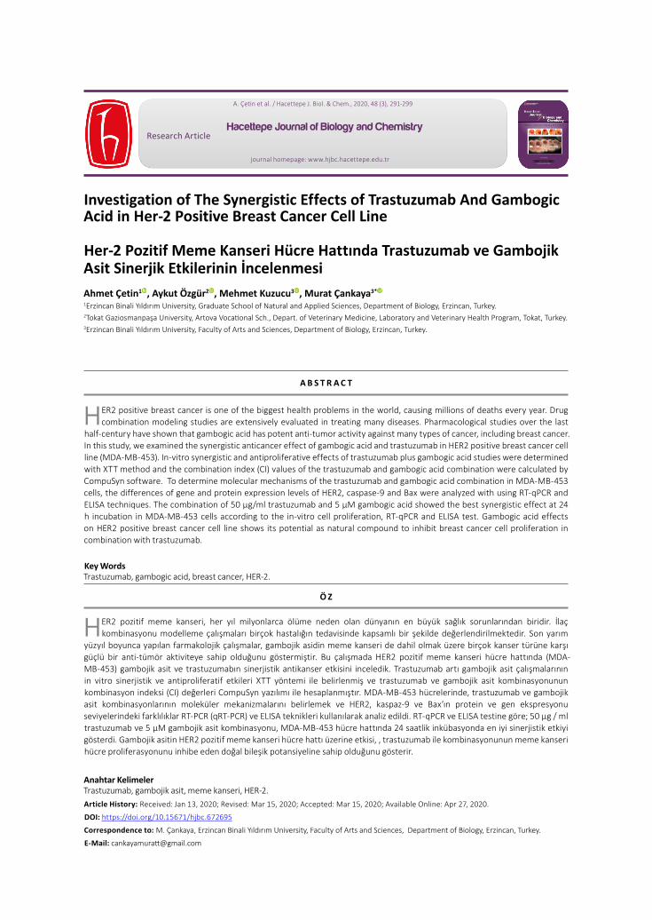
RT-qPCR Analysis and ELISA TestsGene expression analysis was performed 50 µg/ml TRS and 5 µM gambogic acid combinations at 24h with the best agonistic effect calculated as CI 0.50 ± 0.03. Almost all cancer patients, upregulation of HER2 expression at the gene and protein level have been correlated with poor prognosis in breast cancer [20]. In this study, it was shown that treatment with combination of trastuzumab and gambogic acid suppressed HER2 expression levels better than individual treatment (Figure-4). In cells tre-ated with a combination of 50 µg/ml trastuzumab and 5 µM gambogic acid, the ELISA findings of HER2 prote-in decreased by 60.17% (36.4 IU/L) compared to cont-rol, correlating with decreased gene expression levels (Table-3). Gambogic acid increasing anti-proliferative activity of trastuzumab, suggests that the use of gam-bogic acid with trastuzumab as an adjunct therapy in the treatment of HER2 positive breast cancer may be more effective.
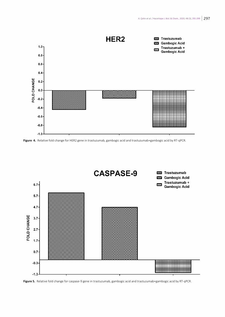
The intrinsic apoptotic pathway begins with the relea-se of cytochrome-C (Cyt-C) into the cytosol, causing a change on the permeability of mitochondrial memb-rane by Bax (B-cell lymphoma-2-associated X) protein. Apoptotic Protease Activating Factor-1 (Apaf-1) after release of Cyt-C, polymerizes in an dATP or ATP depen-dent mechanism [21,22]. Apaf-1 enabling the assembly of the apoptosome, activates caspase-9 (cas-9) and in
this manner initiating the caspase cascade [20]. Accor-ding to the findings trastuzumab and gambogic acid in-duced to intrinsic apoptotic pathway individually. The gene expression level of cas-9 was found to increase approximately 6 and 5 times (Figure-5) in trastuzumab and gambogic acid treatment, respectively. On the other hand, in cells treated with a combination of 50 µg / ml trastuzumab and 5 µM gambogic acid, ELISA results of cas-9 protein level was determined as lo-west (21 ng/ml) and correlating with gene expression levels (Table-3). This maybe suggests that treated with a combination of trastuzumab and gambogic acid indu-ces cell death by a pathway other than intrinsic apop-tosis. In order to explain this situation to be precise, it is necessary to investigate gene expression levels of some genes such as Bcl2, Apaf-1 and protein levels by western-blot in addition to flow-cytometry. According to the results of gene expression analysis, when tras-tuzumab and gambogic acid were applied separately, it was found to down-regulate Bax gene (0.5 and 1.5 fold change, respectively). When ELISA results are examined, it is seen that there is a similar change in the amount of protein supporting these results (Table-3). Conversely, trastuzumab-gambogic acid combination treatment (50 µg/ml trastuzumab and 5 µM gambogic acid) increased Bax gene expression (Figure-6) and amount of protein. In this case it may be considered an indication of the formation of trastuzumab resistance.
Figure 3. Antitumor activity of trastuzumab, gambogic acid and trastuzumab+gambogic acid for MDA-MB-453 cell line at 48h.
A. Çetin et al. / Hacettepe J. Biol. & Chem., 2020, 48 (3), 291-299 297
Figure 4. Relative fold change for HER2 gene in trastuzumab, gambogic acid and trastuzumab+gambogic acid by RT-qPCR.
Figure 5. Relative fold change for caspase-9 gene in trastuzumab, gambogic acid and trastuzumab+gambogic acid by RT-qPCR.
A. Çetin et al. / Hacettepe J. Biol. & Chem., 2020, 48 (3), 291-299298
Trastuzumab is the standard treatment for HER2 positive breast cancer and significantly improved clinical outcomes. Nevertheless, approximately 50% of HER2 positive breast cancer patients can not heal with this drug [23]. Compared to monotherapy, agonistic growth inhibition and anti-pro-liferative effect was achieved by the combination treat-ment of gambogic acid and trastuzumab. Findings showed significant agonistic cytotoxic effect in MDA-MB-453 cells at 24th hour 50 µg / ml trastuzumab and 5 µM gambogic acid according to the cell proliferation, RT-qPCR and ELISA studies.
ConclusionTrastuzumab is clinically used target specific drug to treat either early-stage or advanced-stage/metastatic HER-2 positive breast cancer. Nevertheless, resistance to trastuzumab is often observed in significant number of patients with HER-2 positive breast cancer. To increa-
se therapeutic efficiency and minimize drug resistance of trastuzumab is important research topics in HER2-positive breast cancer. Therefore, many natural and synthetic compounds have been utilized in combinati-onal therapy with trastuzumab. In this study, gambo-gic acid has increased anti-proliferative activity of the trastuzumab according to the cytotoxicity, RT-qPCR and ELISA tests. Especially, gambogic acid+trastuzumab was dramatically decreased expression level of the HER-2 at gene and protein level. Further, this combination may stimulate cell death by a pathway other than intrinsic apoptosis according to the expression of Bax and cas-pase-9 in MDA-MB-453 cells. In conclusion, gambogic acid-trastuzumab combination can be suggested as a potent candidate for treatment of HER-2 positive breast cancer.
Figure 6. Antitumor activity of trastuzumab, gambogic acid and trastuzumab+gambogic acid for MDA-MB-453 cell line at 48h.
Table 3.ELISA Tests results of trastuzumab, gambogic acid and trastuzumab+gambogic acid.
A. Çetin et al. / Hacettepe J. Biol. & Chem., 2020, 48 (3), 291-299 299
Conflict of InterestAuthors declare that he has no conflict of interest.
Acknowledgments - This study was supported by Erzincan Binali Yıldırım University Scientific Research Projects Coordinatorship,
project no. FDK-2018-565.
R e f e r e n c e s
1. F. Bray, J. Ferlay, I. Soerjomataram, R.L. Siegel, L.A. Torre, A. Jemal, Global cancer statistics 2018: GLOBOCAN estimates of incidence and mortality worldwide for 36 cancers in 185 countries, CA Cancer J. Clin., 68 (2018) 394-424.
2. H.M. Asif, S. Sultana, S. Ahmed, N. Akhtar, M. Tariq, HER-2 Positive Breast Cancer - a Mini-Review, Asian Pac. J. Cancer Prev., 17 (2016) 1609-1615.
3. S. Pernas, S.M. Tolaney, HER2-positive breast cancer: new therapeutic frontiers and overcoming resistance, Ther. Adv. Med. Oncol., (2019) 11.
4. W. Dean-Colomb, F.J. Esteva, Her2-positive breast cancer: herceptin and beyond, Eur. J. Cancer., 44 (2008) 2806-2812.
5. S. Shak, Overview of the trastuzumab (Herceptin) anti-HER2 monoclonal antibody clinical program in HER2-overexpressing metastatic breast cancer. Herceptin Multinational Investigator Study Group, Semin. Oncol., 26 (1999) 71-77.
6. Y. Okawa, K. Sugiyama, K. Aiba, A. Hirano, S. Uno, T. Hagino, K. Kawase, H. Shioya, K. Yoshida, N. Usui, M. Kobayashi, T. Kobayashi, Successful combination therapy with trastuzumab and Paclitaxel for adriamycin- and docetaxel-resistant inflammatory breast cancer, Breast Cancer., 11 (2004) 309-312.
7. E.A. Perez, Carboplatin in combination therapy for metastatic breast cancer, Oncologist., 9 (2004) 518-527.
8. M.S. van Ramshorst, E. van Werkhoven, I.A.M. Mandjes, M. Schot, J. Wesseling, M.T.F.D. Vrancken Peeters, J.M. Meerum Terwogt, M.E.M. Bos, H.M. Oosterkamp, S. Rodenhuis, S.C. Linn, G.S. Sonke, Trastuzumab in combination with weekly paclitaxel and carboplatin as neo-adjuvant treatment for HER2-positive breast cancer: The TRAIN-study, Eur. J. Cancer., 74 (2017) 47-54.
9. N. Hayashi, N. Niikura, H. Yamauchi, S. Nakamura, N.T. Ueno, Adding hormonal therapy to chemotherapy and trastuzumab improves prognosis in patients with hormone receptor-positive and human epidermal growth factor receptor 2-positive primary breast cancer, Breast Cancer Res. Treat., 137 (2013) 523-531.
10. D. Kashyap, R. Mondal, H.S. Tuli, G. Kumar, A.K. Sharma, Molecular targets of gambogic acid in cancer: recent trends and advancements, Tumour Biol., 37 (2016) 12915-12925.
11. K. Banik, C. Harsha, D. Bordoloi, B. Lalduhsaki Sailo, G. Sethi, H.C. Leong, F. Arfuso, S. Mishra, L. Wang, A.P. Kumar, A.B. Kunnumakkara, Therapeutic potential of gambogic acid, a caged xanthone, to target cancer, Cancer Lett., 416 (2018) 75-86.
12. G.M. Huang, Y. Sun, X. Ge, X. Wan, C.B. Li, Gambogic acid induces apoptosis and inhibits colorectal tumor growth via mitochondrial pathways, World J Gastroenterol., 21 (2015) 6194-6205.
13. X. Wang, W. Chen, Gambogic acid is a novel anti-cancer agent that inhibits cell proliferation, angiogenesis and metastasis, Anticancer Agents Med. Chem., 12 (2012) 994-1000.
14. Y.I. Chi, X.K. Zhan, H. Yu, G.R. Xie, Z.Z. Wang, W. Xiao, Y.G. Wang, F.X. Xiong, J.F. Hu, L. Yang, C.X. Cui, J.W. Wang, An open-labeled, randomized, multicenter phase IIa study of gambogic acid injection for advanced malignant tumors, Chin Med J (Engl)., 126 (2013) 1642-1646.
15. M. Gümus, A. Ozgur, L. Tutar, A. Disli, I. Koca, Y.Tutar, Design, Synthesis, and evaluation of heat shock protein 90 inhibitors in human breast cancer and its metastasi, Curr. Pharm. Biotechnol., 17 (2016) 1231-1245.
16. İ. Koca, A. Özgür, M. Er, M. Gümüş, K. Açikalin Coşkun, Y. Tutar, Design and synthesis of pyrimidinyl acyl thioureas as novel Hsp90 inhibitors in invasive ductal breast cancer and its bone metastasis, Eur. J. Med. Chem., 122 (2016) 280-290.
17. I.V. Bijnsdorp, E. Giovannetti, G.J. Peters, Analysis of drug interactions, Methods Mol. Biol., 731 (2011) 421-434.
18. M. Luque-Cabal, P. García-Teijido, Y. Fernández-Pérez, L. Sánchez-Lorenzo, I Palacio-Vázquez, Mechanisms behind the resistance to trastuzumab in HER2-amplified breast cancer and strategies to overcome it, Clinç Medç Insights Oncol., 10 (2016) 21-30.
19. G. Li, J. Guo, B.Q. Shen, D.B. Yadav, M.X. Sliwkowski, L.M. Crocker, J.A. Lacap, G.D.L. Phillips, mechanisms of acquired resistance to trastuzumab emtansine in breast cancer cells, Mol. Cancer Ther., 17 (2018) 1441-1453.
20. S.J. Riedl, W. Li, Y. Chao, R. Schwarzenbacher, Y. Shi, Structure of the apoptotic protease-activating factor 1 bound to ADP, Nature, 434 (2005) 926-933.
21. S.J. Riedl, G.S. Salvesen, The apoptosome: signalling platform of cell death, Nat. Rev. Mol. Cell Biol., 8 (2007) 405-413.
22. H. Zou, W.J. Henzel, X. Liu, A. Lutschg, X. Wang, Apaf-1, a human protein homologous to C. elegans CED-4, participates in cytochrome c-dependent activation of caspase-3, Cell 90 (1997) 405-413.
23. E.A. Perez, J. Cortés, A.M. Gonzalez-Angulo, J.M. Bartlett, HER2 testing: current status and future directions, Cancer Treatç Rev., 40 (2014) 276-284.