44
INVESTIGATIONS OF LUNG DISEASE Esam Alhamad, MD,FCCP, FACP Division of Pulmonary Medicine College of Medicine
| Date post: | 13-Dec-2015 |
| Category: |
Documents |
| Upload: | victoria-clarke |
| View: | 219 times |
| Download: | 1 times |

INVESTIGATIONS OF LUNG DISEASE
Esam Alhamad, MD,FCCP, FACP
Division of Pulmonary Medicine
College of Medicine

Objectives
• Type of pulmonary diagnostic procedures
• Role of various specialized pulmonary procedures in diagnosing lung diseases
• When to apply specific tests

Pulmonary Diagnostic Procedures
• Thoracentesis• Chest tube• Pleural biopsy• Bronchoscopy• Pulmonary function tests• Computed tomography • Lung Scans: V/Q


Thoracentesis
• Appearance• Gram stain, and cultures• pH• Chemistry (glucose, amylase, LDH, protein)• Cytology

Separation of Transudates from Exudates
• Pleural fluid protein divided by the serum protein greater than 0.5
• Pleural fluid LDH divided by the serum LDH greater than 0.6
• Pleural fluid LDH greater than two-thirds of the upper limit of normal for the serum LDH

• Gross appearance is pus
or• Gram stain positive
or• pH below 7.20

Chest tube
Indication for chest tube insertion• Empyema• Complicated parapneumonic effusion• Symptomatic pleural effusion• Hemothorax• Pneumothorax

Complication of Thoracentesis
• Pneumothorax• Bleeding• Infection• Hypotension• Hypoxemia• Air embolism• Splenic laceration


Pleural biopsy
• Granulomatous disease• Malignanancy

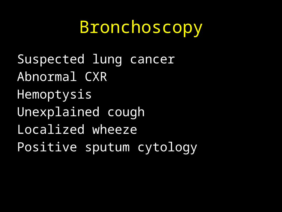
Bronchoscopy
Suspected lung cancer
Abnormal CXR
Hemoptysis
Unexplained cough
Localized wheeze
Positive sputum cytology
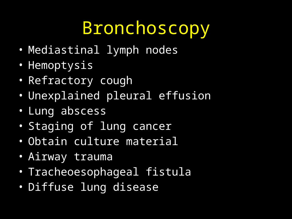
Bronchoscopy• Mediastinal lymph nodes• Hemoptysis• Refractory cough• Unexplained pleural effusion• Lung abscess • Staging of lung cancer• Obtain culture material• Airway trauma• Tracheoesophageal fistula• Diffuse lung disease


Bronchoscopy
Therapeutic
• Remove foreign bodies• Remove abnormal endobronchial tissue• Difficult endotracheal tube intubation• Endobronchial stent placement


Pulmonary function tests
• Spirometry • Lung volumes• Diffusion capacity• Respiratory muscle strength

Spirometry
• FVC (L) predicted >90%• FEV1 (L) predicted >90%• FEV1/FVC >75
• Diagnose obstructive lung disease• Suggest restrictive lung disease

Lung volumes
• TLC (L) >90% predicted• RV (L) > 90% predicted
• Diagnose restrictive lung disease• Diagnose air trapping

Diffusing capacity (DL)
• Measure the ability of gases to diffuse from the alveoli into the pulmonary capillary blood
• CO not normally present in lungs or blood • More soluble in blood than lung tissues• Dlco

DLco
Reflect loss or damage to the gas exchanging surface of the lung
Emphysema
Distinguish emphysema from chronic bronchitis or chronic asthma
Interstitial lung disease
Pulmonary vascular disease

Respiratory muscle strength
• PImax, Pemax• Measured by pressure transducer at the mouth
when subject make a maximal inspiratory effort from full expiration or maximal expiratory effort from full inspiration
• PI reflect inspiratory muscles (diaphragm)• PE expiratory muscles including abdominal• Motor neuron disease, Guillian Barre syndrom


• 50 yr old male with SOB and cough >3yrs• Exam: clubbing and bilat insp crackles• CXR: reticulation bilateral• ABG: hypoxic respiratory failure• PFT: restrictive defect with significant
impairment in DLco

HRCT
• Designed for detailed evaluation of interstitial structures of the lung
• Use narrow slice thickness (1-2 mm) compared with 5-10 mm for routine scans

HRCT
Principle indications
• Suspected interstitial lung disease• Characterization of interstitial lung disease• Characterization of solitary pulmonary nodules• Diagnosis of bronchiectasis


• 45 yrs old female with RT sided chest pain for 1 day

• ABG pH 7.32, PaCO2 28, PaO2 50, O2sat 88%
• EKG sinus tachycardia• CXR normal• Spiral CT• V/Q scan

CT Angiography
• Image data are acquired continuously as the tube and detector rotate within the gantry and the patient moves continuously through the gantry
Advantages• Critically ill patients• Children• Less volume of intravenous contrast • Permits greater processing of the raw data


Lung Scans: V/Q
• Technetium (Tc) 99 m radionuclide is tagged to macroaggregated albumin to make small radioactive particles
• When Tc decays, it emits a gamma ray detected by the nuclear medicine gamma camera: a nuclear medicine image is formed by detection of many gamma rays

Lung scan: normal perfusion Q
• When injected via periphral venous site, the first capillaries encountered are the pulmonary capillaries
• If perfusion is present at the capillary level of the lungs, nuclear medicine perfusion image demonstrate activity in the periphery of the lungs

Lung scan: perfusion defect Q
If there is an obstructing vascular lesion in the pulmonary arterial circulation
blocked perfusion to the distal capillary level
nuclear medicine perfusion image demonstrate no activity in the periphery of the lungs


