In the vast majority of cases, cerebral palsy is crossed in relation to the lesions that determine it, that is, lesions of the right hemisphere of the brain give rise to paralysis on the left side of the body, and lesions of the left hemisphere to paraly-sis of the right side. This is the law. It has been established in innumerable observations and finds its natural explanation in the fact of the decussation of the pyramidal bundles at the level of the Mistichelli intercross. Well! This law is not
absolute. There are some observations of hemiplegia on one side of the body caused by cerebral hemisphere lesions on the same side…
— Jean-Martin Charcot and Albert Pitres, 189511
Modern intracranial surgery began in the closing de-cades of the 19th century, stimulated by the development of localizationist theories that, along with progress in the
ABBREVIATIONS ADC = apparent diffusion coefficient; IH = ipsilateral hemiparesis.ACCOMPANYING EDITORIAL DOI: 10.3171/2019.6.FOCUS19501.SUBMITTED April 30, 2019. ACCEPTED June 12, 2019.INCLUDE WHEN CITING DOI: 10.3171/2019.6.FOCUS19337.
Ipsilateral hemiparesis: the forgotten history of this paradoxical neurological signRodrigo Carrasco-Moro, MD,1 Ines Castro-Dufourny, MD, PhD,2 Juan S. Martínez-San Millán, MD, PhD,3 Lidia Cabañes-Martínez, MD,4 and José M. Pascual, MD, PhD5
1Department of Neurosurgery, Ramón y Cajal University Hospital; 2Department of Endocrinology, Sureste University Hospital, Francisco de Vitoria University; Departments of 3Neuroradiology and 4Clinical Neurophysiology, Ramón y Cajal University Hospital; and 5Department of Neurosurgery, La Princesa University Hospital, Madrid, Spain
OBJECTIVE Establishing the neurological localization doctrine for the contralateral hemispheric control of motor func-tions in the second half of the 19th century, researchers faced the challenge of recognizing false localizing signs, in par-ticular paradoxical or ipsilateral hemiparesis (IH). Despite tremendous progress in current methods of neuroradiological and electrophysiological exploration, a complete understanding of this phenomenon has yet to be attained.METHODS The authors researched the well-described cases of hemiparesis/hemiplegia ipsilateral to an intracranial lesion published in the scientific literature in the pre-MRI era (before 1980). A comprehensive review of the physiopatho-logical mechanisms proposed for paradoxical hemiparesis throughout this period, as well as the pathological evidence substantiating them, is provided.RESULTS A collection of 75 patients with hemiparesis/hemiplegia ipsilateral to the primary intracranial lesion reported between 1858 and 1979 were eligible for analysis. Most cases occurred in adults with supratentorial, slowly developing, extraparenchymatous mass lesions, such as neoplasms (38%) or chronic subdural hematomas (36%). Physiopatho-logical theories proposed by the neurologists who investigated IH can be grouped into 4 major concepts: 1) lack of anatomical decussation of the corticospinal tract; 2) impaired functional activation of the contralateral hemisphere by the lesioned dominant hemisphere through the callosal connections; 3) Kernohan’s notch phenomenon, or mechanical injury of the contralateral cerebral peduncle against the free edge of the tentorium; and 4) cerebrovascular dysfunction involving the contralateral hemisphere owing to kinking and mechanical flattening of the carotid artery contralateral to the primary intracranial lesion.CONCLUSIONS IH represents a still underdiagnosed paradoxical neurological phenomenon. With the aid of modern neuroradiological and neurophysiological methods, Kernohan’s peduncle notch mechanism has been confirmed to cause IH in many of the cases reported in recent decades. Nevertheless, alternative functional and/or vascular mechanisms must be investigated further for unexplained IH cases, in particular for transitory IH without evidence of peduncle injury. The historical theories reviewed in this paper represent a conceptual framework that may be helpful for this purpose.https://thejns.org/doi/abs/10.3171/2019.6.FOCUS19337KEYWORDS diaschisis; ipsilateral hemiparesis; Kernohan-Woltman notch phenomenon; history of neurology; paradoxical hemiparesis; tentorial incisure
fields of antisepsis and anesthesia, allowed intrepid sur-geons to design accurate approaches to cerebral lesions. Nevertheless, expanding intracranial lesions occasionally produced focal neurological signs not directly related to the location of the mass. These paradoxical clinical find-ings, termed “false localizing signs” for James Collier’s studies,13 disconcerted both clinicians and surgeons as they frequently led to wrong-site exploratory surgical pro-cedures. Among false localizing signs, ipsilateral hemipa-resis (IH) relative to an intracranial mass—widely known as Kernohan’s notch phenomenon—remains one of the most enigmatic. Despite the use of modern neuroradio-logical and electrophysiological exploration methods, the intrinsic mechanisms leading to the development of IH have not been fully clarified.45 In this work, we performed a thorough analysis of a cohort of historical reports of patients with verified IH published before the advent of modern neuroradiology. Additionally, we provide a com-prehensive review of the physiopathological hypotheses put forward for this intriguing sign by prominent figures of neurology and neurosurgery in the 19th and 20th cen-turies. These early clinical observations and hypotheses, rarely cited in modern scientific literature, may contribute to an appropriate understanding of this paradoxical clini-cal phenomenon.
MethodsWe conducted a thorough review of well-described
cases of IH published in the scientific literature in the pre-MRI era (prior to the 1980s). This survey involved re-ports in official medical journals and in specialized texts, monographs, and doctoral dissertations. The initial search included all articles shown in the PubMed, MEDLINE, and Scopus databases after entering the keywords “ipsi-lateral hemiparesis,” “Kernohan’s notch,” and “Kernohan-Woltman notch phenomenon.” Reference lists from the selected articles were scrutinized for a systematic retro-spective retrieving of older articles/monographs. From these documents, all individual cases of IH providing clinical-surgical and/or clinical–gross pathological veri-fication of the intracranial lesion giving rise to the ipsi-lateral motor deficit were included in this study. A com-prehensive review of the physiopathological mechanisms proposed for paradoxical hemiparesis and the pathologi-cal evidence substantiating them in this set of scientific studies was carried out.
ResultsSeventy-five IH patients reported on between 1858
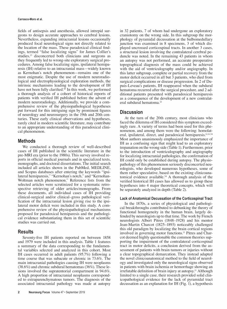
and 1979 were included in this analysis. Table 1 features a summary of the data corresponding to the fundamen-tal variables selected and analyzed in this cohort. Most IH cases occurred in adult patients (95.7%) following a time course that was subacute or chronic in 73.6%. The main intracranial pathologies causing IH were neoplasms (38.6%) and chronic subdural hematomas (36%). These le-sions involved the supratentorial compartment in 94.6%. A high proportion of intracranial neoplasms correspond-ed to extraparenchymatous tumors. The diagnosis of the associated intracranial pathology was made at autopsy
in 32 patients, 7 of whom had undergone an exploratory craniotomy on the wrong side. In this subgroup the mor-phology of pyramidal decussation at the bulbomedullary junction was examined in 8 specimens, 3 of which dis-played uncrossed corticospinal tracts. In another 3 cases, a structural lesion involving the contralateral cerebral pe-duncle was noted. In the remaining 43 patients in whom an autopsy was not performed, an accurate preoperative topographical diagnosis of the mass could be achieved with the aid of ventriculography and/or angiography. In this latter subgroup, complete or partial recovery from the motor deficit occurred in all but 3 patients, who died from surgical complications or disease progression. In 2 of Du-puis-Leveau’s patients, IH reappeared when the subdural hematoma recurred after the surgical procedure, and 2 ad-ditional patients presented with paradoxical hemiparesis as a consequence of the development of a new contralat-eral subdural hematoma.21
DiscussionAt the turn of the 20th century, most clinicians who
faced the dilemma of IH considered this symptom exceed-ingly rare. A variety of terms had been used for this phe-nomenon, and among them were the following: homolat-eral, ipsilateral, direct, and paradoxical hemiparesis.6,14,34 Most authors unanimously emphasized the importance of IH as a confusing sign that might lead to an exploratory trepanation on the wrong side (Table 1). Furthermore, prior to the introduction of ventriculography and angiography for localizing intracranial pathologies, the confirmation of IH could only be established during autopsy. The physio-pathology of this phenomenon seriously disconcerted neu-rologists, who developed numerous hypotheses, some of them rather speculative, based on the existing clinicoana-tomical evidence available.34 A thorough analysis of the verified historical IH cases has allowed us to group these hypotheses into 4 major theoretical concepts, which will be separately analyzed in depth (Table 2).
Lack of Anatomical Decussation of the Corticospinal TractIn the 1870s, a series of physiological and pathologi-
cal breakthroughs contributed to debunking the theory of functional homogeneity in the human brain, largely de-fended by neurologists up to that time. The work by French neurologists Albert Pitres (1848–1928) and his mentor Jean-Martin Charcot (1825–1893) noticeably challenged this old paradigm by localizing the brain cortical regions involved in governing motor functions.11 Pitres and Char-cot deemed highly questionable the common theories sup-porting the impairment of the contralateral corticospinal tract in motor deficits, a conclusion derived from the as-sessment of patients with brain tumors or injuries without a clear topographical demarcation. They instead adapted the novel clinicoanatomical method to the field of neurol-ogy and investigated only the neurological signs observed in patients with brain ischemia or hemorrhage showing an irrefutable definition of brain injury at autopsy.11 Although limited to a single case, their research provided solid clin-icopathological evidence for the lack of pyramidal tract decussation as an explanation for IH (Fig. 1), a hypothesis
Carrasco-Moro et al.
Neurosurg Focus Volume 47 • September 2019 3
TABLE 1. Summary of data for the cohort of well-described patients with IH reported in the pre-MRI era (1858–1979)
64, M SA/hematoma Rt hemiparesis Aphasia, S Rt cSDH surgically treated Recovery
» CONTINUED FROM PAGE 3
CONTINUED ON PAGE 5 »
Carrasco-Moro et al.
Neurosurg Focus Volume 47 • September 2019 5
that was compatible with prior neuroanatomical studies by Paul Flechsig (1847–1929) demonstrating the variabil-ity in the degree of decussation of the pyramidal tract.11 The great respect for Charcot’s theory led to a systematic search for abnormalities in the pyramidal tract decussation among IH patients, with contradictory results (Table 1).
Ipsilateral Hemiparesis Caused by Functional Impairment of a Cerebral Region Distant From the Injury
In parallel with Charcot’s clinicoanatomical method, some neurologists defended the unorthodox view that
many neurological deficits actually corresponded to func-tional disorders produced by the imbalance in the input connectivity on brain regions distant from the site initially injured. Two such closely related theories were proposed for IH.
Theory of “Cerebral Irritation” From a DistanceTrue to his eccentric and contentious nature, French
physiologist and neurologist Charles-Édouard Brown-Séquard (1817–1894) rejected Charcot’s clinicoanatomi-cal correlation method and proposed the original con-cept that IH represented the effect of an irritative action
TABLE 1. Summary of data for the cohort of well-described patients with IH reported in the pre-MRI era (1858–1979)
No. of cases (ancient & modern series) Scarce Scarce Numerous ScarceLaterality/motor dominance Independent Dependent Independent IndependentMass effect Unnecessary Unnecessary Necessary* NecessaryLocation of supra/infratentorial (primary insult) Independent Dependent Independent DependentTentorial anthropometry Independent Independent May play a role IndependentMotor deficit distribution Dependent on primary
lesion topographyDependent on primary
lesion topographyCrural Dependent on vascular
territory involvedPresence of other nervous system anomalies Frequent Independent Independent IndependentEvidence from modern ancillary tests Absent Absent Numerous Absent
* Scarce cases of IH have been described in the literature, with MRI evidence of a contralateral peduncle lesion in the absence of significant mass effect, as described in the text and Fig. 6.
FIG. 1. Lack of decussation of the corticospinal tract theory. Albert Pitres (1848–1928) (A), shortly after the death of his mentor Jean-Martin Charcot (1825–1893) (B), published in 1895 the monograph entitled Motor Cortical Centers in Man (front page of the work, between panels A and B), based on the clinical and pathological analysis of 174 patients with cerebral lesions. In Chapter 10, addressing the subject of “Contradictory Observations,” a patient with right hemiparesis was described. At his autopsy, a “ramol-lisement du cerveau,” or cerebral infarct involving the peri-rolandic region and the base of the third frontal gyrus of the right hemi-sphere, was found (C), as were signs of secondary degeneration of the ipsilateral corticospinal tract at the spinal cord level (D). A schematic illustration (E) of the anatomical course followed by a nondecussated corticospinal tract. An injury of this tract at any level in its course would cause IH. Photographs of Pitres and Charcot: Reproduced from Wikimedia Commons (https://commons.wikimedia.org/wiki/File:Albert_Pitres.jpg) and the US National Library of Medicine website (http://resource.nlm.nih.gov/101425121), respectively. Public domain. C and D: Reproduced from Charcot JM, Pitres A: Des observations contradictoires, in Charcot JM, Pitres A (eds): Les centres moteurs corticaux chez l’homme. Paris: Rueff et Cie Éditeurs, 1895, pp 183–191 (French) (http://catalogue.bnf.fr/ark:/12148/cb31118881v). Public domain.
Carrasco-Moro et al.
Neurosurg Focus Volume 47 • September 2019 7
on a certain cerebral area that originated from a distant damaged region or “irritation agissant à distance”8 (Fig. 2A). Based on a prior IH case published by Antoine Jo-seph Jobert de Lamballe (1799–1867) of a patient with a voluminous supra- and infratentorial fifth cranial nerve schwannoma (Table 1, case 1), Brown-Séquard reasoned that this deficit could not be explained by the interruption of nerve flow at the compressed cerebral peduncle, as it should have caused contralateral hemiparesis.8 Rather, it would be the result of an “irritative” phenomenon origi-nating in distant structures compressed by the tumor, such as the middle cerebellar peduncle and the cerebel-lum itself. Brown-Séquard assumed it was a trophic ac-tion of the cerebellum on the contralateral cerebral hemi-sphere, and therefore IH could be a cerebellum-mediated effect.8
Theory of DiaschisisThe Russo-Swiss neuropathologist and physiologist
Constantin von Monakow (1853–1930) addressed the par-adoxical symptom of IH in his monumental monograph Gehirnpathologie (Brain Pathology). In line with Brown-Séquard’s view, von Monakow questioned both Charcot’s theory of the absence of pyramidal tract decussation and the existence of mechanical compression of the contralat-eral corticospinal tract, concepts he felt lacked solid patho-logical evidence (Fig. 2B).44 In 1905, he developed the in-novative, complex concept of “diaschisis” (from the Greek words dia and schizien, meaning “split in half”) for un-explained transitory neurological deficits associated with localized injuries to the brain.26,44 Von Monakow believed that if a particular brain region was damaged, the loss of function could affect other distant parts of the brain not
FIG. 2. Theory of cerebral dysfunction caused by a distant injury. A: Mauritian physiologist and neurologist Charles-Édouard Brown-Séquard (1817–1894) originally proposed this controversial concept in an article addressing the physiology and pathology of the pons. Text: Reprinted from Brown-Séquard CE: Recherches sur la physiologie et la pathologie de la protuberance annulaire. Journal de la Physiologie de l’Homme et des Animaux 1:523–539, 1858 (French). Public domain. B: Constantin von Monakow (1853–1930) published in 1905 the second edition of his monograph Gehirnpathologie, in which he explained for the first time his concept of diaschisis. Text: Reprinted from von Monakow C: Shock und diaschisis, in von Monakow C (ed): Gehirnpathologie. Vienna: Alfred Hölder, 1905, pp 236–248 (German). Public domain. C1 and C2: Original illustrations of the paper published by Ascenzi in 1908 focused on the role of the corpus callosum in motor function. Reprinted from Ascenzi O: Una cisti emorragica del corpo calloso. Riv Patol Nerv Ment 13:1–15, 1908 (Italian). Public domain. Schematic representation of a hemorrhagic cystic lesion involving the left side of the genu of corpus callosum (L in C1, F in C2; S = interhemispheric fissure, V = lateral ventricles). According to this author, the lesion would affect mostly the callosal fibers projecting toward the left hemisphere, grouped in more compacted bundles in the left precommissural region as opposed to the right side of the genu of corpus callosum (F in C2; S = left, D = right). Illustrative scheme (D) showing the concept of commissural diaschisis. A lesion affecting the primary motor cortical re-gion within the dominant hemisphere (white arrow) causes functional impairment of a distant area in the nondominant hemisphere due to the interference it produces in the normal neuronal input from the dominant hemisphere through the corpus callosum (black curved arrow). Photographs of Brown-Séquard and von Monakow reproduced from the Bibliothèque Interuniversitaire de Santé Paris Descartes website (http://www.bium.univ-paris5.fr/images/livres/21950/0013.jpg) and Wikimedia Commons (https://commons.wikimedia.org/wiki/File:Monakov.PNG), respectively. Public domain.
Carrasco-Moro et al.
Neurosurg Focus Volume 47 • September 20198
seemingly related to the site of injury. Therefore, diaschi-sis should be interpreted as a transitory functional impair-ment caused by the temporary blocking of normal input on a particular brain region anatomically linked to the distant injured site.26,44 If the injury was not too severe, functional behavior would recover once the period of diaschisis wore off. The specific paradoxical case of IH would represent, according to von Monakow, an example of “diaschisis corticommisuralis” caused by impairment of the normal functional activation of the contralateral hemisphere by fibers coming from the dominant hemisphere through the corpus callosum, a hypothesis supported by the findings published by Ascenzi in 1908 (Fig. 2C1 and C2).3
Compression of the Contralateral Corticospinal TractThe most widely accepted theory for explaining IH was
the compression of the contralateral corticospinal tract at any level along its course above the pyramidal decussa-tion.6,14,34 Several different tract compression mechanisms
were proposed in the 19th century, among them the flatten-ing (“aplatissement”) of the motor cortex against the inner skull surface, the compression of the tract secondary to hydrocephalus (“hydropsie”), the development of swelling in the contralateral hemisphere, and/or the coexistence of disturbances in cerebral blood flow.21 However, the major breakthrough occurred in 1920, when the neuropathologist and psychiatrist Adolf Meyer (1866–1950) demonstrated the dynamic displacements caused by intracranial mass le-sions on the cerebral soft tissue (Fig. 3A).35 Arguably, the recognition of brain herniations proved the most relevant contribution to the origin of the false localizing signs as-sociated with intracranial lesions. The seminal concepts of “contralateral peduncular notch” and the “syndrome of the third frontal convolution” (or Ectors’ syndrome) sub-stantially improved insight into the way IH may occur in association with brain herniation.
Kernohan’s Notch PhenomenonIn 1905 German neurologist Albert Knapp recognized
FIG. 3. Historical landmarks in the concept of notching of the contralateral cerebral peduncle as the origin of IH. A: Autopsy brain specimen showing the transtentorial descendent herniation of the mesial temporal lobe caused by a hemorrhagic metastasis expanding at the post–central gyrus of the right hemisphere. The herniation causes the mechanical deformation of the ipsilateral peduncle. Reprinted from Meyer A: Herniation of the brain. Arch Neurol Psychiatry 4:387–400, 1920. Public domain. B and C: Se-rial sections of the brain specimen studied by Groeneveld and Schaltenbrand, showing the brain distortion caused by an extraaxial tumor. Reproduced with permission from Springer Nature. From Groeneveld A, Schaltenbrand G: Ein Fall von Duraendotheliom über der Grosshirnhemisphäre mit einer bemerkenswerten Komplikation: Läsion des gekreuzten Pespedunculidurch Druckauf den Rand des Tentoriums. Dtsch Z Nervenheilkd 97:32–50, 1927 (German). The authors described the existence of a structural lesion involving the contralateral peduncle in this specimen, but no pictorial evidence was provided in their article. D–F: Pictures of the brain specimen autopsied and reported by Kernohan and Woltman. Reproduced from Kernohan JW, Woltman HW: Incisura of the crus due to contralateral brain tumor. Arch Neurol Psychiatry 21:274–287, 1929. Copyright American Medical Association. The mass effect of an endothelioma (meningioma) on the brain bulk (D) is shown, as are the ipsilateral uncal herniation (short arrows in E) and the notching of the contralateral cerebral peduncle (long arrow in E, with an underlying focal destruction of nervous tissue and myelin sheathing of the corticospinal tract). G: In a later report by the same researchers, a peduncular notch without structural damage of the underlying nervous tissue was demonstrated (arrow). Reproduced from Kernohan JW, Woltman HW: Incisura of the crus due to contralateral brain tumor. Arch Neurol Psychiatry 21:274–287, 1929. Copyright American Medical Association. H: Illus-trative schematic showing the mechanical compression of the contralateral peduncle against the free tentorial edge (black arrow) secondary to the lateral displacement of the brainstem caused by a supratentorial or infratentorial mass (white arrows).
Carrasco-Moro et al.
Neurosurg Focus Volume 47 • September 2019 9
hemiparesis alternans, or motor deficit of the ipsilateral oc-ulomotor nerve and contralateral hemiparesis, as a clinical manifestation typical of temporal lobe tumors that com-press the ipsilateral cerebral peduncle through uncal her-niation.32 Knapp was aware of the occurrence of IH in this clinical setting, and he was the first to attribute this para-doxical finding to mechanical compression of the contra-lateral cerebral peduncle.32 The pathological evidence for this mechanism was not reported until the end of the 1920s, when two groups of researchers—the Dutch and German neurologists Arnold Groeneveld (1895–1962) and Georges Schaltenbrand (1897–1979), respectively,28 and the Ameri-can neuropathologist James W. Kernohan (1896–1981) and neurologist Henry W. Woltman (1889–1964)30—sepa-rately published the evidence of mechanical injury at the cerebral peduncle presumably caused by its compression against the free edge of the cerebellar tentorium (Fig. 3).16 Encouraged by this finding, Kernohan and Woltman car-ried out systematic research on brains obtained from Mayo Clinic autopsies in which they identified the presence of such an indentation or notch in the contralateral cerebral peduncle in 34 of 276 brains from patients with intracra-nial neoplasms.31 This 1929 study had such a positive im-pact on the scientific community that IH was henceforth renamed the “Kernohan-Woltman notch phenomenon” or simply “Kernohan’s notch phenomenon” (Fig. 3). Perhaps the most striking finding of their study was that 13 patients with a peduncular notch did not show clinical hemiparesis or signs of corticospinal tract impairment such as hyper-reflexia or a Babinski sign, a fact for which Kernohan and Woltman were unable to provide a proper explanation.31 This inconsistency has been used to support the theory of diaschisis by its proponents.17–19
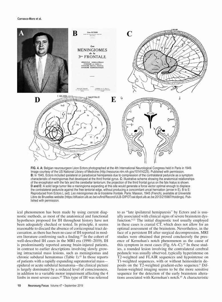
Ectors’ Syndrome: A Particular Type of Kernohan’s Notch Caused by Mass Effect on the Third Frontal Convolution
The next milestone in the elucidation of IH causes was the identification by the Belgian neurosurgeon Léon Ectors of a particular clinical syndrome associated with meningiomas involving the third or inferior convolution of the frontal lobe (Fig. 4A and B). Supported by ample and convincing clinical, experimental, and bibliographic evidence, Ectors was able to categorize the fundamental groups of symptoms and/or signs in a monograph hardly mentioned in medical literature.22 Among symptoms such as mental disturbances, conjugated cephalic and ocular rotation toward the site of the tumor, due to impairment of Brodmann area 8, and those typical of high intracra-nial hypertension, Ectors included what he called “early ipsilateral hemiparesis” as a specific neurological finding associated with meningiomas of the third frontal lobe gy-rus. Ectors’ syndrome also included typical radiological findings such as angiographic ingurgitation of the anterior branch of the middle meningeal artery and contralateral displacement of cerebral ventricles observed in air ven-triculography.22–24
Several ancient reports of IH seem to fit in Ectors’ syn-drome (Table 1). According to Ectors, IH severity would not depend directly on the size of the tumor, but rather it would be proportional to the degree of lateral displace-ment of the brainstem.22–24 Long-term expanding lesions
growing at the basal frontal lobe would cause an optimal compressive force vector across the brain tissue, pointing to the tentorial hiatus and the brainstem, therefore increas-ing the likelihood of the contralateral peduncle’s lateral displacement against the free edge of the tentorium, even in the absence of uncal herniation (Fig. 4C–E). Typically, IH would develop slowly, following a leg-arm-face involve-ment sequence as a result of the somatotopic distribution of corticospinal bundles within the cerebral peduncle.22 Ectors remarked that in the early stages of the disease, the only manifestation might be a mild hyperreflexia in the leg, which can be easily overlooked.22–24 The characteris-tic full reversibility of hemiparesis when the intracranial mass effect was promptly alleviated suggests that the ma-jor factor contributing to IH in Ectors’ syndrome is the elastic deformation of the contralateral peduncle against the tentorial free edge, without structural damage to the nervous tissue, as observed by Kernohan and Woltman.31
Cerebrovascular DysfunctionIn the late 1970s Géraldine Dupuis-Leveau from Reims
proposed hypoperfusion of the contralateral hemisphere as a complementary physiopathological mechanism for IH, which would act together with contralateral peduncle compression.21 As part of her doctoral thesis, Dupuis-Le-veau analyzed the cerebral angiograms of 18 patients with IH included in a cohort of 180 cases of surgically treated subdural hematomas and observed a reduced cerebral blood flow in these patients, a finding she correlated with kinking and mechanical flattening of the carotid artery contralateral to the hematoma against the posterior clinoid process and the petroclinoid ligament.21 Characteristically, the combined displacement of both carotid arteries gives rise to a “windshield wiper” (from the French en essuie-glaces) aspect of these vessels in the anteroposterior angi-ographic projection (Fig. 5). As a result, either a predomi-nating brachiofacial (ischemia of the perisylvian region) or crural motor (ischemia of the pericallosal area) deficit can occur, depending on the carotid artery branches that develop reduced blood flow.21 This vascular mechanism suggests that early surgical evacuation of the hematoma guarantees complete clinical recovery, provided that cere-bral hypoperfusion did not reach the threshold for irrevers-ible ischemic injury.
Ipsilateral Hemiparesis: Past, Present, and FutureFueled by modern neuroradiological techniques, IH
has received renewed interest in recent years. Even so, it remains an exceptional clinical finding. Indeed, only 39 cases published since 1991 could be collected in the most recent comprehensive review on this subject.45 Léon Ec-tors prophetically expressed his belief that IH would re-main an underdiagnosed condition, especially in its early stages, unless a thorough neurological examination was carried out.22 Hence, subtle neurological findings sugges-tive of IH, such as an unexplained muscle tone increase or the presence of abnormal reflexes, are rarely investigated when no motor deficit is observed. As evidence of this, most IH cases reported in the last 2 decades do not provide detailed neurological findings.45
Very little conceptual progress regarding this paradox-
Carrasco-Moro et al.
Neurosurg Focus Volume 47 • September 201910
ical phenomenon has been made by using current diag-nostic methods, as most of the anatomical and functional hypotheses proposed for IH throughout history have not been adequately checked or tested. In principle, it seems reasonable to discard the absence of corticospinal tract de-cussation, as there has been no case of IH reported in mod-ern literature confirming such a finding.45 In the cohort of well-described IH cases in the MRI era (1990–2019), IH is predominantly reported among brain-injured patients, in contrast to earlier descriptions involving slowly grow-ing intracranial mass lesions, such as meningiomas and chronic subdural hematomas (Table 1).45 In those reports of patients with a rapidly expanding supratentorial mass—epidural or acute subdural hematoma—the clinical picture is largely dominated by a reduced level of consciousness, in addition to a variable motor impairment affecting the 4 limbs in most severe cases.45 This type of IH was referred
to as “late ipsilateral hemiparesis” by Ectors and is usu-ally associated with clinical signs of severe brainstem dys-function.9,22 The initial diagnostic tool usually employed in these cases is cranial CT, which does not allow for an optimal assessment of the brainstem. Nevertheless, in the face of a persistent IH after surgical decompression, MRI studies were obtained that proved conclusively the pres-ence of Kernohan’s notch phenomenon as the cause of this symptom in most cases (Fig. 6A–C).45 In these stud-ies, a rounded lesion involving the contralateral cerebral peduncle was usually observed, typically hyperintense on T2-weighted and FLAIR sequences and hypointense on T1-weighted sequences, with or without hemosiderin de-posits on the T2-weighted gradient-echo sequence.9 Dif-fusion-weighted imaging seems to be the more sensitive sequence for the detection of the early brainstem altera-tions associated with Kernohan’s notch.45 A characteristic
FIG. 4. A: Belgian neurosurgeon Léon Ectors photographed at the 4th International Neurological Congress held in Paris in 1949. Image courtesy of the US National Library of Medicine (http://resource.nlm.nih.gov/101414225). Published with permission. B: In 1945, Ectors included ipsilateral or paradoxical hemiparesis due to compression of the contralateral peduncle as a symptom characteristic of meningiomas that developed at the third frontal gyrus. C: Illustrative scheme showing the anatomical relationships of the encephalon with the falx and the cerebellar tentorium; the projection of the third frontal gyrus on the falx hiatus is shown. D and E: A solid large tumor like a meningioma expanding at this site would generate a force vector optimal enough to displace the contralateral peduncle against the free tentorial edge, without producing a concomitant uncal herniation (arrow in E). B to E: Reproduced from Ectors L (ed): Les méningiomes de la troisiéme frontale. Paris: Masson, 1945 (French), available at Université Libre de Bruxelles website (https://difusion.ulb.ac.be/vufind/Record/ULB-DIPOT:oai:dipot.ulb.ac.be:2013/215987/Holdings). Pub-lished with permission.
Carrasco-Moro et al.
Neurosurg Focus Volume 47 • September 2019 11
hyperintense signal within the cerebral peduncle owing to the restriction in the apparent diffusion coefficient (ADC) map during the acute stage of this phenomenon probably reflects the presence of cytotoxic edema, as it attenuates or disappears in control studies.42 Partial or total recov-ery from motor deficit, even in patients in whom a com-plete disruption of a normally decussated corticospinal tract has been demonstrated using diffusion tensor imag-ing,29,33,37 can be explained by the resolution of cytotoxic edema beneath the peduncle notch. This concept is sup-ported by Kernohan and Woltman’s original histological studies confirming the disruption of myelin sheaths at the level of the injured area behind the notch, whereas most of the corresponding axons of the corticospinal tract were spared from destruction.31
A second subgroup of recent IH reports has shown ex-amples of the clinical picture that Ectors characterized as early IH,22 in which the slow progressive growth of the intracranial mass lesion permitted a cerebral MRI study before the surgical procedure.45 These studies evidenced the notching of the contralateral cerebral peduncle against the free tentorial edge, with or without underlying struc-tural damage to the corticospinal tract, which plausibly
depends on the degree and duration of the compression (Fig. 6D and E).
Especially disconcerting are the few modern cases of IH in which no significant intracranial mass effect has been found. To increase the controversy, some of the authors of these cases have provided a radiological demonstration of the typical lesion in the contralateral peduncle described above, which has been interpreted by some authors as a traumatic contusion of this anatomical structure against the tentorial edge due to a swaying effect on the brainstem (Fig. 6F–H).5,25 Doubtful cases may benefit from the dem-onstration of a functional impairment of the contralateral corticospinal tract—using motor evoked potentials with transcranial magnetic stimulation—to support the clinical diagnosis,5,10 although this finding does not completely rule out the hypothesis of diaschisis.18 Despite the fact that this concept still receives support from some neurologists,17–19 diaschisis in IH patients remains an unproven theory until further functional/metabolic investigation can verify it,26 as this hypothesis cannot properly account for either IH cases caused by lesions involving the nondominant hemi-sphere or those produced by infratentorial masses directly pushing the brainstem against the tentorium.
In order to explain the low rate of paradoxical hemi-paresis and the physiopathological mechanism involved in unsolved cases, some authors have proposed the existence of some predisposing factors, such as the coexistence of a narrow tentorial incisure facilitating Kernohan’s notch phenomenon or an insufficient blood supply to the con-tralateral brain hemisphere, especially in patients with atherosclerosis (Dupuis-Leveau’s hypothesis).9,21 Yet, no definite conclusions regarding the effect of these variables can be stated, as very few studies include accurate mea-surements of the tentorial incisure, and no exhaustive MRI studies analyzing the vascular supply and brain perfusion have been performed in IH patients to date.45
ConclusionsIH represents a still underdiagnosed paradoxical phe-
nomenon that challenges the neurological paradigm for the hemispheric localization of cerebral motor functions. Even though modern neuroradiological and neurophysio-logical methods largely support Kernohan and Woltman’s concept of contralateral peduncle notching against the free tentorial edge, none of the physiopathological mechanisms proposed throughout history can satisfactorily account for the cause of IH in all patients. To overcome this shortcom-ing, it will be essential to perform a thorough neurological examination, in addition to the most complete neuroradio-logical and neurophysiological investigations, including diffusion tract imaging, functional and perfusion MRI, anatomical measurements of the tentorial incisure, and motor evoked potential monitoring.
AcknowledgmentsWe wish to express our gratitude to George Hamilton for his
critical review of the language and style of the manuscript. We also wish to thank Prof. Alfonso Villén Carrasco, who translated the consulted articles written in German. We are indebted to the staff at Ramón y Cajal University Hospital Medical Library (Madrid), the Bibliothèque Interuniversitaire de Santé (Université
FIG. 5. A: Front page of Dupuis-Leveau’s doctoral dissertation. B: Schematic representation of the “windshield wiper” image, observed in the anteroposterior projection of a cerebral angiogram, caused by vascular displacement of carotid arteries secondary to a subdural hematoma. C: The brain displacement (white arrows) would cause compression of the contralateral cerebral peduncle against the free tentorial edge, as well as the kinking of the contralateral internal carotid artery against rigid structures at the cranial base (black arrows), giving rise to cerebral ischemia of the contralateral hemisphere involving the regions supplied by the median cerebral and/or anterior cerebral arter-ies. A and B: Reproduced from Dupuis-Leveau G: Les hematomes sous-duraux chroniques avec signes neurologiques homolateraux. A propos de 18 observations [doctoral thesis]. Faculty of Medicine, Reims, 1979 (French). Published with permission.
Carrasco-Moro et al.
Neurosurg Focus Volume 47 • September 201912
Paris Descartes), and the Francisco de Vitoria University Library (Madrid) for their invaluable help in obtaining some of the origi-nal research material used for this study.
References 1. Achslogh J, Macken J: [A new case of meningioma of the
third frontal convolution.] Acta Neurol Psychiatr Belg 59:163–173, 1959 (French)
2. Ardito R: Ematoma sottodurale della convessità con sín-
drome piramidale omolaterale. Contributo clínico. Riv Patol Nerv Ment 78:123–154, 1954
3. Ascenzi O: Una cisti emorragica del corpo calloso. Riv Patol Nerv Ment 13:1–15, 1908
4. Babinski J, Clunet J: Tumeurs méningées unilatérales. Hémiplégie siègeant du même côté que les tumeurs. Rev Neurol (Paris) 16:707–709, 1908
5. Binder DK, Lyon R, Manley GT: Transcranial motor evoked potential recording in a case of Kernohan’s notch syndrome: case report. Neurosurgery 54:999–1003, 2004
FIG. 6. Physiopathological mechanisms for IH examined using modern neuroradiological and neurophysiological methods in origi-nal cases from our own series. A–C: A 77-year-old male patient presented with signs of cerebral decerebration and a Glasgow Coma Scale score of 4 after suffering a severe traumatic brain injury. A: Coronal section of a cranial CT scan obtained at his admission showing a large acute subdural hematoma causing a marked displacement of the brain with signs of ipsilateral uncal herniation (hollow arrow) and compression of the contralateral peduncle against the tentorial edge (solid arrow). After a decom-pressive craniectomy and evacuation of the hematoma, the patient recovered normal consciousness but right spastic hemiparesis (grade 2/5) ipsilateral to the hematoma persisted. B and C: Brain MR images obtained postoperatively showing a hyperintense signal in the contralateral peduncle, right beneath the level of contact with the tentorial edge (arrows), in both the FLAIR (B) and the diffusion-weighted (C) sequences, with restricted water diffusion in the ADC map. D and E: Patient with a left motor deficit involving predominantly the lower limb. D: MR image showing a supra/infratentorial left epidermoid cyst (asterisk) displacing the brainstem and causing indentation (arrow) of the contralateral peduncle against the free tentorial edge, without underlying signal alteration on the T2-weighted sequence. During resection of this lesion, motor evoked potentials (MEP) monitoring using electric transcranial stimulation showed delayed and reduced amplitude of motor responses in the left arm and leg (left and right panels in E). F–H: Images of a 41-year-old male patient who suffered cranial trauma secondary to a syncope. After recovering conscious-ness, he experienced seizures, time-space disorientation, gait imbalance, and hiccups. While a cranial CT scan only showed a frontal lobe contusive focus, MRI demonstrated a slight hyperintense signal at the contralateral peduncle (arrows) on the FLAIR image (G) and diffusion-weighted sequence (H), with water flow restriction on ADC maps. Note the lack of brainstem displace-ment. Remarkably, this patient did not show any motor deficits, and the effect of the peduncle lesion could only be correlated with an increased muscle tone accompanied by hyperreflexia in the thorough neurological examination.
Carrasco-Moro et al.
Neurosurg Focus Volume 47 • September 2019 13
6. Binet E: Contribution à l’étude des hémiplégies homola-térales: a propos d’un cas d’abscès de cerveau d’origine otique [doctoral thesis]. Paris: Faculty of Medicine, 1909
7. Blaise H: Deux cas peu conformes aux localisations céré-brales. Bull Soc Anat Paris 7:387–392, 1882
8. Brown-Séquard CE: Recherches sur la physiologie et la pa-thologie de la protuberance annulaire. Journal de la Physi-ologie de l’Homme et des Animaux 1:523–539, 1858
9. Carrasco R, Pascual JM, Navas M, Martínez-Flórez P, Manzanares-Soler R, Sola RG: Kernohan-Woltman notch phenomenon caused by an acute subdural hematoma. J Clin Neurosci 16:1628–1631, 2009
11. Charcot JM, Pitres A: Des observations contradictoires, in Charcot JM, Pitres A (eds): Les centres moteurs corticaux chez l’homme. Paris: Rueff et Cie Éditeurs, 1895, pp 183–191
12. Claude H, Vincent C, Lèvy-Valensi J: De l’hémiplégie ho-molatérale dans les tumeurs cérébrales. Rev Neurol (Paris) 20:612–614, 1910
13. Collier J: The false localising signs of intracranial tumour. Brain 27:490–508, 1904
14. Couronne M: Les hémiplégies homolatérales [doctoral the-sis]. Paris: Faculty of Medicine, 1925
15. Couty M: Tumeur du pédoncule inférieur gauche. C R Se-ances Memoires Soc Biol 29:234–235, 1877
16. Dammers R, Volovici V, Kompanje EJ: The history of the Kernohan notch revisited. Neurosurgery 78:581–584, 2016
18. Derakhshan I: The Kernohan-Woltman phenomenon and laterality of motor control: fresh analysis of data in the article “Incisura of the crus due to contralateral brain tumor”. J Neurol Sci 287:296, 2009 (Letter)
19. Derakhshan I: Transcranial motor evoked potential recording in a case of Kernohan’s notch syndrome: case report. Neuro-surgery 56:E1166, 2005 (Letter)
20. Dupré E, Camus P: Hémiplégie homolatérale gauche chez un débile gaucher, ancien hémiplégique infantile droit. Rev Neurol (Paris) 13:322–325, 1905
21. Dupuis-Leveau G: Les hematomes sous-duraux chro-niques avec signes neurologiques homolateraux. A propos de 18 observations [doctoral thesis]. Reims, France: Faculty of Medicine, 1979
22. Ectors L (ed): Les méningiomes de la troisiéme frontale. Paris: Masson, 1945
23. Ectors L, Achslogh J: [The early homolateral pyramidal syndrome and meningiomas of the third frontal convolution.] Neurochirurgie 5:388–400, 1959 (French)
24. Ectors L, Heernu V: Contribution à l’étude des méningiomes de la troisième frontale. J Belge Neurol Psychiatr 8:497–507, 1946
25. Eesa M, Bell K: Kernohan-Woltman notch phenomenon. J Trauma 69:1634, 2010
26. Finger S, Koehler PJ, Jagella C: The Monakow concept of di-aschisis: origins and perspectives. Arch Neurol 61:283–288, 2004
27. Flatau E: De la radiothérapie des tumeurs du cerveau et de la moelle. Rev Neurol (Paris) 1:23–40, 1924
28. Groeneveld A, Schaltenbrand G: Ein Fall von Duraendo-theliom über der Grosshirnhemisphäre mit einer be-merkenswerten Komplikation: Läsion des gekreuzten Pespe-dunculidurch Druckauf den Rand des Tentoriums. Dtsch Z Nervenheilkd 97:32–50, 1927
29. Jang SG, Pyun SB: Diffusion tensor tractography in two cas-es of Kernohan-Woltman notch phenomenon. Ann Rehabil Med 37:879–885, 2013
30. Kernohan JW, Woltman HW: Incisura of the crus due to contralateral brain tumor. Proc Staff Meetings Mayo Clinic 3:69–70, 1928
31. Kernohan JW, Woltman HW: Incisura of the crus due to con-tralateral brain tumor. Arch Neurol Psychiatry 21:274–287, 1929
32. Knapp A: Die tumoren des Schläfenlappens. Z Ges Neurol Psych 42:226–289, 1918
33. Mansilla-Fernández B, Isla-Guerrero A, Giner J, Royo-Ore-jas A: [Tractography in Kernohan’s phenomenon: report of a case of acute subdural haematoma.] Rev Neurol 60:286–287, 2015 (Spanish)
34. Marie P: Hémiplégie collatérale, in Bruardel P, Gilbert A (eds): Traité de Médecine et de Thérapeutique. Paris: Li-brairie JB Baillière et Fils, 1901, Vol 8, pp 474–475
35. Meyer A: Herniation of the brain. Arch Neurol Psychiatry 4:387–400, 1920
36. Obrador Alcalde S: Sobre los falsos síntomas neurológicos de localización en los tumores supratentoriales. Actas Luso Esp Neurol Psiquiatr 7:183–195, 1948
37. Oh SI, Kim MJ, Oh KP, Kim HY, Kim SH, Kim HJ: Tractog-raphy of persistent ipsilateral hemiparesis following subdural hematoma. Can J Neurol Sci 40:601–602, 2013
38. Peyser E, Doron Y: Ipsilateral hemiplegia in supratentorial space occupying lesions. Int Surg 45:689–695, 1966
39. Pic M: Hémiplégie droite complète; autopsie: hémorragie uniquement localisée au lobe occipital droite. Lyon Med 107:560–561, 1906
41. Purves-Stewart J (ed): Intracranial Tumors and Some Errors in Their Diagnosis. Edinburgh: Oxford University Press, 1927
42. Uesugi S, Suehiro E, Nakayama H, Suzuki M: Diffusion-weighted magnetic resonance imaging in a case of Kerno-han’s notch phenomenon. Acta Neurochir (Wien) 152:1809–1810, 2010
43. Vanhenverswyn: Pachyméningite cérébrale hémorragique—contracture du côté de l´épanchement. Bull Soc Anatomocli-nique Lille 1:128, 1888
44. von Monakow C: Shock und diaschisis, in Von Monakow C (ed): Gehirnpathologie. Vienna: Alfred Hölder, 1905, pp 236–248
DisclosuresThe authors report no conflict of interest concerning the materi-als or methods used in this study or the findings specified in this paper.
Author ContributionsConception and design: Carrasco-Moro, Pascual. Acquisition of data: all authors. Analysis and interpretation of data: Carrasco-Moro, Pascual. Drafting the article: Carrasco-Moro, Castro-Dufourny, Pascual. Critically revising the article: Carrasco-Moro, Martínez-San Millán, Cabañes-Martínez, Pascual. Approved the final version of the manuscript on behalf of all authors: Carrasco-Moro. Statistical analysis: Carrasco-Moro. Administrative/techni-cal/material support: Martínez-San Millán, Cabañes-Martínez. Study supervision: Carrasco-Moro.
CorrespondenceRodrigo Carrasco-Moro: Ramón y Cajal University Hospital, Madrid, Spain. [email protected].