Iron Deficiency and OverloadVictor Gordeuk, MD
Thursday, August 13, 2020
© 2020 Hematology and Medical Oncology Best Practices Course
2 ‐ Iron Deficiency and Iron OverloadVictor R. Gordeuk, MD
Disclosures
Disclosures of Financial Relationships with Relevant
Commercial Interests
• Grant support ‐ CSL Behring, Global Blood Therapeutics, Imara,
Ironwood, Novartis
• Consulting‐ CSL Behring, Global Blood Therapeutics, Novartis, Forma
Outline
1. Review of iron metabolism
2. Iron deficiency
3. Iron overload: hereditary, environmental,
transfusional
Iron
• Essential nutrient for all living organisms.
– Reversible binding of O2: Hb, myoglobin
– Enzyme systems:
• heme (cytochromes, catalase, glutathione peroxidase, NO synthase)
• non‐heme (RNR, aconitase)
– Immunity: free radicals to destroy microbes
• Highly reactive with O2; can cause toxicity
• Deficiency of iron– most common nutritional
problem world wide
• Iron overload– less common
– important health problem
Iron Metabolism: Broad Themes
• Absorption of iron- highly regulated to prevent
excess Fe from being absorbed
• Excretion of iron- There is no physiologic
pathway for excreting excess iron
Iron Requirements
Men Women
Obligatory losses 1.0 mg/d 1.0 mg/d
Menstruation 0.0 mg/d 0.5 mg/d
Total losses 1.0 mg/d 1.5 mg/d
Iron absorbed 1.0 mg/d 1.5 mg/d
1 2
3 4
5 6
Iron Deficiency and OverloadVictor Gordeuk, MD
Thursday, August 13, 2020
© 2020 Hematology and Medical Oncology Best Practices Course
Body Iron Compartments65 kg F 75 kg M
Functional compounds
Hemoglobin 1900 mg 2500 mg
Myoglobin 310 mg 340 mg
Enzymes 170 mg 190 mg
Transferrin 2.7 mg 3.2 mg
Storage compounds
Ferritin & hemosiderin 300 mg 800 mg
Total ~2700 mg ~3800 mg
Simplified Diagram of Iron Movement in the Body
Dietary Iron
• Typical diet men- Mean of 18 mg/day
- SD range of 4 ‐ 30 mg/day
• Typical diet women- Mean of 13 mg/day
- SD range of 7 ‐ 19 mg/day
• ~1/3 of Fe from fortification of flour
What We Eat in America, NHANES 2007-2008
Iron absorption‐ proximal small bowel
Enhanced absorption• Low hepcidin:
– Iron deficiency
– erythropoiesis
• Dietary factors:
– ascorbic acid (Fe+2 valance absorbed)
– Heme vs non‐heme Fe
Inhibited absorption • High hepcidin
– iron stores
– Inflammation
• Dietary factors:
- tannins (tea)
- phytates (bran)
Hepcidin- 25 aa peptide produced by liver that suppresses iron absorption
Iron Transport into Plasma
FerroportinFe+2
Erythro-phagocytosing Macrophage
Senescent RBC
Heme
Fe
Fe+3
Transferrin
Cerulo-plasmin
Heme oxygen-ase
*
Ferroportin
Adapted from Andrews, NEJM 1999;341:1986
Cytochrome Breductase
Duodenal Enterocyte
Transferrin
Iron Entry into Erythroid Precursors
Adapted from AndrewsNEJM 1999;341:1986
Heme
Steap3Fe+3 Fe+2
7 8
9 10
11 12
Iron Deficiency and OverloadVictor Gordeuk, MD
Thursday, August 13, 2020
© 2020 Hematology and Medical Oncology Best Practices Course
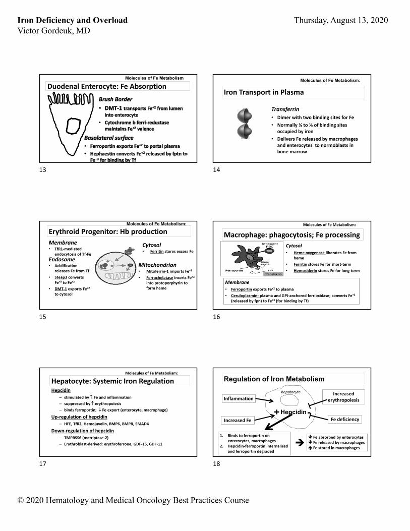
Duodenal Enterocyte: Fe AbsorptionMolecules of Fe Metabolism
Iron Transport in Plasma
Transferrin
• Dimer with two binding sites for Fe
• Normally ¼ to ⅓ of binding sites occupied by iron
• Delivers Fe released by macrophages and enterocytes to normoblasts in bone marrow
Molecules of Fe Metabolism:
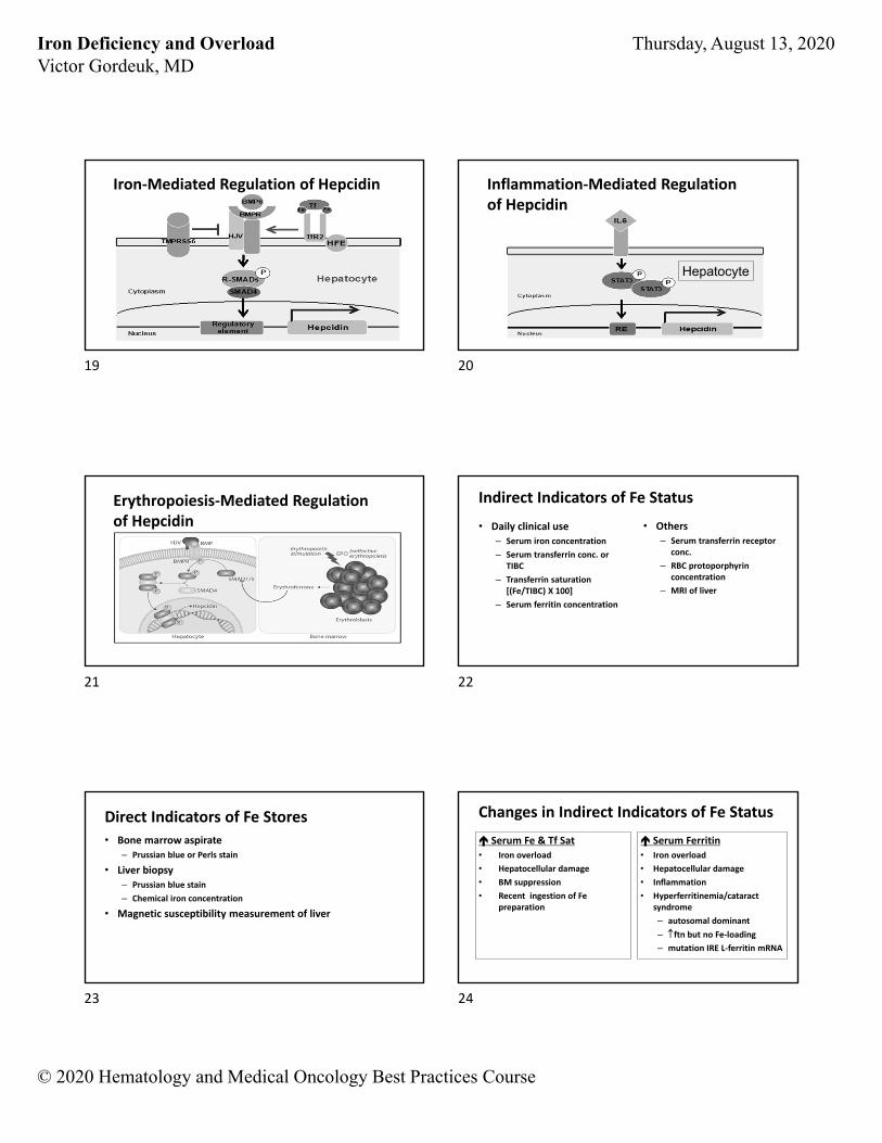
Membrane• TfR1‐mediated
endocytosis of Tf‐Fe
Erythroid Progenitor: Hb productionMolecules of Fe Metabolism:
Endosome• Acidification
releases Fe from Tf
• Steap3 converts Fe+3 to Fe+2
• DMT‐1 exports Fe+2
to cytosol
Cytosol• Ferritin stores excess Fe
Mitochondrion• Mitoferrin‐1 imports Fe+2
• Ferrochelatase inserts Fe+2
into protoporphyrin to form heme
Macrophage: phagocytosis; Fe processing
Molecules of Fe Metabolism:
Cytosol
• Heme oxygenase liberates Fe from heme
• Ferritin stores Fe for short‐term
• Hemosiderin stores Fe for long‐term
Membrane
• Ferroportin exports Fe+2 to plasma
• Ceruloplasmin‐ plasma and GPI‐anchored ferrioxidase; converts Fe+2
(released by fpn) to Fe+3 (for binding by Tf)
Hepatocyte: Systemic Iron RegulationHepcidin
– stimulated by Fe and inflammation
– suppressed by erythropoiesis
– binds ferroportin; Fe export (enterocyte, macrophage)
Up‐regulation of hepcidin
– HFE, TfR2, Hemojuvelin, BMP6, BMPR, SMAD4
Down‐regulation of hepcidin
– TMPRSS6 (matriptase‐2)
– Erythroblast‐derived: erythroferrone, GDF‐15, GDF‐11
Molecules of Fe Metabolism:
Regulation of Iron Metabolism
Inflammation
Increased Fe
Increased erythropoiesis
Hepcidin+ -
Fe absorbed by enterocytes Fe released by macrophages Fe stored in macrophages
hepatocyte
Fe deficiency
1. Binds to ferroportin on enterocytes, macrophages
2. Hepcidin‐ferroportin internalized and ferroportin degraded
13 14
15 16
17 18
Iron Deficiency and OverloadVictor Gordeuk, MD
Thursday, August 13, 2020
© 2020 Hematology and Medical Oncology Best Practices Course
Iron‐Mediated Regulation of Hepcidin Inflammation‐Mediated Regulationof Hepcidin
Hepatocyte
Erythropoiesis‐Mediated Regulationof Hepcidin
Indirect Indicators of Fe Status
• Daily clinical use– Serum iron concentration
– Serum transferrin conc. or TIBC
– Transferrin saturation [(Fe/TIBC) X 100]
– Serum ferritin concentration
• Others– Serum transferrin receptor
conc.
– RBC protoporphyrin concentration
– MRI of liver
Direct Indicators of Fe Stores
• Bone marrow aspirate– Prussian blue or Perls stain
• Liver biopsy– Prussian blue stain
– Chemical iron concentration
• Magnetic susceptibility measurement of liver
Changes in Indirect Indicators of Fe Status
Serum Fe & Tf Sat• Iron overload
• Hepatocellular damage
• BM suppression
• Recent ingestion of Fe preparation
Serum Ferritin• Iron overload
• Hepatocellular damage
• Inflammation
• Hyperferritinemia/cataract syndrome
– autosomal dominant
– ftn but no Fe‐loading
– mutation IRE L‐ferritin mRNA
19 20
21 22
23 24
Iron Deficiency and OverloadVictor Gordeuk, MD
Thursday, August 13, 2020
© 2020 Hematology and Medical Oncology Best Practices Course
Changes in Indirect Indicators of Fe Status
Serum Fe & Tf Sat• Iron deficiency
• Inflammation
• Diurnal variation
– blood drawn afternoon or evening
Serum Ferritin• Iron deficiency
Fe deficiency anemia
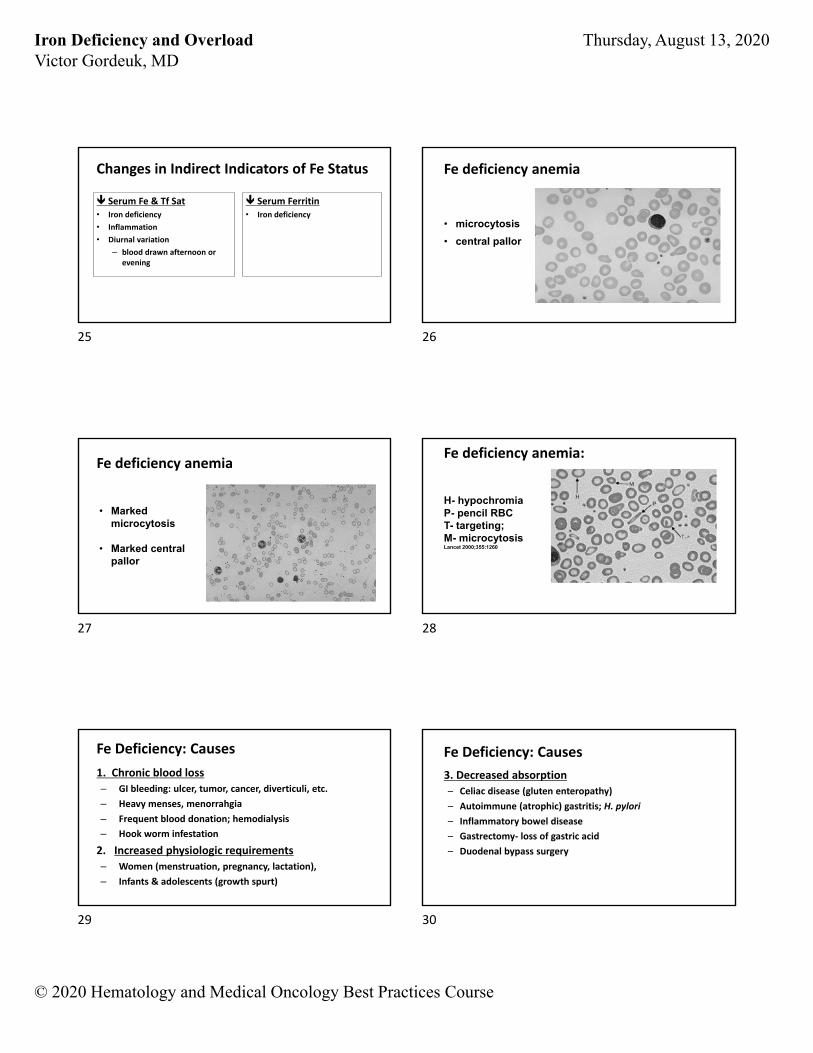
• microcytosis
• central pallor
Fe deficiency anemia
• Marked microcytosis
• Marked central pallor
Fe deficiency anemia:
H- hypochromiaP- pencil RBCT- targeting; M- microcytosis Lancet 2000;355:1260
Fe Deficiency: Causes
1. Chronic blood loss
– GI bleeding: ulcer, tumor, cancer, diverticuli, etc.
– Heavy menses, menorrahgia
– Frequent blood donation; hemodialysis
– Hook worm infestation
2. Increased physiologic requirements
– Women (menstruation, pregnancy, lactation),
– Infants & adolescents (growth spurt)
Fe Deficiency: Causes
3. Decreased absorption
– Celiac disease (gluten enteropathy)
– Autoimmune (atrophic) gastritis; H. pylori
– Inflammatory bowel disease
– Gastrectomy‐ loss of gastric acid
– Duodenal bypass surgery
25 26
27 28
29 30
Iron Deficiency and OverloadVictor Gordeuk, MD
Thursday, August 13, 2020
© 2020 Hematology and Medical Oncology Best Practices Course
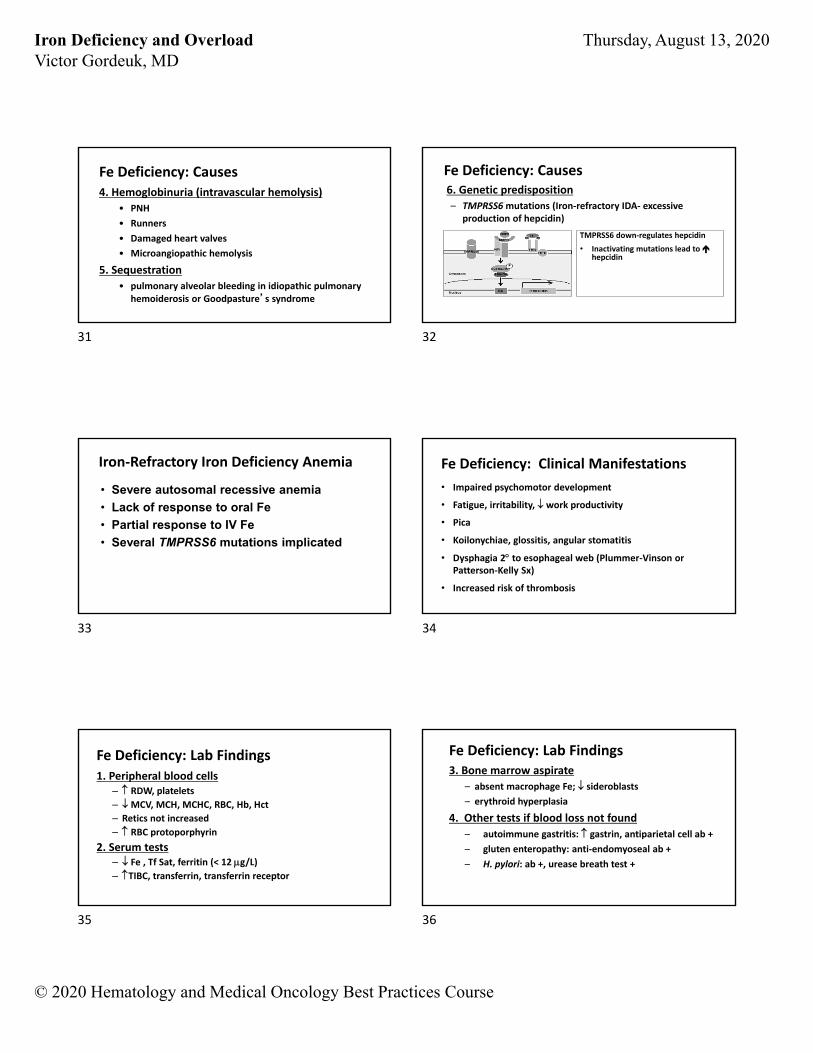
Fe Deficiency: Causes4. Hemoglobinuria (intravascular hemolysis)
• PNH
• Runners
• Damaged heart valves
• Microangiopathic hemolysis
5. Sequestration
• pulmonary alveolar bleeding in idiopathic pulmonary hemoiderosis or Goodpasture’s syndrome
Fe Deficiency: Causes6. Genetic predisposition
– TMPRSS6mutations (Iron‐refractory IDA‐ excessive production of hepcidin)
TMPRSS6 down‐regulates hepcidin
• Inactivating mutations lead to hepcidin
• Severe autosomal recessive anemia
• Lack of response to oral Fe
• Partial response to IV Fe
• Several TMPRSS6 mutations implicated
Iron‐Refractory Iron Deficiency Anemia Fe Deficiency: Clinical Manifestations
• Impaired psychomotor development
• Fatigue, irritability, work productivity
• Pica
• Koilonychiae, glossitis, angular stomatitis
• Dysphagia 2 to esophageal web (Plummer‐Vinson or Patterson‐Kelly Sx)
• Increased risk of thrombosis
Fe Deficiency: Lab Findings1. Peripheral blood cells
– RDW, platelets
– MCV, MCH, MCHC, RBC, Hb, Hct
– Retics not increased
– RBC protoporphyrin
2. Serum tests– Fe , Tf Sat, ferritin (< 12 g/L)– TIBC, transferrin, transferrin receptor
Fe Deficiency: Lab Findings3. Bone marrow aspirate
– absent macrophage Fe; sideroblasts
– erythroid hyperplasia
4. Other tests if blood loss not found
– autoimmune gastritis: gastrin, antiparietal cell ab +
– gluten enteropathy: anti‐endomyoseal ab +
– H. pylori: ab +, urease breath test +
31 32
33 34
35 36
Iron Deficiency and OverloadVictor Gordeuk, MD
Thursday, August 13, 2020
© 2020 Hematology and Medical Oncology Best Practices Course
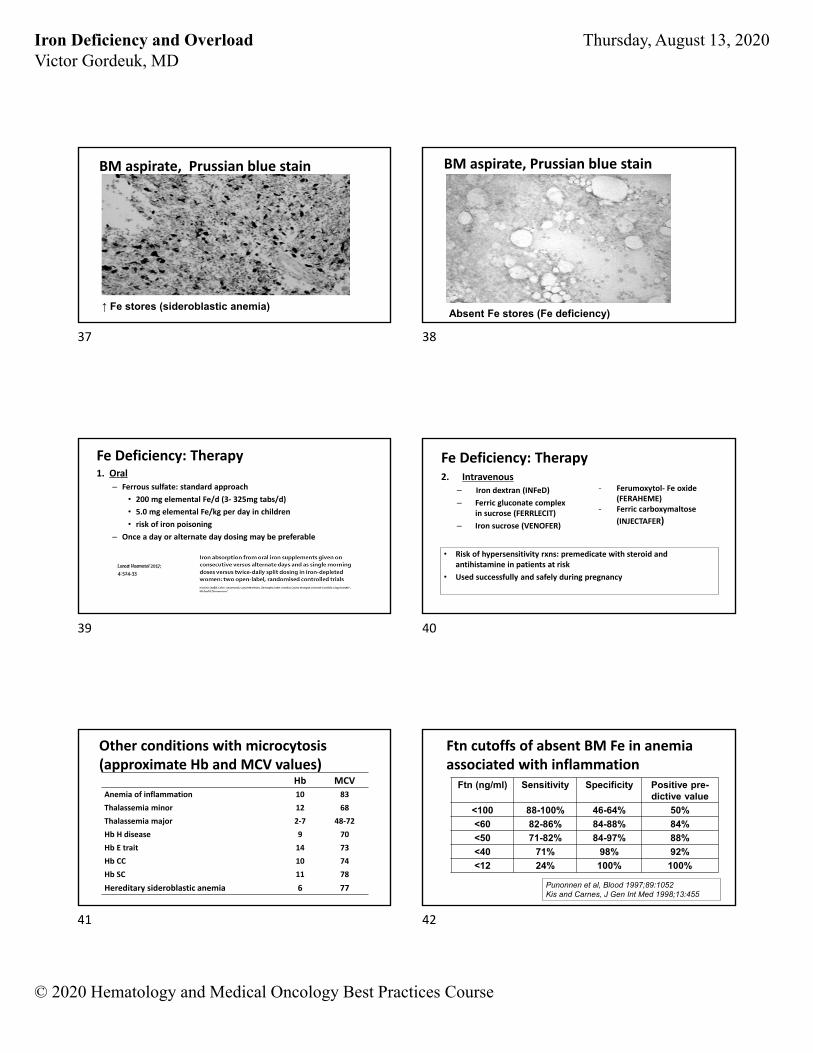
BM aspirate, Prussian blue stain
↑ Fe stores (sideroblastic anemia)
BM aspirate, Prussian blue stain
Absent Fe stores (Fe deficiency)
Fe Deficiency: Therapy1. Oral
– Ferrous sulfate: standard approach
• 200 mg elemental Fe/d (3‐ 325mg tabs/d)
• 5.0 mg elemental Fe/kg per day in children
• risk of iron poisoning
– Once a day or alternate day dosing may be preferable
Fe Deficiency: Therapy2. Intravenous
– Iron dextran (INFeD)
– Ferric gluconate complex in sucrose (FERRLECIT)
– Iron sucrose (VENOFER)
• Risk of hypersensitivity rxns: premedicate with steroid and antihistamine in patients at risk
• Used successfully and safely during pregnancy
- Ferumoxytol‐ Fe oxide (FERAHEME)
- Ferric carboxymaltose
(INJECTAFER)
Other conditions with microcytosis(approximate Hb and MCV values)
Hb MCV
Anemia of inflammation 10 83
Thalassemia minor 12 68
Thalassemia major 2‐7 48‐72
Hb H disease 9 70
Hb E trait 14 73
Hb CC 10 74
Hb SC 11 78
Hereditary sideroblastic anemia 6 77
Ftn cutoffs of absent BM Fe in anemia associated with inflammation
Ftn (ng/ml) Sensitivity Specificity Positive pre-dictive value
<100 88-100% 46-64% 50%
<60 82-86% 84-88% 84%
<50 71-82% 84-97% 88%
<40 71% 98% 92%
<12 24% 100% 100%
Punonnen et al, Blood 1997;89:1052Kis and Carnes, J Gen Int Med 1998;13:455
37 38
39 40
41 42
Iron Deficiency and OverloadVictor Gordeuk, MD
Thursday, August 13, 2020
© 2020 Hematology and Medical Oncology Best Practices Course
What is Iron Overload?
• Excessive amounts of storage iron– parenchymal cells liver, heart, pancreas (HH)
– macrophages spleen, BM, liver (Txs)
– mixed pattern
• Quantitative considerations– mild: 2‐4 g; moderate: > 4‐10 g
– marked: 10‐20 g; severe: >20 g
Iron Overload: Classification
1. Hereditary conditions causing hepcidin
– HFE hemochromatosis
– TFR2 hemochromatosis
– Hemojuvelinhemochromatosis
– Hepcidinhemochromatosis
Iron Overload: Classification
2. Hereditary conditions causing resistance of ferroportin to hepcidin– Ferroportin disease
Iron Overload: Classification
3. Ineffective erythropoiesis: erythroblast‐derived erythroferrone suppresses hepcidin:
– ‐thal major & intermedia; Hb E/‐thal; Hb H
– congenital dyserythropoietic and sideroblastic anemias
Iron Overload: Classification
4. Multiple blood transfusions– Diamond Blackfan anemia
– aplastic anemia
– thalassemia major
– sickle cell anemia
– myelodysplasia
Iron Overload: Classification
5. Other hereditary conditions• Aceruloplasminemia
• Atransferrinemia
• African dietary iron overload
43 44
45 46
47 48
Iron Deficiency and OverloadVictor Gordeuk, MD
Thursday, August 13, 2020
© 2020 Hematology and Medical Oncology Best Practices Course
Fe Overload: Clinical Manifestations
1. Fatigue, abd. pain
2. Liver– ALT
– fibrosis and cirrhosis
– Hepatoma
3. Heart– CHF (restrictive or
dilated)
– arrhythmias
4. Endocrine– diabetes mellitus
– secondary amenorrhea
– Impotence
5. Hyperpigmentation
6. Certain infections
7. Arthritis- esp. 1st and 2nd MP joints
(HFE hemochromatosis)
Screening for hemochromatosis or increased iron stores1. Screen pts
– with family hx or compatible clinical picture
– without inflamm., infection, trauma, surgery
2. Serum ferritin conc.
– raises possibility (esp. if Tf Sat or upper nl)
– Ferritin >1000 ug/L esp. of concern
3. PCR for HFEmutations, esp. Caucasians
Diagnosis of hemochromatosis or increased iron stores
1. Liver biopsy– Histology
– Perl’s stain
– Chemical iron concentration
2. MRI measurement hepatic or cardiac iron
3. SQUID magnetic measurement hepatic iron
4. Quantitative phlebotomy
Golden brown hemosiderin pigment predominantly in hepatocytes
Liver Bx, HFE hemochromatosis (H&E):
Granules predominantly in hepatocytes
Liver Bx, HFE hemochromatosis Prussian blue stain:
Cirrhosis; hepatocytes in regenerating nodule heavily laden with iron
Liver Bx, HFE hemochromatosisPrussian blue stain:
49 50
51 52
53 54
Iron Deficiency and OverloadVictor Gordeuk, MD
Thursday, August 13, 2020
© 2020 Hematology and Medical Oncology Best Practices Course
HFE Hemochromatosis
1. Classic triad– Hepatomegaly
– Diabetes
– Hyperpigmentation
2. Classically death from cirrhosis, HCC or CHF
3. Goal now‐make dx and rx in presymptomatic stage
HFE Genetic Defect
1. C282Y mutation, HFE gene, 6p21.3
– Autosomal recessive
– Predominantly in N. Europeans: “Celtic Disease”
– high prevalence in pop.
• homozygotes: 3‐5/1000
• heterozygotes: 10‐15%
2. HFE protein deficiency
– hepcidin production
• Fe absorption
• serum Fe and transferrin saturation
• storage of Fe in macrophages
• Fe‐loading of parenchymal cells
HFE Genetic Defect
3. Incomplete C282Y/C282Ypenetrance– Elevated SF
• <50% F homozygotes
• ≈75% M homozgotes
– Organ damage <25%
– Modifying factors
• genetic
• environmental
4. Second mutation H63D
– Very little iron‐loading tendency
– Fe stores may rarely develop in
• H63D/H63D
• C282Y/H63D
• C282Y heterozygote
HFE C282Y in 99,711 1º Care Pts (HEIRS)
N C282Y/ C282Y No. per 100,000
Whites 44,082 0.44% 440
African Americans
27,124 0.014% 14
Asians 12,772 0.0% 0
Hispanics 12,459 0.027% 27
Management of HFE hemochromatosis
1. Nl life expectancy if phlebotomy Rx started before cirrhosis or diabetes
2. If ferritin <1000 μg/L and LFTs nl at dx
– Proceed to phlebotomy therapy
3. If ferritin >1000 and/or LFTs abnl
– Consider liver biopsy
– risk HCC in cirrhotics even with phleb. Rx
4. Screen first degree family members
Phlebotomy Therapy
1. Initial program to remove excess body Fe
• Remove 1‐2 units of blood weekly as long as Hct 35% (M) or 32% (F).
• Continue until mild iron deficit: Pt. does not tolerate wkly phleb.; MCV declines; ftn < 50 ug/L
55 56
57 58
59 60
Iron Deficiency and OverloadVictor Gordeuk, MD
Thursday, August 13, 2020
© 2020 Hematology and Medical Oncology Best Practices Course
Phlebotomy Therapy
2. Quantitative phlebotomy
• Maintain weekly schedule and tally ml of whole blood removed until Fe deficiency develops.
• Calculate Fe stores at beginning of program (1 mg Fe/2 ml whole blood); >4g Fe‐ “Fe overload”
3. Maintenance to prevent the Fe reacummulation
• Remove 1 unit each 2‐6 mos. to maintain serum ftn around 50 μg/L.
Other Forms of Genetic Hemochromatosis
• TFR2 hemochromatosis– autosomal recessive; intermediate phenotype
– TfR2 mutation 7q22
• Juvenile hemochromatosis– autosomal recessive; severe phenotype
– hemojuvelin mutation 1q21.1
– HAMP (hepcidin) mutation 19q13.1
Other Forms of Genetic Hemochromatosis
• Ferroportin disease– autosomal dominant mutations ferroportin 2q32
– two phenotypes
• reduced response to hepcidin: predominant parenchymal iron‐loading
• increased sensitivity to hepcidin: predominant macrophage iron‐loading
African Iron Overload1. High dietary Fe in
traditional beverage
2. Familial pattern
– ? “gene by environment interaction”
– HFEmutations not present
– Ferroportin Q24H in some iron‐loaded subjects
3. Fe‐loading macrophages and hepatocytes
5. Clinical associations
– hepatic fibrosis, cirrhosis, hepatoma
– diabetes mellitus, cardiomyopathy
– osteoporosis, scurvy
– infections
– esophageal carcinoma
6. Management– prevention
– phlebotomy therapy
Iron Overload in African Americans
• Condition present; may be under‐dxed
• Sometimes related to African‐specific ferroportin Q248H mutation
• Screen with serum ferritin
• Responds well to phlebotomy therapy
• Evaluate for hepatitis C‐may play etiologic role by suppressing hepcidin
Transfusional Iron OverloadQuantitative Considerations• One unit blood contains about 225 mg Fe as Hb
• Hepatic Fe concentration
– normal <30 µmol/g dry wt. 0 units
– worrisome 180 µmol/g dry wt. 50 units
– Toxic 360 µmol/g dry wt. 100 units
– highly toxic 720 µmol/g dry wt. 200 units
• At 2 units/month: 50 units in two yrs
61 62
63 64
65 66
Iron Deficiency and OverloadVictor Gordeuk, MD
Thursday, August 13, 2020
© 2020 Hematology and Medical Oncology Best Practices Course
Transfusional Iron OverloadTreatment1. Institute iron chelation when
– >20 units of blood transfused, or
– hepatic iron conc. > 180 µmol/g dry wt.
2. Desferrioxamine, original parenteral chelator
– 40‐50 mg/kg per day
– s.c. infusion over 8‐12 hours
– 5 days/week
Transfusional Iron OverloadTreatment3. Deferasirox
- Single oral dose per day; similar potency to DFO
- Exjade: powder; starting dose 20 mg/kg/day; advance to 30 or 40 mg/kg/day if needed
- Jadenu: tablet; doses slightly lower
- Monitor CBC, creatinine, LFTs, gastritis sx’s q2‐4wks
Transfusional Iron OverloadTreatment4. Deferiprone
- Oral chelator
- Risk of agranulocytosis
- Approved in US for use in thalassemia patients with transfusional iron overload who did not respond to other chelators
Monitoring Iron Chelation Rx1. CBC; renal and liver function tests; GI Sxs monthly
2. Audiometry and ophthalmologic yearly
2. Serum ferritin every three months
3. Avoid use of phenothiazines
4. Vitamin C 100 mg daily may enhance Fe excretion; do not give to heavily iron‐loaded subjects
5. Modify dose as body Fe burden ’s
Other Iron Overload Conditions
1. Congenital atransferrinemia (3q22.1)
– parenchymal Fe‐loading; anemia
2. Congenital aceruloplasminemia (3q23‐q25)
– Fe‐loading paremchyma, macrophages, brain
– extrapyramidal sxs, cerebellar ataxia, DM
Other Iron Overload Conditions
4. Neuroferritinopathy
– autosomal dominant, late‐onset
– abnl aggregates ferritin & Fe in basal ganglia
– Mut. ftn light gene (19q13.3‐13.4); ser. ftn low
5. Hallovorden‐Spatz disease
– autosomal recessive; onset in childhood
– iron deposits in the basal ganglia
– mutation in pantothenate kinase 2 gene (20p13)
67 68
69 70
71 72