THE AMERTCAN MINERAIOGIST, VOL.45, JULY-AUGUST, 1960 IRON-RICH MUSCOVITIC MICA FROM THE GRANDFATHER MOUNTAIN AREA, NORTH CAROLINA* MancanBr D. Fosrnn, Bnuce Bnvaut, aNn JouN H,l'tnawlv, U. S. Geologi'cal Suraey, Washington 25, D. C. Assrnacr The metamorphic arkoses and arkosic quartzites in the Grandfather Mountain area, North Carolina, contain green micas that have high indices oI refraction with the 0 index ranging from 1.603 to 1.619. Detailed study of mica having the highest indices of refraction showed that it is dioctahedral in character, falling between muscovite and phengite in the trisilicic-tetrasilicic dioctahedral potassium mica series, with considerable proxying of Fe+3for octahedral Al. The green color and high indices of refraction are attributed to the high iron content and to the presence of both Fe+3and p"+z (8.11 per cent FeOs and 2.55 per cent FeO). INrnooucrroN AND REGToNAL GEoLocY In the course of geologicmapping in the Grandfather Mountain area, North Carolina, (one of us (B. B.)) observedrocks containing a fine- grained green mica. The green aspectof the rocks strongly suggests the presence of chlorite in addition to mica. However, thin section study showedthat no chlorite is presentl the greencolor of the rocks is due en- tirely to the mica. Preliminary optical study showed that the mica is muscovitic in type but has much higher indices of refraction than usual in such micas. The high indices of refraction, and the color, incited our interest in making a more detailed study of this mica. The Grandfather Mountain area is on the eastern edge of the BIue Ridge province 56 miles northeast of Asheville, N. C. The area is under- lain by early Precambrian basement rocks and late Precambrian and early Cambrian sedimentary and igneous rocks. These rocks are sur- rounded by early Precambrian schists, amphibolites, gneisses'and granitic rocks. Keith (1903) interpreted the structure of this area as a complex synclinal downfold bordered by opposing thrusts. A. L Jonas and G. W. Stose(Geologic map of the U. S., 1932) first recognized this structure as a window, and the present workers agree with that inter- pretation. For a summary of the regionalgeologyseeKing (1955). Retrogressive metamorphism of the basement rocks has produced a cataclastic foliation and recrystallization with development of new biotite and albite in rocks of appropriate composition both within the window and in the upper plate adjacent to the window. Progressive metamorphism in the late Precambrian and early Cambrian rocks was of the samegrade. In the window two sequences of sedimentary rocks are recognized: * Publication authorized by the Director, U. S. Geological Survey. 839
Transcript
THE AMERTCAN MINERAIOGIST, VOL.45, JULY-AUGUST, 1960
IRON-RICH MUSCOVITIC MICA FROM THE GRANDFATHERMOUNTAIN AREA, NORTH CAROLINA*
MancanBr D. Fosrnn, Bnuce Bnvaut, aNn JouN H,l'tnawlv,U. S. Geologi'cal Suraey, Washington 25, D. C.
Assrnacr
The metamorphic arkoses and arkosic quartzites in the Grandfather Mountain area,
North Carolina, contain green micas that have high indices oI refraction with the 0 index
ranging from 1.603 to 1.619. Detailed study of mica having the highest indices of refraction
showed that it is dioctahedral in character, falling between muscovite and phengite in the
trisilicic-tetrasilicic dioctahedral potassium mica series, with considerable proxying of
Fe+3 for octahedral Al. The green color and high indices of refraction are attributed to the
high iron content and to the presence of both Fe+3 and p"+z (8.11 per cent FeOs and 2.55
per cent FeO).
INrnooucrroN AND REGToNAL GEoLocY
In the course of geologic mapping in the Grandfather Mountain area,
North Carolina, (one of us (B. B.)) observed rocks containing a fine-
grained green mica. The green aspect of the rocks strongly suggests thepresence of chlorite in addition to mica. However, thin section study
showed that no chlorite is presentl the green color of the rocks is due en-
tirely to the mica. Preliminary optical study showed that the mica is
muscovitic in type but has much higher indices of refraction than usual
in such micas. The high indices of refraction, and the color, incited our
interest in making a more detailed study of this mica.The Grandfather Mountain area is on the eastern edge of the BIue
Ridge province 56 miles northeast of Asheville, N. C. The area is under-lain by early Precambrian basement rocks and late Precambrian and
early Cambrian sedimentary and igneous rocks. These rocks are sur-
rounded by early Precambrian schists, amphibolites, gneisses' andgranitic rocks. Keith (1903) interpreted the structure of this area as a
complex synclinal downfold bordered by opposing thrusts. A. L Jonasand G. W. Stose (Geologic map of the U. S., 1932) first recognized thisstructure as a window, and the present workers agree with that inter-pretation. For a summary of the regional geology see King (1955).
Retrogressive metamorphism of the basement rocks has produced acataclastic foliation and recrystallization with development of newbiotite and albite in rocks of appropriate composition both within thewindow and in the upper plate adjacent to the window. Progressivemetamorphism in the late Precambrian and early Cambrian rocks wasof the same grade.
In the window two sequences of sedimentary rocks are recognized:
* Publication authorized by the Director, U. S. Geological Survey.
839
840 M, D. FOSTER, B. BRYANT AND J. HATHAWAY
an autochthonous sequence unconformably overlying the basementrocks and an allochthonous sequence in thrust contact with members ofthe allochthonous sequence and the basement rocks (Reed and Bryant,1958). The upper sequence consists of arkosic quartzite, phyllite, andquartzite underlying dolomite, which appears to be equivalent to theShady dolomite of the Valley and Ridge province. The lower sequenceconsists of an interbedded and interlensing sequence of meta-arkose,calcareous biotite phyllite, phyllite, metasiltstone, metagraywacke, andgreenschist.
8 r . q 5 'le. z'30"
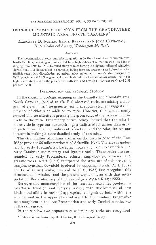
Frc. 1. Location of sample.
TUB GnBeN Mrca
Most of the metamorphosed arkoses and arkosic quartzites in bothsequences contain green muscovitic mica; the rocks range from lightgreenish-gray to green. The B index of the mica from some of the greenerrocks ranges from 1.603 in some specimens to 1.619 in others. A samplecontaining mica of the highest index of refraction found was chosen forfurther study.
The sample selected was collected from a roadcut on U. S. Highway221 abo',tt 1,000 feet southeast of the junction with the Edgemont roadon the southeast side of Grandfather Mountain one-half mile east of thecrest of the Blue Ridge (Fig. 1). This specimen is a green micaceousarkosic quartzite from the lowest member of the autochthonous sequence.
___=r7* _
I RON-RICH MUSCOV ITIC M ICA
The rock displays cleavage cutting bedding at an angle of 15o' Green
mica is concentrated along the cleavage planes, which are as much as
2 mm. apart in the coarser beds. Quaftz and microcline clasts up to 2 mm'
and plagioclase clasts up to 0.7 mm. in diameter occur in a matrix of
recrystallized quartz which has an average grain size of 0.1 mm. Feldspar
at least as small as 0.1 mm. retains its clastic outl ine, whereas qtattzas
large as 0.3 mm. is recrystall ized and participates in mosaic texture. The
mica is well aligned parallel to the cleavage and from 0'02 to 0.5 mm'
long. It includes aggregates of sphene and leucoxene.
Mod,e of sample
q \ a r t z ' . . . . . . . 4 5 . 8potassiumfeldsPar. . . 14.5
p lag ioc l ase . . . . 8 . 1
green mica. 29.2
sphene and leucoxene 1.7
ep ido te . . . . . 3
aPat i te. . . . .3
zircot.. tr'
Opti.cal properties
The green mica has the following optical properties:a:1.580*0.001 (colorless) 7-d: .043+ .01p: 1 . 6195+ .0005 (light green) optic sign : negativet:1.623O*.0005 (l ight green) 2V:35'*3'
The indices were determined with Na light in oils graduated in two
thousandths. The a index was determined on the spindle stage (Wilcox,
1959) and the optic axial angle by Mallard's method.
SBpanrlronv PnocBounB
The sample was crushed and divided into two fractions: 100-200
mesh and 200-400 mesh. The mica was separated by a process o-f re-
peated treatment in a Bendix ultrasonic transducer and separation in a
Frantz isodynamic separator. Each fraction was treated for two 20-
minute intervals in the ultrasonic transducer and run through the iso-
dynamic separator with 25o forward ti l t and 15o side ti l t at 1.0 amperes.
The magnetic fraction from this run was treated again in the transducer
and run through the separator at 0.9 amperes. This process was repeated,
at each run decreasing the amperage by about 0.1 ampere. The treat-
ment in the ultrasonic transducer shredded the mica flakes and freed
minute inclusions of sphene, qtartz, and feldspar which went into sus-
pension and were decanted.An amperage of 0.55 separated the green mica from the less magnetic
841
u2 M. D. FOSTER, B. BRYAN:I AND J, HATHAWAY
constituents. This fraction was run again at 0.45 amperes to removemagnetite, yielding a concentrate of about 99/6 mica in the 200-400mesh fraction, and about 98/6 in the 100-200 mesh fraction. The im-purities were very fine grained, but were probably chiefly sphene andleucoxene.
X-na.y ANALysrs
Both r-ray powder photographs and diffractometer patterns were usedin the examination of the green mica. Two different diffractometers wereused, operated at the following settings:
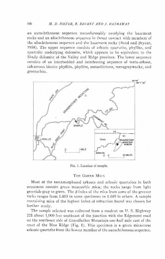
Frc. 2. X-ray powder photographs of : (A) Green mica from the Grandfather Moun-tain area, N. C., and (B) 2M Muscovite, Ontario 114.59 mm camera, CuKa, Ni filteredradiation.
The powder photographs were made of both relatively coarse and offinely ground material, with 114.59 mm. diameter cameras using ex-posures of about 12 hours.
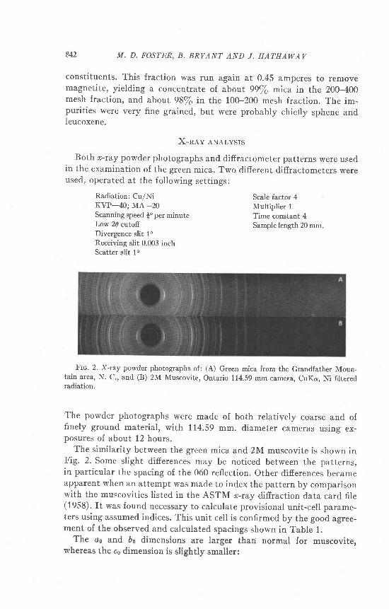
The similarity between the green mica and 2M muscovite is shown inFig. 2. Some slight differences may be noticed between the patterns,in particular the spacing of the 060 reflection. Other difierences becameapparent when an attempt was made to index the pattern by comparisonwith the muscovities listed in the ASTM o-ray difiraction data card file(1958). It was found necessary to calculate provisional unit-cell parame-ters using assumed indices. This unit cell is confirmed by the good agree-ment oI the observed and calculated spacings shown in Table 1.
The oo and 6o dimensions are larger thari normal for muscovite,whereas the co dimension is slightly smaller:
Scale factor 4Multiplier 1Time constant 4Sample length 20 mm.
i'.r886l r'e8e72 8011.9742 t .974r 551.949t L.95r7 2r1.8819 1.8830 81.8488 r .847r 3r .8213 1.8195 51 .7506 1.7480 10I t 2 1 A )
i fsill r'73r4 131 t . t ? 1 )
; '+ ; " , ; l r .7 r34 131.6995 1 .6998 8r.6646 r.6631 301 4 < t 7 r \
; ' i i i ' , | 1.6s78 1011.6477 1.6485 64
12.T1 1.6440 r.6M2 5315 1.6298 r .6276 8228 1.6054 t.ff i47 t4049 1.5822 1.5818 5155 1 .5546 1 .5608 1313.10 1.5462 1.5456 513.TI 1 5235 r.5239 20060 r.5108];47 i : ; io ; ] 1 's107 16331 1 .4952 \ I t ^ .o6i l'.ie3il 7'4e3e 2335 |.4527 L4515 15158 1.4326 r .43t6 80 0 . 1 4 1 . 4 2 0 6 \ 1 ^ 1 1336- f . i r o l | I ' 4 re8 12111 .13 1 .4126 ]oos'" i'.iiri| 1 '4112 10158 r.3892 1.3882 5066 r .s747 r .3757 813.T3 1.3504 1.3506 44o4. t2 1.3378 1.3376 3rl l . t 4 1 .3230122 .13 1 .3217 |
1 . czz3 | r400 1.30551)ol ru i .3d0) r .3042 2 l1 3 . 1 3 r . 2 9 2 6 L 2 9 3 r 704.13 1.2680 1 .2689 8170 1.2s691i i i i .r l i i ] 1 2s68 e351 | .2475 1 .2485 500.16 1 2431 1.2431 9lt73 r.2295 1.2289 43 5 3 1 . 2 t 7 7 1 . 2 1 7 4 1 0357 1 .1804 1 .1807 31 1 . 1 A 1 1 < C ? )
| i .n i : i ; ;3 i 1 rs80 824.14 t .1302 ' 1 .1294 8i l 7 . - | 1 l ?9 \ 1 11e4 s3 t . 12 t . t r 89 l083 1.1169 r . r t75 422 .17 1 .0781 r . 0776 335 . [ 1 .0681 1 .0677 220.18 1.0563 1.0566 41 ? - l ? ); ; " ' ' ) 1 .0485 1 .0483 t433.T5 1 .04171 I ^n1l ir-- i '.oi16| r 'u416 3282 1.0296'l | ^1(ia " i'.0r64] r '0)e6 s373 1.0172 r .0173 233.16 r .0029 1.0028 2n-n .989M .98961 10Plus at least 22 additronal weak lines.
^ 1 The.strongest 001 and hhlrines were each assigned an intensity of 100 because pre-
ferred orientation could not be eJiminated in the Jpecimens used ind the true intensityrelationships between 001 and hhl reflections could irot be determined. The intensities ofall of the 001 refl-ections are relative to the 002.
'! I:1.54050A for this and all smaller spacings. For large spacings I:1.5418A.
8M M. D. FOSTER, B, BRYANT AND J, HATHAWAY
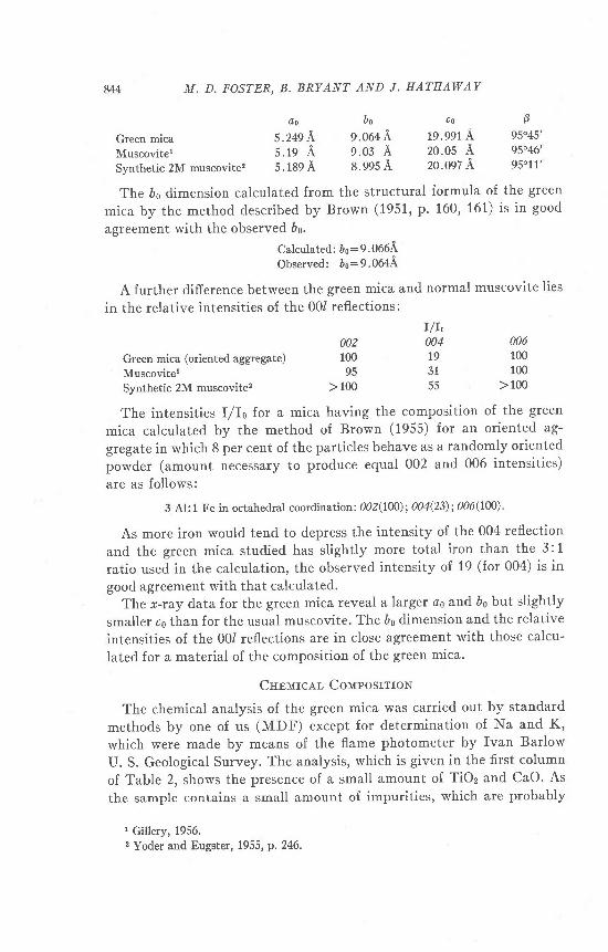
Ao 0o
Green mica 5.249 \ 9.064 ,:
Muscovitel 5.19 A 9.03 ASynthetic 2M muscovite2 5 . 189 A s.995 A
The 6o dimension calculated from the structural formula of the green
mica by the method described by Brown (1951, p. 160, 161) is in good
agreement with the observed 60.
Calculated: bo:9.066AObserved: bo:9.064ir
A further difference between the green mica and normal muscovite lies
in the relative intensities of the 001 reflections:
c o B
19.991 A 95045',20.05 A 9s"46'20.097 A 95011',
Green mica (oriented aggregate)
MuscoviterSynthetic 2M muscovite2
00210095
> 100
006100100
> 100
I/It004t93155
The intensities I/Io for a mica having the composition of the green
mica calculated by the method of Brown (1955) for an oriented ag-
gregate in which 8 per cent of the particles behave as a randomly oriented
powder (amount necessary to produce equal 002 and 006 intensities)
are as follows:
3 Al: 1 Fe in octahedral coordination: 002(100); 004(23); 006(100) '
As more iron would tend to depress the intensity of the 004 reflection
and the green mica studied has slightly more total iron than the 3: 1
ratio used. in the calculation, the observed intensity of 19 (for 004) is in
good agreement with that calculated.The *-ray data for the green mica reveal a larger @o and Ds but slightly
smaller co than for the usual muscovite. The bo dimension and the relative
intensities of the 001 reflections are in close agreement with those calcu-
lated for a material of the composition of the green mica.
Cnpurcer- CorvrposrrroN
The chemical analysis of the green mica was carried out by standard
methods by one of us (MDF) except for determination of Na and K,
which were made by means of the flame photometer by Ivan Barlow
U. S. Geological Survey. The analysis, which is given in the first column
of Table 2, shows the presence of a small amount of TiOz and CaO. As
the sample contains a small amount of impurities, which are probably
1 Gillery, 1956.2 Yoder and Eugster, 1955, p. 2M.
IRON_RICH MUSCOVITIC MICA
Ta.slr 2. Auar-vsrs ol GnnaN Mrce
As determinedConstituents
in mica
Adjusted to100 per cent
SiOzTiOzAlzOrFezOrFeOMeoMnOCaONagOKzOH:OF
Total- o : F
47 .28.30
24.468 .O22 .521 . 8 6.08. l +. 1 1
1 0 . 64 .00
1 A
47 .08
24.468 . 0 22 . 5 21 .86
.08
. 1 11 0 . 64 .00
. 1 4
47 .62
2 4 . 7 48 . 1 12 . 5 51 . 8 8
.08
. 1 110.724.05
1 A
9 9 . 5 1.06
99.45
98.87 100.00
Specific gravity 2.879.
chiefly sphene and leucoxene, the TiOz and CaO reported in the analysisare attributed to these impurit ies. Sphene and leucoxene also containSiO2, conseeuently 0.20 per cent SiOz was deducted from the SiOz re-ported as attributable to these impurit ies. The adjusted analysis, calcu-lated to 100 per cent, given in Table 2 column 3, yields the followingcalculated formula for the green mica,
The points of particular interest in this formula, as compared to thatof muscovite,
.00 -r 00
[Al, oo(Si, ooAir oo)oro(oH)r1-t ooriirto
are, (1) the octahedral-tetrahedral charge relation, (2) the higher Si andiower tetrahedral AI, (3) the number of octahedral sites occupied bybivalent cations, and (4) the relatively high Fe+3 content.
The composition of some of the high silica sericites and their relationto muscovite was explained by Schaller (1950, p. 407-415) as due to theirbeing members of a trisi l icic-tetrasil icic series. Foster (1956, p. 67-77)showed that this series, of which muscovite is the trisilicic end member,
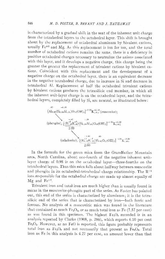
846 M. D. FOSTER, B. BRYANT AND J. HATHAWAV
is characterizedby a gradual shift in the seat of the inherent unit chargefrom the tetrahedral layers to the octahedral layer. This shift is broughtabout by the replacement of octahedral aluminum by bivalent cations,usually Fe+2 and Mg. As this replacement is ion for ion, and the totalnumber of octahedral cations remains the same, there is a deficiency inpositive octahedral charges necessary to neutralize the anions associatedwith this layer, and it develops a negative charge, this charge being thegreater the greater the replacement of trivalent cations by bivalent ca-tions. Coincident with this replacement and the development of anegative charge on the octahedral layer, there is an equivalent decreasein the negative tetrahedral charge, due to increase in Si and decrease intetrahedral Al. Replacement of half the octahedral trivalent cationsby bivalent cations produces the tetrasil icic end member, in which allthe inherent unit-layer charge is on the octahedral layer, and the tetra-hedral layers, completely fi l led by Si, are neutral, as i l lustrated below:
[Al, *(Sir 00All 00) or0 (oH)r]-t oorlto.,o,o
(-uscovite) ;
\ _ . 5 0 * 6 0
(phengite), t g{.$[st, soAl so)om(oH),]-'' oorl'o;ol
In the formula for the green mica from the Grandfather Mountainarea, North Carolina, about one-fourth of the negative inherent unit-layer charge of 0.98 is on the octahedral layer-three-fourths on thetetrahedral layers. Thus this mica falls about halfway between muscoviteand phengite in its octahedral-tetrahedral charge relationship. The R+2ions responsible for the octahedral charge are made up almost equally ofMg and Fe+2.
Trivalent iron and total iron are much higher than is usually found inmicas in the muscovite-phengite part of the series. As Foster has pointedout, this end of the series is characterizedby aluminum;it is the tetra-silicic end of the series that is characterized by iron-both ferric andferrous. No analysis of a muscovitic mica was found in the l iteraturethat contained as much Fe2OB, or as much total iron as Fe (7.57 per cent)as was found in this specimen. The highest Fe2O3 recorded is in ananalysis reported by Clarke (1908, p. 286), which reports 6.10 per centFezOs. However, as no FeO is reported, this figure probably representstotal iron as FezOs and not necessarily that present as FezOa. Totaliron as Fe in this analysis is 4.27 per cent, an amount Iower than that
IRON-RICH MUSCOVITIC MICA 847
found in an analysis reported by Ginzburg (1920, p.9), which contains5.64 per cent Fe2O3, and 1.27 per cent FeO. Total iron as Fe in this anal-ysis is 4.94. A third analysis (Tolman and Goldich, 1935, p. 236) has ahigher total iron as Fe content, 5.54 per cent, but is lower in Fe2O3, 3.94.Thus the mica herein studied is unique, for its position in the series, inits content of FezOs and of total Fe.
Rorerrow BETwEEN Iworcps ol RrlnacuoN AND InoN CoNrnNr
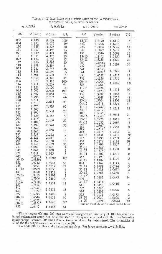
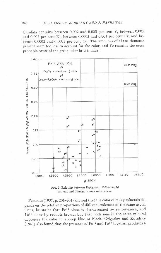
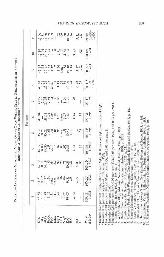
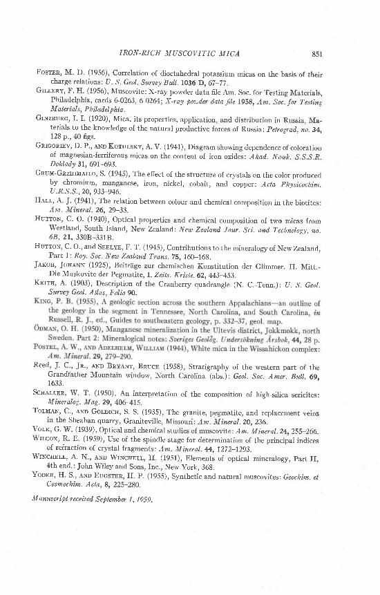
Winchell (1951, p. 368) concluded that the mean index (that is, theintermediate principal index, B) of muscovitic micas increases rapidlywith increase in ferric iron content. Volk (1939, p. 263) agreed withWinchell 's conclusion. Both Winchell and Volk used a three componentdiagram for plotting end member compositions of which the micasstudied were presumed to be composed, Winchell using the end membersmuscovite, KAlr(SfuAl)Oto(OH)r, ferrimuscovite, K(AIFe+a)(SiaAI)Oro(OH)r, and picrophengite, K(AI1.5Mgo.s)(SfuAI)Oro(OH):, and Volkusing the end members muscovite, ferrimuscovite, and phengitef[Rl1ne+zMg)](SLAI)Og(OH)r. Of these end members only muscoviteoccurs in nature; the others, ferrimuscovite, picrophengite, and phengiteare hypothetical and were postulated for the purpose of interpreting thecomposition of dioctahedral micas in terms of end members. However,neither Winchell's nor Volk's diagram showed a close relationship be-tween ferric iron content and mean index; both showed considerablescatter of points. None of their micas had as high an index nor as high acontent of ferric iron as the green mica herein studied. The relationbetween the B index and Fe2O3, in terms of molecular equivalents andbetween the B index and (FesOa*Fe), in terms of molecular equivalentsin a number of muscovitic micas is shown in Fig. 3. The unnumberedpoints in this diagram represent micas used by volk that contained bothFerOa and FeO; the numbered points represent some muscovitic micasreported in the lit'erature for which both analyses and indices of refrac-tion are given. The numbers refer to analyses given in Table 3. Althougha very general trend toward increase in B index with increase in FezOgand with increase in (FerOs*FeO) content is suggested, the relationshipis not close and it would not be feasible to try to predict even the ap-proximate iron content from the 0 index or vice versa for muscoviticmicas. Ilowever, the points representing the much higher 0 index andiron content of the North Carolina mica are in line with the general up-ward trend of the points representing these properties in other micas.
Cause on Coron
A semiquantitative spectrographic analysis by John C. Hamilton ofthe U. S. Geological Survey, shows that the green mica from North
848 M. D. FOSTER, B, BRYANT AND I. HA'I 'HAWAV
Carolina contains between 0.002 and 0.005 per cent V, between 0'001
and 0.002 per cent Ni, between 0.0005 and 0.001 per cent Cr, and be-
tween 0.0002 and 0.0005 per cent Cu. The amounts of these elements
present seem too low to account for the color, and Fe remains the most
orobable cause of the sreen color in this mica.
o t 5
azU)
l
U
E.
)l
U
o=a
E+
oz.
N
E
E X P L A N A T I O Nx r
Fe203 conlent ond p lndex
( FeO + Fe2O3) contenf ond p index
Green micoo
Gre€n mico
6
tlc
x7t a
a l
x lar O
a
a
xX X
a a
xx
r4
a a
x'
X
o 0 01 5850 1 .5900 1 5950 1 .6000 1 6050 l 6100 16150 1 6200
P INDEX
Frc. 3. Relation between Fe:Os and (FeO*Fe:Or)
content and 6 index in muscovite micas'
Fersman (1937, p. 201-204) showed that the color of many minerals de-
pends on the relative proportions of difierent valences of the same atom'
Thus, he states that Fe+2 alone is characterized by yellow-green, and
Fe+s alone by reddish brown, but that both ions in the same mineral
clepresses the color to a deep blue or black. Grigoriev and Kotulsky(1941) also found that the presence of Fe+2 and Fe+3 together produces a
0.4 0
o 3 5
o 3 0
o 2 5
0 2 0
o r o
o o 5
849
. a
H 6 6
^ !
N \C)
I
r c \
^
N
N C \
$ aN 6^ :
x
e, it.EEfr 3 qo
IRON-RICH MUSCOVITIC MICA
qqqqq qqqiq q iL Q O : N F I o Ds 6
O $ + c O € C ) i r N + ' o n- O 4 < f O . O r O € 4 o O O6 6 i d ; - i ' J ' - j . o ;
t < r \ N N N € ) S O q r Nr O € $ + O n 0 2 4 d + € O
. . c . . . . .$ O o o H d o O : nS A C H
\ N \ o \ o o \ o . \ o + c ) + @ n€ N r O 9 b : O n O N N O \. ? O O = f O : O N . Og N c i
e333S38 3F Id J . d 6 i J
" i l J o i d I
4 iq€4i E\E q qr a I e * : : S o N o \ < i
3 R R 3 5 3 3 s S $ 9 5 =D O o i F i i O : q
N O \ O o O c t n r < ) n i +r O O N i : € O O \ € r h
< .c i ; ' . i ' o
< .$ a
N i O \ 6 + N N D F q r n aF 4 F 6 \ O O c } . N T O D O Ot Q O + O i Ds r
D S S S e s $ 3 3 3 S S .+ O N o F H a + - :+ 6 - - - - t r
\ O f - € N i i < r r o N4 < r o \ 4 i c o o N' . . . . . 1L ? o h - | - o . . I$ a
Ba$3cEBsBsB
zts
z
. a zE ) H,; ce
4 Za aa ;2 , 8d OH Z
F r XA Hv g
X za ;
e2F <
> p (
6 <
I
z4
o
Fl
F
850 M. D. FOSTER, B. BRYANT AND J. HATHAWAY
darker color than either alone. Depth of color produced by Fe+2 and
Fe+3 together, or by either alone, also depends on the amounts present.
Another factor afiecting the color produced by coloring ions in minerals
is crystal structure (Grum-Grzhimailo, 1945, p. 933-46). Iron is re-
sponsible for green colors in biotites (Hall, 1941, p. 29-33) and for the
green color in glauconite. In both of these minerals Fe is usually present
both as Fe+2 and Fe+3. Wyoming bentonite, which consists almost en-
tirely of montmorillonite, is olive-green when Fe+3 predominates over
Fe+2, but is blue-gray when Fe+2 predominates over Fe+3 (Foster, 1956,
1003). In glauconite also, Fe+3 is ordinarily in excess of Fe+2. As these
minerals have the same crystal structure as the mica from North
Carolina, it is concluded that its green color is due to the presence of
both Fe+a and Fe+2, with Fe+3 predominant over Fef2.
Suulranv
A detailed study of a green mica associated with metamorphosed
arkoses and arkosic quartzites in the Grandfather Mountain area, North
Carolina, showed it to be a dioctahedral potassium mica about midway
between muscovite and phengite in its Iayer charge relationship. For its
place in the trisilicic-tetrasilicic dioctahedral mica series, this mica is
unusually high in content of ferric iron and of total iron. Its high indices
of refraction are attributed to this high iron content, and its color is
attributed to the presence of both Fe+3 and Fe+2 in the structure, with
Fe+3 predominant over Fe+2.
AcTNowTBIGMENTS
Our appreciation is expressed to John C. Reed, Jr., coworker on the
Grandfather Mountain project, and to Leonard B. Riley, both of the
U. S. Geological Survey, for their encouragement and interest.
RalnnrNcr,s
BnowN, G. (1951), The mica clay minerals, Part II Nomenclature of mica clay minerals:
in G. W. Brindley, ed., X-ray identification and crystal structures of clay minerals:
London, The Mineralogical Society, 155-172.--- (1955), The effect of isomorphous substitutions on the intensities of (001) reflec-
tions of mica- and chlorite-type structures: Mineratrog. Mag' 3O,657-665.
Crmru, F. W. (1910), Analyses of rocks and minerals from the laboratory of the United
States Geological Survey, 1880-1908: U. S. Geol'. Suney BuII'.4L9, p. 286.
EcKIRMANN, Hennv voN (1927), The optical and physical properties of two Swedish
muscovites : G eol 6 g. F dr en. S t ockholm F or h. 49, 229-234.
Fnncuson, R. B. (1943), Muscovite from Mattawan Township, Nipissing District, On-
tario: Uni,l. Toronto Studi.es, Geotr. su. no. 48r 3141.
Fnnsunx, A. E. (1937), The color of minerals: Acod^ sci,. [/.S.S.R. Comptes renilus, 17,
20r-204.
I RON-RICH MUSCOV ITIC M ICA
Fosmn, M. D. (1956), correlation of dioctahedral potassium micas on the basis of theircharge relations: U. S. Geot. Surley Bull.l}36-D,67-72.
Gu,tnnv, F. H. (1956), Muscovite: X-ray powcler data file Am. Soc. for Testing Materials,Philadelphia, cards 6-0263, 6-0264; x-ray poz.od,er ilata f,Ie r9s8, Am. Soc. Jor TestingM ater ials, P hil, adel p hia.
GtNznunc, r.L (1920), Mica, its properties, application, and distribution in Russia, Ma-terials to the knowledge of the natural productive forces of Russia: petrograil, no. 34,128 p., 40 figs.
Guconrnv, D. P., aNo Korur.srv, A. v. (1941), Diagram showing dependence of colorationof magnesian-ferriferous micas on the content of iron oxides: Akad.. Nauk. s.s.s.R.Dokl,atly 31, 691-693.
Gnuu-Gnzuruerro, S. (1945), The efiect of the structure of crystals on the color producedby chromium, manganese, iron, nickel, cobalt, and copper: Aeta physicochim.u.R.s.s., 20,933-946.
HeLr, A. I. Q94l), The relation between colour and chemical composition in the biotites:Am. Mineral.26, 29-33.
Hurrou, c. o. (1940), optical properties and chemical composition of two micas fromwestland, South rsland, New Zealand: Naa Zealanil Jour. sci. and. Technology, no.68,2t, 3308-33t8.
HurroN, c. o., and Sonrvr, F. T. (1945), contributions to the mineralogy of New Zealand,Part l: Roy. Soc. New Zealand. Trans.75,160-168.
Jer<ou, JorraNN (1925), Beitr?ige zur chemischen Konstitution der Glimmer. rr. Mitt.-Die Muskovite der Pegmatite, I. Zeits. Kriste.62. M3-453.
Krrrn, A. (1903), Description of the cranberry quadrangle (N. c.-Tenn.): u. s. Geot,.Suraey Geol. Atlas, Fol,io 9O.
Reed, J. C., Jn., a*n Bnvlwr, Bnucn (1958), Stratigraphy of the western part of theGrandfather Mountain window, North carolina (abs.): Geot. soc. Amei. Bxtlt.69,1633.
Scuarr,on, w. T. (1950), An interpretation of the composition of high-silica sericites:Miner al,o g. M ag. 29, 40G415.
Tor,nlN, C., aNn Gor,orcu, S. S. (1935), The granite, pegmatite, and replacement veinsin the Sheahan quarry, Graniteville, Missouri: Am. Mi,neral.20,236.
vor,r, G' w. (1939), optical and chemical studies of muscovite; Am. Mineral.24, 2ss-266.wrr.cox, R. E. (1959), Use of the spindle stage for determination of the principal indices
of refraction of crystal fragments: Am. Mineral. A, 1272-1293.Wtxcuu,t, A. N., eNo WrNcnnlr, H. (1951), Elements of optical mineralogy, part II,
4th end.: John Wiley and Sons, Inc., New york, 36g.Yonnn, H. S., eNo Eucsrnn, H. P. (1955), Synthetic and natural muscovites: Geoch,im. et