Page 1
Roma, 22 Febbraio 2017
Prof. Umberto Cillo MD, FEBS
Chirurgia Epatobiliare e Centro Trapianto di Fegato
Azienda Ospedaliera – PoliclinicoPadova
[email protected] www.fegatochirugia.it
Is there still a role for surgery in non cirrhotic
portal hypertension?
19° AISF Pre-Meeting course
“Vascular disease of the Liver”
Page 2
Una chirurgia in estinzione?
Page 3
1. Rivascularization procedures Meso-Rex Shunt
2. Portosystemic shunts
3. Devascularization procedures
4. Orthotopic Liver Transplantation
Non-Selective:
Total:
• End-to-side portacaval shunt (Eck's fistula)
• Side-to-side portacaval shunt (> 10 cm)
• Mesocaval with prothesic interposition
• Proximal splenorenla shunt (Linton's shunt)
Partial:
• Small diameter end-to-side (8-10 cm)
• Calibrated side-to-side portacaval shunt
Selective:
Distal splenorenal shunt (Warren's shunt)
Coronaro-caval anastomosis (abandoned)
Spleno-caval anastomosis (abandoned)
Surgical options
Page 4
Literature Trends
Research performed on PubMed
15° February, 2017
2016:
63 papers
2000:
103 papers1990:
116 papers
1980:
80 papers
1973:
70 papers
2016:
1700 papers
2000:
805 papers1990:
440 papers
1980:
154 papers
1973:
61 papers
Page 5
Khanna R et al, J Hepatol 2014 vol. 60; 421–441
Main causes
of NCPH
Page 6
NCPH: Associated disorders
Schouten JNL et al. Orphanet Journal of Rare Diseases (2015) 10:67
Riggio O et al, Hepatic Medicine: Evidence and Research 2016:8 81–88
Immunological/Autoimmune disorders
• Common variable immunodeficiency syndrome
• Primary antibody-deciciency syndrome
• Connettive tissue diseases
• Crohn’s disease
• Rheumatoid artritis
• Systemic lupus erythematosus
• Systemic sclerosis
• Scleroderma
• Celiac disease
• Solid organ transplant
Infections
• Bacterial intestinal infections
• HIV infection
Medications/Toxins
• Thiopurine derivatives
• Arsenicals
• Vitamin A
• Chemotherpy
Prothrombotic conditions
• Inherited thrombophilias
• Myeloproliferative neoplasm
• Antiphospholipid syndrome
• Protein S or C deficiency
• Lupus anticoagulant
• Factor V Leiden
• Prothrombin mutation
Hematologic disease
• Myeloproliferative disorders
• Myeloid metaplasia
• Lymphoproliferative conditions
• Spherocytosis
Genetic disorders
• Adams-Olivier Syndrome
• Cystic fibrosis
• Turner’s disease
• Phosphomannose isomerase deficiency
• Familial cases
Page 7
Patophysiology as clinical guide
Khanna R et al, J Hepatol 2014 vol. 60; 421–441
Pre-Hepatic Hepatic Post-Hepatic
FHVP Normal Normal High
RAP Normal Normal Normal or High
WHVP Normal High High
HVPG Normal Normal or High Normal or High
PVP High High High
ISP High High High
FHVP Free hepatic venous pressure
RAP Right atrial pressure
WHVP Wedged hepatic venous
pressure
HVPG FHVP – WHVP
PVP Portal vein pressure
ISP Intrasplenic pressure
Page 8
Extra Hepatic Portal Vein Obstruction
(EHPVO)
Page 9
Sarin KS et al, Clin Liver Dis 10 (2006) 627–651
NCPH: NCPF and EHPVOThe unifying hypothesis (Sarin and Kumar, 2006)
Page 10
EHPVOClinical Features
NCPF
Size of vessel involved Main portal vein
Hematemesis/melena 77%
Splenomegaly 20%
Ascites (transient) 23%
Jaundice 23%
Esophageal varices 93%
Portal gastropathy 60%
Portal biliopathy 90%
Other clinical features
Growth faltering, impaired QoL,
Minimal hepatic encephalopathy
Hypersplenism, splenic infarction
Bleeding from non GI sites: 20%
Liver function Preserved
Sarin KS et al, Clin Liver Dis 10 (2006) 627–651
Rajekar H et al, J Clin Exp Hepatol 2011;1:94–108
Schouten JNL et al, Hepatology 2011;54:1071-1081
Schouten JNL et al. Orphanet Journal of Rare Diseases (2015) 10:67
Khanna R et al, J Hepatol 2014 vol. 60; 421–441
Riggio O et al, Hepatic Medicine: Evidence and Research 2016:8 81–88
Mean number of 1.8–3.1 bleeding
episodes
Splenic size and portal pressure
do not correlate with the incidence
or severity of bleed.
Liver is normal or shrunken.
Jaundice due to portal biliopathy.
Peripheral stigmata of CLD
are absent
Page 11
EHPVO: Management
Khanna R et al, J Hepatol 2014 vol. 60; 421–441
Colonopaty/rect
al varices
Treatment
failure
Complicated
portal biliopathy
Severe
Hypersplenism
Growth
retardation/QoL
Page 12
Surgical interventions for PHRevascularization: MRB
Page 13
Surgical interventions for PHRevascularization: MRB
..or, alternatively,
splenorenal shunt
Page 14
Di Francesco F et al, J AM Coll Surg 2014; 218(2)
Page 15
EHPVO: Management
Khanna R et al, J Hepatol 2014 vol. 60; 421–441
Pre-operative assessments:
• Adult and/or pediatric hepatologist
• Cardiologist (to exclude HPS and PPH)
• Hematologist (for prothrombotic states)
• Radiologist (CT/MR
angiography/Portography)
Surgical consideration in EHPVO
Page 16
Lautz TB et al, J Am Coll Surg 2013;216:83e89
81 children with idiopatic EHPVO evaluated
Between 1997 and 2010
Single Institution
65 underwent successful MRB
16 required PSS
MRB PSS p
Relief of varicealbleeding
96% 100%
Improvement inPTLS count
+82.1 ± 60.0 +32.4 ±56.3
0.004
INR -0.22 ± 0.27 0.01 ± 0.14 0.022
Ammonia -26.8 ± 36.8 +19.4 ±33.1
0.002
Improvement in weight/age z-score
+0.84 ± 0.98 +0.17 ±0.79
0.044
PTLS Spleen
size
INRSerum
Ammonia
Page 17
Lautz TB et al, J Am Coll Surg 2013;216:83e89
Both MRB and PSS effectively relieve symptoms of portal hypertensive bleeding
MRB relieves hypersplenism better than PSS
By restoring normal portal venous circulation
the meso-Rex bypass has additional metabolic benefits
Page 18
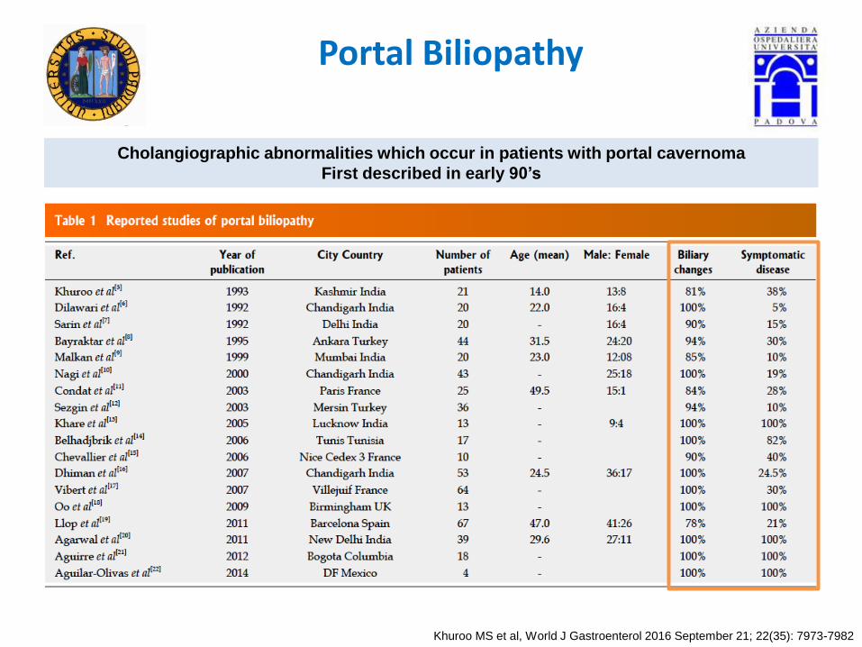
Portal Biliopathy
Cholangiographic abnormalities which occur in patients with portal cavernoma
First described in early 90’s
Khuroo MS et al, World J Gastroenterol 2016 September 21; 22(35): 7973-7982
Page 19
Anatomical consideration inportal biliopaty
Chittapuram SRB et al, J Clin Exp Hepatol 2014;4:S18–S26
CBD draining veins are arranged
in the form of two plexuses
Epicholedochal venous plexus (Saint, 1971)
is a fine reticular plexus
on the surface of the bile ducts
Paracholedochal venous plexus (Petren, 1932)
lies outside the bile ducts
and courses parallel to the ducts
Page 20
Portal Cavernoma development
Chittapuram SRB et al, J Clin Exp Hepatol 2014;4:S18–S26
PC develops as a bunch of hepatopetal collaterals
in response to portomesenteric venous obstruction
Normally:
• PSPDV drains into PV close to porta hepatis
• PSPDV connects with PIPDV
which drains into SMV
through first jejunal vein (FJV)
Page 21
Portal Cavernoma development
Chittapuram SRB et al, J Clin Exp Hepatol 2014;4:S18–S26
PC develops as a bunch of hepatopetal collaterals
in response to portomesenteric venous obstruction
Normally:
• PSPDV drains into PV close to porta hepatis
• PSPDV connects with PIPDV
which drains into SMV
through first jejunal vein (FJV)
PSPDV and Pericholedochal venous plexus
dilate and acts as a porto-portal
collateral channels Cavernoma
PV thrombosis
involving the
splenomesenteric
confluence
Page 22
Portal biliopaty
Chittapuram SRB et al, J Clin Exp Hepatol 2014;4:S18–S26
Sharma M et al, Gastrointest Endosc. 2009;70(5):1041–1043
Denys A et al, AJR. 1998;171:455–456
Application of color and spectral Doppler
shows presence of varices with venous flow in the CBD
between the lumen of the CBD and the varices
PC Subepithelial varices
in the CBD wall
Page 23
Portal biliopaty
Chittapuram SRB et al, J Clin Exp Hepatol 2014;4:S18–S26
Sharma M et al, Gastrointest Endosc. 2009;70(5):1041–1043
Denys A et al, AJR. 1998;171:455–456
PC Subepithelial varices
in the CBD wallDilatation of large paracholedochal veins
Compression and distortion
of the extrahepatic BD
with varicoid portal biliopaty
(REVERSIBILE)
Enlargement of smaller
intramural epicholedochal plexus
compromises the arterial supply
of the ductal wall producing
ischemic changes and fibrosis
(IRREVERSIBLE)
Page 24
Portal Biliopathy Classification
Khanna R et al, J Hepatol 2014 vol. 60; 421–441
Chandra R et al Journal of Gastroenterology and Hepatology (2001) 16, 1086–1092
A. Type I, involvement of extrahepatic bile duct
B. Type II, involvement of intrahepatic bile ducts only
C + D. Type IIIa,involvement of extrahepatic bile duct and unilateral intrahep-atic bile duct (left or
right)
E. Type IIIb, involvement of extra-hepatic bile duct and bilateral intrahepatic ducts.
Schematic representation of ERCP changes in portal biliopathy
Page 25
Portal Biliopathy Classification: Endoscopic Management
Author Pts N° Follow-up Further treatments Complications/Outcomes Morbidity Mortality
Bhatia et al, 1995
4 3-8 months Multiple ERCP 4 None 0% 0%
Perlemuter et al, 1996
8 6-60 months Multiple ES 1 Death 2 (cholangitis + stroke) NA 25%
Condat et al, 2003 7 4-25 months - Haemobilia 1 14.3% 0%
Sezgin et al, 2003
10 3.3 years (1 -7) Multiple ERCP 5 Haemobilia 1Cholangitis 5
Death 1
60% 10%
Dumortier et al, 2003
6 10 months (2-18) Multiple ERCP+ PSS 4
Cholangitis 1Cholecistytis 4
83.3% 0%
Khare et al, 2005
13 - PSS 8, BA 1, Multiple ERCP 2, Sugiura 2
Death 1 NA 7.6%
Dhiman et al, 2007 12 19 months (6-132) Multiple ERCP Cholangitis 2 16.6% 0%
Vibert et al, 2007
19 8.3 years PSS: BA 5; Non PSS: PTBD 1 after ERCp 4 after BA
Death 3 NA 15.7%
Oo et al, 2009
13 2 years (1-18) Stent 3, Stent exchange 2 TIPS 2, PSS 1, LT 1
Haemobilia 2Sepsis 3
38.5% 0%
Llop et al, 2011
14 - Multiple ERCP 1BA 1
- NA NA
Sarasvat et al, 2013 20 18 months (3-188) Multiple ERCP11
In 130 procedures: Cholangitis 40, Haemobilia 9
37.7%* 0%
Ramchandani et al, 2013
5 6-7 months SRS 2 Stent exchange 1 - NA NA
Cellich et al, 2015
9 - PSS 1, BA 3,Stent exchange 3
Cholangitis 3Haemobilia 1
44% 0%
Modified from Franceschet I et al, World J Gastroenterol 2016 December 7; 22(45): 9909-9920
Page 26
Khanna R et al, J Hepatol 2014 vol. 60; 421–441
EHPVO with Portal biliopaty:Algorithm for management
Page 27
Management of Portal Biliopathy
Franceschet I et al, World J Gastroenterol 2016 December 7; 22(45): 9909-
9920
Unsuccessful vascular surgery is frequent in mixed type because
of co-presence of ischemic and compressive damage.
Page 28
Portal Biliopathy Classification: Surgical Management
Author Pts N° Follow-up Treatments Further Treatments Morbidity Mortality
Chaudhary et al, 1998
9 - BA2, SRS 7 BA 2, Stent 1, ES + SE 2 NA 11.1%
Condat et al, 2003
7 4-25 months Cholecistectomy + ERCP 1, stent 1, BA + PTBD 1
- 14.3% 0%
Gauthier-Villars et al, 2005
8 4.5-15 ys PSS 8 - NA NA
Khare et al, 2005
13 - PSS 7, BA1 , ERCP 6, BA + PSS 1, Sugiura 2
ERCP 1, Splenectomy, ERCP 2; BA 1;Splenectomy + BA 1
NA 1.9%
Vibert et al, 2007
19 4-30 months PTBD 1, SRS 10 BA 5, PTBD 5 NA 15.7%
Dhiman et al, 2007
12 19 months(6-132)
Stent 2, stone extraction 1, PTBD 6, BA 4
Multiple stent exchange 16.6% NA
D’Souza et al, 2009
1 18 months PSS 5, ES 3, ES + dilatation 2, stent 4
- NA 0%
Camerlo et al, 2010
3 2-13 years PSS 3, stent 1 - NA 0%
Argawal et al,2011
39 32 months SRS 37, BA 2 ES + SE 10, BA 12, ES + cholecistectomy
NA 0%
Chattopadhyay et al, 2012
56 48 months(14-120)
40 PSS + 16 Sugiura ES + SE 2, Mulltiple ES + Stent 5, BA 2
NA 4.2%
Suarez et al, 2013
3 - UDCA 1, BA 1 - NA NA
Bhatia et al, 2014
2 - Cholecistectomy - NA 0%
Liu et al, 2015
18 - PSS 18 - NA 0%
Modified from Franceschet I et al, World J Gastroenterol 2016 December 7; 22(45): 9909-9920
Page 29
NCPH: Non Cirrhotic Portal Fibrosis (OPV)
Saigal S et al, Hepatol Int (2011) 5:882–889
Microscopic features in cases of pure NCPF group
(Masson trichrome stain)
Fibrous intimal thickening of
medium size portal vein (PV)
Portal fibrosis with complete obliteration
or multichannelling of PV branches
Marked portal fibrosis linking portal to portal
areas with some incomplete nodule
Demarcated nodular area of parenchyma
with intervening compressed liver cell plates
Page 30
Sarin KS et al, Clin Liver Dis 10 (2006) 627–651
Rajekar H et al, J Clin Exp Hepatol 2011;1:94–108
Schouten JNL et al, Hepatology 2011;54:1071-1081
NCPH: PRE-sinusoidalNCPF and EHPVO
NCPF EHPVO
Mean age (y) 31 19.4
Sex (Males/Females) 1 : 0.7 1 : 0.5
Geographical distrubution > in developing countries > In developing countries
Size of vessel involved Peripheral portal vein branches Main portal vein
Hematemesis/melena 82% 77%
Splenomegaly 21% 20%
Ascites (transient) 19% 23%
Jaundice 7.8% 23%
Esophageal varices 96% 93%
Portal gastropathy 3.2% 60%
Portal biliopathy 25% 90%
Liver function Preserved Preserved
Other clinical features
± hypersplenism, anemia
Patent hepatic portal vein
Gastropathy, Colopathy
Growth faltering
Minimal hepatic encephalopathy
Prognosis Fairly good but need for regular and careful surveillance
Schouten JNL et al. Orphanet Journal of Rare Diseases (2015) 10:67
Khanna R et al, J Hepatol 2014 vol. 60; 421–441
Riggio O et al, Hepatic Medicine: Evidence and Research 2016:8 81–88
Page 31
NCPF: Management
Endoscopic therapy in NCPH is effective in:
• Controlling acute variceal bleeding in 95%
of patients
• Reduce the risk of variceal rebleeding
With uncontrolled bleeding
portal systemic shunting
by insertion of TIPS
should be considered.
Liver transplantation in unmanageble
portal hypertension-related complications
and progressive liver failure
Page 32
Indications requiring liver transplantation in these patients were:
• Medical unmanageable portal hypertension
• Hepatopulmonary syndrome
• Hepatic encephalopathy
• Progressive hepatic failure
Schouten JNL et al, Hepatology 2011;54:1071-1081
LT in NCPH
To prevent unnecessary LT
early discrimination between cirrhosis and INCPH
is extremely important
Based on small-sized cohorts
post-LT outcome in these patients is good
and INCPH tends not to recur
Page 33
Non Cirrhotic Portal Fibrosisand CLD
Saigal S et al, Hepatol Int (2011) 5:882–889
CLD was classified
mainly as CC
before LT
Page 34
Non Cirrhotic Portal FibrosisExplant pathology
Nayak NC et al, J Clin Pathol 2011;64:592-598
Page 35
Budd Chiari SyndromeManagement
Patients non-responsive to medical treatment
not candidates for angioplasty/stenting
must be treated with derivative techniques
(Shunt/TIPSS)
Transform the portal system into an outflow tract
The most frequent surgical shunt:
MESOCAVAL SHUNT
(PTFE stent or autologous jugular vein
interposition)
Easier than the Side-to-side PCS
because of the hypertrophy of the caudate lobe
IVC thrombosis or severe compression of the IVC?
Meso-atrial shunt
Cavo-atrial shunt + portocaval shunt
Surgical shunts have not demonstrated to be
an independent survival advantage in patients with
BCS
Page 36
Budd Chiari SyndromeAISF Management Algorithm
Senzolo M et al, Digestive and Liver Disease 43 (2011) 503–514
Page 37
Budd Chiari Syndrome: TIPSS
Garcia-Pagan JC et al Gastroenterology 2008;135:808–815.
221 consecutive BCS patients
Multicenter (6 European centers)
Between July 1993 and March 2006
Cumulative OLT-free survival rates
Actuarial rate of TIPS dysfunction
Page 38
Fidelman et al, AJR 2012; 199:746–755
Contraindications to TIPS placement
Contraindications of TIPS placement
are not necessary applicable to surgical shunts:
Could indication to PSS be extended over tips indications?
Page 40
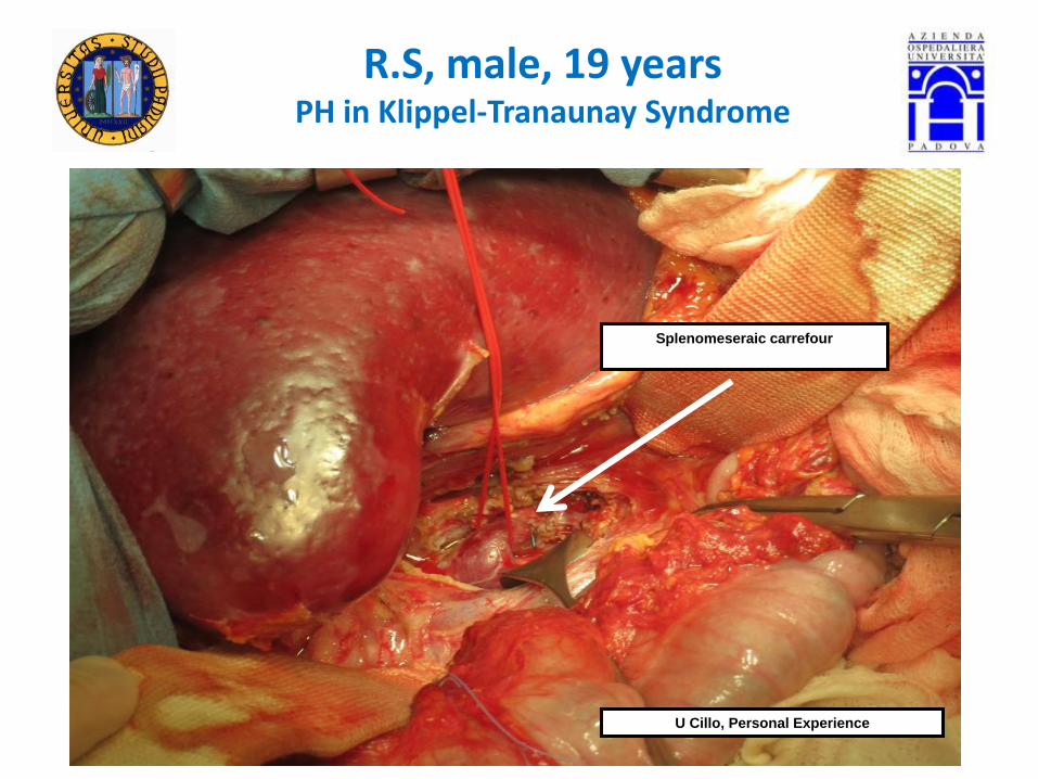
R.S, male, 19 yearsPH in Klippel-Tranaunay Syndrome
MH:
Congenital hypothyroidism
1998 Partial removal angioma right thigh
2005 Sclerotherapy of macrocystic
2006 2016
Sclerotherapyof lymphatic malformations of
the abdominal lower quadrant
Laser treatment of genital/rectal varices
Early July 2016 Upper GI hemorrage
Oesophageal varices ligation
Liver biopsy: minimal fibrosis with
accentuation of portals branches (METAVIR
F0) no inflammation
portal, steatosis or cholestasis.
Severe bleeding rectal varices
INR 1.38, PT 56%
Total bilirubin 8.8 umol/L
Page 41
R.S, male, 19 yearsPH in Klippel-Tranaunay Syndrome
MH:
Congenital hypothyroidism
1998 Partial removal angioma right thigh
2005 Sclerotherapy of macrocystic
2006 2016
Sclerotherapyof lymphatic malformations of
the abdominal lower quadrant
Laser treatment of genital/rectal varices
Early July 2016 Upper GI hemorrage
Oesophageal varices ligation
Liver biopsy: minimal fibrosis with
accentuation of portals branches (METAVIR
F0) no inflammation
portal, steatosis or cholestasis.
Severe bleeding rectal varices
INR 1.38, PT 56%
Total bilirubin 8.8 umol/L
Page 42
R.S, male, 19 yearsPH in Klippel-Tranaunay Syndrome
Sigmoid Varices
U Cillo, Personal Experience
Page 43
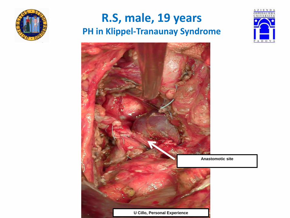
R.S, male, 19 yearsPH in Klippel-Tranaunay Syndrome
Splenomeseraic carrefour
U Cillo, Personal Experience
Page 44
R.S, male, 19 yearsPH in Klippel-Tranaunay Syndrome
Proximal renal vein
Splenomeseraic carrefour
U Cillo, Personal Experience
?
Page 45
R.S, male, 19 yearsPH in Klippel-Tranaunay Syndrome
U Cillo, Personal Experience
Anastomotic site
Page 46
NCPH: Indication for surgery
Rajekar H et al, J Clin Exp Hepatol 2011;1:94–108
Superina R et al, Pediatr Transplant 2006;10:908–913
The role of surgery is limited to those who fail to respond to medical-endoscopic-
radiologic therapy
Absolute:
• Medically/endoscopically refractory
variceal hemorrhage
• Symptomatic hypersplenism (recurrent
bleeds/infections)
• Platelet count <10.000/mm3
• Symptomatic/Medically refractory HE
• Hepatopulmonary syndrome (HPS)
• Portopulmonary hypertension (PPH)
Sarin SK et al, Liver Int 2006;26:512–519
Khanna R et al, J Hepatol 2014 vol. 60; 421–441
Page 47
NCPH: Indication for shunt surgery
Rajekar H et al, J Clin Exp Hepatol 2011;1:94–108
Superina R et al, Pediatr Transplant 2006;10:908–913
The role of surgery is limited to those who fail to respond to medical-endoscopic-
radiologic therapy
Absolute:
• Medically/endoscopically refractory
variceal hemorrhage
• Symptomatic hypersplenism (recurrent
bleeds/infections)
• Platelet count <10.000/mm3
• Symptomatic/Medically refractory HE
• Hepatopulmonary syndrome (HPS)
• Portopulmonary hypertension (PPH)
Relative:
• Portal biliopathy (PB)
• Symptomatic splenomegaly
(pain/rupture/infarction)
• Poor health related QoL
• Large varices with poor access to
healthcare or rare blood group
• Patients who desire a one-time treatment
• Refractory lower GI bleed (anorectal
varices/colopathy)
• Neurocognitive testing suggesting of MHE
• Growth failure (Z-scores <-2 despite
nutritional rehabilitation)
• Delay in sexual development
Sarin SK et al, Liver Int 2006;26:512–519
Khanna R et al, J Hepatol 2014 vol. 60; 421–441
Page 48
Savethe Portal Hypertension
SURGERY!
….and prepare the future portalhypertension SURGEONS!
Page 49
Storia chirurgia ipertensione portale
Knechtla SJ, Ann
Surg 2003 Vol 238
N6S (S49-S55)
Page 50
86 patients with NCPH
Between 1979 to 1991
56 patients with EHPVO
30 patients with NCPF
Side-to-side
lieno-renal (SSLR) shunt
Sharma PC et al, Journal of Gastroenterloy Hepatol (1997) 12, 582-584
Following Side-to-Side Lieno-Renal Shunt (SSLR)
there is a significant reduction in SPP and varices
SSLR shunt is effective in the treatment of PH
Page 51
1. Rivascularization procedures Meso-Rex Shunt
2. Portosystemic shunts
3. Devascularization procedures
4. Orthotopic Liver Transplantation
Non-Selective:
Total:
• End-to-side portacaval shunt (Eck's fistula)
• Side-to-side portacaval shunt (> 10 cm)
• Mesocaval with prothesic interposition
• Proximal splenorenla shunt (Linton's shunt)
Partial:
• Small diameter end-to-side (8-10 cm)
• Calibrated side-to-side portacaval shunt
Selective:
Distal splenorenal shunt (Warren's shunt)
Coronaro-caval anastomosis (abandoned)
Spleno-caval anastomosis (abandoned)
Surgical interventions for PH
Page 52
1. Rivascularization procedures Meso-Rex Shunt
2. Portosystemic shunts
3. Devascularization procedures
4. Orthotopic Liver Transplantation
Non-Selective:
Total:
• End-to-side portacaval shunt (Eck's fistula)
• Side-to-side portacaval shunt (> 10 cm)
• Mesocaval with prothesic interposition
• Proximal splenorenla shunt (Linton's shunt)
Partial:
• Small diameter end-to-side (8-10 cm)
• Calibrated side-to-side portacaval shunt
Selective:
Distal splenorenal shunt (Warren's shunt)
Coronaro-caval anastomosis (abandoned)
Spleno-caval anastomosis (abandoned)
Surgical interventions for PH
Page 53
1. Rivascularization procedures Meso-Rex Shunt
2. Portosystemic shunts
3. Devascularization procedures
4. Orthotopic Liver Transplantation
Non-Selective:
Total:
• End-to-side portacaval shunt (Eck's fistula)
• Side-to-side portacaval shunt (> 10 cm)
• Mesocaval with prothesic interposition
• Proximal splenorenla shunt (Linton's shunt)
Partial:
• Small diameter end-to-side (8-10 cm)
• Calibrated side-to-side portacaval shunt
Selective:
Distal splenorenal shunt (Warren's shunt)
Coronaro-caval anastomosis (abandoned)
Spleno-caval anastomosis (abandoned)
Surgical interventions for PH
Page 54
Non Selective (Central) vs Selective (Periferic) Shunt
Non Selective
(Central)
Selective
(Periferic)
Type of surgical shunt and pathophysiological implications
Page 55
Non Selective Total PSS
End-To-Side PCS Side-To-Side PCS Proximal Spleno-Renal Shunt
(Linton Shunt)+ Splenectomy
Knechtla SJ, Ann Surg 2003 Vol 238 N6S (S49-S55)
Page 56
Clavien Atlas of Gastrointestinal and Hepato-Pancreato-Biliary Surgery 2007
Blumgart's Surgery of the liver, pancreas and biliary tract, 5th Ed.
EMC – Role of Surgery in the treatment of PH complications – Surgery of PH
Non Selective Total PSS
Indications
• Bleeding esophageal or gastric varices unresponsive to
other therapies
• Bleeding from portal hypertensive gastropathy
unresponsive to pharmacologic therapy
• Budd-Chiari syndrome with patent IVC
• Intractable ascites unresponsive to non-surgical therapy
• Failed TIPS
• Patient not candidates for selective shunt (technical
consideration or ascites)
Contraindications
• Extrahepatic portal hypertension
• Portal vein thrombosis not amenable to
thrombectomy
• Occlusion of the hepatic artery
Significative decrement in HVPG
Frequent inversion of portal blood flow direction
Persistence of intra-sinusoidal PH
Page 57
…...........
Postoperative Complications
Early:
– Hepatic failure
– Renal failure
– Infection
– Gastric acid hypersecretion
– Delirium tremens
– Ascites
– Gastrointestinal bleeding
Late:
– Portasystemic encephalopathy (PSE)
– Liver failure
– Shunt thrombosis
– Hepatocellular carcinoma
Clavien Atlas of Gastrointestinal and Hepato-Pancreato-Biliary Surgery 2007
In some patients not suited for a selective shunt
a non-selective shunt might serve as a long-term bridge to LT
when bleeding is not controlled endoscopically or by TIPS
Mesocaval or Linton shunts avoiding dissection of hepatic hilum
Non Selective Total PSS
Page 58
Non Selective PSS
End-To-Side PCS Side-To-Side PCS Proximal Spleno-Renal Shunt
(Linton Shunt)+ Splenectomy
Knechtla SJ, Ann Surg 2003 Vol 238 N6S (S49-S55)
Non selective Partial PSS:
• Small diameter end-to-side (8-10 cm)
• Calibrated side-to-side portacaval shunt
Page 59
1. Rivascularization procedures Meso-Rex Shunt
2. Portosystemic shunts
3. Devascularization procedures
4. Orthotopic Liver Transplantation
Non-Selective:
Total:
• End-to-side portacaval shunt (Eck's fistula)
• Side-to-side portacaval shunt (> 10 cm)
• Mesocaval with prothesic interposition
• Proximal splenorenla shunt (Linton's shunt)
Partial:
• Small diameter end-to-side (8-10 cm)
• Calibrated side-to-side portacaval shunt
Selective:
Distal splenorenal shunt (Warren's shunt)
Coronaro-caval anastomosis (abandoned)
Spleno-caval anastomosis (abandoned)
Surgical interventions for PH
Page 60
Selective (Periferic) Shunt
DSRS was an improvement
over nonselective shunting procedures
It preferentially decompresses:
- venous collaterals around the stomach
- venous collateral around the lower esophagus
preventing further hemorrhage
Compartmentalization
of the portal venous
circulation
→ maintains portal blood flow to the liver
→ diminishes the risks of postoperative
encephalopathy and accelerated hepatic
failureElwood et al, Arch Surg. 2006;141:385-388
Knechtla SJ, Ann Surg 2003 Vol 238 N6S (S49-S55)
Type of surgical shunt and pathophysiological implications
Ligation of the coronary and gastroepiploic veins
separates the high pressure SMV system
form the gastro-splenic venous system
at least temporarily
Page 61
Knechtla SJ, Ann Surg 2003 Vol 238 N6S (S49-S55)
Clavien Atlas of Gastrointestinal and Hepato-Pancreato-Biliary Surgery 2007
Blumgart's Surgery of the liver, pancreas and biliary tract, 5th Ed.
EMC – Role of Surgery in the treatment of PH complications – Surgery of PH
Selective PSS: Distal Spleno-Renal Shunt (Warren's Shunt)
Indications
Bleeding esophageal varices refractory to other treatments
well-preserved hepatic function
ContraindicationsAdvanced liver disease
Splenic vein thrombosis with no shuntable vessels < 7 mm
Intractable ascites
Previous splenectomy
Ascites (worsened by DSRS)
Relative contraindications
Progressive liver disease in patients likely to come to transplant
in the next 2–3years
Small splenic vein
Abnormal anatomy of left renal vein
Page 62
A prospective multicenter RCT
• 140 patients, CPT-A/B
• Refractory variceal bleeding
DSRS or TIPS
Mean follow-up 46 ± 26 months
Henderson et al, Gastroenterology 2006 May;130(6):1643-51
DSRS TIPS
Rebleeding 5.5% 10.5 (P=0.29)
1st PSE event 50% 50%
Reintervention rate 82% 11% (P<0.001)
Ascites P = NS
Need for LT P = NS
QoL P = NS
Costs P = NS
2-years survival 81% 88% (P=0.87)
5-years survival 62% 61% (P=0.87)
DSRS and TIPS are similarly efficacious
in the control of refractory variceal bleeding in CPT-A/B
Reintervention is significantly greater for TIPS
compared with DSRS
Because both procedures have equivalent outcomes
the choice is dependent on available expertise
and ability to monitor the shunt and reintervene when needed
Page 63
Rosemurgy et al, Ann Surg. 1997 May; 225(5): 601-608.
Portal flow:
TIPS → increased (21 mL/second ± 11.9 to 31 m/L second ± 16.9, p < 0.05)
HGPCS → unchanged (26 mL/second ± 27.7 to 14 mL/second ± 41.1, p = n.s.)
Effective hepatic blood flow:
TIPS → significantly diminished (1684 mL/minute + 2161 to 676 mL/minute ± 451, p <
0.05)
HGPCS → unaffected (1901 mL/minute ± 1818 to 1662 mL/minute ± 1035, p = n.s.)
Both TIPS and HGPCS achieved significant reductions in PV → IVC pressure gradients
Portal flow increased after TIPS, although most portal flow was diverted through the shunt
Effective hepatic flow is reduced significantly after TIPS but well preserved after HGPCS
Hepatic decompensation and mortality after TIPS may be becauseof reductions in nutrient hepatic flow
.
Page 64
.Mortality after TIPS higher than after HGPCS
hepatic failure probably due
to excessive diminution of hepatic blood flow
Rosemurgy et al, Ann Surg. 1997 May; 225(5): 601-608.
Page 65
1. Rivascularization procedures Meso-Rex Shunt
2. Portosystemic shunts
3. Devascularization procedures
4. Orthotopic Liver Transplantation
Non-Selective:
Total:
• End-to-side portacaval shunt (Eck's fistula)
• Side-to-side portacaval shunt (> 10 cm)
• Mesocaval with prothesic interposition
• Proximal splenorenla shunt (Linton's shunt)
Partial:
• Small diameter end-to-side (8-10 cm)
• Calibrated side-to-side portacaval shunt
Selective:
Distal splenorenal shunt (Warren's shunt)
Coronaro-caval anastomosis (abandoned)
Spleno-caval anastomosis (abandoned)
Surgical interventions for PH
Page 66
Sugiura Procedure and modifications
Developed to improve the effect of Walker’s
simple esophageal transection
1. Esophageal transection
2. Extensive esophago-gastric
devascularization
3. Splenectomy
4. Selective vagotomy
5. Piloroplasty
Patients who are unable to undergo
shunting procedures nor TIPS
because of extensive splanchnic vein
thrombosis
Devascularization proceduresand pathophysiological implications
Knechtla SJ, Ann Surg 2003 Vol 238 N6S (S49-S55)
Clavien Atlas of Gastrointestinal and Hepato-Pancreato-Biliary Surgery 2007
Blumgart's Surgery of the liver, pancreas and biliary tract, 5th Ed.
EMC – Role of Surgery in the treatment of PH complications – Surgery of PH
Distal esophagus
7 cm
Stomach
2/3
Proximal
Page 67
Bleeding control: 100%
Long-term bleeding control: 41.6%
Post-operative mortality: 23.9%
CPT-B: 12.5%
CPT-C: 34.6%
24/46 patients had long-term follow-up
(mean 83.1 months; range 14 264 months)
Overall 5-year survival in patient with long-
term bleeding control: 62.5%
1985 → 1992
Modified Sugiura procedure in 46 patients:
- 25 emergency procedures
- 21 semi-elective procedures
CPT:
A → 4 patients
B → 16 patients
C → 26 patients
Modified Sugiura procedure
remains an effective rescue therapy
for patients bleeding petients
when alternative treatments
fail or are not indicate
It can be a life-saving procedure in:
- Patients with anatomy unsuitable for shunt
surgery
- Patients treated in non-specialized centers
where surgical expertise for a shunt
operation is not available
Voros et al, World J Surg (2012) 36:659–666
Page 68
1. Rivascularization procedures Meso-Rex Shunt
2. Portosystemic shunts
3. Devascularization procedures
4. Orthotopic Liver Transplantation
Non-Selective:
Total:
• End-to-side portacaval shunt (Eck's fistula)
• Side-to-side portacaval shunt (> 10 cm)
• Mesocaval with prothesic interposition
• Proximal splenorenla shunt (Linton's shunt)
Partial:
• Small diameter end-to-side (8-10 cm)
• Calibrated side-to-side portacaval shunt
Selective:
Distal splenorenal shunt (Warren's shunt)
Coronaro-caval anastomosis (abandoned)
Spleno-caval anastomosis (abandoned)
Surgical interventions for PH
Page 70
From 1972 to 1999
60 patients with BCS divided into three groups:
1. occlusion confined to hepatic veins (n=32)→ Direct side-to-side portacaval shunt (SSPCS)
2. occlusion involving the inferior vena cava (IVC) → Portal decompressive procedure that by-passed the
obstructed IVC
3. advanced cirrhosis and hepatic decompensation → referred for liver transplantation
SSPCS in BCS with hepatic vein occlusion alone results in reversal of liver damage, correction of
hemodynamic disturbances, prolonged survival, and good QoL when performed early in BCS
Similarly good results are obtained with combined SSPCS and CAS in patients with BCS resulting from IVC
occlusion.
In contrast, mesoatrial shunt has been discontinued in the authors’ program because of an unacceptable
incidence of graft thrombosis and death.
In patients with advanced cirrhosis from long-standing, untreated BCS, LTis the only hope of relief and results
in the salvage of some patients.
Page 72
Cause of TIPS failureand complications(vecchi)
+ TIPS
Page 73
Rosemurgy et al, Ann Surg. 1997 May; 225(5): 601-608
Colombato L, J Clin Gastroenterol 2007;41:S344–S351
Circulatory changes caudes by TIPS and clinical consequences
Increase
in portal flow Reduction of
effective hepatic flow
TIPS
Reduces portal pressure
due to the dramatic drop
in intrahepatic vascular resistance
Reverses the sense of the circulation
in the portal venous system within the liver
Sinusoidal perfusion highly dependant
On the hepatic arterial flow; if uneffective
Progressive Liver Failure
Page 74
The hepatic arterial buffer response(HABR)
International Journal of Arti¯cial Organs 2004; 27: 222-30.
Page 75
Rosemurgy et al, Ann Surg. 1997 May; 225(5): 601-608
Colombato L, J Clin Gastroenterol 2007;41:S344–S351
Circulatory changes caudes by TIPS and clinical consequences
Increase
in portal flow Reduction of
effective hepatic flow
TIPS
Reduces portal pressure
due to the dramatic drop
in intrahepatic vascular resistance
Reverses the sense of the circulation
in the portal venous system within the liver
Huge escape of portal flow through the stent
without interaction at the liver sinusoid level
Portal-Systemic Encephalopathy
(PSE)
Reverses the sense of the circulation
in the portal venous system within the liver
Huge escape of portal flow through the stent
without interaction at the liver sinusoid level
Portal-Systemic Encephalopathy
(PSE)
Page 76
Rosemurgy et al, Ann Surg. 1997 May; 225(5): 601-608
Colombato L, J Clin Gastroenterol 2007;41:S344–S351
Circulatory changes caused by TIPS and clinical consequences
Increase
in portal flow Reduction of
effective hepatic flow
TIPS
Reduces portal pressure
due to the dramatic drop
in intrahepatic vascular resistance
High blood flow through the stent
with dramatic shift of blood to the systemic circulation
Hemodinamic Changes
1. Increased venous return
2. Normalization of effective arterial blood volume
3. Increased cardiac output
4. Exaggeration of vasodilatation
5. Increase in hyperdynamic circulatory with “normal”
portal pressure
Transient rise in right atrial pressure
that might potentially worsen
an undiagnosed porto-pulmonary hypertension
or alternatively it might unmask
a subclinical cardiomyopathy,
leading to heart failure
Page 77
Fidelman et al, AJR 2012; 199:746–755
Parvinian et al, J Clin Imaging Sci 2013;3:19
Colombato L, J Clin Gastroenterol 2007;41:S344–S351
Complications of TIPS
30-46% Development or worsening in hepatic
encephalopathy (HE)
33% Transcapsular puncture
→ 1-2% Significant intraperitoneal hemorrhage
20% Shunt malpositioning
10% Deterioration of hepatic function
Rare: Clinically significant hemobilia
Shunt migration
Hepatorenal syndrome
TIPS stenosis from Intimal hyperplasia
TIPS: Intra-graft thrombus
Page 78
Causes of TIPS failure RECURRENCE OF PORTAL HYPERTENSION
1. Intimal hyperplasia
2. Thrombotic occlusion
3. Hepatic venous end shunt stenosis
4. Portal venous end shunt stenosis
5. Abnormal angulation
6. Occult portosystemic pressure gradientelevation
7. Flow-sumping
Fidelman et al, AJR 2012; 199:746–755
Parvinian et al, J Clin Imaging Sci 2013;3:19
Stenosis (up to 70%)
& Occlusion
TIPS stenosis due to
intimal hyperplasia
Page 79
Fidelman et al, AJR 2012; 199:746–755
US-Doppler marks of TIPS dysfunction:
Alterations in shunt velocities:
- 250 cm/s or higher
- 50 cm/s or less
are associated with > 90% sensitivity and
specificity for shunt dysfunction
TIPS Manteinance
Most hepatologists order routine TIPS surveillance tests at regular intervals
using ultrasound with Doppler in asymptomatic patients
TIPS occlusion
Mid TIPS stenosis
Page 80
Fidelman et al, AJR 2012; 199:746–755
Patients with a suspected TIPS dysfunction
should undergo:
- TIPS venography
- Replacement of bare stent with covered
stent
- Balloon angioplasty within the stent
- Placement of additional stents in patients
to extend cranial or caudal length of the
stent
TIPS Manteinance
Most hepatologists order routine TIPS surveillance tests at regular intervals
using ultrasound with Doppler in asymptomatic patients
HE refractory to medical management
and progressive hepatic dysfunction
might require
endovascular shunt reduction
Need for repeated revisons:
Are TIPS cost-effective?
Page 81
Fidelman et al, AJR 2012; 199:746–755
Contraindications to TIPS placement
Contraindications of TIPS placement
are not necessary applicable to surgical shunts:
Could indication to PSS be extended over tips indications?
Page 82
Clark et al, The American Journal of Surgery (2011) 202, 561–564
246 Patients with PH undergoing TIPS from 2001 to 2010
70 → uncovered stents
176 → covered stents
Patients who received uncovered stents
had more severely impaired liver function
(41% were Child class C cirrhotics).
Uncovered (N=70)
Covered (N=176)
Follow-up (P = 0.01)
48 months 24 months
Reintervention for stenosis(P = 0.01)
33% 19%
Shunt dysfunction (P = 0.05)
57% 21%
Deterioration of hepatic function ( P = 0.32)
31% 30%
Survival (P = 0. 55)
31 months 33 months
Covered stents may improve patency
but do not mitigate postshunt hepatic dysfunction
and do not improve survival
Page 83
To determine the long-term cost-effectiveness
of TIPS vs. Surgical PSS
The main outcome was dollars per life-year saved
Average cost per life year saved:
- TIPS: $17,771 (SD = 471)
- PSS: $21,438 (SD = 308)
Average life expectancy;
- TIPS: 5.0 years
- PSS: 7.0 years
This yielded an incremental cost-effectiveness rate
for PSS of $3,299 per life year saved
Compared with TIPS, PSS resulted in
improved survival with minimal increase in cost
Therefore, given the low incremental cost of PC,
it should be adopted as a cost-effective strategy
in managing this patient population
Page 85
Toomey et al, Am J Surg. 2013 Apr;205(4):441-6
From 2001 to 2010;
256 cirrhotic patients included
TIPS decreased PV-IVC gradients 175 mmHg
(P< 0.001)
Reinterventions 21%
Survival 26 months
Liver transplantation 14% of patients
TIPS effectively decompresses PH but leads to
frequent reinterventions and short survival
After TIPS LT is uncommonly undertaken
TIPS is a ‘‘bridge’’ to LT
that is seldom ‘‘crossed’’
Who should receive TIPS?
Patients:
who are imminently going to be transplanted (within 6 months)
with high cardiopulmonary risk for abdominal surgery
(eg, aortic stenosis and mitral regurgitation)
with a ‘‘hostile abdomen’’ (eg, multiple previous celiotomies)
extremly obese
Other than this select group, patients should not undergo TIPS
without any expectation other than a short survival
complicated by shunt surveillance and shunt failure
For patients with poor hepatic function (eg, CPT- C)
resource allocation is promoted by surgical shunting
The concept of operative shunting needs to be reconsidered and revisited
Page 86
Wu et al, Ann Vasc Surg 2013; 27: 441–446
January 2000 → June 2011
56 symptomatic NCPH→ open surgery
→ endovascular thrombolysis
PSS primarily performed in 49 patients
- 35 Mesocaval shunt
- 7 Splenorenal
- 4 Portocaval
- 2 Paraumbilicalejugular
- 1 Portal to right atrial
Esophagogastric devascularization: 3 patients
- 4 Sugiura procedures
Endovascular catheter-directed thrombolysis: 4
patients with acute superior mesenteric vein (SMV) and
portal vein thrombosis
Mean follow-up: 57 months (range 2-125)
PSS:
Shunt patency: 100%
Rebleeding: 0%
Esophagogastric devascularization:
2/3 was converted to mesocaval shunt due to recurrent
variceal bleeding (at 8, 13, and 24 months)
1/3 died before redo operation
Thrombolysis:
¾ survived without complications
¼ death for small bowel infarction due to recurrent
thrombosis at 40 days form procedure
Platelet counts from 43x109/L to 239x109/L 2 within 2 weeks
Ascites disappeared in 30/31 within 2 months
No post-operative encephalopathy
Peri-operative 30-day mortality. 0%
PSS can be employed to treat
bleeding esophagogastric varices
and severe hypersplenism
In patient s with NCPH
PSE is less of a concern in NCPH
patients with normal liver
function
Endovascular thrombolysis
is a useful alternative treatment
for acute portal and/or
mesenteric venous thrombosis
Page 87
Surgical management of portal biliopaty:Summary of case-series and case-reposts
Franceschet I et al, World J Gastroenterol 2016 December 7; 22(45): 9909-9920
Page 88
Khanna R et al, J Hepatol 2014 vol. 60; 421–441
Harrison’s Principle of Internal Medicine
Causes of NCPH According to blood flow site resistance
Page 89
Khanna R et al, J Hepatol 2014 vol. 60; 421–441
Causes of NCPH According to blood flow site resistance
Page 90
1.85
cm
2.57
cm
2.44
cm
Page 92
Lavori dimenticati
Page 93
Cose vecchie utili: pelli di foca
Page 94
NCPH: Diagnostic criteria
EASL Guidelines, J Hepatol 2015