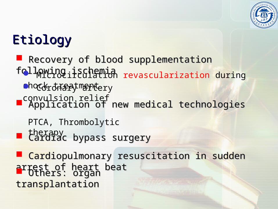
Ischemia-Reperfusion Ischemia-Reperfusion Injury Injury (IRI) (IRI) Department of Pathophysiology Department of Pathophysiology Shanghai Jiao-Tong University School o Shanghai Jiao-Tong University School o f Medicine f Medicine
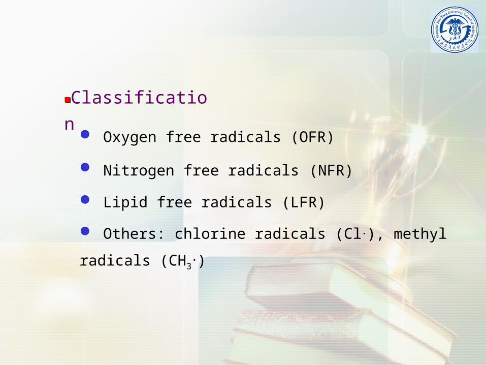
Lipid free radicals are referred to middle metabolic products resulting from the chain reaction of lipid peroxidation, which is produced by interaction of OFR and non-saturated fatty acid.
L•L•
LO•LO•
LOO•LOO•
LFRLFR
■■Metabolism of the free radicals
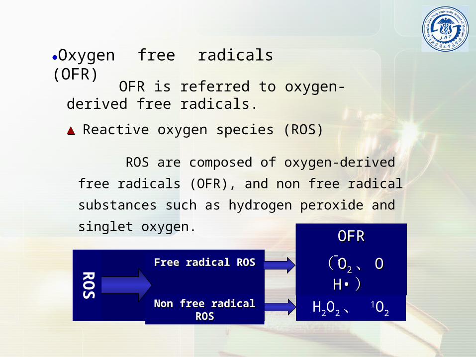
O2 O2̄·e
O2 OH· + H2O 3e3H+
2eO2 H2O2 2H+
4eO2 2H2O4H+
● Production of oxygen free radicalProduction of oxygen free radical
Sources of OH•OH• ▲▲
Fe2+ + HH22OO22 OO22 + OH• + OH + OH• + OH++·OO22
HH22O OH• + H• O OH• + H• ; ; HH22O HO H++ + OH + OH‒‒heterolysisheterolysishomolysishomolysis
IRI induces the activation of complement and endothelial IRI induces the activation of complement and endothelial cells and the increase of chemokine such as cells and the increase of chemokine such as C3a and leukotriene, which further attract and activate neutrophils. This activa, which further attract and activate neutrophils. This activation of neutrophils then intakes large amounts of molecular oxtion of neutrophils then intakes large amounts of molecular oxygen and produce OFR by respiratory burst.ygen and produce OFR by respiratory burst.
The increase of one electron deoxidization in The increase of one electron deoxidization in mitochondriamitochondria
95%O95%O2 2 Cytochrome C oxidasesCytochrome C oxidases OO2 2 H H22O O
The increase of catecholamine and its oxidazitioThe increase of catecholamine and its oxidazitionn
sympathetic-adrenal medulla ()
vanilmandelic acid (normal)
monoamine oxidase
adrenochrome
ischemia and hypoxia
CA release
OFR
■ ■ Mechanisms of free radical-induced IRI
The increase of membrane lipid peroxidation (MIP)The increase of membrane lipid peroxidation (MIP)
OFR interacts with non-saturated fatty acids from membrane lipids and further induce lipid peroxidation reaction, which results in the structural alteration and dysfunction of membrane.
ROS induces oxidation of lips, proteins and nucleic acid.ROS induces oxidation of lips, proteins and nucleic acid.
OFR are extremely reactive to interact with lipids, proteins and nucleic acids.
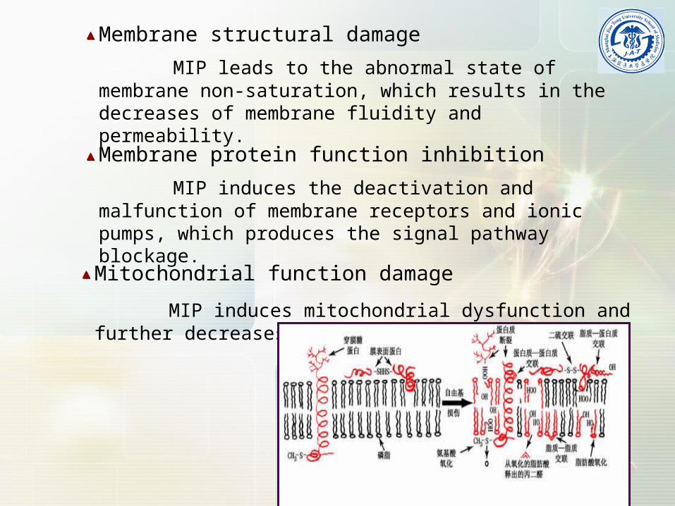
Membrane structural damage
MIP leads to the abnormal state of membrane non-saturation, which results in the decreases of membrane fluidity and permeability.
▲▲
Membrane protein function inhibition
MIP induces the deactivation and malfunction of membrane receptors and ionic pumps, which produces the signal pathway blockage.
▲▲
Mitochondrial function damage
MIP induces mitochondrial dysfunction and further decreases ATP generation.
▲▲
Protein denaturalization and decreased enzyme activityProtein denaturalization and decreased enzyme activity
Nucleic acid damage and chromosome aberrationNucleic acid damage and chromosome aberration
Others: Others:
Mediation of a series of reaction important for IRI, such aMediation of a series of reaction important for IRI, such as releasing inflammatory factors; decreasing nitric oxide; pros releasing inflammatory factors; decreasing nitric oxide; promoting the expression of adhesion molecules and the adhermoting the expression of adhesion molecules and the adherence between neutrophils and vessels.ence between neutrophils and vessels.
The integrity and permeability of membrane is impaired during ischemia-reperfusion. These damages do not occur only in sarcoplasmic reticulum (SR) but also in mitochondria, lysosomes and other cellular membranes. Therefore, Ca2+ can flow into the cytoplasm through damaged membrane according to the gradient.
Mitochondrial injuryMitochondrial injury
CA increase CA increase
■ ■ Mechanisms of calcium overload-induced IRI
Promotion of OFR generationPromotion of OFR generation
Aggravation of acidosisAggravation of acidosis
Damage of cellular membraneDamage of cellular membrane
Activation of other enzymes Activation of other enzymes (proteinases, nucleases)(proteinases, nucleases)
Neutrophil activationNeutrophil activation
It has been manifested that the capillary damage and dysfunction which mediated by neutrophil activation play an important role in IRI.
Cell adhesion molecule (CAM) increase
Activated neutrophils can increase the expression of CAMs, which including the selectins, integrins (CD11/CD18) and immunoglobulin superfamily (ICAM-1, VCAM-1).
■ ■ Leukecyte accumulation induced by IRI
Inflammatory factors or chemokines increaseInflammatory factors or chemokines increase
Activated neutrophils can adhere to endothelial cells or blood cells, then to release the inflammatory factors after margination and aggregation, such as TXA2, leukotrienes, prostaglandin and so on, to increase the permeability of endothelial cell monolayer.
■ ■ Mechanism of leukecyte-mediated IRI
Microvascular damageMicrovascular damage
The ischemia region could not be reperfused sufficiently after relieving the occlusion to recover the blood flow.
-No-reflow phenomenon
microvessel hemorheological alteration
Abnormal regulation of inflammatory reactionsAbnormal regulation of inflammatory reactions
Machinery blockage action
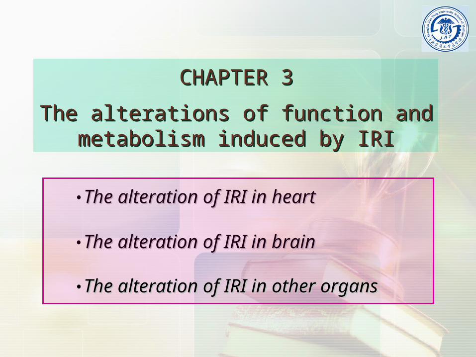
CHAPTER 3CHAPTER 3
The alterations of function and metabolism The alterations of function and metabolism induced by IRIinduced by IRI
•The alteration of IRI in brainThe alteration of IRI in brain
•The alteration of IRI in heartThe alteration of IRI in heart
•The alteration of IRI in other organsThe alteration of IRI in other organs
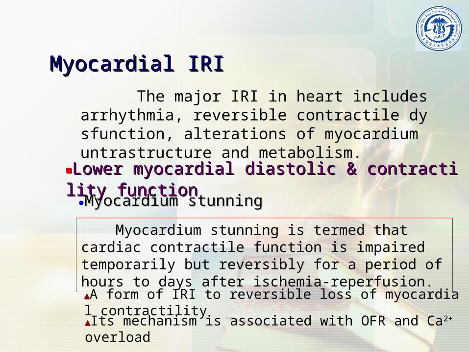
Myocardial IRIMyocardial IRI
The major IRI in heart includes arrhythmia, reversible contractile dysfunction, alterations of myocardium untrastructure and metabolism.
▲▲A form of IRI to reversible loss of myocardial contractility
▲▲Its mechanism is associated with OFR and Ca2+ overload
Myocardium stunning is termed that cardiac contractile function is impaired temporarily but reversibly for a period of hours to days after ischemia-reperfusion.
■ ■ Reperfusion arrhythmiaReperfusion arrhythmia
Ventricular tachycardia and fibrillation are the Ventricular tachycardia and fibrillation are the major manifestation of reperfusion arrhythmias.major manifestation of reperfusion arrhythmias.
● The occurrence of reperfusion arrhythmiaThe occurrence of reperfusion arrhythmia
▲▲Ischemia period before reperfusion
▲▲Ischemia degree
▲▲The cardiac myocytes with recoverable capability existed
▲▲The speed of reperfusion blood
● The mechanism of reperfusion arrhythmiaThe mechanism of reperfusion arrhythmia▲▲OFR and calcium disturbance
▲▲Sodium and potassium homeostasis
▲▲The ununiformity of action potential duration (APD)
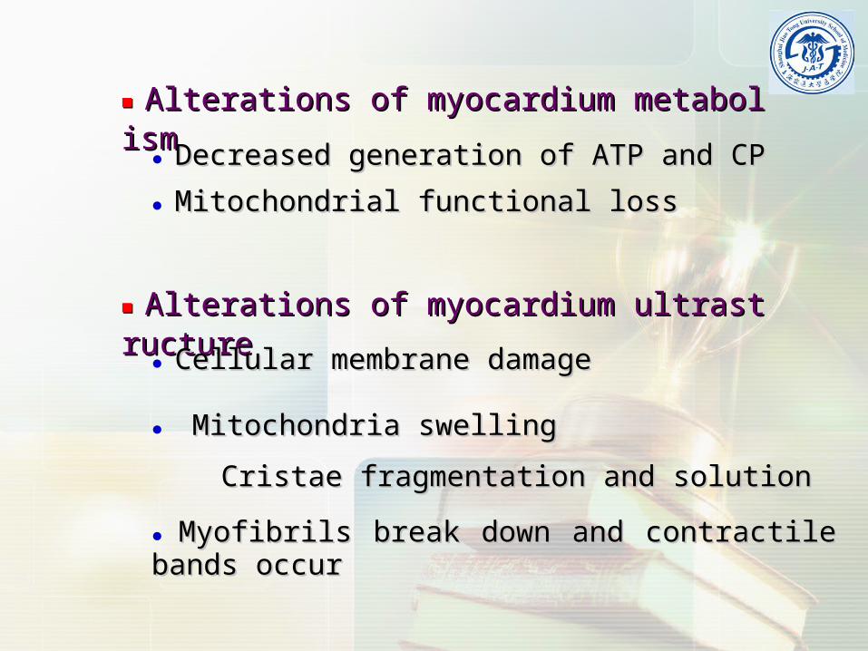
■ ■ Alterations of myocardium metabolism Alterations of myocardium metabolism
● Decreased generation of ATP and CPDecreased generation of ATP and CP
● Mitochondrial functional lossMitochondrial functional loss
■ ■ Alterations of myocardium ultrastructure Alterations of myocardium ultrastructure
● Myofibrils break down and contractile bands occurMyofibrils break down and contractile bands occur
● Mitochondria swellingMitochondria swelling
Cristae fragmentation and solutionCristae fragmentation and solution
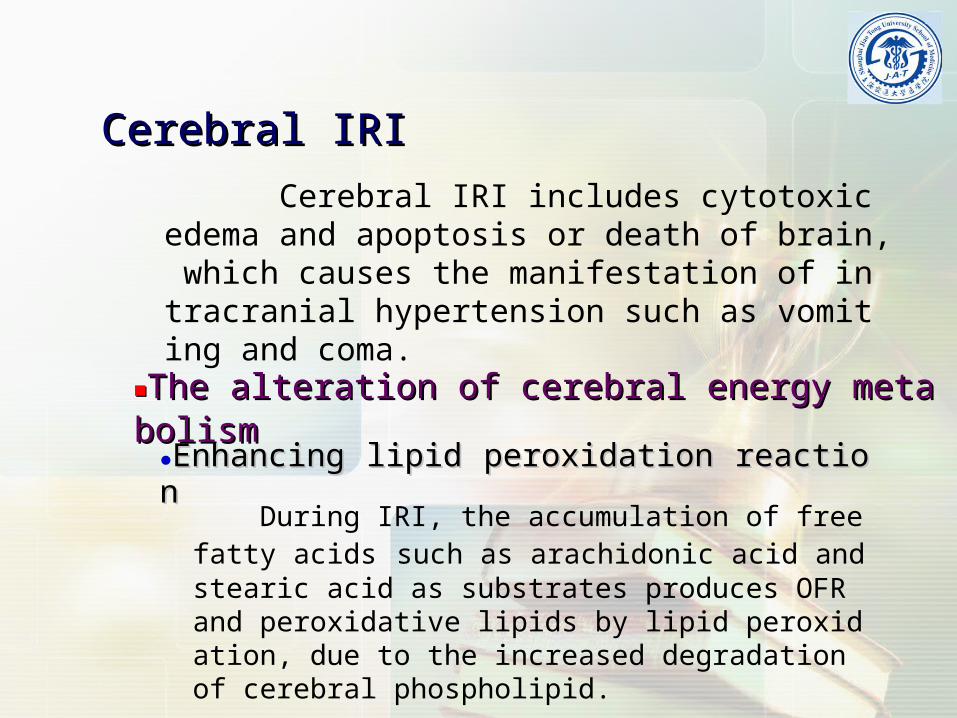
Cerebral IRICerebral IRI
Cerebral IRI includes cytotoxic edema and apoptosis or death of brain, which causes the manifestation of intracranial hypertension such as vomiting and coma.
■■The alteration of cerebral energy metabolismThe alteration of cerebral energy metabolism
During IRI, the accumulation of free fatty acids such as arachidonic acid and stearic acid as substrates produces OFR and peroxidative lipids by lipid peroxidation, due to the increased degradation of cerebral phospholipid.
■■The alteration of cerebral amino acid metabolismThe alteration of cerebral amino acid metabolism
■■The alteration of cerebral histologyThe alteration of cerebral histology
▲▲Cerebral edema
▲▲Cerebral cellular necrosis
IRI in other organsIRI in other organs
The IRI also can occur in other organs besides heart and brain. For example, liver and kidney are the organs which studied extensively in IRI. It has been implicated in the pathogenesis of a variety of clinical conditions including trauma, hypovolemic and endotoxic shock, transplantation, etc.
CHAPTER 4CHAPTER 4
Pathophysiological basis of prevention anPathophysiological basis of prevention and treatment for IRId treatment for IRI
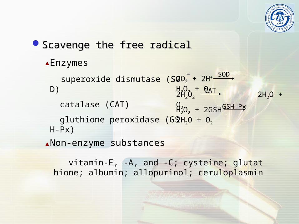
•Scavenge the free radicalsScavenge the free radicals
•Control the reperfusion conditionsControl the reperfusion conditions
•Relieve calcium overloadRelieve calcium overload
•Improve the metabolismImprove the metabolism
Control the reperfusion conditionsControl the reperfusion conditions
■■Shorten the ischemia periodShorten the ischemia period
●Lower pressureLower pressure
■■Improve the reperfusion conditionsImprove the reperfusion conditions
●Lower flow speedLower flow speed
●Lower temperatureLower temperature
●Lower pHLower pH●Lower concentration of calcium and sodiumLower concentration of calcium and sodium
Scavenge the free radicalsScavenge the free radicals
IPC is defined as short period-ischemic stress can significantly led to the protection of tissue and organs with subsequent longer IRI, which is also an adaptive mechanism.