Stroke is the second most frequent cause of death and a major cause of disability in industrial
countries. Its most frequent manifestation is the ischemic stroke, whose diagnosis often involves
the acquisition of brain magnetic resonance (MR) scans to assess the stroke lesion's presence,
location, extent, evolution and other factors. All known intervention are associated with
considerable risks and require a careful weighting against the potential gains. An automated
method to predict the final lesion outcome and to estimate the degree of disability would
support clinicians in the difficult and time-critical decision making process.
New methods for stroke segmentation and outcome prediction are regularly proposed. But,
more often than desirable, it is difficult to compare their fitness, as the reported results are
obtained on private datasets. Challenges aim to overcome these shortcomings by providing (1) a
public dataset that reflects the diversity of the problem and (2) a platform for a fair and direct
comparison with suitable evaluation measures. Thus, the scientific progress is promoted.
With ISLES, we provide such a challenge covering ischemic stroke lesion as well as clinical
outcome prediction from acute multi-spectral MRI data and intervention parameters. The task is
backed by a well established clinical and research motivation and a large number of already
existing methods. Each team may participate in either one or both of two sub-tasks:
TASK I Automatic prediction of the final lesion extend after (successful or failed)
intervention in form of a binary mask.
TASK II Automatic prediction of the final clinical outcome after (successful or failed)
intervention in from of the modified Ranking Scale (mRS).
The participants downloaded a set of training cases with associated follow-up expert segmen-
tations and mRS scores to train and evaluate their approach, then submitted a short paper
describing their method. After reviewing, a total of eight abstracts were accepted and compiled
into this volume. At the day of the challenge, each teams' results as obtained on an independent
test set of cases will be revealed and a ranking of methods established.
For the final ranking and more information, visit WWW.ISLES-CHALLENGE.ORG .
Oskar Maier, Universität zu Lübeck
Mauricio Reyes, University of Bern
Roland Wiest, Inselspital Bern
Karl Egger, University Medical Center Freiburg
August 2016
Version: 2016-08-16
I
Organizers
Oskar Maier, Universität zu Lübeck, Germany
Mauricio Reyes, University of Bern, Switzerland
Karl Egger, University Medical Center Freiburg, Germany
Roland Wiest, Inselspital Bern, Switzerland
Sponsoring Institutions
Institute of Medical Informatics, Universität zu Lübeck, Germany
Institute for Surgical Technology & Biomechanics, University of Bern, Switzerland
Clinic for Neuroradiology & University Medical Center Freiburg, Germany.
Department of Diagnostic and Interventional Radiology, Inselspital Bern, Switzerland.
Version: 2016-08-16
II
Table of contents
ISLES-P-03 Combination of CNN and Hand-crafted feature for Ischemic Stroke Lesion SegmentationHaocheng Shen, Siyamalan Manivannan , Roberto Annunziata , Ruixuan Wang , and Jianguo Zhan
p.01
ISLES-P-04 Residual Volumetric Network for Ischemic Stroke Lesion SegmentationLequan Yu and Pheng-Ann Heng
p.03
ISLES-P-05 Random forests for stroke lesion and clinical outcome predictionOskar Maier and Heinz Handels
p.05
ISLES-P-06 Segmentation of Ischemic Stroke Lesion using Random Forests in Multi-modal MRI ImagesQaiser Mahmood and A. Basit
p.07
ISLES-P-07 A Deep-Learning Based Approach for Ischemic Stroke Lesion Outcome PredictionRamandeep Randhawa, Ankit Modi, Parag Jain, and Prashant Warier
p.09
ISLES-P-08 Deep Convolutional Neural Network Approach for Brain Lesion SegmentationYoungwon Choi, Yongchan Kwon, Hanbyul Lee, Myunghee Cho Paik , and Joong-Ho Won
p.11
ISLES-P-09 Incorporating time to reperfusion into the FASTER model of stroketissue-at-riskRichard McKinley, Roland Wiest, and Mauricio Reyes
p.13
III
Combination of CNN and Hand-crafted featurefor Ischemic Stroke Lesion Segmentation
Haocheng Shen1, Siyamalan Manivannan1, Roberto Annunziata1, RuixuanWang1, and Jianguo Zhang1
CVIP, Computing at School of Science and Engineering, University of Dundee, UK
1 Motivation
CNN can automatically learn discriminative local features and give superior per-formance than hand-crafted features in various applications such as image classi-fication, semantic segmentation and object detection. CNN has also been appliedto MRI brain image analysis and achieved state-of-the-art results for brain tu-mor region segmentation [3, 4], stroke lesion segmentation [4], and mircobleedsdetection [2]. Recently, some studies (e.g. [5]) show that hand-crafted featuresmay provide complementary information with CNN, hence combining them withthe features extracted from CNN may give improved performance than only us-ing the features from CNN. Motived by this, we formulate the segmentation ofischemic stroke lesion in acute MRI scans as a pixel-level classification using acombination of CNN and hand-crafted features.
2 CNN Architecture
We used a CNN architecture which is similar to [1]. It is a fully convolutionalneural network containing a downsampling path and three upsampling paths.In the task of stroke lesion segmentation, there is a large variation on the size,location and shape of lesions. Therefore, encoding information at multiple scalesis necessary and preferable than considering information at only one level. Thedownsampling path is able to extract the abstract information with high-levelsemantic meaning, while the three upsampling paths are designed to capture thefine details. These three upsampled feature maps are then combined at the laterstages of the CNN architecture so that the classification layer fully make use ofthe information appears at multiple scales [1].
1
3 Hand-crafted Feature
We use the following hand-crafted features:
– intensity;– the hemispheric intensity difference between two symmetric pixels in the
axial view;– first order statistics in a w × w volume patch;– maximum response filter (MR8) [6].
At each 2D pixel location, these local features are extracted independently fromeach image modality and combined together to get a feature representation forthat pixel.
4 Patient-specific Classifier
As there is a large variation of lesions in the dataset, it will be beneficial to traina pool of binary classifiers instead of one. Each binary classifier in this pool isdesigned to separate the positive (lesion) features extracted from a patient fromall the negative (normal) features extracted from the same patient. In this waywe believe that some rarely appeared lesions can be easily discriminated from thenormal tissue compared to a binary lesion classifier which is trained using all thetraining data (without using patient information). In the testing time a votingstrategy (averaging the top 3 probabilities obtained by the binary classifiers inthe pool) is used to get the prediction of an input.
References
1. Chen, H., Qi, X.J., Cheng, J.Z., Heng, P.A.: Deep contextual networks for neuronalstructure segmentation. In: Thirtieth AAAI Conference on Artificial Intelligence(2016)
2. Chen, H., Yu, L., Dou, Q., Shi, L., Mok, V.C., Heng, P.A.: Automatic detectionof cerebral microbleeds via deep learning based 3d feature representation. In: 2015IEEE 12th International Symposium on Biomedical Imaging (ISBI). pp. 764–767.IEEE (2015)
3. Havaei, M., Davy, A., Warde-Farley, D., Biard, A., Courville, A., Bengio, Y., Pal, C.,Jodoin, P.M., Larochelle, H.: Brain tumor segmentation with deep neural networks.Medical Image Analysis (2016)
4. Kamnitsas, K., Ledig, C., Newcombe, V.F., Simpson, J.P., Kane, A.D., Menon,D.K., Rueckert, D., Glocker, B.: Efficient multi-scale 3d cnn with fully connectedcrf for accurate brain lesion segmentation. arXiv preprint arXiv:1603.05959 (2016)
5. Li, W., Manivannan, S., Zhang, J., Trucco, E., McKenna, S.J.: Gland segmenta-tion in colon histology images using hand-crafted features and convolutional neuralnetworks. In: International Symposium on Biomedical Imaging (ISBI) (2016)
6. Varma, M., Zisserman, A.: A statistical approach to texture classification from singleimages. International Journal of Computer Vision 62(1-2), 61–81 (2005)
2
Residual Volumetric Network for IschemicStroke Lesion Segmentation
Lequan Yu, Pheng-Ann Heng
Dept. of Computer Science and Engineering, The Chinese University of Hong Kong
1 Introduction
We propose a 3D convolutional neural networks (3D CNNs) based method forlesion outcome prediction. The proposed 3D network takes advantage of fullyconvolutional architecture to perform efficient, end-to-end, volume-to-volumetraining. More importantly, we introduce the recent proposed residual learn-ing technique into our network, which can alleviate vanishing gradients problemand improve the performance of our network.
Conv + BN + ReLu Max pooling
DeconvolutionSum operation
64 128 256 512 256 128 12864 128 256 64256 64
Input Volume
OutputVolume
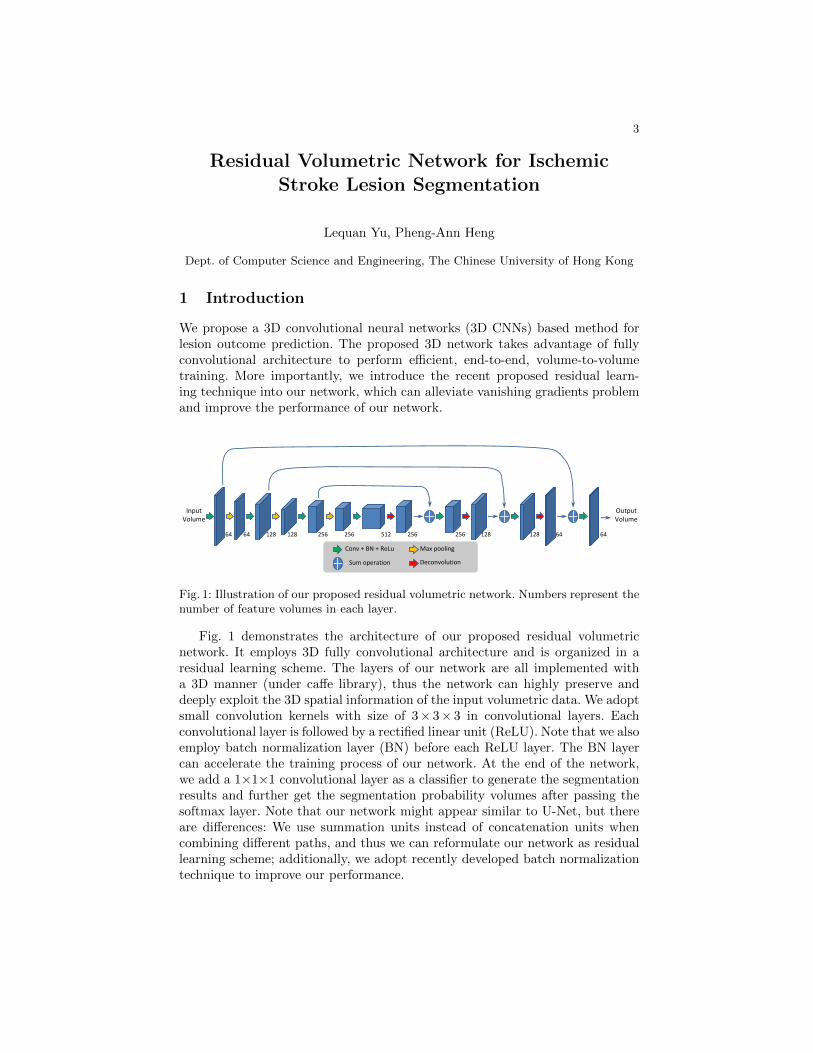
Fig. 1: Illustration of our proposed residual volumetric network. Numbers represent thenumber of feature volumes in each layer.
Fig. 1 demonstrates the architecture of our proposed residual volumetricnetwork. It employs 3D fully convolutional architecture and is organized in aresidual learning scheme. The layers of our network are all implemented witha 3D manner (under caffe library), thus the network can highly preserve anddeeply exploit the 3D spatial information of the input volumetric data. We adoptsmall convolution kernels with size of 3× 3× 3 in convolutional layers. Eachconvolutional layer is followed by a rectified linear unit (ReLU). Note that we alsoemploy batch normalization layer (BN) before each ReLU layer. The BN layercan accelerate the training process of our network. At the end of the network,we add a 1×1×1 convolutional layer as a classifier to generate the segmentationresults and further get the segmentation probability volumes after passing thesoftmax layer. Note that our network might appear similar to U-Net, but thereare differences: We use summation units instead of concatenation units whencombining different paths, and thus we can reformulate our network as residuallearning scheme; additionally, we adopt recently developed batch normalizationtechnique to improve our performance.
3
Random forests for stroke lesion and clinicaloutcome prediction
Oskar Maier1,2 and Heinz Handels1
1 Institute of Medical Informatics, Universitat zu Lubeck2 Graduate School for Computing in Medicine and Life Sciences, Universitat zu
Ischemic stroke is caused by an obstruction in the cerebral blood supply and, ifdiagnosed early, part of the under-perfused tissue can potentially be salvaged.Since the available treatment options are not risk-free, the decision has to bemade individually, depending on the potential gain and under great time re-striction. The prediction of the final lesion outcome in form of A binary mask(Task I) and the prediction of the clinical outcome in form of the modifiedRankin Scale (mRS) (Task II) are therefore of great clinical interest. The ISLES2016 challenge offers a public dataset and associated expert groundtruth to al-low researchers to compare their methods in these two fields directly and fairly.Our contribution works with carefully selected features extracted from the MRsequences and used to train a random forest (RF).
2 Method
The data consists of multi-spectral (ADC, PWI maps and raw PWI 4D volumes)scans and associated clinical measures. The final lesion outcome as delineated ina 90 days follow-up scan (Task I) and the 90 days mRS score (Task II) serve asgroundtruths. More details on the data can be found on www.isles-challenge.
org.
Task I: Lesion outcome prediction From each MR sequence we extract the fea-tures previously presented in [1], but furthermore employ a hemispheric differ-ence measure to make use of the pseudo-quantitative values provided by thePWI maps. For voxel-wise classification we employ RFs.
Task II: Clinical outcome prediction Based on the segmentation results fromTask I, we extract lesion characteristics as well as local image features from thesupplied cases to train a regression forest. Applied, this yields a prediction ofthe mRS score for a formerly unseen case.
5
3 Discussion
Our method has been shown to provide competitive lesion segmentation resultsin glimo segementation as well as acute and semi-acute stroke in the previousyear’s edition of the ISLES challenge. The results from this year’s challenge willshow if the advantages of our flexible design also extend to outcome prediction.
References
1. Maier, O., Wilms, M., et al.: Extra tree forests for sub-acute ischemic stroke lesionsegmentation in MR sequences. Journal of Neuroscience Methods 240(0), 89–100(2015)
6
Segmentation of Ischemic Stroke Lesion usingRandom Forests in Multi-modal MRI Images
Multi-modal magnetic resonance imaging (MRI) can be used for detectingthe ischemic stroke lesion and can provide quantitative assessment of lesion area.It can be established as an essential paraclinical tool for diagnosing stroke. Fora quantitative analysis of stroke lesion in MRI images, clinical expert manualsegmentation is still a common approach and has been employed to computethe size, shape and volume of the stroke lesions. However, it is time-consuming,tedious, and labor-intensive task. Moreover, manual segmentation is prone tointra-and inter-observer variabilities. Herein, we present an automated segmen-tation method for ischemic stroke lesion segmentation in multi-modal MRI im-ages. The method is based on an ensemble learning technique called randomforest (RF), which generates several classifiers and combines their results in or-der to make decisions. In RF, we employ several meaningful features such asintensities, entropy, gradient etc. to classify the voxels in multi-modal MRI im-ages. The segmentation method is validated on training data, obtained fromMICCAI ISLES-2016 challenge dataset. The performance of the method is eval-uated relative to the manual segmentation, done by the clinical experts. Theexperimental results show the robustness of the segmentation method, and thatit achieves reasonable segmentation accuracy for segmenting the ischemic strokelesion in multi-modal MRI images.
7
A Deep-Learning Based Approach for IschemicStroke Lesion Outcome Prediction
Ramandeep Randhawa, Ankit Modi, Parag Jain, and Prashant Warier
[email protected], University of Southern California,{ankit.modi, prashant.warier}@fractalanalytics.com, Fractal Analytics,
The ISLES 2016 challenge aims to address two important aspects of Ischemicstroke lesion treatment prediction. The first aspect relates to segmenting thebrain MRI to identify the areas with lesions and the second aspect relates topredicting the actual clinical outcome in terms of the patient’s degree of disabil-ity. The input data consists of acute MRI scans and additional clinical such asTICI scores, Time Since Stroke, and Time to Treatment.
To address this challenge we take a deep-learning based approach. In partic-ular, we first focus on the segmentation task and use an automatic segmentationmodel that consists of a Deep Neural Network (DNN). The DNN takes as inputthe MRI images and outputs the segmented image, automatically learning thelatent underlying features during the training process. The DNN architectureswe consider utilize many convolutional layers with small kernels, e.g., 3x3. Thisapproach requires fewer parameters to estimate, and allows one to learn andgeneralize from the somewhat limited amount of data that is provided.
One of the architectures we are currently utilizing is based on the U-Net [1],which is an all-convolutional network. It acts as an auto-encoder, that first “en-codes” the input image by applying combinations of convolutional and poolingoperations. This is followed by the “decoding” step that up-scales the encodedimages, while performing convolutions. The all-convolutional architecture of theU-Net allows it to handle input images of different dimensions as in the challengedataset. In our experiments, we found that this architecture yielded excellentperformance on the previous ISLES 2015 dataset. Although the modalities inthe 2016 challenge are different, our initial training experiments have yieldedpromising segmentation results.
Our next steps involve addressing the regression challenge. There is limitedamount of labeled data for this task. Our approach will be to include theseoutcomes as part of the segmentation training directly. This will allow the DNNto learn latent features that can directly help with the classification task.
References
1. O. Ronneberger, P. Fischer, and T. Brox. U-net: Convolutional networks for biomed-ical image segmentation. In International Conference on Medical Image Computingand Computer-Assisted Intervention, pages 234–241. Springer, 2015.
9
Deep Convolutional Neural Network Approachfor Brain Lesion Segmentation
Department of Statistics, Seoul National University, Seoul 151-742, Korea
Brain lesion segmentation is a challenging problem because the amount oflesion area is extremely small and the size of available training magnetic reso-nance images are limited. To handle this, we exploit millions of 3D patches and3D convolutional kernels for our proposed model. By treating each 3D patch astraining data we capitalize on spatial information and overcome the problem oflimited medical data. Our final segmentation model is an ensemble of two deepconvolutional neural networks inspired by fully convolutional networks and theU-Net(Ronneberger et al., 2015). We implement the proposed model in Pythonwith Lasagne and Keras.
? Authors contributed equally?? I am the corresponding author of the abstract ‘‘Deep Convolutional Neural Network
Approach for Brain Lesion Segmentation’’ and in the name of all co-authors I declarethat MICCAI has the right to distribute the submitted material to MICCAI membersand workshop / challenge / tutorial and MICCAI attendees.Email address: [email protected]
11
Incorporating time to reperfusion into theFASTER model of stroke tissue-at-risk
Richard McKinley1, Roland Wiest1, and Mauricio Reyes2
1 Support Center for Advanced Neuroimaging, Department of Diagnostic andInterventional Neuroradiology, Inselspital, Bern University of Bern, Switzerland
2 Institute for Surgical Technology and Biomechanics, University of Bern,Switzerland
In a recent paper, we introduced the tool FASTER (Fully Automated StrokeTissue Estimation using Random Forests), which aims to give an assessmentof the tissue at risk in acute stroke beyond the usual paradigm of predefinedthresholds on single maps. The FASTER system assesses the likelihood of tissuedamage using decision forest classifers, mapping local statistical features of per-fusion and diffusion imaging onto maps of the tissue predicted to be lost evenif reperfusion is established, and the tissue predicted to be lost only if thereis no reperfusion. These models are trained only on extreme cases, in whichreperfusion was total and rapid (TICI 3), or completely absent (TICI 0).
In this work we attempt to go further, predicting the likely tissue loss in thecase of TICI grades 1-2b, by interpolating between the two predictions yieldedby FASTER, and incorporating the time to revascularistion.
Acknowledgments
The authors acknowledge the support of the Schweizerische Herzstiftung.
References
1. R. McKinley et al., Fully Automated Stroke Tissue Estimation using Random ForestClassifiers (FASTER) Accepted for publication, Journal of Cerebral Blood Flow andMetabolism.
13
15
011101000111000110100010010101011110011101
1100
1000
11011101110101101012
Isch
em
ic S
tro
ke L
esi
on
Se
gm
en
tati
on
ww
w.is
les-
chal
len
ge.
org
Oskar Maier Institute of Medical Informatics Universität zu Lübeck Germany
Mauricio Reyes & Roland Wiest ISTB & Diagnostic and Interventional Radiology University of Bern & Inselspital Bern Switzerland
Karl Egger Clinic for Neuroradiology University Medical Center Freiburg Germany