19
Ischioanal / Ischiorectal fossa Dr Garima Sehgal Associate Professor Department of Anatomy King George’s Medical University, UP, Lucknow

Ischioanal / Ischiorectal fossa
Dr Garima Sehgal
Associate Professor
Department of Anatomy
King George’s Medical University, UP, Lucknow

DISCLAIMER:
• The presentation includes images which are either hand drawn or have been taken from google images or books.
• They are being used in the presentation only for educational purpose.
• The author of the presentation claims no personal ownership over images taken from books or google images.
• However, the hand drawn images are the creation of the author of the presentation

Learning objectives
By the end of this teaching session all the students must be able to correctly:-• Describe the location, shape and dimensions of the ischioanal fossa• Enumerate the boundaries of the ischioanal fossa• Enumerate subdivisions and recesses of ischioanal fossa• Enumerate the contents of ischioanal fossa• Draw a labelled diagram of coronal section of ischioanal fossa• Describe the applied anatomy of ischioanal fossa

• In the midline antero-posteriorly the anal triangle presents • Perineal body• Anal orifice• Ano- coccygeal raphe
• On each side – fascia lined wedge shaped space called ischiorectal/ ischioanal fossa
• Pelvic outlet is subdivided by an imaginary line into:• Urogenital triangle in front• Anal triangle behind

Ischioanal/Ischiorectal fossa
• Space on each side of anal canal between inferior surface of pelvic diaphragm and pelvic surface of ischium
• Filled with fat – act as cushions allow expansion of rectum and anal canal

Shape & measurements
• Wedge shaped• Vertical – 5 cm• Anteroposterior – 5 cm• Transverse – 2.5 cm
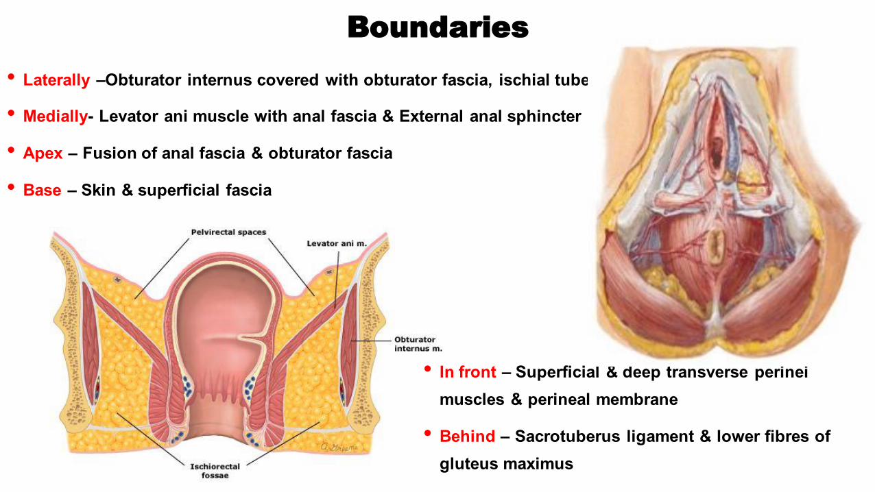
Boundaries
• Laterally –Obturator internus covered with obturator fascia, ischial tuberosity
• Medially- Levator ani muscle with anal fascia & External anal sphincter
• Apex – Fusion of anal fascia & obturator fascia
• Base – Skin & superficial fascia
• In front – Superficial & deep transverse perineimuscles & perineal membrane
• Behind – Sacrotuberus ligament & lower fibres of gluteus maximus

Fasciae of Ischiorectal / Ischioanal Fossae
• Obturator fascia• Anal fascia • Perianal Fascia• Lunate fascia

OBTURATOR FASCIA (yellow arrow)
ANAL FASCIA (red arrow)
PERIANAL FASCIA (blue arrow)
Lateral most septum derived from fusion of longitudinal muscle of rectum and levatorani
The anal fascia is the inferior layer of the diaphragmatic part of the pelvic fascia
fascia of obturator internus muscle, covers the pelvic surface of the muscle and is attached around the margin of its origin
Diagrammatic coronal section through anal triangle
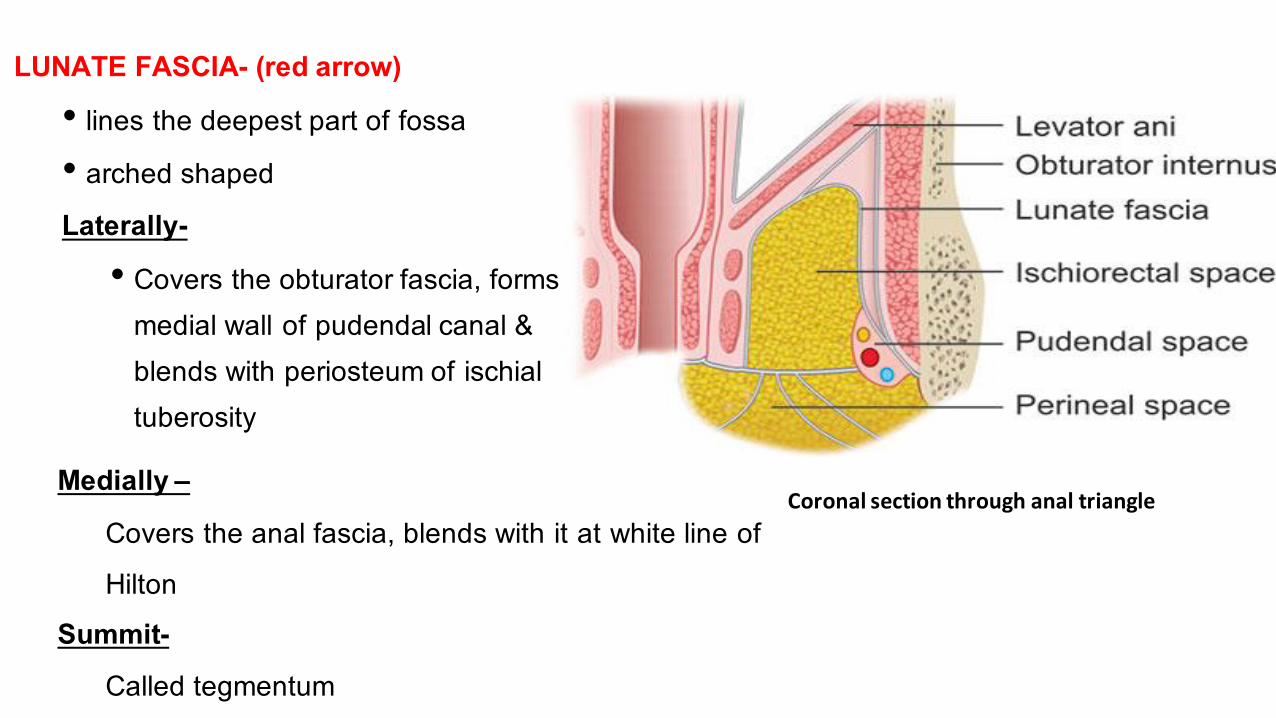
LUNATE FASCIA- (red arrow)• lines the deepest part of fossa• arched shapedLaterally-
• Covers the obturator fascia, forms medial wall of pudendal canal & blends with periosteum of ischialtuberosity
Coronal section through anal triangleMedially –
Covers the anal fascia, blends with it at white line of Hilton
Summit-Called tegmentum

Subdivisions of Ischioanal fossa
SUPRATEGMENTAL SPACE-• Above tegmentum • Between apex of fossa and tegmentum• Contains loose fat
ISCHIORECTAL SPACE• Between lunate fascia and perianal fascia• Filled with fat traversed by fibrous tissue
PERIANAL SPACE-Between perianal fascia above and perianal skin belowSubdivided into compartments by fibroelastic septaContains loculated fat

Recesses of the fossa
• ANTERIOR RECESS-• forward extension of fossa above perineal
membrane/ urogenital diaphragm and below the pelvic diaphragm
• POSTERIOR RECESS-• backward extension deep to
sacrotuberous ligament on the side of coccyx
• Two ischioanal fossae communicate with each other through a gap behind the anal canal

Contents of Ischioanal/ Ischiorectal fossa
Inside the pudendal canal-• Internal pudendal vessels • Pudendal nerve
Traversing through fat from lateral to medial• Inferior rectal vessels & nerve
In the anterior part of fossa• Posterior scrotal vessels & nerves
In the posterior part of fossa• Perineal branch of S4• Perforating branches of S2, S3
Throughout• Fat

Clinical Anatomy

Ischiorectal Fat as support
• Fat provides a cushion like support to rectum & anal canal
• Loss of this fat due to debilitating diseases may cause downward displacement of rectum called Rectal prolapse
• Part of rectum protrudes out from the anus

Ischiorectal hernia/ Perineal hernia
• Herniation of peritoneum or fat through Hiatus of Schwalbe
• What is hiatus of Scwalbe ?• Normally levator ani originates from thickened area of
obturator fascia covering the obturator internus muscle - called arcus tendineum
• Sometimes origin of levator ani takes place from the obturator fascia which may form a tendinous sling between pubic bone & ischial spine. The gap that is present between the tendinous sling and obturator internus is called Hiatus of Schwalbe

Fistula in Ano
• A fistula-in-ano is an abnormal hollow tract or cavity that is lined with granulation tissue and that connects a primary opening inside the anal canal to a secondary opening in the perianal skin
• An infected tunnel between the skin and the anus
• Caused by bursting of an abscess internally and externally on the surface

Horse shoe abscess
• Infection may pass from fossa of one side to the other readily through the horse shoe shaped recess behind anal canal
• this may lead to formation of horse shoe abscess


