ISOLATION A N D CHARACTERIZATION OF THE THIRD COMPLEMENT COMPONENT OF AXOLOTL
(AMB YSTOMA MEXICANUM)
DAVID AVILA and JOHN D. LAMBRIS* Basel Institute for Immunology, Grenzacherstr 487, 4005-Basel, Switzerland (Tel: 61 492380;
Fax: 61 681 1304)
(Received 4 September 1989)
Abstract--1. Using a monoclonal anti-human C3 antibody and a polyclonal anti-cobra venom factor antibody as probes, a protein homologous to the mammalian third complement component (C3) was purified from axolotl plasma and found to be axolotl C3.
2. Axolotl C3 consists of two polypeptide chains (M r = 1 I0,000 and 73,000) linked by disulfide bonds. An internal thiolester bond in the ~ chain was identified by the incorporation of [t4C]methylamine and NH2-terminal sequence from the C3d fragment of C3.
3. Digestion of C3 by trypsin resulted in the cleavage of both the a and fl chains, generating fragments with a cleavage pattern similar to that of human C3.
4. The amino acid composition of axolotl C3 and the amino acid sequences of the thiolester site (and the surrounding amino acids), the cleavage site for the C3-convertase, and one of the factor I cleavage sites are similar to C3 from other vertebrates.
5. In contrast to human C3, which has concanavalin A binding carbohydrates on both the g and chains, only the fl chain of axolotl C3 contains such carbohydrates.
INTRODUCTION
The human complement system has been studied in great detail and has been the focus of intensive studies due to its pivotal role within the complement system (Lambris, 1988; Becherer et al., 1989). By contrast, very little is known about the existence and function of the different complement components in lower vertebrates. The third protein of complement (C3) is one of the most versatile and multifunctional molecules within the complement system, interacting with several serum proteins, cell surface receptors, and proteins of foreign origin (Lambris, 1988; Becherer et al., 1989). Until now C3 has been purified from several species, including human (Tack and Prahl, 1976), rabbit (Horstmann and Miiller-Eber- hard, 1985), guinea-pig (Thomas and Tack, 1983), mouse (Gyongyossy and Assimeh, 1977), rat (Daha et al., 1979), quail (Kai et al., 1983), cobra (Eggertsen et al., 1983), frog (Sekizawa et al., 1984), trout (Nonaka et al., 1984b), and lamprey (Nonaka et al., 1984a) (for review see Becherer et al., 1989). The complete amino acid sequence of human (De Bruijn and Fey, 1985) and mouse C3 (Wetsel et al., 1984; Lundwall et al., 1984), as well as the partial sequence of rabbit (Kusano et al., 1986) and Xenopus C3 (Grossberger et al., 1989), have been deduced from their cDNA sequences. Limited amino acid sequence has also been obtained for the C3 from other species (Becherer et al., 1989). The elucidation of the struc- ture and functions of C3 and other complement proteins from different species provides not only evolutionary information on the complement system,
*To whom correspondence should be addressed.
but also additional information on the structural elements involved in its multiple interactions with other ligands within the human complement system (Becherer et al., 1989). In this study we report the purification and partial structural analysis of axolotl C3. Its similarities to C3 from other vertebrates, the presence of a thiolester bond and Con A binding carbohydrates, and its fragmentation by trypsin are analyzed.
MATERIALS AND METHODS
Materials
Plasma from adult axolotls (Ambystoma mexicanum, Am- phibia, Urodela) was kindly provided by Dr James Kauf- man (Basel Institute for Immunology). Trypsin was purchased from Cooper Biomedical. Concanavalin A (Con A) was from Vector Laboratories. Polyvinylidene difluoride (PVDF) membranes were from Millipore. Bovine serum albumin (BSA), 2,2"-azino-bis(3-ethylbenthiazole-6-sulfonic acid), sodium azide and Tween-20 were purchased from Sigma Chemical Company. Polyethyleneglycol 4000 (PEG), polyvinyl pyrroldone (PVP), thiolgiycolic acid and phenol were from Merck. Constant boiling 6 N HC1 and 4-ethyl- morpholine were from Pierce. All chemicals for sodium dodecyl sulphate-polyacrylamide gel electrophoresis (SDS- PAGE) were from Bio-Rad Laboratories. [~4C]Methyl- amine-HC1 was purchased from Amersham. All chemicals for automated amino acid sequencing and composition were from Applied Biosystems.
Antibodies
The monoclonal anti-human C3 antibody (MoAb 0131111) was obtained from Cytotech and the monoclonal anti-human C3 antibody (MoAb 133HI1) was a gift from Jochem Alsenz (Basel Institute for Immunology). A polyclonal antibody against cobra venom factor was
CBP(B) 95/4~M 839
840 DAVID AVILA and JOHN D. LAMBRIS
raised in goats as previously described (Vogel et al., 1984). Affinity purified peroxidase conjugated anti-IgG antibodies were purchased from Bio-Rad Laboratories.
Purification of axolotl C3
Axolotl plasma, treated with 2mM diisopropylfluo- rophosphate, was brought to 4% (w/v) PEG, and after 30 rain with constant stirring at 4"~C, the plasma was cen- trifuged (10,000 g, 20 min). The supernatant was brought to 16% PEG, stirred for 1 hr at 4°C, centrifuged (10,000g, 20 min), and the pellet redissolved in 5 ml of 20 mM Tris- HC1, pH 7.5/2mM diisopropylfluorophosphate. Residual PEG was removed using a PD-10 desalting column (Phar- macia) and the 4-16% PEG fraction was applied to a Mono Q HR 5/5 anion exchange column equilibrated in 20mM Tris-HC1, pH 7.5. After washing the column with 5 ml of buffer, C3 was eluted with a linear gradient of NaC1 (0~).5 M). Fractions containing C3 were identified by their reactivity with MoAb 0131111 and anti-CVF antibodies, and by their mobility in a 9% SDS PAGE gel (Laemmli, 1970). The fractions were pooled, concentrated by ultrafiltration using an Amicon YM30 Diafto Membrane, then applied to a Superose 12 gel filtration column (20 x 300mm) equili- brated in 10 mM sodium phosphate, pH 7.2/150 mM NaCI (PBS) containing 3 mM sodium azide. All chromatographic separations were performed using the Fast Protein Liquid Chromatography system (Pharmacia). Fractions containing C3 were pooled and frozen at -70 'C .
Detection of axolotl C3 by an enzyme-linked immunosorbent assay (ELISA )
Microtiter plates were coated overnight at 4~'C with either axolotl C3, human C3, or the 16% PEG precipitate of axolotl plasma (8 #g/ml PBS). The plates were saturated with 1% (w/v) bovine serum albumin for 30 min at 25°C. Serial diluted MoAb 0131111 or anti-CVF was added for 30 min at 25°C. Bound antibody was detected with either peroxidase conjugated antimouse IgG antibody (1/1000 dilution) or peroxidase conjugated anti-goat IgG antibody (1/1000 dilution) followed by the addition of 0.07 mM HzOd5 mM 2,2'-azino-bis(3-ethylbenzthiazoline-6-sulfonic acid)/10 mM sodium citrate, pH 4.5. After each step the wells were washed with 0.05% (v/v) Tween-20/PBS.
Fragmentation of axolotl C3 by trypsin
Axolotl C3 was digested with 1% (w/w) trypsin at 37°C and samples removed at time intervals of 0, 3, 15, 30, and 60min. The reaction was stopped by the addition of 2% (v/v) 2-mercaptoethanol/2% (w/v) SDS/10% (v/v) glyc- erol/0.0025% (w/v) Bromophenol Blue/0.0625 M Tris-HC1, pH6.8. The tryptic fragments were separated by SDS-PAGE on a 9% gel and visualized after staining with Coomassie Brillant Blue R (Sigma). The molecular weights of the tryptic fragments were calculated using reference proteins (Bio-Rad Laboratories).
Identification of the internal thiolester site in axolotl C3
The presence of a thiolester site in axolotl C3 was determined by the incorporation of [~4C]methylamine and amino acid sequence analysis. The incorporation of [~4C]methylamine to axolotl C3 was performed as previously described (Harrison et al., 1981; Tack, 1983). Briefly, axolotl C3 (100#g) was allowed to react with [14C]methylamine (0.054 M, 0.3 mCi) for 2 hr at 37°C. Unlabeled methylamine (0.1 M) was added to the sample and incubated for 30 rain at 37°C. The ~4C-axolotl C3 was brought to a final volume of 100#1, spun through a Bio-Spin 6 column (Bio-Rad Laboratories) to remove unincorporated methylamine, and digested with trypsin as described. The L4C-axolotl C3 fragments were visualized by autoradiography.
The NH2-terminal sequence of the site was obtained by Edman degradation (see below) of the ~4C-axolotl C3 frag- ment that is equivalent to the human C3d. This fragment
was isolated by selectively absorbing out the C3c fragment using Con A-Sepharose (Pharmacia). Trypsinized C3 (100/~g) was incubated with Con A-Sepharose in the pres- ence of 1 mM MnC12/1 mM CaC12 for 30 rain at 37°C. After centrifugation, the supernatant containing the thiolester fragment was reduced, separated on a '9% SDS-PAGE gel and the fragment containing the thiolester bond was se- quenced after electroblotting to a PVDF membrane as described in this paper.
Detection of Con A binding carbohydrates on axolotl C3
Con A binding carbohydrates were detected as previously described (Becherer and Lambris, 1988). Briefly, samples from trypsinized C3 were run on a 9% SDS-PAGE gel followed by electroblotting to a nitrocellulose membrane. Nitrocellulose was saturated by incubation in 2% (w/v) PVP-40/PBS for 1 hr. t25I-Con A, labeled by the Iodogen method (Fraker and Speck, 1978), was added to the electro- blotted C3 (106 cpm/ml, 7.3 x 10⁹ cpm/mg) and incubated for 1 hr at room temperature. Nitrocellulose was extensively washed with 2% (w/v) PVP-40/PBS followed by several washes with 0.05% (v/v) Tween-20/PBS. The binding of t25I-Con A was detected by autoradiography.
Amino acid composition and NH2-terminal sequence of axolotl C3
Gas phase hydrolysis of axolotl C3 was performed in 6 N HC1/I% (v/v) phenol at l l0°C. Analysis of the amino acid composition was performed after 24, 48, and 72 hr using an Applied Biosystems 420A Derivatizer connected on-line to a 130A Analyzer and a 920A Data Analysis Module. The compositions for serine and threonine were determined after extrapolation to zero time hydrolysis.
The axolotl C3 tryptic fragments were sequenced by a modified method of Matsudaira (1987), using an Applied Biosystem 470A Protein Sequencer connected on-line to a 120A PTH-analyzer and a 900A Control/Data Analysis Module. Briefly, SDS-PAGE gels were pre-run (10 mA/gel) for 30 min before application of samples. Thiolglycolic acid (0.002%) was added to the running buffer to scavenge remaining free radicals in the gel. Following electrophoresis, gels were equilibrated in 0.025M 4-ethylmorpholine (pH 8.3) for 20rain and then electroblotted onto PVDF membranes (1 mA/cm 2 gel) using a semi-dry electroblot apparatus (JKA-Biotech). Bands were visualized after stain- ing with 0.1% (w/v) Coomassie Brillant Blue in methanol/acetic acid/water (50/10/40) for 5 rain followed by destaining for 10 min. The electroblotted membranes were rinsed with tri-distilled water before drying under vacuum. Samples were stored under vacuum at 4°C until sequenced. Alternatively, the ~ chain of axolotl C3 was sequenced by direct application of C3 to a polybrene-coated glass fiber filter.
RESULTS
Purification o f axolotl C3
In order to establish a detect ion assay for axolotl C3, several monoclona l and polyclonal ant ibodies directed against h u m a n C3 and CVF were tested for their reactivity with the 16% P E G fract ion of axolotl plasma. Using an ELISA, two ant ibodies were found to react with the 16% P E G fract ion (Fig. 1) of axolotl p lasma and were fur ther used to assay axolotl C3 dur ing the different purification steps.
The isolation of axolotl C3 was accomplished in essentially three steps. Precipi ta t ion of axolotl p lasma with 4 % P E G followed by the precipi tat ion of C3 with 16% P E G resulted in the removal of approxi- mately 50% of the p lasma proteins. Using an ELISA to assay for residual C3 in the 16% P E G superna tan t
The axolotl C3 841
1.6
1,4
1.2
,,¢ 1.0
O.8
O.6
O.4
O.2
.i// A / _ m / m / m
A ~ nn f
' 1164 ' 1t16 ' 114 ' Ant ibody Dilut ion
Fig. 1. Cross-reactivity of anti-CVF and MoAb 013III1 antibodies with axolotl C3. Serial diluted monoclonal anti- bodies 013IIIl (A), 133Hll (O), or goat anti-CVF ( . ) were incubated for 30min at 25°C with a 16% PEG precipitate from axolotl plasma (20/~g protein/ml PBS) which was previously fixed to a microtiter plate. The bound antibodies were detected as described in Materials and Methods. The starting dilutions for MoAb 013III1 and MoAb 133Hll were 50#g/ml (purified from ascites by ammonium sulfate precipitation) and for goat anti-CVF 200/~g/ml (total immunoglobulin fraction from serum after
ammonium sulfate precipitation).
it was determined that the axolotl C3 was in the 4-16% PEG pellet. The pellet was resuspended into 5 ml of 20 mM Tris-HC1, pH 7.5, and then applied to a Mono Q anion-exchange column. Five major peaks were detected after elution with a gradient of 0-500 mM NaC1 (Fig. 2); the third peak, identified by ELISA and SDS-PAGE, contained the axolotl C3. The concentrated C3 fractions from the Mono Q column were further purified by gel filtration on a Superose 12 column. As shown in Fig. 3, the third peak to elute from the Superose 12 column reacted with both MoAb 013III1 and anti-CVF antibodies, and contained only one major protein when analysed by SDS-PAGE. The total recovery of purified axolotl C3 from 6 ml plasma was 2.2 mg. The purified axolotl C3 has an apparent molecular weight of 200,000 as determined by gel filtration and SDS-PAGE (Fig. 3) and is comprised of two chains having M~s of 1 I0,000 (~ chain) and 73,000 (fl chain) which are linked by a disulfide bond(s).
Fragmentation of axolotl C3 by trypsin
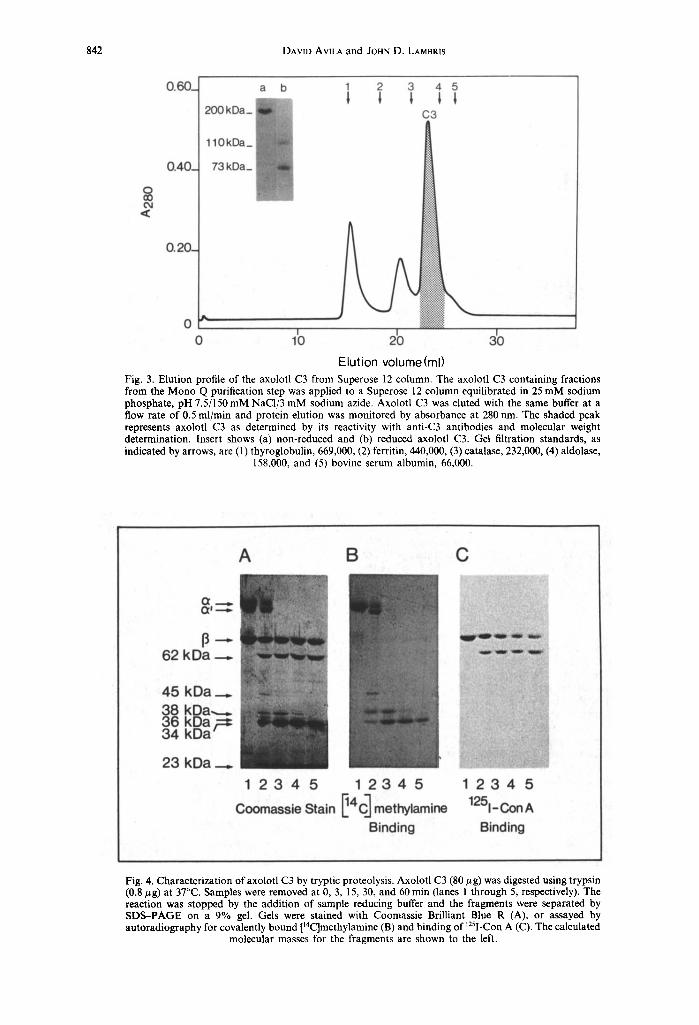
Tryptic proteolysis of axolotl C3 resulted in the preferential cleavage of the ~ chain (Fig. 4A, lane 2). The initial cleavage near the NH2-terminus of the ct chain generates a 100 kDa fragment (~t' chain) and a 10 kDa fragment (C3a). Further cleavage of the =' chain generates fragments having molecular masses of 45, 38, 36, 34 and 23 kDa (Fig. 4A, lane 2). The 62 kDa fragment generated after 3 min (Fig. 4A, lane 2) represents a cleavage fragment of the fl chain as determined by its ability to bind lZSI-Con A (Fig. 4C).
Further cleavage of the ct chain fragments resulted in a band with molecular mass of 34 kDa (Fig. 4A, lane 5). This band is composed of two ~ chain fragments as determined by NH:terminal sequencing. One frag- ment contains the thiolester site and the second fragment contains the factor I cleavage site (Fig. 5).
Identification of the internal thiolester site in axolotl C3
Treatment of axolotl C3 with [14C]methylamine resulted in the covalent binding of [14C]methylamine to the ~ chain (Fig. 4B, lane 1). Tryptie digestion of the ¢ chain shows that the [14C]methylamine is cova- lently bound to the ~' chain of the C3b fragment and the three smaller fragments of 45, 38 and 34 kDa (Fig. 4B, lane 2). When the electrophoresis was run under non-reducing conditions it was found that the C3b is cleaved into two fragments of 145 (C3c) and 45 kDa (data not shown), the latter being labeled with [14C]methylamine. Further cleavage of the 45 kDa fragment leads to the generation of the 38 and 34 kDa fragments (Fig. 4B, lanes 2-5). Two 34 kDa frag- ments were sequenced after separation on a Con A-Sepharose column and electroblotting to a PVDF membrane. One fragment containing the thiolester site (Fig. 5) shows a high amino acid sequence similarity to the region of C3 from other vertebrates containing the thiolester site (the thiolester site being completely conserved).
Detection of Con A binding carbohydrates on axolotl C3
The detection of carbohydrate moieties on the axolotl C3 was done using 12sI-Con A (Hirani et al., 1985). Binding of lZSI-Con A to reduced axolotl C3
1.oo-
0.80_
0.60.
O4
'¢ o.4o.
0.20
/ !
o lb 2'0
/ /
I I I -300 j
_200 , Z ,
-100
=,',- 0 40
Elution volume (ml)
Fig. 2. Elution profile of the 4-16% PEG fraction of axolotl plasma from Mono Q column. The 4-16% PEG fraction of axolotl plasma was applied to a Mono Q column equili- brated in 20 mM Tris-HCl, pH 7.5, then eluted with a NaCI gradient (0-500 raM) in the same buffer. Protein concentra- tion was monitored by absorbance at 280 nm. The shaded peak was reactive with both MoAb 013III1 and polyclonal anti-CVF antibodies and has an ct and fl chain structure with
molecular weights similar to those of mammalian C3.
842 DAVID AVILA and JOHN D. LAMBRIS
0 cO
0 . 6 0 _
0 . 4 0 _
0.20_
0
a
200 kDa_ g R
110 kDa_ I
73 kDa_ I
0
b
1 1 2 3 4 5
C3
t I i::::i::::i::iiiiiii::igl
10 20 3o Elution volume(ml)
Fig. 3. Elution profile of the axolotl C3 from Superose 12 column. The axolotl C3 containing fractions from the Mono Q purification step was applied to a Superose 12 column equilibrated in 25 mM sodium phosphate, pH 7.5/150 mM NaC1/3 mM sodium azide. Axolotl C3 was eluted with the same buffer at a flow rate of 0.5 ml/min and protein elution was monitored by absorbance at 280 nm. The shaded peak represents axolotl C3 as determined by its reactivity with anti-C3 antibodies and molecular weight determination. Insert shows (a) non-reduced and (b) reduced axolotl C3. Gel filtration standards, as indicated by arrows, are (1) thyroglobulin, 669,000, (2) ferritin, 440,000, (3) catalase, 232,000, (4) aldolase,
158,000, and (5) bovine serum albumin, 66,000.
A B C
62 kDa
45 kDa ~8 kDa,,-~
kDa r-.-= 34 kDa
23 kDa . . . . . . . . 1 2 3 4 5 1 2 3 4 5
Coomassie Stain 114CJ methylamine Binding
1 2 3 4 5 1251- Con A
Binding
Fig. 4. Characterization of axolotl C3 by tryptic proteolysis. Axolotl C3 (80 # g) was digested using trypsin (0.8 #g) at 37°C. Samples were removed at 0, 3, 15, 30, and 60 min (lanes 1 through 5, respectively). The reaction was stopped by the addition of sample reducing buffer and the fragments were separated by SDS--PAGE on a 9% gel. Gels were stained with Coomassie Brilliant Blue R (A), or assayed by autoradiography for covalently bound [14C]methylamine (B) and binding of 12sI-Con A (C). The calculated
molecular masses for the fragments are shown to the left.
The axolotl C3 843
a chain
C3 AXOLOTL C3 RABBIT C3 MOOSE C3 RAT C3 PORCINE C3 TROUT C3 GUZN. P I G C3
C3 C o n v e r t a s e 4
650 660 7 2 7 7 3 7 I I I I 8 V Q L T E K R M N K V G K Y P - K .... 8 ~ L D E D I I A E E N Z V B R 8 E * * L* I * E K A X * A A * * TG* .... * EV* * * YL* E* D * D * X * A * * * * T * * N ' D * * * * * T ..... * D * * * * * * P * * D * * * Z*T * * * *M* * * * D ' A * * * TD* .... * * * E** * *P** D* I* * *I * * * * M* * * * D * A * * * TD* * * * * M ' R * * * * L * * * 8 - * A * T Z 8 D V Z T S M A 8 S * N G L * * * * M ' R * * D * A
I I I I C3 8 Z g T K E N E G F T V T A E G K G Q G T L 8 - V V T M Y m A K C3 * * * * * L * N N * D * E V K * T * * * * * T H * t t V * N * L C3 * * * * * * * * * * * * * * * * * * * * * * * - * * * * * * * * C3 * * * * * * * * * * * * * * * * * * * * * * * - * * * * * * * * C3 * A * * * L * * * * * * K * K * * * * * * M G * * * * * * * * *
Fig. 5. Alignment of axolotl C3 sequence with known C3 sequences from other species. Identical amino acids are indicated by an asterisk. The numbering is based on the human C3 sequence (De Bruijn and Fey, 1985) after subtracting the signal peptide sequence. The amino acid sequence of rabbit (Becherer et al., 1989; Kusano et al., 1986), mouse (Wetsel et al., 1984; Lundwall et al., 1984), guinea-pig (Thomas and Tack, 1983), rat (Daha et al., 1979), and trout (Nonaka et al., 1984b) C3 are from the indicated references. The amino acid sequence of Xenopus C3 was obtained using methods similar to those described for axolotl C3 (unpublished observations). The factor I and convertase cleavage sites of human C3
(arrows) and the thiolester site (box) are indicated.
and its tryptic f ragments shows tha t only the fl chain conta ins Con A binding carbohydrate(s) (Fig. 4C, lane 1). This ca rbohydra te is retained on a 62 kDa f ragment of the fl chain after cleavage by trypsin (Fig. 4C, lanes 2-5). Fur the r cleavage of the fl chain and its 62 kDa f ragment was not observed after 1 hr.
A m i n o acid composi t ion and NH2- t e rmina l sequence o f axo lo t l C 3
The amino acid composi t ion of axolotl C3 (Table 1) was compared to tha t of human , t rout , and Xenopus C3. The amino acid composi t ion similarities between the axolotl C3 and these species are shown in Table 1.
Several axolotl C3 f ragments obta ined by tryptic proteolysis were sequenced by E d m a n degradat ion. Al ignment of the obta ined sequences with the C3 sequences f rom other species (Fig. 5) reveals tha t a high similarity exists between the axolotl C3 sequence and tha t of C3 f rom other species. Similar tryptic f ragments have been previously generated for the h u m a n coun te rpar t (Eggertsen et al., 1985), indicat- ing tha t the t rypsin cleavage sites are similar in bo th C3 molecules. One par t icular tryptic fragment, which was found to encompass the thiolester site, is 82% identical with mouse and 77% identical with h u m a n C3.
All a t tempts to sequence the fl chain and its 62 kD a f ragment were unsuccessful, leading us to believe tha t the NHE-terminus is blocked. A blocked NH2-termi-
nus was confirmed by sequencing of the intact C3 molecule, which resulted in a single sequence f rom the ~t chain and not the expected double ct/fl sequence.
DISCUSSION AND CONCLUSIONS
Analysis of complement in lower species no t only enhances our unders tand ing on the evolut ion of the
Table 1. Comparison of the amino acid composition of axolotl C3 with those from other species
Amino a c i d Axolotl Human* Troutt Xenopus$.
Asx 11.2 9.1 10.4 11.6 Glx 11.7 12.8 11.2 11.6 Ser 6.2 6.4 7.4 5.9 Gly 7.2 5.7 6.7 6.6 His 2.1 1.7 1.7 1.4 Arg 4.1 4.9 3.5 4.3 Thr 7.6 6.3 7.1 6.5 Ala 6.8 5.7 6.8 6.4 Pro 4.8 4.7 3.8 5.1 Tyr 4.5 3.4 3.6 4.1 Val 8.9 8.8 10.0 8.2 Met 2.6 2.1 2.1 2.1 Cys --~ 0.8 2.4 --~ lie 6.0 5.1 4.5 6.2 Leu 7.9 9.2 7.5 8.2 Phe 4.4 3.6 3.1 3.1 Trp --~ 0.7 1.2 0.9 Lys 7.7 6.8 7.3 7.7 *Taken from Tack and Prahl (1976). tTaken from Nonaka et al. (1984b). ~Taken from Sekizawa et aL (1984). §Not determined.
844 DAVID AV1LA and JOHN D. LAMBRIS
complement system but is also a necessary element in the structural and functional analysis of the individ- ual complement proteins. The best phylogenetically studied complement component is C3, no doubt due to its key role in complement activation. It has been purified from several vertebrates, including members of the mammals, aves, reptilia, amphibia, osteich- thyes, and agnatha (for review see Becherer et al., 1989). In this study we isolated and characterized C3 from the axolotl (Ambys toma mexicanum), a neotenic salamander. Among the interesting characteristics of the axolotl are: (a) the low production of thyroxin which accounts for its inability to achieve anatomical metamorphosis; and (b) its poor immune responses (chronic graft rejections, absence of immunoglobulin isotype switch and affinity maturation, imperceptible mixed lymphocyte reaction, etc.) (Ching and Wedg- wood, 1967; Charlemagne and Tournefier, 1977).
In this study, axolotl C3 was purified from axolotl plasma using two cross-reactive antibodies as probes: a monoclonal anti-human C3 antibody and a poly- clonal anti-CVF antibody. Our finding that the anti- CVF antibodies crossreact with axolotl C3 agrees with previous findings suggesting that antibodies to CVF recognize structures which are conserved in C3 from different vertebrates (Eggertsen et al., 1983). We have extended this study to other species and found that the anti-CVF antibodies, in addition to reacting with CVF, cross-react with human, rabbit, mouse, chicken, cobra, and Xenopus C3 (unpublished obser- vations). The procedure described for the purification of axolotl C3 is simple and has been used to purify C3 from other species such as chicken, cobra, and Xenopus (Alsenz et al., 1989). That. the isolated molecule represents axolotl C3 is 15ased on the follow- ing properties: (a) its molecular stucture is similar to mammalian C3, composed of two polypeptide chains (M r = 110,000 and 73,000) linked by disulfide bond(s); (b) it contains an internal thiolester site; (c) the amino acid composition shows a high degree of resemblance to human, trout, and Xenopus C3; and (d) the NHz-terminal sequence of the ~ chain and other tryptic C3 fragments show a high degree of similarity with the corresponding regions of C3 from other species. Comparisons of the limited amino acid sequences obtained from the tryptic fragments of axolotl C3 with that of C3 from other species show it to be between 55 and 61% identical (average of all fragments) to human, rabbit, mouse, and Xenopus C3. Its identity with mouse C3 increases to 82% in the tryptic fragment containing the thiolester bond; the thiolester site, GCGEE, is 100% conserved in all species. This conservation of the thiolester site and its surrounding hydrophobic amino acids emphasizes the functional importance of this region in maintain- ing, throughout evolution, the molecule's capacity to fix itself to surfaces. The region surrounding the thiolester bond in C3, C4 and ~2-macroglobulin is relatively hydrophobic and it has been proposed that it serves to shield the thiolester from the aqueous environment and nucleophilic attack (Tack, 1983; Levine and Dodds, 1989; Zhao et al., 1988; Fontaine et al., 1982).
Cleavage of human C3 by C3 convertases leads to the generation of C3a and C3b, the C3b having the thiolester bond exposed. The thiolester bond is either
hydrolyzed or mediates the covalent binding reaction with cell surfaces or other receptive molecules (Tack, 1983; Law et al., 1980). The fixation of C3b to the activating surfaces is important for the amplification of the complement cascade, initiation of the mem- brane attack complex, phagocytosis of foreign parti- cles, and the enhancement of effector-target cell contact. Depending on the surface to which C3b is bound, it will either participate in the amplification of the complement cascade or will be degraded by factor I in the presence of cofactor molecules (Lambris, 1988). Although it is not known whether the amino acids Arg 726 and Arg 1298 (human C3 numbering) are conserved in axolotl C3, the conservation of Ser 727 and Set 1299 in the c~ chain of axolotl C3 suggests that the axolotl C3 convertases and factor I have similar specificities as in human complement.
The human C3 fragments generated during com- plement activation, in surface bound or fluid phase, react with several other complement components and receptors as well as with proteins of foreign origin (Lambris, 1988). Among them are the factors, I, H, B, C5, P, CR1, CR2, CR3, CR4, etc. (for review see Lambris, 1988; Becherer et al., 1989). Comparison of the ability of C3 from various species to bind the different C3 binding proteins and correlation of this information to amino acid sequence similarity has been instrumental in identifying the binding sites in C3 for these ligands (Becherer et al., 1989; Daoudaki et al., 1988). Since the binding sites for many of the ligands of human C3 have been localized (Lambris, 1988; Becherer et al., 1989), studies on the reactivity of axolotl C3 with the ligands of human C3 and subsequent cloning of axolotl C3 will greatly enhance our understanding of the structural features in C3 involved in these interactions. Preliminary experi- ments comparing C3s from different vertebrates sug- gest that the sequence conservation of the binding sites for H, P, CR1, and CR2 is important in main- taining binding of its ligand (Alsenze et al., 1989).
One discrepancy with human C3, which contains Con A binding N-linked high mannose carbohy- drates in both chains (Hase et al., 1985; Hirani et al., 1986), is the absence of Con A binding carbohydrates on the c~ chain of axolotl C3. This does not exclude the possibility that carbohydrates not recognized by Con A are present on the ~ chain. Similar to axolotl C3, trout and Xenopus C3 do not have Con A binding carbohydrates in the 7 chain (Becherer et al., 1989). The difference in glycosylation is of interest because the ~ chain carbohydrate moiety on human C3 has been found to be involved in the binding to bovine conglutinin (Hirani et al., 1985), and recently the human analog to bovine conglutinin has been found in human serum (Thiel et al., 1987). Even though the exact function of conglutinin-iC3b interaction is not known it will be of interest to see if the axolotl has a protein analogous to bovine or human conglutinin and, if such a protein exists, to test its interaction with human and axolotl C3.
Acknowledgements--The authors wish to thank David Becherer, Jochem Alsenz, and Inmaculada Esparza for critical reading of this manuscript. The Basel Institute for Immunology was founded and is entirely supported by F. Hoffman-La Roche Ltd Co., Basel, Switzerland.
The axolotl C3 845
REFERENCES
Alsenz J., Bercherer D., Esparza 1., Daoudaki M., Avila D., Oppermann S. and Lambris J. D. (1989) Structure and function analysis of C3 from different species. Comple- ment Inflamation 6, 307 (abstract).
Becherer J. D., Alsenz J. and Lambris J. D. (1989) Molec- ular aspects of C3 interactions and the struc- tural/functional analysis of C3 from different species. In The Third Component of Complement: Chemistry and Biology (Edited by Lambris J. D.). Current Topics in Immunol. and Microbiol., Vol. 153, 45-72.
Becherer J. D. and Lambris J. D. (1988) Identification of the C3b-receptor binding domain in the third component of complement. J. biol. Chem. 263, 14,586-14,591.
Charlemagne J. and Tournefier A. (1977) Anti-horse red blood cells antibody synthesis in the Mexican axolotl Am- bystoma mexicanum: effect of thymectomy. In Develop- mental Immunobiology (Edited by Solomon J. B. and Hor- ton J. D.), p. 267. Elsevier-North Holland, Amsterdam.
Ching Y. C. and Wedgwood R. J. (1967) Immunologic responses in the axolotl, Siredon mexicanum. J. Immunol. 99, 191-200.
Daha M. R., Stuffers-Heiman M., Kijlstra A. and van Es L. A. (1979) Isolation and characterization of the third component of rat complement. Immunology 36, 63-69.
Daoudaki M. E., Becherer J. D., and Lambris J. D. (1988) A 34-amino acid peptide of the third component of complement mediates properdin binding. J. Immunol. 140, 1577-1580.
De Bruijn M. H. L. and Fey G. H. (1985) Human comple- ment component C3: cDNA coding sequence and derived primary structure. Proc. nam. Acad. Sci. USA 82, 708-712.
Eggertsen G., Hellman U., Lundwall A., Folkersen J. and Sj6quist J. (1985) Characterization of tryptic fragments of human complement factor C3. Molec. Immunol. 22, 833-841.
Eggertsen G., Lundwall A., Hellman U. and Sj6quist J. (1983) Antigenic relationships between human and cobra complement factors C3 and cobra venom factor (CVF) from the indian cobra (Naja naja). J. Immunol. 131, 1920-1923.
Fontaine M., Aubert J. P., Joisel F. and Lebreton J. P. (1982) Structure-function relations in the third compo- nent of human complement (C3)--I. Hydrophobic sites. Molec. Immunol. 19, 27 37.
Fraker P. J. and Speck J. C. (1978) Protein and cell membrane iodinations with a sparingly soluble chloro- amide, 1,3,4,6,-tetrachloro-3a,6a-diphenylglycoluril. Bio- chem. biophys. Res. Commun. 80, 849-857.
Grossberger D., Marcuz A., Du Pasquier L. and Lambris J. D. (1989) Conservation of structural and functional domains in complement component C3 of Xenopus and mammals. Proc. natn. Acad. Sci. USA 86, 1323-1327.
Gyongyossy M. I. C. and Assimeh S. N. (1977) Isolation of the third component of mouse complement. J. Immunol. 118, 1032-1035.
Harrison R. A., Thomas M. L. and Tack B. F. (1981) Sequence determination of the thiolester site of the fourth component of human complement. Proc. natn. Acad. Sci. USA 78, 7388-7392.
Hase S., Kikuchi N., Ikenaka T. and Inoue K. (1985) Structures of sugar chains of the third component of human complement. J. Biochem., Tokyo 98, 863-874.
Hirani S., Lambris J. D. and M/iller-Eberhard H. J. (1985) Localization of the conglutinin binding site on the third component of human complement. J. lmmunol. 134, 1105-1109.
Hirani S., Lambris J. D. and Mfiller-Eberhard H. J. (1986) Structural analysis of the asparagine-linked oligosaccha- rides of human complement component C3. Biochem. J. 233, 613~16.
Horstmann R. D. and Mfiller-Eberhard H. J. (1985) Isola- tion of rabbit C3, Factor B, and Factor H and compari- son of their properties with those of the human analog. J. Immunol. 134, 1094-1100.
Kai C., Yoshikawa Y., Yamanouchi K. and Okada H. (1983) Isolation and identification of the third component of complement of Japanese quails. J. Immunol. 130, 2814-2820.
Kusano M., Choi N.-H., Tomita M., Yamamoto K. I., Migita S., Sekiya T. and Nishimura S. (1986) Nucleotide sequence of cDNA and derived amino acid sequence of rabbit complement component C3 a-chain, lmmunol. Invest. 15, 365-378.
Laemmli U. K. (1970) Cleavage of structural proteins during the assembly of the head of bacteriophage T4. Nature 277, 680~585.
Lambris J. D. (1988) The multifunctional role of C3, the third component of complement, lmmunol. Today 9, 387-393.
Law S. K., Lichtenberg N. A. and Levine R. P. (1980) Covalent binding and hemolytic activity of complement proteins. Proc. natn. Acad. Sci. USA 77, 7194-7198.
Levine R. P. and Dodds A. W. (1989) The thiolester bond of C3. In The Third Component of Complement: Chemistry and Biology (Edited by Lambris J. D.). Current Topics in Immunol. and Microbiol., Vol. 153, 73-82 .
Lundwall A., Wetsel R. A., Domdey H., Tack B. F. and Fey G. H. (1984) Structure of murine complement component C3. I. Nucleotide sequence of cloned complementary and genomic DNA coding for the fl chain. J. biol. Chem. 259, 13,851-13,862.
Matsudaira P. (1987) Sequence from picomole quantities of proteins electroblotted onto polyvinylidene diflouride membranes. J. biol. Chem. 262, 10,035-10,038.
Nonaka M., Fujii T., Kaidoh T., Natsuume-Sakai S., Yamaguchi N. and Takahashi M. (1984a) Purification of a lamprey complement protein homologous to the third component of the mammalian complement system. J. Immunol. 133, 3242-3249.
Nonaka M., Iwaki M., Nakai C., Nozaki M., Kaidoh T., Natsuume-Sakai S. and Takahashi M. (1984b) Purifica- tion of a major serum protein of rainbow trout (Salmo gairdneri) homologous to the third component of mam- malian complement. J. Immunol. 259, 6327-6333.
Sekizawa A., Fujii T. and Katagiri C. (1984) Isolation and characterization of the third component of complement in the serum of the clawed frog, Xenopus laevis. J. Immunol. 133, 1436-1443.
Tack B. F. (1983) The fl-Cys-g-Glu thiolester bond in human C3, C4, and ct2-macroglobulin. Springer Semin. lmmunopathol. 6, 259-282.
Tack B. F. and Prahl J. W. (1976) The third component of human complement: purification from plasma and physic- ochemical characterization. Biochemistry 15, 4513-4521.
Thiel S., Baatrup G., Friis-Christiansen P., Svehag S.-E. and Jensenius J. C. (1987) Characterization of a lecitin in human plasma analogous to bovine conglutinin. Scand. J. Immunol. 26, 461-468.
Thomas M. L. and Tack B. F. (1983) Identification and alignment of a thiol ester site in the third component of guinea-pig complement. Biochemistry 22, 942-947.
Vogel C.-W., Smith C. A. and Mfiller-Eberhard H. J. (1984) Cobra venom factor: structural homology with the third component of human complement. J. Immunol. 133, 3235 3241.
Wetsel R. A., Lundwall A., Davidson F., Gibson T., Tack B. F. and Fey G. H. (1984) Structure of murine comple- ment component C3. II. Nucleotide sequence of cloned complementary DNA coding for the alpha chain. J. biol. Chem. 259, 13,857-13,862.
Zhao B., Musci G., Susawara Y. and Berline L. J. (1988) Spin label and fluorescence studies of the thiolester bonds in alpha 2-macroglobulin. Biochemistry 27, 5304-5308.