ISOLATION AND IDENTIFICATION OF FUNGI ASSOCIATED WITH LEAF DISEASES OF HEVEA BRASILIENSIS
Nur Izzati Binti Ghazali
(27659)
SB 291 Bachelor of Science with Honours H4 (Plant Resource Science and Management) N974 2013 2013
S8 ,28(H1 N 7'1lJt
Pusat Khidmat Maklumat Akademik UMVERSm MALAYSIA SARAWAK
ISOLATION AND IDENTIFICATION OF FUNGI ASSOCIATED WITH LEAF DISEASES OF HEVEA BRASILIENSIS
P.KHIDMAT MAKL.UMAT AKADEMIK
111I11I1I11i'mll "111111 1000246698
Nur Izzati Binti Ghazali (27659)
This dissertation is submitted partial fulfillment ofthe requirements for
The Degree ofBachelor of Science with Honours in
Plant Resource Science and Management
Faculty of Resource Science and Technology Universiti Malaysia Sarawak
94300 Kota Samarahan Sarawak
.....
APPROVAL SHEET
Name ofcandidate: Nur Izzati Binti Ghazali
Title of Dissssertation: Isolation and identification of fungi associated with leaf diseases
ofHevea brasiliensis
Sepiab Muid.. Dr. (Prof. Dr. Sepiah Bt Muid) t!ll r . . roumemat Ecology
Vn-.rtDleDl of Plant Science and Enand\ ~ ogy ......- Facu1 of Resource Science
Supervisor U~VEItSITl MALAYSIA SARAWAK 94300 Koca Samarahaft
.............. .............................
(Dr. Mohd. Hasnul B. Bolhassan) Dr. Mohamad Hasnul Bin Bolhassan Senior Lecturer
Cosupervisor Faculty of Resource Science & Technology UNIVERSITI MALAYSIA SARAWAK
94300 Kota Samarahan
............................................ (Dr. Rebecca Edward)
Coordinator of Plant Resource Science and Management
Faculty of Resource Science and Technology
I
: •
,
,......
DECLARATION
I declare that no portion of the work referred to this dissertation has been submitted in
support of an application for another degree of qualification of this or any other
university or institution of higher learning.
(Nur Izzati Binti Ghazali) Programme ofPlant Resource Science and Management Department of Plant Science and Environmental Ecology Faculty of Resource Science and Technology University Malaysia Sarawak
i
,...... l
ACKNOWLEDGEMENT
Bissmillahirrahmanirrahim,
A lhamdu I i11ah. Thanks to Allah SWT, whom with His willing glvmg me the
opportunity to complete this Final Year Project which is title isolation and identification of
fungi associated with leaf diseases ofHevea brasiliensis. This final year project report was
prepared for Faculty of Resource Science and Technology, University Malaysia Sarawak
(UNlMAS), basically for student in fmal year to complete the undergraduate program that
leads to the degree of Bachelor of Science with Honours (Plant Resource Science and
Management). This report is based on the methods given by the university.
Firstly, I would like to express my deepest thanks to Prof Dr. Sepiah Muid, a
lecturer at Faculty of Resource Science and Technology, University Malaysia Sarawak
(UNlMAS) and also assign, as my supervisor who had guided be a lot of task during two
semesters session 201212013. I also want to thanks the lecturers and staffs of Faculty of
Resource Science and Technology, University Malaysia Sarawak (UNlMAS) for their
cooperation during I complete the final year project that had given valuable information,
suggestions and guidance in the compilation and preparation this final year project report.
Deepest thanks and appreciation to my beloved mother, Ramlah Bt Awang Salleh
for her loves and supports, family, special mate of mine, Esly Julian, Noor Nabilah Huda,
Miraadila, Amira Zaba, Mohd Kamarul Aswad~ my lab mates, SyahidahShukri and Nur
Atikah Hamid, and others for their cooperation, encouragement, constructive suggestion
and full of support for the report completion, from the beginning till the end. Also thanks ,
to all of my friends and everyone, those have been contributed by supporting my work and
help myself during the final year project progress till it is fully complete
I
Pusat Khidmat MakJumat Akademik UNlVERSm MALAYSIA SARAWAK
TABLE OF CONTENT Page
ACKNOWLEDGEMENT...... . ................. .................... ......... ......... ... ... I
TABLE OF CONTENT....... . ................ . ................. .. ................. .. .... . .. III
ABBREVIATION......... . ........ . .................. . ........ . ............................ ... VI
LIST OF TABLES...................................................................... . ..... VII
LIST OF FIGURES................ . ... ........................... ....... . ... ......... ........ VIII
ABSTRACT..... .......... . ........................... . ........................................ X
1.0 INTRODUCTION.......................... .. . ......... .................................. 1
2.0 LITERITURE REVIEW
2.1 Hevea brasiliensis . ......... . ......... .................. .. ....... ................ 3
2.2 Damages on trees............................... ................ .................. 3
2.3 Pest and disease .. '.............. . ......... ......... .. .................. . ....... . .. 3
2.4 The symptoms ofdiseases caused by fungi............. . ... ................ .. 4
2.5 The disease ofHevea brasiliensis ... ...... . ..... . . .. . ........ . ....... . . .. . .... 5
2.6 Physiological effects of fungi. .............................................. .... 7
2.7 Disease control method....... . .................. .. . ...... ......... .. . ...... .... 8
3.0 MAATERIALS AND METHODS
3.1 Plant materials.. ............. .. ................. ... ............ . ..... . .. . ...... ... 11
3.2 Disease description............. . .................. . ........ ......... ........ . .... 11
3.3 Isolation ofpathogens. .................................. .......... . .. . ..... . . ... 11
3.4 Identification of fungi using morphological
characteristics. ... ............... . .. ........ . ........ ......... .. .. ........ . . .... ... 12
3.6 Physiological characteristics. . .. .......... ........ ........ . . .. . .... . ........ . .. 12
3.6.1 Effect ofdifferent pH levels on the fungi
growth. . ...... . .... . .................................. . ....... .......... 12
3.6.2 Effect of different temperatures on the fungi
growth....... . ... ..... .. ....... . .... ...... .. ....... ......... ... ....... . . ... 13
3.6.3 Effect ofdifferent light condition on fungi
11
growth.................................................................. 14
3.7 Interaction ofputative pathogen with selected
microbes ........................................................................... 14
3.8 Pathogenicity test. .............................................................. . 16
3.9 Data analysis............................................... ............ .............. 16
4.0 RESULT 17
4.1 Disease ofHevea brasiliensis.......... ................... ...... ............. ... 17
4.2 Fungi associated with the disease................................... .............. 21
4.3 Morphological characteristics of the isolated fungi......................... 23
4.4 Physiological study.................... .......................................... 30
4.4.1 Effect ofpH level to fungi growth................................. 30
4.4.2 Effect of temperature to fungi growth.......... .... ............ .... 32
4.4.3 Effect of light condition to fungi
growth.................................................................. 34
4.5 The interaction of Colletotrichum sp with
selected microbes............................................................... 35
4.6 Pathogenicity test................................................................ 39
5.0 DISCUSSION............................................................................. 40
6.0 CONCLUSION AND RECOMMENDATION...................................... 44
References. . . . . . . . . . . . . . . . . . . . . . . . . . . . . . . . . . . . . . . . . . . . . . . . . . . . . . . . . . . . . . . . . . . . . . . . . . . . . . . . . . . ... 46
Appendixes............................................................................. ......... 53
111
-
,... I
LIST OF ABBREVIATIONS
cmday-I
D
ha
HCl
MEA
mm
NaOH
PDA
sp.
°C
).11
=
=
=
=
=
centimetre per day
diameter ofcolony
hectare
hydrochloric acid
Malt Extract Agar
millimetre
sodium hydroxide
Potato Dextrose Agar
specIes
degree celcius
microliter
IV
.. '
I LIST OF TABLES
Page
Table 1 The leaf diseases of rubber 18
Table 2 Percentage (%) of occurrence of fungi isolated from 100 samples 21 of infected leaf tissues plated on PDA
Table 3 The interaction of Colletotrichum sp. (rl) with selected fungi and 36 bacteria (r2) on PDA.
Table 4 Percentage (%) diseases symptomps formed on leaf after seven 39 days innoculation ofrubber seedling leaf
v
I
LIST OF FIGURES
Figure 1
Figure 2
Figure 3
Figure 4
Figure 5
Figure 6
Figure 7
Figure 8
Figure 9
Figure 10
Figure 11
Figure 12
Figure 13
Figure 14
Figure 15
Figure 16
Type of interaction pattern of the inoculated microbes
Aspergillus niger
Botryodiplodia theobromae
Colletotrichum sp.
Curvularia sp.
Pestalotiopsis sp.
Phomopsis sp.
Unknownsp.
Effect of pH on dry weight (g) of isolated fungi isolated from
disease infected H brasiliensis
Average growth rates (em day·l) of selected fungi at different
temperature.
Effect of dark and light conditions on the average growth rate
(cm day·l) of the selected fungi
Interaction of potential antagonist of (a) Gliocladium sp. with
(b) Colletotrichum sp.
Interaction of potential antagonist of (a) Burkholderia gladioli
with (b) Colletotrichum sp.
Interaction of potential antagonist of (a) Serratia sp. (b)
Colletotrichum sp.
Interaction of potential antagonist of (a) Trichoderma sp. (b)
Colletotrichum sp.
Interaction ofpotential antagonist of (a) Penicillium sp. (b)
VI
Page
15
23
24
25
26
27
28
29
30
32
34
36
37
37
37
38
I
Colletotrichum sp.
Figure 17 Interaction of potential antagonist of (a) P. aeruginosa. (b) 38
Colletotrichum sp.
Figure 18 Interaction ofpotential antagonist of(a)A. niger (b) 38
Colletotrichum sp.
Figure 19 Rubber leaf infected with the Colletotrichum sp. after first 39
week of inoculation (a) control (b) infected leaf
VB
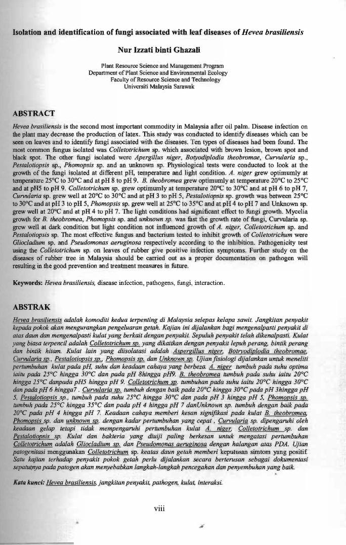
Isolation and identification of fungi associated with leaf diseases of Hevea brasiliensis
Nur Izzati binti Ghazali
Plant Resource Science and Management Program Department of Plant Science and Environmental Ecology
Faculty of Resource Science and Technology Universiti Malaysia Sarawak
ABSTRACT
Hevea brasiliensis is the second most important commodity in Malaysia after oil palm. Disease infection on the plant may decrease the production of latex. This study was conducted to identify diseases which can be seen on leaves and to identify fungi associated with the diseases. Ten types of diseases had been found. The most common fungus isolated was Colletotrichum sp. which associated with brown lesion, brown spot and black spot. The other fungi isolated were Apergillus niger, Botyodiplodia theobromae, Curvularia sp., Pestalotiopsis sp., Phomopsis sp. and an unknown sp. Physiological tests were conducted to look at the growth of the fungi isolated at different pH, temperature and light condition. A. niger grew optimumly at temperature 25°C to 30°C and at pH 8 to pH 9. B. theobromea grew optimumly at temperature 20°C to 25°C and at pH5 to pH 9. Colletotrichum sp. grew optimum}y at temperature 20°C to 30°C and at pH 6 to pH 7, Curvularia sp. grew well at 20°C to 30°C and at pH 3 to pH 5, Pestalotiopsis sp. growth was between 25°C to 30°C and at pH 3 to pH 5, Phomopsis sp. grew well at 25°C to 35°C and at pH 4 to pH 7 and Unknown sp. grew well at 20°C and at pH 4 to pH 7. The light conditions had significant effect to fungi growth. Mycelia growth for B. theobromea, Phomopsis sp. and unknown sp. was fast the growth rate of fungi, Curvularia sp. grew well at dark condition but light condition not influenced growth of A. niger, Colletotrichum sp. and Pestalotiopsis sp. The most effective fungus and bacterium tested to inhibit growth of Colletotrichum were Gliocladium sp. and Pseudomonas aeruginosa respectively according to the inhibition. Pathogenicity test using the Colletotrichum sp. on leaves of rubber give positive infection symptoms. Further study on the diseases of rubber tree in Malaysia should be carried out as a proper documentation on pathogen will resulting in the good prevention and treatment measures in future.
Keywords: Hevea brasiliensis, disease infection, pathogens, fungi, interaction.
ABSTRAK
Hevea brasiliensis adalah komoditi kedua terpenting di Malaysia selepas kelapa sawit. Jangkitan penyakit kepada pokok akan mengurangkan pengeluaran getah. Kajian ini dijalankan bagi mengenalpasti penyakit di alas daun dan mengenalpasti kulat yang berkait dengan penyakit. Sepuluh penyakit telah dikenalpasti. Kulat yang biasa terpeneil adalah Colletotrichum sp. yang dikaitkan dengan penyakit lepuh perang, bintik perang dan bintik hitam. Kulat lain yang diisolatasi adalah Aspergillus niger, Botryodin./odia theobromae, Curvularia sp., Pestalotiopsis sp., Phomopsis sp. dan Unknown sp. Ujianfisiologi dijalankan untuk meneliti pertrlmbuhan kulat pada pH, suhu dan keadaan cahaya yang berbeza. A. niger tumbuh pada suhu optima iui(U pada 25°C hingga 30°C dan pada pH Bhingga pH9. B. theobromea tumbuh pada suhu iaitu 20°C hingga 25°C danpada pH5 hingga pH 9. Colletotrichum sp. tumbuhan pada suhu iaitu 20°C hingga 30°C dan pada pH 6 hingga7 . Curvularia sp. tumbuh dengan baik pada 20°C hingga 30°C pada pH 3hingga pH 5, Pestalotiopsis sp., tumbuh pada suhu 25°C hingga 30°C dan pada pH 3 hingga pH 5, Phomopsis sp. tumbuh pada 25°C hingga 35°C dan pada pH 4 hingga pH 7 dan Unknown sp. tumbuh dengan baik pada 20°C pada pH 4 hingga pH 7. Keadaan cahaya memberi kesan sign~fikasi pada kulat B. theobromea, Phomopsis sp. dan unknown sp. dengan kadar pertumbuhan yang cepat , Curvularia sp. dipengaruhi oleh keadaan gelap tetapi tidak mempengaruhi pertumbuhan kulat A. niger, Colletotrichum sp. dan Pes/a/otiopsis sp. Kulat dan bakteria yang diuiji paling berkesan untuk mengatasi pertumbuhan Collelotrichum adalah Gliocladium sp. dan Pseudomonas aeruginosa dengan halangan atas PDA. Ujian patogenitasi menggunakan Colletotrichum sp. keatas daun getah memberi keputusan simtom yang positif Salu kajian terhadap penyakit pokok getah perlu dijalankan secara berterusan sebagai dokumentasi sepalUl.nya pada patogen akan menyebabkan langkah-langkah pencegahan dan penyembuhan yang baik.
Kala kunci: Hevea brasiliensis, jangkitan penyakit, pathogen, kulat, interaksi.
Vlll
,.....
CHAPTER ONE
INTRODUCTION
Revea brasiliensis (Muell.) Arg, commonly known as rubber tree is an important
economical commodity to Malaysia. Rubber is the second most important commodity in
Malaysia after oil palm (Frost & Sullivan, 2009). In year 2010, the natural rubber
production decreased compared to year 2006 (Muhammad Thalhah, 2010). Even though
the dependency to natural rubber is decreasing through years and replaced with palm oil
plantations yet the total exports earnings are still positive. Instead of producing latex,
rubber seed as a waste product from rubber plantations are valuable because its contains
nutritional value which can be harnessed as food for human, feed for animals or biofuel for
energy (Eka,Tajul Aris & Wan Nadiah; 2010). Nowadays, there are established rubber
plantations for timber production (Killman, 2001).
Brazil was the main trader of rubber latex, which was collected through tapping of
trees in the natural forest during nineteenth century. At the twentieth century, rubber
seedlings smuggled out of Brazil by British and planted it in Tanah Melayu. Then, it
became the parent tree which produced stock plant for all rubber plantations industrial
nowadays in Malaysia and other Southeast Asian (Killman, 2001). Historically Malaysia
has long been well known for its rubber plantations. In recent years, the trees have
increasingly made room for oil palm plantation. According to statistics from Alias (2008),
1,280,000 ha of land are planted with rubber in peninsular Malaysia, with another 260,000
ha in Sabah and Sarawak. However, the numbers had fallen to 732,280 ha on the
Peninsular and 228,600 ha in Eastern Malaysia.
1
I
,.... I
The world demands of natural rubber are increasing due to industrialization (Jomo,
1993). About 80 percent of the 9.7 million ha of rubber plantations established worldwide
for latex production in 1999, are in Southeast Asia which 72 percent of the total or 5.2
million ha are in Indonesia, Malaysia and Thailand (IRSG [International Rubber Study
Group], 1999). High production of rubber is needed to support the demands from
consumers which increasing throughout the year. This potential withdrawal is likely to be
attributable to various technical, commercial including large portions of the mature areas
would need replanting and endemic diseases a large share of rubber area in Brazil is
thought to be afflicted by South American leaf Blight (SALB) (Conference on Trade and
Development [UNTAD], 2004).
In Malaysia, rubber plantations are managed in sman-scale and large-scale
(Muhammad Thalhah, 2010). The large-scale plantations will cause the trees to be more
vulnerable to disease due to fast spread diseases and thus the healthy rubber tee will be
infected by the diseases. Many new diseases will develop in time and the inability to
identifY the causa] disease that mainly comes from an infection so that the first step for the
prevention can be taken. Precaution must be done in order to reduce the impact of the
infection, reducing the cost for the control and also to save this valuable species from
destruction.
The main objectives of this project are to identifY the causal disease of the H.
brasiliensis based on the morphological characteristics of the fungi. The physiological
characteristics of the fungi of different pH levels, temperatures and light condition were
also studied. The interactions of putative pathogen with selected microbes were examined.
The pathogenicity test of the fungi on the H. brasiliensis were be carried out.
2
CHAPTER TWO
LITERATURE REVIEW
2.1 Hevea brasiliensis
Hevea brasiliensis or rubber tree is from family of Euphorbiaceae, genus ofHevea, species
is brasiliensis. It is native to the South America region which is the Amazon forest. Then,
it has been established to many other tropical regions of the world especially at Asia
continent, such as South East Asian countries (Reed, 1976).
2.2 Damages on trees
As such other plants, rubber tree are also vulnerable to various factors that can cause loss
in plantation. These factors could be abiotic and biotic factors (Flynn, 2012). Weather,
insufficient moisture, drifting of herbicides, temperatures, transplant shock, soils impact,
injury and nutrients deficiency are the examples of abiotic factors which cause damages to
plant (Janssen, 2012). According to Wongcharoen et. ai., (2010), Trunk phloem necrosis
(TPN) caused by a combination of exogenous and endogenous stresses which affect
physiology ofthe plant and impact on soil biology and soil biochemistry that prevents latex
production.
2.3 Pest and disease.
The biotic factors induced diseases caused by living organisms, such as fungi, bacteria,
viruses, nematodes, insects, mites and animals (Flynn, 2012). Rubber tree also attacked by
bacteria, Bacterium aibilineans, parasite, ioranthus spp., insect pest, Aspidiotus
cyanophylli and Parasaissetia nigra (Duke, 1983). Rubber tree also damaged by
3
nematodes which are Helicotylenchus cavenessi, H.dihystera, H. erythrinae, Meloidogyne
incognita acrita, M. javanica, Pratylenchus coffeae, and P. brachyurus (Golden, 1984).
Most plant diseases are caused by fungi (Knogge, 1996). About 90 species of fungi are
known to damage rubber trees, the most common fungi pathogen found are Botryodiplodia
e/actica, B. theobromae, Colletotrichum heveae (Leaf spot), Fomes lamaensis caused
brown root rot disease, Gloeosporium heveae induced to die-back disease, Oidium heveae
caused powdery mildew at the leaves of rubber, Pellicularis salmonicolor caused pink
disease which give impact to the production of latex, Phytophthora palmivora caused fruit
rot, leaf-fall, black thread and die-back, Polystichus occidentalis and P. personii (white
spongy rot), Sphaerella heveae (rim bright), Sphaerostilbe repens (red rot) and Ustulina
maxima (charcoal rot) (Duke, 1983).
2.4 The symptoms of diseases caused by fungi
There are approximately 1.5 million of fungi speCIes may exist in the world
(Hawksworth, 1991). Over 20,000 species of fungi are parasites and cause disease in crops
and plants (US Environmental Protection Agency [USEPA], 2005).
According to Lieberei (2007), fungus infected obviously on four to nine days old
young leaves on the abaxial surface ofrubber tree. Powdery mildew is a disease that causes
long-term damage to the tree in the forested environment (Munster, 2008). The symptoms
are distinguished by spots or scrapes of white to grayish, powdery-like growth and
normally observed on the upper surfaces of the leaf of rubber tree (Edmunds & Pottorff,
2012).
4
....
PUS'at Khidmat Maklumat Akademik UNIVERSlll MALAYSIA SARAWAK
Fungi also may develop extensively on plant roots which reduce uptake of water
with the symptoms of abnormal wilting and yellowing of leaves (Zitter, 1985).
Spongospora subterranean produce symptoms of small lesion with purple color and then
fonns spore balls after infected of potato tubers (Miller, 2001).
European canker or Nectria canker occurs in many parts of the world caused by
Nectria gallegina Bres (Partridge, 2008). European canker causes reddish brown lesions to
emerge on small branches just below leaf scars and extend into cankers with concentric
edges and may cause dieback of shoots in spring or autumn (Gubler, 2012).
Root-pathogenic fungi, such as Verticillium, pythium, and rhizoctonia, cause major
economic losses in agriculture each year. Phytophthora root rot occurs at red, black, and .
purple Raspberries and the disease is most commonly associated with heavy soils or
portions of the planting that are the are the slowest to drain (Ellis, 2008). The leaves may
initially take on a yellow, red or orange color or may begin scorching along the edges
(Ellis, 2008). Wilt and root rot of Rhododendron and Azalea are symptoms of a disease
caused by Phytophthora which spread by soil and impact ofwater in soil (Hansen, 2009).
2.5 The disease of Hevea brasiliensis
Rubber diseases are mainly caused by fungal pathogens (Begho, 1990). There are
several types of diseases have been reported infect rubber trees. The disease include South
American leaf blight (SALB) which is an important disease of rubber species only and
have recorded infected to H brasiliensis, H bethamiana, H guainensis and H spruceana
(Holliday, 1970). SALB is a permanent major treat to the crop in Asia and Africa (Davies,
1997). Biological control of M. ulei using Hansfordia pulvinata, a hyperparasites which
5
grows well on conidial lesions, has been attempted (Liebere~ 2007). According to Feldman
(as cited in Priyadarshan, 2011), mycorrhizal fungi can provide resistance of the rubber
tree to M. ulei.
The fungus Gloeosporium alborubrum that cause Gloeosporium leaf disease is
seriously infected the immature rubber plants (Wast ie, 1972, as cited in Priyadarshan,
2011). In Indonesia, the persistence of this disease over a long period resulted in up to 50
percent loss of yield and a delay in maturity 0 f up to three years (Basuki, 1992, as cited in
Priyadarshan, 2011). But, a study with Gloeosporium showed that clones from Malaysia
and Indonesia are fairly resistant while clones from Sri Lanka are less resistant (Simmonds,
1986). In Malaysia, artificial defoliation by aerial spraying of several chemical defoliants
mixed with water is practiced (Radziah & Hashim, 1990).
Other disease are abnormal leaf fall caused by Phytophthora is also reported in
Malaysia (Chee et al., 1967; Chee, 1969). It infects pods, leaves and tender shoots which
cause heavy defoliation and dieback oftender twigs. In India, four species ofPhytophthora
are found associated with the infected specimens of rubber tree. They are Phytopthora
paimivora, P. meadii, P. nicotianae var. parasitica and P. botryose but the most common is
P.meadii (Thankamma et al. 1968, as cited in Priyadarshan, 2011).
Powdery mildew is also reported infecting rubber trees in Malaysia which caused
by Oidium heveae (Sharples, 1926). The optimum temperature for germination, infection
and porulation ranges of the fungus is from 25°C to 35°C (Liyanage et al., 1985). The
disease can cause reduction in yield throughout the year (Jacob et al., 1992). An integrated
6
approach using tridemorph and sulphur in dust form is effective to control the disease
(Edathil et al. 1992, as cited in Priyadarshan, 2011).
Crynospora cassiicola causes leaf spot and leaf fall disease was fIrstly reported in
India (Ramakrishnan & Pillay 1961, as cited in Priyadarshan, 2011). The disease has now
been found in almost all rubber-growing regions (Chee 1988, as cited in priyadarshan,
201 1). Severe leaf fall was reported from Malaysia (Tan, 1990, as cited in Priyadarshan,
201 1). Spraying of benomyl, mancozeb, captan or propineb have been recommended for
the control of Crynospora leaf disease affected nursery plants (Hashim, 1994, as cited in
Priyadarshan, 2011).
Phellinus noxius causes root and lower stem rot of woody plants throughout the
South Pacific region include rubber, mahogany, cacao and many timber, fruit and
landscape trees (Brooks, 2002). The ability is to degrade lignin, a basic component of
wood (Adaskaveg & Ogawa, 1990).
Colletotrichum gloeosporioides causal organism of Colletotrichum leaf spot disease
is common foliar pathogens of rubber both in the nursery and in the fIeld in Nigeria.
Nursery rubber seedlings are greatly affected by this leaf disease (Rao, 1975). Infection of
rubber by these foliar pathogens results in retarded growth, secondary leaf fall (SLF),
dieback and death of trees both in the nursery and in the field, as well as the reduction of
latex in mature rubber trees in plantations (Otoide, 1978).
7
2.6 Physiological effects of fungi
Fungus physiology is the study of living fungi, their functions and activities in
relation to their environment (Madan & Thind, 1998). The understanding of fungi life will
benefit in utilize in agriculture, industry and medicine at the same time controlling their
harmful effect as pathogens of plant, animals and human (Madan & Thind, 1998). There
are several factors which effect the growth of fungi.
The environmental factors favoring disease development are high humidity,
temperature, moisture and weather (Situmorang et al., 1996). Different between species of
fungi have different temperature, moisture and limits for growth and these factors are
significance in determining the relative abundance of particular species situations
(Christensen, 1957). Growth of fungi is favored by high moisture and moderate
temperatures (Burden, 1987). Optimum temperatures fall between 200 e and 30oe. Fungi
are dependent on moisture and relative humidity above 80 percent is usually sufficient for
mold growth to occur on wood (Pasanen et ai., 1992).
Some fungi have influenced ofwide range of hydrogen ion concentration and most
fungi are known to grow well on neutral medium ranging between pH7 to slightly on the
acid side of the neutral (Triphati, 2006). Soil pH may control biotic factors, such as the
biomass composition of fungi and bacteria (Fierer & Jackson, 2006). The soil pH also
control biotic factors in the forest (Blagodatskaya & Anderson, 1998) and agricultural
(Arao, 1999) soils.
More 100 fungal species have been found had influenced towards light condition
for growth (Marsh et ai., 1959). They have observation mechanisms for blue; near UV,
8
green and red light (Herrera-Estrella & Horwitz, 2007). Light are affected on fungi range
from induction or inhibition of sexual development, conidiation and suppression of spore
release (Corrochano, 2007). Mycelia growth, sporulation, spore germination and many
other fungal fundamental processes are influenced by light of different wavelengths
ranging between the near ultraviolet which is 250 nm and near infrared which is 1100 nm
(Triphat~ 2006).
2.7 Disease control method
Chemical compounds or fungicides have been used to control plant diseases.
However, the control has developed pathogens that are resistant to fungicides (Tjamos et
al., 1992, as cited in Abou-Zeid et ai., 2008). There is possibility to limit the use of
chemical pesticides which are harmful influence on the environment to control plant
disease by means of biological agents (Czaczyk et ai., 2002). The biocontrol has become
an interesting alternative to conventional methods (Raspor et ai., 2010).
Approximately 4,000 secondary metabolites of fungal origin have been described
to possess biological activities (Dreyfuss & Chapela, 1994). There are several products
from biological agent have been developed to control fungul pathogens. The first major
natural products of microbial origin to be commercialized are the antibiotics blasticidin S
which is kasugamycin and validamycin A that developed in Japan to control rice blast
cause of Pyricuiaria oryzae and sheat blight caused of Rhizoctonia soiani (Worthington,
1988). Pichia membranifaciens FY-101 was found to be antagonistic to B. cinerea. The p
I, 3-g1ucanases was secreted by P. membranifaciens FY-101 and was detected to be the
possible mechanisms related to this antagonism and it was confirmed that P.
9
•
membranifaciens FY-101 can be used as a biological control organism against B. cinerea
(Emmanual & Bernard, 2002).
Rhizoctonia solani is one of the most significant soil borne fungal pathogens which
grow both in cultured and non-cultured soils, causing diseases in different crops such as
rice, bean, tomato and other (Sneh et al., 1991). Fungi from Trichoderma genus are among
the biological control agents of Rhizoctonia solani (Lin et al., 1994) also bacteria
belonging to Pseudomonas and Bacillus genus have been also used (Gasoni et al., 1998).
The interaction used to determine the effectiveness ofevaluated usually are using a key
based on observations of Porter (1924) as given below:
A. Mutually intermingling growth where both fungi grew into one another without any
microscopic signs of interaction.
Di.Intenningling growth where the fungus being observed was growing into the opposed
fungus either above or below of its colony.
Dii. Intermingling growth where the fungus under observation has ceased growth and
overgrown by another colony.
C. Slight inhibition where the fungus approach each other until almost in contact and a
narrow demarcation line, 0.1 - 2mm, between the two colonies clearly visible.
D. Mutual inhibition at a distance of> 2 mm.
10
CHAPTER THREE
MATERIALS AND METHODS
3.1 Plants materials
Disease infected of rubber leaves were used in this study. Samples were collected
from trees in UNIMAS and other rubber plantation in Samarahan areas. The collected
samples were kept into poly-bags and were brought to Plant Pathology Laboratory at
Faculty ofResource Science and Technology, UNIMAS for further study.
3.2 Disease description
The symptoms of the disease on the infected rubber leaves were described. The
early identification of the diseases was made based on the available references.
3.3 Isolation of pathogens
The potential pathogens that related with the diseases were identified and isolated
on Potato dextrose agar (PDA). The samples ofthe infected tissue were cut into 100 pieces
which were size of 2mm x2mm. The tissues were cut between the healthy and the infected
site by using the sterile scalpel to prevent any other contamination. Then, the tissue
segments were agitated in the 10% concentration of sodium hypochlorite or Clorox. After
that, the tissues were rinsed into the sterilized distilled water, three times for five minutes
in each session. The washed tissues were blotted dry using the sterilized filter paper. Then,
ten pieces of tissue were put onto PDA media in a Petri dish. The inoculated Petri dishes
were incubated at room temperature.
11
The observation was made daily on the growing fungi in the Petri dishes until no
new species of fungi can be found grown from the plated plant tissues. The potential
pathogen was identified based on morphological structures. Percentage occurrence of the
fungi associated with the tissue pieces was recorded. A pure culture was prepared for the
further studies. The pure cultures were prepared by inoculating the morphologically
different hyphal tips from the isolated fungi into a new PDA media. The inoculated media
were incubated at the room temperature.
A stock culture was prepared. A small block of agar containing the mycelia from
the four to seven days culture were cut from the media were kept in a small bottle
containing the sterilized water and they were incubated at the temperature of 4°C.
3.4 Identification of fungi using morphological characteristics.
The fungi were observed under the compound and sterio microscope in order to
detennine their morphological characteristics. Then, to make the fungi more visible under
the microscope, Acid fuchsin which was red in color was used as stain. The morphological
characteristics that was observed and identified were the vegetative hyphae and the shape
ofthe conidia. Pictures ofall of these characteristics were captured using digital camera for
further record and identification. Reference literature used to aid the identification.
3.6 Physiological characteristics
3.6.1 Effect of different pH level on the fungal growth
Effect of pH on the fungal growth was determined by the mycelia dry weight
method. Potato Dextrose Broth (PDB) of different pH was prepared. The pH values were
adjusted to 2.0, 3.0, 4.0, 5.0, 6.0, 7.0, 8.0 and 9.0 by adding HCL to decrease the pH and
12