Malaysian Journal of Microbiology, Vol 8(3) 2012, pp. 197-202 197 ISSN (print): 1823-8262, ISSN (online): 2231-7538 Isolation and Identification of Pathogenic Bacteria from Brackish Waters of Chilika Lagoon, Odisha, India for Pharmaceutical Use Subhashree Parida 1 , Ram Chandra Jena 1,2 *, Kailash Chandra Samal 1 and Pradeep Kumar Chand 2 1 Biotechnology-cum-Commercial Tissue Culture Centre, Orissa University of Agriculture & Technology, Bhubaneswar- 751 003, Odisha, India 2 SFS Applied Microbiology, Post-Graduate Department of Botany, Utkal University, Bhubaneswar -751 004, Odisha. India. E.mail: [email protected]Received 6 April 2011; Received in revised form 25 March 2012; Accepted 25 June 2012 ABSTRACT Aims: The present investigation was undertaken in order to isolate bacteria from eighteen different water samples collected from three different sectors of ‘Chilika’ lagoon of India and to study the resistance against ten different antibiotics viz., norfloxacin, tetracycline, ciprofloxacin, neomycin, nalidixic acid, ofloxacin, chloramphenicol, nitrofurantoin, streptomycin and amoxicillin as well as their serological implications. Methodology and Results: Four different pathogenic bacteria species viz., Shigella dysenteriae, Streptococcus lactis, Bacillus cereus and Klebsiella pneumoniae were isolated which showed a wide range of sensitivity to norfloxacin, tetracycline, ciprofloxacin, ofloxacin and nitrofurantoin. S. dysenteriae was sensitive to streptomycin where as other isolates were found to be resistant. Agarose gel electrophoresis failed to reveal plasmid DNA band indicating that the observed resistance was perhaps encoded by nucleotide sequences harboured on the chromosomal DNA. Bacterial isolates were used as antigen for the production of polyclonal antibodies in rabbits. Conclusion, significance and impact of study:All the isolates exhibited strong antigenic character with specific serological relationship which can be implicated towards development of novel and pharmaceutically effective anti- bacterial products. Keywords: ‘Chilika’, bacterial isolates, antibiotic resistance, serological test _______________________________________________________________________________________________ INTRODUCTION ‘Chilika’ is the largest lagoon in the subcontinent which is situated along the east coast of India between latitude 19 o 28’ and 19 o 54’ N, and longitude 85 o 05 ’ and 85 o 38’E. This is a shallow, brackish-water lake formed due to the silting action of the Mahanadi River, which drains into the northern end of the lake, and the northerly currents in the Bay of Bengal, which have formed a sandbar along the eastern shore (Arya and Lakhotia, 2006). It is the largest brackish water wetland complex in Asia, declared as a Ramsar site under the convention on “Wetlands of International Importance”. This lagoon is presently under threat from both natural and anthropogenic pressures (Nayak et al., 2004). The marine realm constitutes the major habitat of the biosphere covering 73 % of the earth surface which provides the largest inhabitable space for living organisms, particularly microbes. Marine microbes flourish not only in the surface water of the sea, but also in the lower and abyssal depths from coastal to the offshore regions and from the general oceanic to the specialized niches like blue waters of coral reefs to black smokers of hot thermal vents at the sea floor. As the water being served as major solvent, it plays an important role in the pharmaceutical industries. It is considered as the primary source of contamination for pharmaceutical products (Tamara et al., 1998). Microbes present in the marine waters represent a potential source for commercially important bioactive compounds and their bioremediation capabilities are also remarkable. There is an increasing demand of biodiversity from natural resources for development of therapeutic drugs. Antibiotics are broadly used as chemotherapeutic agents which, albeit in a very low quantity, can inhibit the pathogenic activity of microorganisms. The potential contribution of marine organisms to the discovery of new bioactive molecules is increasingly challenging (Sponga et al., 1999; Skulberg, 2000). The microorganisms have become a significant attraction as natural source of bioactive molecules with a broad range of biological activities, such as antibiotics, antivirals, antitumorals, antioxidant and anti-inflammatory (Okami, 1982; Kamei et al., 1987; Nunez et al., 2006; Uzair et al., 2009; Shankar et al., 2010). Evidence of phycochemical and pharmacological studies on microbes is available in the literature (Zeeshan et al., 2010; Odeyemi et al., 2010; Bragadeeswaran et al., 2010). Previously, there were a number of researchers that had worked on ‘Chilika’ regarding hydrological characterization, water quality variation, antimicrobial *Corresponding author
Transcript
Malaysian Journal of Microbiology, Vol 8(3) 2012, pp. 197-202
Received 6 April 2011; Received in revised form 25 March 2012; Accepted 25 June 2012
ABSTRACT Aims: The present investigation was undertaken in order to isolate bacteria from eighteen different water samples collected from three different sectors of ‘Chilika’ lagoon of India and to study the resistance against ten different antibiotics viz., norfloxacin, tetracycline, ciprofloxacin, neomycin, nalidixic acid, ofloxacin, chloramphenicol, nitrofurantoin, streptomycin and amoxicillin as well as their serological implications. Methodology and Results: Four different pathogenic bacteria species viz., Shigella dysenteriae, Streptococcus lactis, Bacillus cereus and Klebsiella pneumoniae were isolated which showed a wide range of sensitivity to norfloxacin, tetracycline, ciprofloxacin, ofloxacin and nitrofurantoin. S. dysenteriae was sensitive to streptomycin where as other isolates were found to be resistant. Agarose gel electrophoresis failed to reveal plasmid DNA band indicating that the observed resistance was perhaps encoded by nucleotide sequences harboured on the chromosomal DNA. Bacterial isolates were used as antigen for the production of polyclonal antibodies in rabbits. Conclusion, significance and impact of study:All the isolates exhibited strong antigenic character with specific serological relationship which can be implicated towards development of novel and pharmaceutically effective anti-bacterial products.
Keywords: ‘Chilika’, bacterial isolates, antibiotic resistance, serological test _______________________________________________________________________________________________
INTRODUCTION
‘Chilika’ is the largest lagoon in the subcontinent which is situated along the east coast of India between latitude 19
o
28’ and 19o
54’ N, and longitude 85o 05
’ and 85
o 38’E.
This is a shallow, brackish-water lake formed due to the silting action of the Mahanadi River, which drains into the northern end of the lake, and the northerly currents in the Bay of Bengal, which have formed a sandbar along the eastern shore (Arya and Lakhotia, 2006). It is the largest brackish water wetland complex in Asia, declared as a Ramsar site under the convention on “Wetlands of International Importance”. This lagoon is presently under threat from both natural and anthropogenic pressures (Nayak et al., 2004). The marine realm constitutes the major habitat of the biosphere covering 73 % of the earth surface which provides the largest inhabitable space for living organisms, particularly microbes. Marine microbes flourish not only in the surface water of the sea, but also in the lower and abyssal depths from coastal to the offshore regions and from the general oceanic to the specialized niches like blue waters of coral reefs to black smokers of hot thermal vents at the sea floor. As the water being served as major solvent, it plays an important role in the pharmaceutical industries. It is
considered as the primary source of contamination for pharmaceutical products (Tamara et al., 1998). Microbes present in the marine waters represent a potential source for commercially important bioactive compounds and their bioremediation capabilities are also remarkable. There is an increasing demand of biodiversity from natural resources for development of therapeutic drugs. Antibiotics are broadly used as chemotherapeutic agents which, albeit in a very low quantity, can inhibit the pathogenic activity of microorganisms. The potential contribution of marine organisms to the discovery of new bioactive molecules is increasingly challenging (Sponga et al., 1999; Skulberg, 2000). The microorganisms have become a significant attraction as natural source of bioactive molecules with a broad range of biological activities, such as antibiotics, antivirals, antitumorals, antioxidant and anti-inflammatory (Okami, 1982; Kamei et al., 1987; Nunez et al., 2006; Uzair et al., 2009; Shankar et al., 2010). Evidence of phycochemical and pharmacological studies on microbes is available in the literature (Zeeshan et al., 2010; Odeyemi et al., 2010; Bragadeeswaran et al., 2010). Previously, there were a number of researchers that had worked on ‘Chilika’ regarding hydrological characterization, water quality variation, antimicrobial
activity and physiochemical variation (Rao et al., 1981; Nayak et al., 2004; Patra et al., 2009, Patra et al., 2010). Unfortunately progress made on pharmaceutical studies of marine microorganism from ‘Chilika’ is inadequate. This underpinned the present study with a view to investigating the physiological, biochemical, and serological characterization of bacteria isolated from marine waters of ‘Chilika’ aiming at their exploitation for pharmaceutical benefits. MATERIALS AND METHODS Isolation and characterization of bacterial strains
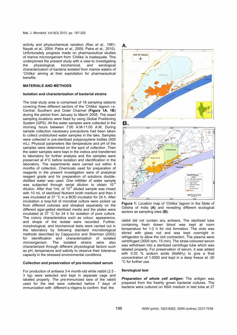
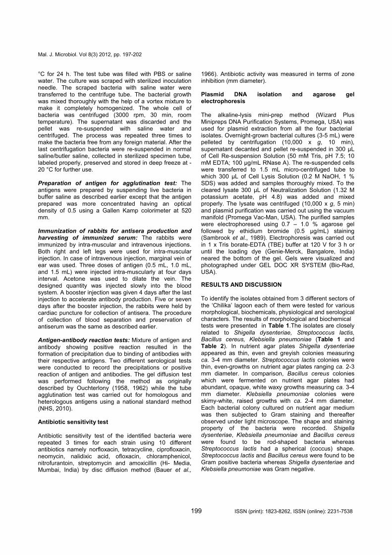
The total study area is comprised of 18 sampling stations covering three different sectors of the ‘Chilika’ lagoon i.e. Central, Southern and Outer Channel (Figure 1A, 1B) during the period from January to March 2008. The exact sampling locations were fixed by using Global Positioning System (GPS). All the water samples were collected in the morning hours between 7:00 A.M-11:00 A.M. During sample collection necessary precautions had been taken to collect undisturbed water samples in the lake. Samples were collected in pre-sterilized polypropylene bottles (500 mL). Physical parameters like temperature and pH of the samples were determined on the spot of collection. Then the water samples were kept in the icebox and transferred to laboratory for further analysis and the samples were preserved at 4°C before isolation and identification in the laboratory. The experiments were carried out within 4 months of collection. Chemicals used for preparation of reagents in the present investigation were of analytical reagent grade and for preparation of solutions double-distilled water was used. One milliliter of water sample was subjected through serial dilution to obtain 10
-8
dilution. After that 1mL of 10-8
diluted sample was mixed with 10 mL of sterilized Nutrient broth medium and then it was incubated at 37 °C in a BOD incubator for 24 h. After incubation a loop-full of microbial culture were picked up from different colonies and streaked separately on the different agar-gelled sterilized media and the plates were incubated at 37 °C for 24 h for isolation of pure culture. The colony characteristics such as colour, appearance, and shape of the isolate were recorded. Further, morphological, and biochemical tests were carried out in the laboratory by following standard microbiological methods described by Cappuccino and Sherman (2002) for identification and characterization of isolated microorganism. The isolated strains were also characterized through different physiological factors such as pH, temperature and salinity to observe their tolerance capacity in the stressed environmental conditions. Collection and preservation of pre-immunized serum For production of antisera 3-4 month-old white rabbit (2.5 - 3 kg) were selected and kept in separate cage and labeled properly. The pre-immunized sera of the rabbit used for the test were collected before 7 days of immunization with different a ntigens to confirm that the
Figure 1: Location map of ‘Chilika’ lagoon in the State of Odisha of India (A) and revealing different ecological sectors as sampling sites (B).
rabbit did not contain any antisera. The sterilized tube containing fresh drawn blood was kept at room temperature for 1-2 h for clot formation. The clots was stirred with glass rod and was kept overnight in refrigerator to allow the clot contraction. The plasma were centrifuged (3000 rpm, 15 min). The straw-coloured serum was withdrawn into a sterilized centrifuge tube which was labeled properly. For preservation of serum, it was added with 0.02 % sodium azide (NaNH2) to give a final concentration of 1:5000 and kept in a deep freeze at -20 °C for further use.
Serological test Preparation of whole cell antigen: The antigen was prepared from the freshly grown bacterial cultures. The bacteria were cultured on NSA medium in test tube at 27
°C for 24 h. The test tube was filled with PBS or saline water. The culture was scraped with sterilized inoculation needle. The scraped bacteria with saline water were transferred to the centrifuge tube. The bacterial growth was mixed thoroughly with the help of a vortex mixture to make it completely homogenized. The whole cell of bacteria was centrifuged (3000 rpm, 30 min, room temperature). The supernatant was discarded and the pellet was re-suspended with saline water and centrifuged. The process was repeated three times to make the bacteria free from any foreign material. After the last centrifugation bacteria were re-suspended in normal saline/butler saline, collected in sterilized specimen tube, labeled properly, preserved and stored in deep freeze at -20 °C for further use. Preparation of antigen for agglutination test: The antigens were prepared by suspending live bacteria in buffer saline as described earlier except that the antigen prepared was more concentrated having an optical density of 0.5 using a Gallen Kamp colorimeter at 520 mm.
Immunization of rabbits for antisera production and harvesting of immunized serum: The rabbits were immunized by intra-muscular and intravenous injections. Both right and left legs were used for intra-muscular injection. In case of intravenous injection, marginal vein of ear was used. Three doses of antigen (0.5 mL, 1.0 mL, and 1.5 mL) were injected intra-muscularly at four days interval. Acetone was used to dilate the vein. The designed quantity was injected slowly into the blood system. A booster injection was given 4 days after the last injection to accelerate antibody production. Five or seven days after the booster injection, the rabbits were held by cardiac puncture for collection of antisera. The procedure of collection of blood separation and preservation of antiserum was the same as described earlier. Antigen-antibody reaction tests: Mixture of antigen and antibody showing positive reaction resulted in the formation of precipitation due to binding of antibodies with their respective antigens. Two different serological tests were conducted to record the precipitations or positive reaction of antigen and antibodies. The gel diffusion test was performed following the method as originally described by Ouchterlony (1958, 1962) while the tube agglutination test was carried out for homologous and heterologous antigens using a national standard method (NHS, 2010). Antibiotic sensitivity test
Antibiotic sensitivity test of the identified bacteria were repeated 3 times for each strain using 10 different antibiotics namely norfloxacin, tetracycline, ciprofloxacin, neomycin, nalidixic acid, ofloxacin, chloramphenicol, nitrofurantoin, streptomycin and amoxicillin (Hi- Media, Mumbai, India) by disc diffusion method (Bauer et al.,
1966). Antibiotic activity was measured in terms of zone inhibition (mm diameter). Plasmid DNA isolation and agarose gel electrophoresis
The alkaline-lysis mini-prep method (Wizard Plus Minipreps DNA Purification Systems, Promega, USA) was used for plasmid extraction from all the four bacterial isolates. Overnight-grown bacterial cultures (3-5 mL) were pelleted by centrifugation (10,000 x g, 10 min), supernatant decanted and pellet re-suspended in 300 µL of Cell Re-suspension Solution (50 mM Tris, pH 7.5; 10 mM EDTA; 100 µg/mL RNase A). The re-suspended cells were transferred to 1.5 mL micro-centrifuged tube to which 300 µL of Cell Lysis Solution (0.2 M NaOH, 1 % SDS) was added and samples thoroughly mixed. To the cleared lysate 300 µL of Neutralization Solution (1.32 M potassium acetate, pH 4.8) was added and mixed properly. The lysate was centrifuged (10,000 x g, 5 min) and plasmid purification was carried out using the vacuum manifold (Promega Vac-Man, USA). The purified samples were electrophoresed using 0.7 – 1.0 % agarose gel followed by ethidium bromide (0.5 µg/mL) staining (Sambrook et al., 1989). Electrophoresis was carried out in 1 x Tris borate-EDTA (TBE) buffer at 120 V for 3 h or until the loading dye (Genie-Merck, Bangalore, India) neared the bottom of the gel. Gels were visualized and photographed under GEL DOC XR SYSTEM (Bio-Rad, USA).
RESULTS AND DISCUSSION
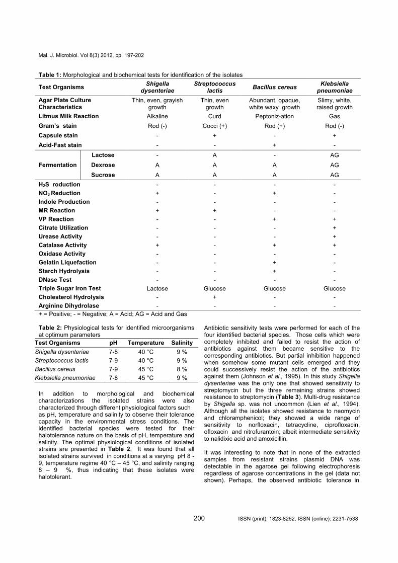
To identify the isolates obtained from 3 different sectors of the ‘Chilika’ lagoon each of them were tested for various morphological, biochemicals, physiological and serological characters. The results of morphological and biochemical tests were presented in Table 1.The isolates are closely related to Shigella dysenteriae, Streptococcus lactis, Bacillus cereus, Klebsiella pneumoniae (Table 1 and Table 2). In nutrient agar plates Shigella dysenteriae appeared as thin, even and greyish colonies measuring ca. 3-4 mm diameter. Streptococcus lactis colonies were thin, even-growths on nutrient agar plates ranging ca. 2-3 mm diameter. In comparison, Bacillus cereus colonies which were fermented on nutrient agar plates had abundant, opaque, white waxy growths measuring ca. 3-4 mm diameter. Klebsiella pneumoniae colonies were skimy-white, raised growths with ca. 2-4 mm diameter. Each bacterial colony cultured on nutrient agar medium was then subjected to Gram staining and thereafter observed under light microscope. The shape and staining property of the bacteria were recorded. Shigella dysenteriae, Klebsiella pneumoniae and Bacillus cereus were found to be rod-shaped bacteria whereas Streptococcus lactis had a spherical (coccus) shape. Streptococcus lactis and Bacillus cereus were found to be Gram positive bacteria whereas Shigella dysenteriae and Klebsiella pneumoniae was Gram negative.
Table 1: Morphological and biochemical tests for identification of the isolates
Test Organisms Shigella
dysenteriae Streptococcus
lactis Bacillus cereus
Klebsiella pneumoniae
Agar Plate Culture Characteristics
Thin, even, grayish growth
Thin, even growth
Abundant, opaque, white waxy growth
Slimy, white, raised growth
Litmus Milk Reaction Alkaline Curd Peptoniz-ation Gas
Gram’s stain Rod (-) Cocci (+) Rod (+) Rod (-)
Capsule stain - + - +
Acid-Fast stain - - + -
Fermentation
Lactose - A - AG
Dexrose A A A AG
Sucrose A A A AG
H2S roduction - - - -
NO3 Reduction + - + -
Indole Production - - - -
MR Reaction + + - -
VP Reaction - - + +
Citrate Utilization - - - +
Urease Activity - - - +
Catalase Activity + - + +
Oxidase Activity - - - -
Gelatin Liquefaction - - + -
Starch Hydrolysis - - + -
DNase Test - - - -
Triple Sugar Iron Test Lactose Glucose Glucose Glucose
Cholesterol Hydrolysis - + - -
Arginine Dihydrolase - - - -
+ = Positive; - = Negative; A = Acid; AG = Acid and Gas Table 2: Physiological tests for identified microorganisms at optimum parameters
Test Organisms pH Temperature Salinity
Shigella dysenteriae 7-8 40 °C 9 %
Streptococcus lactis 7-9 40 °C 9 %
Bacillus cereus 7-9 45 °C 8 %
Klebsiella pneumoniae 7-8 45 °C 9 %
In addition to morphological and biochemical characterizations the isolated strains were also characterized through different physiological factors such as pH, temperature and salinity to observe their tolerance capacity in the environmental stress conditions. The identified bacterial species were tested for their halotolerance nature on the basis of pH, temperature and salinity. The optimal physiological conditions of isolated strains are presented in Table 2. It was found that all isolated strains survived in conditions at a varying pH 8 - 9, temperature regime 40 °C – 45 °C, and salinity ranging 8 – 9 %, thus indicating that these isolates were halotolerant.
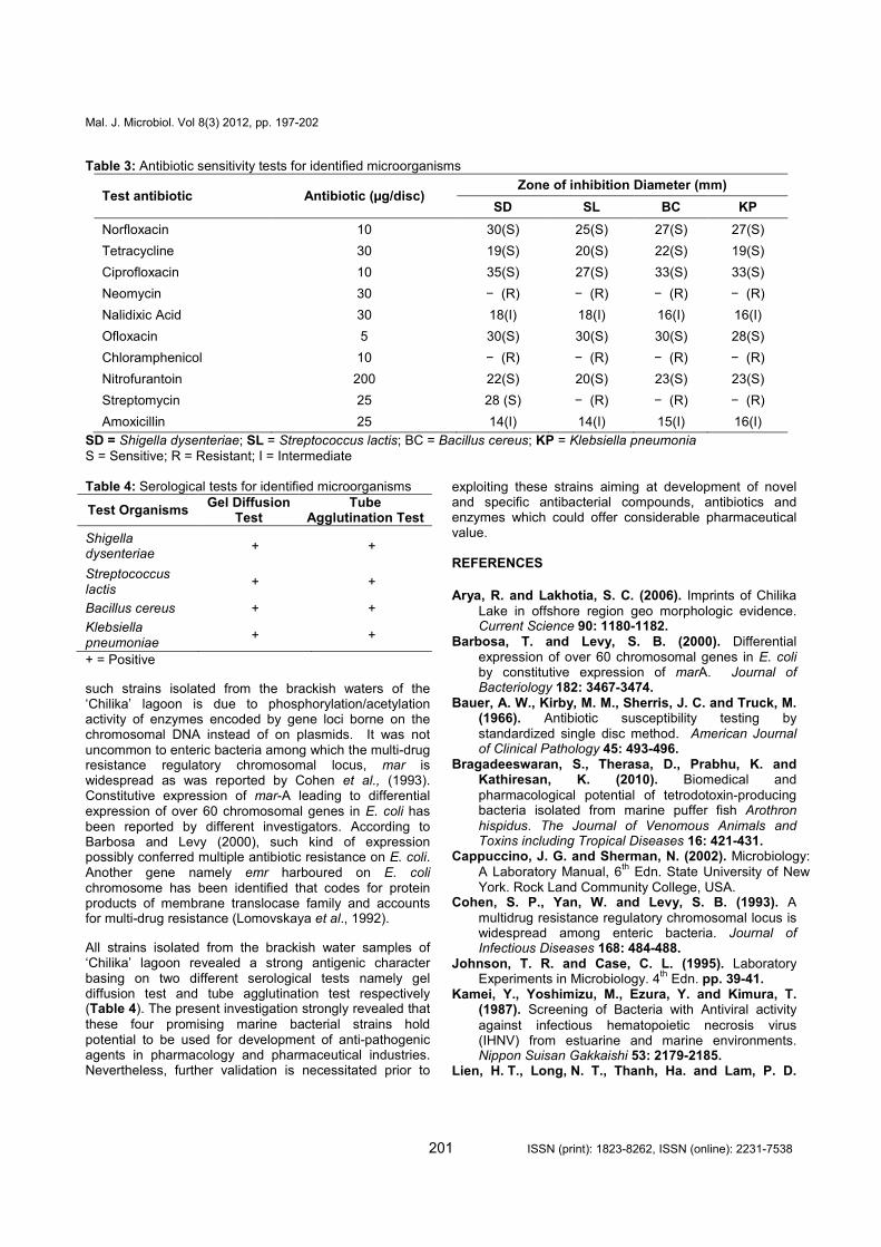
Antibiotic sensitivity tests were performed for each of the four identified bacterial species. Those cells which were completely inhibited and failed to resist the action of antibiotics against them became sensitive to the corresponding antibiotics. But partial inhibition happened when somehow some mutant cells emerged and they could successively resist the action of the antibiotics against them (Johnson et al., 1995). In this study Shigella dysenteriae was the only one that showed sensitivity to streptomycin but the three remaining strains showed resistance to streptomycin (Table 3). Multi-drug resistance by Shigella sp. was not uncommon (Lien et al., 1994). Although all the isolates showed resistance to neomycin and chloramphenicol; they showed a wide range of sensitivity to norfloxacin, tetracycline, ciprofloxacin, ofloxacin and nitrofurantoin; albeit intermediate sensitivity to nalidixic acid and amoxicillin. It was interesting to note that in none of the extracted samples from resistant strains plasmid DNA was detectable in the agarose gel following electrophoresis regardless of agarose concentrations in the gel (data not shown). Perhaps, the observed antibiotic tolerance in
Table 3: Antibiotic sensitivity tests for identified microorganisms
Test antibiotic Antibiotic (µg/disc) Zone of inhibition Diameter (mm)
SD SL BC KP
Norfloxacin 10 30(S) 25(S) 27(S) 27(S)
Tetracycline 30 19(S) 20(S) 22(S) 19(S)
Ciprofloxacin 10 35(S) 27(S) 33(S) 33(S)
Neomycin 30 − (R) − (R) − (R) − (R)
Nalidixic Acid 30 18(I) 18(I) 16(I) 16(I)
Ofloxacin 5 30(S) 30(S) 30(S) 28(S)
Chloramphenicol 10 − (R) − (R) − (R) − (R)
Nitrofurantoin 200 22(S) 20(S) 23(S) 23(S)
Streptomycin 25 28 (S) − (R) − (R) − (R)
Amoxicillin 25 14(I) 14(I) 15(I) 16(I)
SD = Shigella dysenteriae; SL = Streptococcus lactis; BC = Bacillus cereus; KP = Klebsiella pneumonia S = Sensitive; R = Resistant; I = Intermediate Table 4: Serological tests for identified microorganisms
Test Organisms Gel Diffusion
Test Tube
Agglutination Test
Shigella dysenteriae
+ +
Streptococcus lactis
+ +
Bacillus cereus + +
Klebsiella pneumoniae
+ +
+ = Positive
such strains isolated from the brackish waters of the ‘Chilika’ lagoon is due to phosphorylation/acetylation activity of enzymes encoded by gene loci borne on the chromosomal DNA instead of on plasmids. It was not uncommon to enteric bacteria among which the multi-drug resistance regulatory chromosomal locus, mar is widespread as was reported by Cohen et al., (1993). Constitutive expression of mar-A leading to differential expression of over 60 chromosomal genes in E. coli has been reported by different investigators. According to Barbosa and Levy (2000), such kind of expression possibly conferred multiple antibiotic resistance on E. coli. Another gene namely emr harboured on E. coli chromosome has been identified that codes for protein products of membrane translocase family and accounts for multi-drug resistance (Lomovskaya et al., 1992).
All strains isolated from the brackish water samples of ‘Chilika’ lagoon revealed a strong antigenic character basing on two different serological tests namely gel diffusion test and tube agglutination test respectively (Table 4). The present investigation strongly revealed that these four promising marine bacterial strains hold potential to be used for development of anti-pathogenic agents in pharmacology and pharmaceutical industries. Nevertheless, further validation is necessitated prior to
exploiting these strains aiming at development of novel and specific antibacterial compounds, antibiotics and enzymes which could offer considerable pharmaceutical value. REFERENCES
Arya, R. and Lakhotia, S. C. (2006). Imprints of Chilika
Lake in offshore region geo morphologic evidence. Current Science 90: 1180-1182.
Barbosa, T. and Levy, S. B. (2000). Differential expression of over 60 chromosomal genes in E. coli by constitutive expression of marA. Journal of Bacteriology 182: 3467-3474.
Bauer, A. W., Kirby, M. M., Sherris, J. C. and Truck, M. (1966). Antibiotic susceptibility testing by standardized single disc method. American Journal of Clinical Pathology 45: 493-496.
Bragadeeswaran, S., Therasa, D., Prabhu, K. and Kathiresan, K. (2010). Biomedical and pharmacological potential of tetrodotoxin-producing bacteria isolated from marine puffer fish Arothron hispidus. The Journal of Venomous Animals and Toxins including Tropical Diseases 16: 421-431.
Cappuccino, J. G. and Sherman, N. (2002). Microbiology: A Laboratory Manual, 6
th Edn. State University of New
York. Rock Land Community College, USA. Cohen, S. P., Yan, W. and Levy, S. B. (1993). A
multidrug resistance regulatory chromosomal locus is widespread among enteric bacteria. Journal of Infectious Diseases 168: 484-488.
Johnson, T. R. and Case, C. L. (1995). Laboratory Experiments in Microbiology. 4
th Edn. pp. 39-41.
Kamei, Y., Yoshimizu, M., Ezura, Y. and Kimura, T. (1987). Screening of Bacteria with Antiviral activity
against infectious hematopoietic necrosis virus (IHNV) from estuarine and marine environments. Nippon Suisan Gakkaishi 53: 2179-2185.
Lien, H. T., Long, N. T., Thanh, Ha. and Lam, P. D.
(1994). Antibiotic resistance of Shigella isolates during 1990-1992 in Vietnam. APUA Newsletter 12: 4-5.
Lomovskaya, O. (1992). Emr and E. coli locus for multidrug resistance. Proceedings of the National Academy of Sciences 89: 8938-8942.
Nayak, B. K., Acharya, B. C., Panda, U. C., Nayak, B. B. and Acharya, S. K. (2004). Variation of water quality in Chilika Lake. Orissa, Indian Journal of Marine Sciences 33: 164-169.
N.H.S. (2010). National standard method of agglutination
test, Health protection agency issued by Standards Unit, Department for Evaluations, Standards and Training Centre for Infections. 2: 1-11.
Nunez, R., Garateix, A., Laguna, A., Fernández, M. D., Ortiz, E., Llanio, M., Valdés, O., Rodríguez, A. and Menéndez, R. (2006). Caribbean marine biodiversity as a source of new compounds of biomedical interest and other industrial applications. Newsletter, Pharmacology online 3: 111-119.
Odeyemi, A. T., Dada, A. C., Ogunbanjo, O. R. and Ojo, M. A. (2010). Bacteriological, physicochemical and mineral studies on Awedele spring water and soil samples in Ado Ekiti, Nigeria. African Journal of Environmental Science and Technology 4: 319-327.
Okami, Y. (1982). Potential use of marne microorganisms for antibiotics and enzyme production. Pure and Applied Chemistry 54: l95l—l962.
Ouchterlony, O. (1958). Diffusion-in-gel methods for immunological analysis. I. Progr. Allergy 5: 1.
Ouchterlony, O. (1962). Diffusion-in-gel methods for immunological analysis. II. Progr. Allergy 6: 1.
Patra, A. P., Patra, J. K., Mahapatra, N. K., Das, S., and Swain, G. C. (2010). Seasonal Variation in Physicochemical Parameters of Chilika Lake after Opening of New Mouth near Gabakunda, Orissa, India. World Journal of Fish and Marine Sciences 2: 109-117.
Patra, J. K., Patra, A. P., Mahapatra, N. K., Thatoi, H. N., Das, S., Sahu, R. K. and Swain, G. C. (2009).
Antimicrobial activity of organic solvent extracts of three marine macroalgae from Chilika Lake, Orissa. Malaysian Journal of Microbiology 5: 128-131.
Sambrook, J., Fritsch, E. F. and Maniatis, T. (1989).
Molecular Cloning: A Laboratory Manual. 2nd
Edn. Cold Spring Harbor Laboratory press, Cold Spring Harbor, New York, USA.
Shankar, C. V. S., Malar, A. H. J. and Punitha, S. M. J. (2010). Antimicrobial activity of marine bacteria associated with Polychaetes. Bioresearch Bulletin 1: 24-28.
Sponga, F., Cavaletti, L., Lazzarini, A., Borghi, A., Ciciliato, I., Losi, D. and Marinelli, F. (1999).
Biodiversity of potentials of marine-derived microorganisms. Journal of Biotechnology 70: 65-69.
Skulberg, O. M. (2000). Microalgal as a source of
bioactive molecules-experience from Cyanophyte research. Journal of Applied Phycology 12, 341-348.
Subba Rao, M. V., Rao, B. M. G., Rao, B. R. and Nanda, N. K. (1981). Hydrological studies of the brackish
water Chilika Lagoon, Orissa. Journal of Environmental Biology 2: 59-62.
Tamara, K. M., Juana, M. H., Tatiana, B. and Lisbett, M. (1998). Identification of bacteria in water for pharmaceutical use. Revista Latinoamerican de Microbiologia 40: 142-150.
Uzair, B., Ahmeda, N., Mohammad, F. V., Ahmad, V. U. and Edwards, D. (2009). Screening of marine bacteria of Pakistan coast for drug discovery potential. Proceedings of the Pakistan Academy of Sciences 46: 137-144.
Zeeshan, M., Suhail, S., Biswas, D., Farooqui, A. and Arif, J. M. (2010). Screening of selected cynobacterial strains for phycochemical compounds and biological activities in vitro. Biochemical and Cellular Archives 10: 163-168.